Speciation analysis of inorganic arsenic in food and water samples by electrothermal atomic absorption spectrometry after magnetic solid phase extraction by a novel MOF-199/modified magnetite nanoparticle composite

Abolfazl Abbaszadeh and Azadeh Tadjarodi*
Research Laboratory of Inorganic Materials Synthesis, Department of Chemistry, Iran University of Science and Technology, Narmak, Tehran 16846-13114, Iran. E-mail: tajarodi@iust.ac.ir; Fax: +98 2177491204; Tel: +98 2177240516

Received 31st August 2016 , Accepted 28th November 2016

First published on 28th November 2016


Abstract

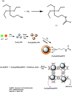
In this work a novel magnetic metal–organic framework (MOF-199/dithiocarbamate modified magnetite nanoparticle composite) was synthesized and utilized for speciation analysis of As(III) and As(V) via determination by electrothermal atomic absorption spectrometry. The synthesized sorbent represented selectivity toward As(III) at pH = 3 while As(V) remained in the initial solution. Total amount of arsenic in the samples was determined after reduction of the As(V) species to As(III) ions with the mixture of Na2S2O3 and KI. A design of experiments approach was employed to find the best extraction conditions by evaluating the parameters affecting the preconcentration procedure. Under the optimal conditions the limit of detection, linearity and relative standard deviation of the method for As(III) were 1.2 ng L−1, 4–300 ng L−1 and <8.4%, respectively. The developed method was validated by analyzing two certified reference materials. Finally, the outlined method was successfully employed to the rapid extraction and speciation analysis of As(III) and As(V) in water samples and total arsenic in rice and canned tuna samples.


1. Introduction

Metal–organic frameworks (MOFs) or porous coordination polymers are a new class of porous crystalline materials which are assembled via bonding of organic bridging ligands with metal centers.1 Owing to their unique structural diversity and excellent properties, MOFs have attracted considerable attention in analytical chemistry and have been investigated as excellent sorbents for the analysis of various environmental pollutants such as polybrominated diphenyl ethers,1 pesticides,2 polycyclic aromatic hydrocarbons,3 parabens,4 plasticizers5 and heavy metal ions.6–10 MOFs can be employed as ideal sorbents owing to their unique properties such as very high surface areas, flexible and highly porous structure and great diversity in surface functionalization.11 These unique properties make the MOFs excellent sorbents in extraction and preconcentration of various analytes.

Environmental pollution of heavy metals and metalloids has considered great attention due to their high toxicity. Toxic metal ions have a high tendency to bond with various ligands, especially biomolecules containing N, S, and O moieties.12 The complex formation may cause changes in the molecular conformation of proteins, enzymes inhibition and hydrogen bonds breaking. The toxicity and carcinogenic nature of the heavy metals and metalloids such as those influencing the central nervous system (Hg(II), Pb(II), As(III)); the kidneys or liver (Cu(II), Cd(II), Hg(II), Pb(II)); or skin, bones, or teeth (Ni(II), Cu(II), Cd(II), Cr(III))13–15 can be explained by these interactions. Thereby, it is necessary to monitor and quantify very low levels of these toxic elements, as it is a proper tool for the identification, monitoring and quantification of toxicants in water and food samples. In this regards, development of fast, easy, sensitive, selective and environmentally friendly methods for monitoring and quantification of toxic metals are of particular significance.

Arsenic is one of the abundant elements in environment since it is released into environment through diverse sources such as wood preservatives, electronics industries, pesticides, glassware production, waste incineration, volcanic emissions and fossil fuels.16 However, arsenic is toxic to human beings and its toxicity depends on the existing forms where soluble inorganic arsenic species are more toxic than organic counterparts.17,18 As arsenic contamination in natural waters is considered a global concern, the World Health Organization (WHO) guideline value set for arsenic in drinking water is 10 μg L−1 for daily intake.18 Arsenic as a ubiquitous element is found in soils, rocks, air, natural waters and organisms.19,20 Nowadays, arsenic speciation in complex sample matrixes is critical since the quantification of total arsenic in diverse sample matrixes is no longer sufficient to indicate the risks of arsenic to the environment and also human health.21 Various oxidation states of arsenic are as following: As(0), ionic species such as arsenate (As(V)), arsenite (As(III)) and arsine (AsH3). The toxicity of inorganic As(III) species is more than the inorganic As(V). Therefore, differentiation of As(III) and As(V) in the case of toxicity assessment is necessary. As(V) and/or As(III) are the two chief species of As found in water derived from natural sources and based on the environmental conditions the distribution of As(III)/As(V) fluctuates.22

The concentration of arsenic in real samples is often very low and in most cases the samples have complicated matrices; thereby a separation and preconcentration step is vital for precise and accurate quantification.23 Accordingly, various preconcentration and extraction methods such as solvent extraction,24 co-precipitation,25 cloud point extraction26 and solid phase extraction (SPE)27–30 have been reported for preconcentration and speciation analysis of As(III) and As(V) ions. Owing to its simplicity, rapidity, low consumption of reagents and minimal cost, SPE is the most universally employed extraction method for preconcentration of metal species in different environmental and food samples.31 However, in most cases the fast and complete isolation and removal of sorbents from extraction media is difficult, time-consuming, and labor-intensive which can cause to additional environmental problems and affect the precision and accuracy of the method.32 Utilizing a magnetic sorbent facilitates the separation of solid material from solution, by employing an external magnetic field, accelerates sample preparation procedures and can solve the mentioned problems.32–34

The aim of this work is speciation analysis of As(III) and As(V) by means of a novel magnetic metal–organic framework. Initially, MOF-199 was synthesized and then it was immobilized with Fe3O4@dithiocarbamate nanoparticles (Fe3O4@DTC NPs). The synthesized nanosorbent was characterized by Fourier transform infrared spectroscopy (FT-IR), BET analysis, scanning electron microscopy (SEM), CHNS analysis and vibrating sample magnetometry (VSM). The magnetic characteristic of nanosorbent simplified and accelerated the separation and recovery of the sorbent from extraction medium. Functionalization with dithiocarbamate moieties led to its selectivity towards As(II) species. Design of experiments (DOE) approach through response surface methodology was employed in order to find the optimum operating conditions of the extraction method. Finally, the developed magnetic solid phase extraction (MSPE) method was used for determination of As(III) and As(V) species in water and total arsenic in canned tuna and rice and satisfactory results were obtained.

2. Experimental

2.1. Reagents and solutions

All reagents (FeCl3, (NH4)2Fe(SO4)2·6H2O, HCl, HNO3, thiourea (TU), NaOH, Na2S2O3, KI, carbon disulfide (CS2), (3-aminopropyl)-triethoxysilane (3-APS), benzene-1,3,5-tricarboxylate (H3BTC), toluene, ammonium hydroxide (28% w/v), tetraethyl orthosilicate (TEOS), dimethylformamide (DMF), ethanol, and methanol) were of analytical grade and purchased from Merck (Darmstadt, Germany) or from Fluka and were used without any purification. Standard solutions of 1000 mg L−1 of As(III) and As(V) were purchased from Merck. All working solutions were prepared using deionized water.

2.2. Instrumentation

A Varian (Varian Company, USA, www.varian.com) Spectra model AA 220 with a graphite furnace atomizer (GTA-110) equipped with an ASC-6100 auto sampler was used for determination of arsenic ions in the optimization studies and real sample analysis. Deuterium background correction was employed in order to correct non-specific absorbance. An arsenic hollow cathode lamp with wavelength of 193.7 nm with a spectral bandwidth of 1.0 nm and a pyrolytic graphite coated graphite tube was used too. In all tests, the injection volume was 20 μL. A solution containing 300 mg L−1 Mg(NO3)2 and 1000 mg L−1 Pd(NO3)2 was used as chemical modifier. The operating conditions of the instrument are tabulated in Table 1. Ar with 99.995% purity (Roham Gas Company, Tehran, Iran) was employed as protective and purges gas. The pH of the solutions were measured at 25 ± 1 °C with a digital Metrohm 827 pH meter (Herisau, Switzerland) equipped with a combined glass-calomel electrode. A Bruker IFS-66 FT-IR spectrophotometer (Karlsruhe, Germany, www.brukeroptics.com) was used for FT-IR spectra recording. Scanning electron microscopy (SEM) was conducted using a KYKY-3200SEM (Zhongguancun Beijing, China, www.kyky.com.cn) instrument. Transmission electron microscopy (TEM) analysis was conducted by employing a LEO 912AB electron microscope (Leo Ltd., Germany, www.zeiss.com). Magnetic properties of nanocomposites were measured by a vibrating sample magnetometer (VSM) model AGFM/VSM 117 3886 (Kashan, Iran) with a magnetic field strength of 1 Tesla and at room temperature. Elemental contents of nanocomposite were determined by an elemental analyzer model Thermo Finnigan Flash EA112 (Okehampton, UK, www.thermoscientific.com).
Table 1 Operating condition of ETAAS for determination of arsenic
Step Temperature (°C) Time (s) Argon gas flow rate (mL min−1)
Ramp Hold
Injection of modifier 75 5 25 250
[thin space (1/6-em)]
Injection of sample
Drying 120 5 20 250
Drying 260 2 10 250
Ashing 1000 1 20 250
Atomization 2000 0 3 0 (read)
Cleaning 2400 0 5 250


2.3. Preparation of standard solutions

Stock solutions (1000 mg L−1) of potentially interfering ions such as K+, Na+, Mg(II), Ca(II), Mn(II), Pb(II), Cr(III), Cr(VI), Al(III), Cd(II), Cu(II), Zn(II), Co(II), Cl, NO3, SO42− and CH3COO were prepared in a 2% (v/v) HNO3 solution. The working standard solutions were prepared by diluting an appropriate amount of the stock solution with double distilled water. All of these solutions were stored at ambient temperature prior to use.

2.4. Synthesis of magnetic MOF nanocomposite

2.4.1. Synthesis of dithiocarbamate functionalized Fe3O4 NPs. Initially, 2 mL 3-APS and 0.8 mL CS2 were mixed and the obtained solution was stirred at room temperature for 3 h to obtain 3-APS/CS2 compound (Fig. 1). Afterwards, Fe3O4 NPs were synthesized according to our previously reported procedure10 and were dried at room temperature. In the next step, 1.0 g of synthesized Fe3O4 NPs was suspended in a solution of 250 mL deionized water, 75 mL ethanol and 4 mL NH4OH (28%). Thereafter, 3.0 mL TEOS was added slowly to the mixture under vigorous stirring.35,36 After 10 h stirring at 40 °C, the prepared Fe3O4@SiO2 NPs were gathered by a strong magnet (15 cm × 12 cm × 5 cm, 1.4 T), washed with ethanol and finally dried at room temperature.36
image file: c6ra21819a-f1.tif
Fig. 1 (a) A schematic representation for synthesis of Fe3O4@DTC NPs. (b) A schematic illustration for the synthesis of MOF-199/Fe3O4@DTC nanocomposite.

To synthesis dithiocarbamate functionalized Fe3O4 NPs (Fe3O4@DTC), 1.0 g of Fe3O4@SiO2 NPs was suspended in 75 mL dry toluene, and the obtained mixture was stirred for 45 min at 700 rpm. Afterwards, 3-APS/CS2 (0.5 g) was added to the mentioned mixture and it was refluxed for 15 h.10 Fe3O4@DTC NPs was dried at 50 °C after washing with methanol and magnetic decantation. The synthesis of Fe3O4@DTC NPs was investigated by FT-IR spectroscopy, high-angle X-ray diffraction, TEM, and SEM methods.

2.4.2. Synthesis of MOF-199/dithiocarbamate magnetite nanoparticles composite. MOF-199/Fe3O4@DTC nanocomposite was prepared according to the following procedure (Fig. 1b). Briefly, 2.38 mmol trimesic acid (H3BTC, 0.5 g) was dissolved in 80 mL of DMF/ethanol (1[thin space (1/6-em)]:[thin space (1/6-em)]1) and 0.4 g Fe3O4@DTC NPs was suspended in 30 mL ethanol and added to the reaction mixture under magnetic stirring, and its temperature was increased up to 70 °C. Afterwards, 40 mL Cu(OAc)2·H2O (0.86 g, 4.31 mmol) aqueous solution was added to the reaction mixture and the reaction was continued for a further 4 h at 70 °C under magnetic stirring.10 Ultimately, MOF-199/Fe3O4@DTC nanocomposite was recovered from the supernatant solution by magnetic decantation and washed with water and ethanol, respectively and dried at room temperature. The obtained nanocomposite was characterized by FT-IR spectroscopy, VSM, CHNS analysis, SEM and BET analysis.

2.5. Extraction procedure

Extraction process was performed in test tubes containing 30 mL 0.5 μg L−1 solution of As(III). The pH of the solutions was adjusted to 3.0 using 0.1 mol L−1 HCl solution. Afterwards, 14.5 mg of magnetic nanocomposite was added to the mixture and it was stirred for 13 min in order to extract As(III) ions from the solution, completely. Then the sorbent was separated from the solution by using the strong magnet. The sorbed amount of As(II) ions was determined using ETAAS based on its concentration change after the sorption process. In the next step, 2.5 mL of 1.1 mol L−1 thiourea in 0.065 mol L−1 HCl solution as an eluent was added to the test tube containing the sorbent and it was stirred for 11 min. Finally, the mixture was again exposed to the strong magnet to separate the sorbent from the eluent media. The obtained clear solution containing eluted As(III) ions was introduced to ETAAS for subsequent analysis. In order to determine As(V) ions, 1 mL of a 1% (w/v) of Na2S2O3 solution and 1 mL 0.5% (w/v) KI solution were added to the sample, before adjusting the pH and performing the extraction process.

2.6. Real sample pretreatment

2.6.1. Canned tuna and rice samples. Rice and canned tuna samples were purchased from local supermarkets in Tehran, Iran. Briefly, 1.0 g of each sample was placed in a digestion cell and 6 mL concentrated HNO3 (65% w/w)–H2O2 (30% w/w) solution (3[thin space (1/6-em)]:[thin space (1/6-em)]1 v/v) was added to the sample. Digestion was performed according to the following procedure: 2 min at 250 W, 2 min for 0 W, 6 min at 250 W, 5 min at 400 W, 8 min at 550 W, and ventilation for 8 min, respectively.28 Afterwards, the obtained solution was cooled to room temperature, filtered into a 500 mL volumetric flask and diluted with ultrapure water.
2.6.2. Water samples. River water sample (south of Tehran, Iran) and drinking water sample (Tehran, Iran) were filtered into cleaned polyethylene bottles. The pH of each sample was adjusted to 3.0 according to the optimized experimental conditions before the extraction procedure.
2.6.3. Reference material. A water standard reference material (SRM, 1643e, NIST) and NIST SRM 1568a Rice Flour were analyzed for validation test of the suggested extraction method. NIST SRM 1568a Rice Flour was digested according to the procedure mentioned for rice and canned tuna samples. The pH of all pretreated samples adjusted to 3.0 before performing the preconcentration procedure.

2.7. Design of experiments approach

Various factors may influence the extraction efficiency of arsenic. To obtain precise optimum value for each affecting factor, design of experiments (DOE) through response surface methodology can be employed.37,38 DOE approach reduces the number of test and required time which terminates in the reduction of the overall required costs. Box–Behnken design (BBD) as the most widely used response surface method was used to fit a second-order response surface and optimization study.10 This design is a spherical, rotatable, or nearly rotatable second-order design and consists of linear, quadratic and interaction terms.39,40

In this study, the StatGraphics plus 5.1 package was used for designing of experiments, analyzing the obtained data and calculating the predicted responses.

3. Results and discussion

3.1. Characterization

3.1.1. FT-IR spectroscopy and CHNS analysis. The FT-IR spectrum of MOF-199/Fe3O4@DTC nanocomposite was obtained by employing KBr pellet method. The presence of the absorption peaks related to C–H aliphatic (2946 and 2878 cm−1), C[double bond, length as m-dash]C (1458 cm−1), C–N (1439 cm−1), C[double bond, length as m-dash]S (1238 cm−1), N–H (796 cm−1), Fe–O (578 cm−1) and Si–O–Si (1031 cm−1) confirmed the functionalization of MOF-199 with Fe3O4@DTC NPs. The CHNS analysis was conducted to study the elemental contents of MOF-199/Fe3O4@DTC nanocomposite. The results demonstrate 32.4% C, 1.6% H, 2.2% N and 3.7% S in the structure of the magnetic nanocomposite which indicate that Fe3O4@DTC NPs have been sufficiently immobilized in the structure of MOF-199.
3.1.2. SEM and TEM characterization. To investigate the surface morphology of Fe3O4@DTC NPs and MOF-199/Fe3O4@DTC nanocomposite, the samples were characterized by TEM or SEM techniques (Fig. 2). The crystals of MOF-199 sample have a smooth surface and their average size was 10 μm (Fig. 2a) while the surface of MOF-199/Fe3O4@DTC nanocomposite was rough after modification with Fe3O4@DTC (Fig. 2b) and revealed the successful immobilization of Fe3O4@DTC on the surface of MOF-199. The TEM image of Fe3O4@DTC NPs exhibited a core–shell structure (Fig. 2c) with an electron dense region which is related to nanosized Fe3O4 cores (10–20 nm) and a less dense and more transparent layer around Fe3O4 cores. This outer layer is corresponds to SiO2 and dithiocarbamate coating shell (5–15 nm).
image file: c6ra21819a-f2.tif
Fig. 2 (a) The SEM micrographs of MOF-199 and (b) MOF-199/Fe3O4@DTC nanocomposite. (c) TEM image of Fe3O4@DTC NPs and (d) VSM curves of Fe3O4 NPs, Fe3O4@DTC NPs and MOF-199/Fe3O4@DTC nanocomposite.
3.1.3. VSM and BET analyses. The magnetic property of Fe3O4 NPs, Fe3O4@DTC NPs and MOF-199/Fe3O4@DTC nanocomposite were studied by employing a magnetic property measurement system at room temperature. As represented in Fig. 2d, the saturation magnetization intensity of Fe3O4 NPs, Fe3O4@DTC NPs and MOF-199/Fe3O4@DTC nanocomposite were 62.2, 43.5 and 24.8 emu g−1, respectively which are enough for magnetic isolation with a conventional magnet in MSPE.41

The BET analysis was conducted to calculate the specific surface area and pore volume of MOF-199 and MOF-199/Fe3O4@DTC nanocomposite. Surface and pore volume of MOF-199 were 1104 m2 g−1 and 0.62 cm3 g−1, respectively while the specific surface area and pore volume of MOF-199/Fe3O4@DTC were 689 m2 g−1 and 0.45 cm3 g−1, respectively. This decrease in surface area and pore volume demonstrates the successful synthesis of MOF-199/Fe3O4@DTC nanocomposite.10

3.2. Effect of pH

The pH is a very important factor that affects the surface charge of solid sorbent along with complex formation between the metal species and pair-electron donor heteroatoms. To study the effect of pH on sorption efficiency of As(III) and As(V) ions the pH was varied in the range of 1–8 (Fig. 3). The results revealed that As(III) ions sorbed quantitatively on the sorbent surface in the range of 2.5–6, while As(V) ions retained on the sorbent surface at pH = 6.0. Besides, at pH = 3.0 only As(III) is sorbed completely, while As(V) sorption percentage was negligible. Hence, pH = 3.0 was selected for the selective extraction of As(III) species.
image file: c6ra21819a-f3.tif
Fig. 3 Effect of pH on the sorption efficiency of As(III) and As(V) into MOF-199/Fe3O4@DTC nanocomposite.

3.3. Design of experiments and optimization study

3.3.1. Sorption step. Box–Behnken design was employed to study the effect of various factors such as pH of sample, nanosorbent amount, and uptake time in sorption step. Experimental design approach permits the simultaneous optimization of these factors along with study their interaction effect.

The number of experimental runs (N) can be obtained from the following expression:42

N = 2K(K − 1) + C0
where K is the number of affecting factor and C0 is the number of experiment in center point. In this study K and C0 were selected as 3 and 6 respectively that mean 18 trials should be performed. The levels of each factor are tabulated in Table 1S (ESI). The Pareto chart of main effects along with interaction effects, as result of analysis of variance (ANOVA), is depicted in Fig. 4a. The vertical line on the Pareto chart determines statistically significant effects. The bar exceeding beyond the vertical line are significant at 95% confidence level.42–44 Based on the Pareto chart uptake time has the most significant positive effect on sorption efficiency. The uptake of As(III) ions was increased as the sorption time value increased. The pH of sample had a negative significant effect on the sorption efficiency which means lower pH condition is favorable for complex formation between As(III) as a soft acid and dithiocarbamate moieties as a soft base.30 Moreover, sorbent amount exhibited a positive non-significant effect on the sorption efficiency. Response surface and two-dimensional contour plots (Fig. 4b) was employed for analyzing concurrent effects of the uptake time and pH on the sorption efficiency. Based on the obtained results of BBD, the best sorption conditions were pH of 3.0; sorbent amount of 14.5 mg and uptake time of 13.0 min.


image file: c6ra21819a-f4.tif
Fig. 4 (a) Pareto chart obtained from BBD in the sorption step. AA, BB and CC are the quadratic effects of pH, sorbent amount and uptake time, respectively. (b) Response surface and two-dimensional contour plots obtained by uptake time vs. pH of sample.
3.3.2. Optimization of elution step. At first effect of eluent type was studied by employing several acidic eluents including HCl, HNO3, thiourea and mixture of them. The results exhibited that mixture of thiourea and diluted HCl can recover As(III) ions without degradation of the nanocomposite. In the next step, four factors including thiourea concentration (mol L−l), HCl concentration (mol L−l), eluent volume (mL) and elution time (min) were optimized by performing a BBD. BBD was selected owing to its requirement to the least number of experiments (18 runs). The obtained results of conducting experiments were evaluated based on ANOVA and Pareto chart. The Pareto chart revealed that HCl and thiourea concentrations along with eluent volume have positive and significant effect on the extraction recovery while elution time has a non-significant positive effect (Fig. 5a). HCl and thiourea concentration has the greatest effect on the extraction recovery among the studied factors. Fig. 5b exhibits the simultaneous effect of the elution time and eluent volume on extraction efficiency of As(III) ions. According to the obtained results of BBD study, the best extraction conditions for recovery of As(III) ions were selected as: HCl concentration, 0.065 mol L−l; thiourea concentration, 1.1 mol L−l; eluent volume, 2.5 mL; and elution time, 11.0 min.
image file: c6ra21819a-f5.tif
Fig. 5 (a) Pareto chart obtained from BBD in the elution step. AA, BB, CC and DD are the quadratic effects of thiourea concentration, HCl concentration, eluent volume and elution time, respectively. (b) Response surface and two-dimensional contour plots obtained by eluent volume vs. elution time.

3.4. Effect of the potentially interfering ions

The effect of some potentially interfering ions such as K+, Na+, Mg(II), Ca(II), Mn(II), Pb(II), Cr(III), Cr(VI), Al(III), Cd(II), Cu(II), Zn(II), Co(II), Cl, NO3, SO42− and CH3COO on extraction efficiency of As(III) was investigated by addition of each mentioned ion to 250 mL of a solution containing 0.25 μg As(III) ions. It should be noted that after extraction process the eluent was diluted in the ratio of 1[thin space (1/6-em)]:[thin space (1/6-em)]4 to calculate concentration of recovered As(III) ions. The extraction recoveries of As(III) in the individual binary mixture solutions are listed in Table 2S (ESI). From the obtained results, it can be concluded that even high concentration of the potentially interfering ions did not influence the extraction recovery of As(III), hence selective extraction of As(III) in the case of real sample analysis can be obtained.

3.5. Effect of sample volume and sorption capacity study

In the case of real samples, the sample volume is one of the very important factors influencing the preconcentration factor. Thereby, the effect of sample volume was studied by dissolving 1 mg of As(III) ions in 100, 200, 400, 600, 800 and 1000 mL distilled water. Afterwards, the developed method was conducted and the results exhibited that As(III) ions can be recovered for sample volumes of 600 mL. Hence, a preconcentration factor of 240 was achieved for As(III) extraction using MOF-199/Fe3O4@DTC nanocomposite.

The sorption capacity of MOF-199/Fe3O4@DTC nanosorbent was studied using a standard solution containing 30 mg L−1 of As(III) ions under the optimum extraction conditions and the equilibrium concentration of As(III) ions after sorption process was determined by ETAAS. The maximum sorption capacity is defined as the total amount of As(III) sorbed ions (mg) per gram of the nanosorbent which was 235 mg g−1 and demonstrated the high capacity of synthesized sorbent owing to its porous and nano-sized structure.

3.6. Analytical figures of merit of the developed method

Under the opted operating conditions, linearity was obtained within the range of 4–300 ng L−1 for As(III) in initial solution with a correlation of determination (r2) equal to 0.9926. The limit of detection was calculated as LOD = 3Sb/m, where Sb is the standard deviation of 7 replicate blank signals and m is the slope of the calibration curve after extraction process. LOD was found to be 1.2 ng L−1 for a sample volume of 600 mL. The precision of the developed method for a standard solution containing 50 and 200 ng L−1 of As(III) ions (n = 5) was evaluated as the relative standard deviation (RSD%) and was found to be 8.4% and 6.0%, respectively.

3.7. Validation of the developed method

The accuracy of developed method was evaluated by using two certified reference materials (NIST SRM 1643e trace elements in water, and NIST SRM 1568a rice flour). In this regards, the concentration of As(III), As(V) ions or total arsenic was determined under the optimized operating conditions. As summarized in Table 2, the obtained results are in good accordance with the real amount of arsenic in two SRM. The value of texperimental was obtained using the following equation:
image file: c6ra21819a-t1.tif
where [X with combining macron] is the sample mean, s is the standard deviation of sample and n is the number of replicate. The obtained t-values for the analysis of arsenic in two SRMs were lower than 4.30 (t0.05,2 = 4.30), which confirm that there was no significant difference between the measured values and the certified values reported for the SRM. Thereby, the developed extraction method can be utilized as a reliable method for the preconcentration, determination and speciation of arsenic in various samples.
Table 2 Determination of As(III) and As(V) in certified reference materials
SRM Concentration Recovery (%) Observed t-valuea
Ion Certified Found (n = 3)
a t0.05,2 = 4.30(tcritical).b Total arsenic.
NIST SRM 1643e As(III) 60.45b (ng mL−1) 9.4 96.2 0.97
As(V) 48.8
NIST SRM 1568a rice flour As(V) + As(III) 0.29 (μg g−1) 0.27 93.1 2.3


3.8. Determination of As(III) and As(V) and total arsenic in real samples

The applicability of the proposed extraction method was investigated by analyzing water, rice and canned tuna samples under the optimized operating conditions in order to reduce undesirable matrix effect. It is necessary to note that in the case of water samples As(III) and As(V) was determined, while in the analysis of rice and canned tuna samples total amount of arsenic was quantified.

The water samples were split into two aliquots, As(III) was determined in one portion according to the mentioned procedure, while total inorganic arsenic was determined in the other portion after reduction of As(V) to As(III). Briefly, 1 mL of a 1% (w/v) of Na2S2O3 solution and 1 mL 0.5% (w/v) KI solution were added to each water sample, before adjusting the pH and performing the extraction process.45–47 This measurement determines the total inorganic arsenic. The concentration of As(V) was determined by subtracting the As(III) concentration from the total arsenic concentration. Table 3 depicts the arsenic recoveries in various samples which in all cases, were almost quantitative (84.2–103%) and are in good agreement with spiked levels. This new magnetic solid phase extraction exhibited the advantages such as high sorption capacity (235 mg g−1), high enrichment factor (240 times), fast extraction time (25 min) and low detection limit (1.2 ng L−1) compared to previously reported methods as shown in Table 4.

Table 3 Determination of As(III) and As(V) ions in different real samples (mean ± SDa)c,d,e
(a)
Sample Real value Added value Found Total arsenic Recovery (%)
As(III) As(V) As(III) As(V) As(III) As(V) As(III) As(V)
a Standard deviation.b Total arsenic.c For water samples concentration are based on μg L−1.d For rice and tuna fish samples concentration are based on ng g−1.e ND: not detected.
Drinking water ND ND 1.0 1.0 0.98 ± 0.04 0.95 ± 0.06 1.93 98.0 95.0
River water 2.1 ± 0.2 1.2 ± 0.1 2.0 1.0 4.0 ± 0.3 2.1 ± 0.2 3.3 95.0 90.0

(b)
Sample Real value Added value Found valueb Recovery (%)
Rice 1 10.2 ± 1.1 10.0 19.6 ± 1.2 94.0
Rice 2 23.4 ± 1.5 20.0 41.4 ± 4.0 90.0
Rice 3 48.5 ± 3.6 50.0 100 ± 6 103
Canned tuna 1 67.9 ± 5.7 50.0 110 ± 8.4 84.2
Canned tuna 2 54.1 ± 4.8 50.0 97.2 ± 5.8 86.2


Table 4 Comparison of the proposed method with previously reported works for As(III) determination
Method Instrument LODa SCb PFc Ref.
a ng L−1.b Sorption capacity (mg g−1).c Preconcentration factor.d These values are for As(V) ions.
MOF-199/Fe3O4@DTC nanocomposite ETAAS 1.2 235 240 This work
Streptococcus pyogenes immobilized on Sepabeads SP 70 HGAAS 13 7.3 36 28
Modified Al2O3 nanoparticles ETAAS 1.81 0.287 750 29
Alternaria solani coated Diaion HP-2MG resin HGAAS 11 8.5 35 27
3-(2-Aminoethylamino) propyltrimethoxysilane modified MWCNTs ICP-MS 15d 8.01d 10 48


4. Conclusion

In this study, a novel MOF-199/Fe3O4@dithiocarbamate nanocomposite was employed as a viable sorbent for preconcentration, extraction and speciation analysis of As(III) and As(V) ions. The developed method was fast, simple, selective, accurate and precise. Immobilization of Fe3O4@dithiocarbamate nanoparticles at MOF-199 surface led to it selectivity towards As(III) ions. The magnetic characteristic of nanosorbent simplified and accelerated the separation and recovery of the sorbent from extraction medium. This new magnetic solid phase extraction exhibited the advantages of high sorption capacity (235 mg g−1), high enrichment factor (240 times), fast extraction time (25 min) and low detection limit (1.2 ng L−1) compared to previously reported methods. Hence, the analytical performance of the developed method is desirable. The proposed method was validated using two certified reference materials (NIST SRM 1643e trace elements in water, and NIST SRM 1568a rice flour) to confirm its accuracy. Ultimately, the method in combination with ETAAS was employed for the fast extraction and speciation of ultra trace arsenic in various real samples. Besides, this new magnetic MOF nanocomposite can be employed for the monitoring and determination of various organic pollutants and also as a potential drug carrier in drug delivery systems.

References

  1. C. Y. Zhang, Z. G. Yan, Y. Y. Zhou, L. Wang, Y. B. Xie, L. P. Bai, H. Y. Zhou and F. S. Li, J. Chromatogr. A, 2015, 1383, 18–24 CrossRef CAS PubMed.
  2. S. Zhang, Z. Jiao and W. Yao, J. Chromatogr. A, 2014, 1371, 74–81 CrossRef CAS PubMed.
  3. S. Huo and X. Yan, Analyst, 2012, 137, 3445–3451 RSC.
  4. P. Rocío-Bautista, C. Martínez-Benito, V. Pino, J. Pasán, J. H. Ayala, C. Ruiz Pérez and A. M. Afonso, Talanta, 2015, 139, 13–20 CrossRef PubMed.
  5. E. Tahmasebi, M. Y. Masoomi, Y. Yamini and A. Morsali, RSC Adv., 2016, 6, 40211–40218 RSC.
  6. M. Babazadeh, R. Hosseinzadeh-Khanmiri, J. Abolhasani, E. Ghorbani-Kalhor and A. Hassanpour, RSC Adv., 2015, 5, 19884–19892 RSC.
  7. E. Ghorbani-Kalhor, R. Hosseinzadeh-Khanmiri, M. Babazadeh, J. Abolhasani and A. Hassanpour, Can. J. Chem., 2015, 93, 518–525 CrossRef CAS.
  8. M. R. Sohrabi, Z. Matbouie, A. A. Asgharinezhad and A. Dehghani, Microchim. Acta, 2013, 180, 589–597 CrossRef CAS.
  9. M. Taghizadeh, A. A. Asgharinezhad, M. Pooladi, M. Barzin, A. Abbaszadeh and A. Tadjarodi, Microchim. Acta, 2013, 180, 1073–1084 CrossRef CAS.
  10. A. Tadjarodi and A. Abbaszadeh, Microchim. Acta, 2016, 183, 1391–1399 CrossRef CAS.
  11. Y. Wang, H. Chen, J. Tang, G. Ye, H. Ge and X. Hu, Food Chem., 2015, 181, 191–197 CrossRef CAS PubMed.
  12. H. Bagheri, A. Afkhami, M. Saber-Tehrani and H. Khoshsafar, Talanta, 2012, 97, 87–95 CrossRef CAS PubMed.
  13. J. W. Hamilton, R. C. Kaltreider, O. V. Bajenova, M. A. Ihnat, J. McCaffrey, B. W. Turpie, E. E. Rowell, J. Oh, M. J. Nemeth, C. A. Pesce and J. P. Lariviere, Environ. Health Perspect., 1998, 106, 1005–1015 CrossRef CAS PubMed.
  14. B. L. Vallee and D. D. Ulmer, Annu. Rev. Biochem., 1972, 41, 91–128 CrossRef CAS PubMed.
  15. T. Partanen, P. Heikkilä, S. Hernberg, T. Kauppinen, G. Moneta and A. Ojajärvi, Scand. J. Work, Environ. Health, 1991, 1, 231–239 Search PubMed.
  16. Y. Zhang, W. Wang, L. Li, Y. Huang and J. Cao, Talanta, 2010, 80, 1907–1912 CrossRef CAS PubMed.
  17. W. A. Shaban, O. J. Richard, J. W. Michael and I. H. Parvez, Environ. Health, 2010, 9, 79–85 CrossRef PubMed.
  18. V. K. Sharma and M. Sohn, Environ. Int., 2009, 35, 743–759 CrossRef CAS PubMed.
  19. E. Terlecka, Environ. Monit. Assess., 2005, 107, 259–284 CrossRef CAS PubMed.
  20. T. Yang, M. L. Chen, L. H. Liu, J. H. Wang and P. K. Dasgupta, Environ. Sci. Technol., 2012, 46, 2251–2256 CrossRef CAS PubMed.
  21. M. L. Chen, Y. M. Huo and J. H. Wang, Talanta, 2009, 78, 88–93 CrossRef CAS PubMed.
  22. B. E. dos Santos Costa, N. M. M. Coelho and L. M. Coelho, Food Chem., 2015, 178, 89–95 CrossRef PubMed.
  23. M. Tuzen, D. Çıtak, D. Mendil and M. Soylak, Talanta, 2009, 78, 52–56 CrossRef CAS PubMed.
  24. M. Shamsipur, N. Fattahi, Y. Assadi, M. Sadeghi and K. Sharafi, Talanta, 2014, 130, 26–32 CrossRef CAS PubMed.
  25. L. Elci, U. Divrikli and M. Soylak, Int. J. Environ. Anal. Chem., 2008, 88, 711–723 CrossRef CAS.
  26. J. A. Baig, T. G. Kazi, A. Q. Shah, M. B. Arain, H. I. Afridi, G. A. Kandhro and S. Khan, Anal. Chim. Acta, 2009, 651, 57–63 CrossRef CAS PubMed.
  27. M. Tuzen, K. O. Saygi, I. Karaman and M. Soylak, Food Chem. Toxicol., 2010, 48, 41–46 CrossRef CAS PubMed.
  28. O. D. Uluozlu, M. Tuzen, D. Mendil and M. Soylak, Food Chem. Toxicol., 2010, 48, 1393–1398 CrossRef CAS PubMed.
  29. H. Abdolmohammad-Zadeh and Z. Talleb, Talanta, 2014, 128, 147–155 CrossRef CAS PubMed.
  30. S. Hassanpoor, G. Khayatian and A. R. J. Azar, Microchim. Acta, 2015, 182, 1957–1965 CrossRef CAS.
  31. M. Tuzen, K. O. Saygi and M. Soylak, J. Hazard. Mater., 2008, 152, 632–639 CrossRef CAS PubMed.
  32. A. A. Asgharinezhad and H. Ebrahimzadeh, J. Chromatogr. A, 2016, 1435, 18–29 CrossRef CAS PubMed.
  33. Z. Panjali, A. A. Asgharinezhad, H. Ebrahimzadeh, S. Karami, M. Loni, M. Rezvani, R. Yarahmadi and S. J. Shahtaheri, Anal. Methods, 2015, 7, 3618–3624 RSC.
  34. B. Hu, M. He and B. Chen, Anal. Bioanal. Chem., 2015, 407, 2685–2710 CrossRef CAS PubMed.
  35. S. Sadeghi and E. Aboobakri, Microchim. Acta, 2012, 178, 89–97 CrossRef CAS.
  36. J. Abolhasani, R. Hosseinzadeh-Khanmiri, E. Ghorbani-Kalhor, A. Hassanpour, A. A. Asgharinezhad, N. Shekari and A. Fathi, Anal. Methods, 2015, 7, 313–320 RSC.
  37. A. A. Asgharinezhad, M. Rezvani, H. Ebrahimzadeh, N. Shekari, N. Ahmadinasab and M. Loni, Anal. Methods, 2015, 7, 10350–10358 RSC.
  38. A. A. Asgharinezhad, N. Jalilian, H. Ebrahimzadeh and Z. Panjali, RSC Adv., 2015, 5, 45510–45519 RSC.
  39. A. A. Asgharinezhad and H. Ebrahimzadeh, J. Chromatogr. A, 2015, 1412, 1–11 CrossRef CAS PubMed.
  40. J. Zolgharnein, A. Shahmoradi and J. B. Ghasemi, J. Chemom., 2013, 27, 12–20 CrossRef CAS.
  41. A. A. Asgharinezhad and H. Ebrahimzadeh, Anal. Bioanal. Chem., 2016, 408, 473–486 CrossRef CAS PubMed.
  42. H. Ebrahimzadeh, N. Shekari, Z. Saharkhiz and A. A. Asgharinezhad, Talanta, 2012, 94, 77–83 CrossRef CAS PubMed.
  43. H. Ebrahimzadeh, N. Mollazadeh, A. A. Asgharinezhad, N. Shekari and F. Mirbabaei, J. Sep. Sci., 2013, 36, 3783–3790 CrossRef CAS PubMed.
  44. H. Ebrahimzadeh, F. Mirbabaei, A. A. Asgharinezhad, N. Shekari and N. Mollazadeh, J. Chromatogr. B: Anal. Technol. Biomed. Life Sci., 2014, 947–948, 75–82 CrossRef CAS PubMed.
  45. M. Ghambarian, M. R. Khalili-Zanjani, Y. Yamini, A. Esrafili and N. Yazdanfar, Talanta, 2010, 81, 197–201 CrossRef CAS PubMed.
  46. J. T. Van Elteren, H. A. Das, C. L. De Ligny and J. Agterdenbos, Anal. Chim. Acta, 1989, 222, 159–167 CrossRef CAS.
  47. M. Asadollahzadeh, H. Tavakoli, M. Torab-Mostaedi, G. Hosseini and A. Hemmati, Talanta, 2014, 123, 25–31 CrossRef CAS PubMed.
  48. H. Peng, N. Zhang, M. He, B. Chen and B. Hu, Talanta, 2015, 131, 266–272 CrossRef CAS PubMed.

Footnote

Electronic supplementary information (ESI) available. See DOI: 10.1039/c6ra21819a

This journal is © The Royal Society of Chemistry 2016
Click here to see how this site uses Cookies. View our privacy policy here.