DOI:
10.1039/C6RA21693E
(Paper)
RSC Adv., 2016,
6, 100230-100238
Fe doped LaGaO3: good white light emitters†
Received
30th August 2016
, Accepted 14th October 2016
First published on 17th October 2016
Abstract
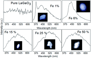
A polycrystalline sample of LaGa1−xFexO3 has been prepared by solid state reaction. The structural, optical and electronic structure of Fe doped LaGaO3 have been investigated. It is observed that with Fe doping the lattice parameter of the prepared samples systematically increases whereas the optical band gap decreases. First-principles density functional theory (DFT) calculations were carried out to understand the effect of Fe doping on the electronic structure and to understand the origin of systematic decrease in optical band gap. DFT studies suggests that there exists an indirect band gap to direct band gap transition with Fe doping which is further confirmed using photoluminescence measurements. Photoluminescence studies suggest that Fe doped LaGaO3 are potential candidates as white light emitting materials.
Introduction
The search for new materials for various electronic applications has dominated research worldwide particularly after the development of silicon based tiny transistors/integrated circuits. During the last five decades the nature of electronic devices has witnessed huge changes. Tiny as well as energy efficient electronic devices are now dominating research worldwide and replacing conventional electronic circuit elements. White light emitting diodes, spin valves and signal transmission using optical cables are examples.1 The design and development of direct band gap materials for light emitting diodes (LED), field emission display and florescent displays etc. are of great scientific and technological interest. In this connection the indium gallium arsenide (InGaAs) is one of the most successful candidates.2 One of the main problems associated with the InGaAs based structure is degradation due to oxidation (ageing effect).3–6 In this connection the direct band gap oxides have attracted much attention.7–11 The LaGaO3 is well known wide band gap perovskite oxide material having band gap close to 4.0 eV in its bulk form;12,13 further doped LaGaO3 has been widely explored for the phosphor/luminescence/field emission properties by doping of Yb, Eu, Ce, Cr, Sm, Tb, Tm etc. and found to be a potential candidate for the above mentioned luminescence applications.7,14–16 However systematic studies such as effect of doping concentration and type of dopants on structural properties, electronic band gap and photoluminescent properties of doped LaGaO3 may add new information in this regard. Despite its luminescence applications, LaGaO3 is one of the most widely studied materials for solid state fuel cells.17 The effect of Fe, Ni, Co, Mg, Sr etc. doping on the ionic conductivity of this material has been investigated.18–20 Recently Rai et al.21,22 have demonstrated magneto-dielectric effect and relatively high dielectric constant in Mn doped LaGaO3 at room temperature; whereas Natile et al.23 studied the gas sensing properties of Fe doped LaGaO3. Thus it appears that the ABO3 type perovskite gallates are potential candidates for various electronic applications. As discussed above the doped LaGaO3 is widely explored for the luminescence applications and it is observed that with different dopants it is possible to tune the emission properties of doped LaGaO3. At this juncture it is important to note that the end compound LaGaO3 and LaFeO3 and solid solution i.e. intermediate compound possesses octahedral symmetry around transition metal ion24 with space group Pnma.25–27 Thus the Fe doping in LaGaO3 expected to create minimum structural disorder and may be ideal candidate to study the effect of Fe doping on various properties. Further due to difference in the ionic radius of Ga3+ and Fe3+ it is expected that with Fe doping in LaGaO3 may lead to systematic variation in lattice parameters as observed in similar perovskites oxides28 and consequently variation in band gap.10 As LaGaO3 is already know to show the excellent luminescence properties7,14–16 hence it may be beneficial to investigate the photoluminescence properties of Fe doped LaGaO3. Keeping above all in view, here we aim to study the effect of Fe doping on the structural, optical, density functional theory (DFT) based first principles electronic and photoluminescence, properties of LaGa1−xFexO3.
It is observed that with Fe doping the volume of the unit cell systematically increases whereas the band gap decreases from 3.50 eV to 2.10 eV. DFT studies confirmed that Fe doing creates some impurity states (with respect to pure un-doped LaGaO3) in the band gap, which in turn reduces the band gap of the system. The spin polarized DFT calculations suggests that the antiferromagnetic is the ground state for Fe doped LaGaO3 samples, whereas ferromagnetic state is the ground state for LaFeO3, these calculations further suggests that there exist indirect band gap to direct band gap transition with Fe doping which is further confirmed using photoluminescence measurements. The photoluminescence studies suggest that the Fe doped LaGaO3 are the potential candidates as a white light emitting material.
Experimental
(i) Sample preparation
The polycrystalline samples of LaGa1−xFexO3 (0 < x < 1) were prepared by conventional solid-state reaction route28 with starting materials; La2O3 (99.99%), Ga2O3 (99.99%), and Fe2O3 (99.98%). These starting materials were mixed in stoichiometric amount and homogeneously mixed with propanol as a mixing medium. The resulting homogenous mixture was calcined in air ambient at 1000 °C, 1110 °C, and 1200 °C each time for 24 hours and final sintering was carried out at 1350 °C in air for 24 hours.
(ii) Structural characterizations
In order to examine the structural phase purity of the prepared samples the powder X-ray diffraction (XRD) experiments were carried out on Bruker D8 diffractometer equipped with Cu target. The obtain X-ray diffraction data is analyze using Fullprof-2k Rietveld refinement program.29
(iii) Optical band gap/diffuse reflectance measurements
The optical band gap of prepared samples has been measured using diffuse reflectivity measurements. These measurements have been performed in the 190 nm to 800 nm wavelength range using Cary-60 UV-VIS-NIR spectrophotometer having Harrick Video-Barrelino diffuse reflectance probe. The beam spot size on the sample was around 1.5 mm in diameter and an integral sphere detector is used for diffuse signal detection.
Electronic structure calculations. We have investigated the structural and electronic properties of pure and Fe-doped LaGaO3 systems by employing first-principles calculations to elucidate the effect of Fe doping in LaGaO3. We have used spin-polarized density functional theory (DFT) calculations as implemented in the Vienna Ab initio Simulation Package (VASP).30 Projected augmented wave (PAW) method31,32 is employed using an energy cut-off of 470 eV to describe the electronic wave functions. The local density approximation (LDA) + U method33 is used to account for the strong on-site correlated electrons in the partially filled d orbitals. We have employed correlation energy (U) of 4 eV and exchange energy (J) of 0 eV for Fe d-orbitals. These U and J values have been well tested and used in similar systems.34,35 40 (4 × 2 × 1) and 20 atoms (2 × 2 × 1) supercells are used to study pure and doped LaGaO3 systems to achieve different Fe-doping concentrations. Our optimized structural parameters are very much in agreement with the respective crystal structures. The Monkhorst–Pack36 generated set of 5 × 5 × 5 (for 40 atoms supercell) and 7 × 7 × 7 (for 20 atoms supercell) K-points are used to optimize the structures. The convergence criteria for energy and force are set at 10−6 eV and 10−3 eV Å−1, respectively. 7 × 7 × 7 (for 40 atoms supercell) and 15 × 15 × 15 (for 20 atoms supercell) Monkhorst–Pack36 generated K-points are used for the spin-polarized density of state calculations (DOS). Band structures of pure and Fe-doped LaGaO3 systems are calculated along the M → X → T → Γ → M path in Brillouin zone.
Photoluminescence measurements. Photoluminescence (PL) spectra were recorded using Dongwoo Optron DM 500i, having He–Cd laser as an excitation source consisting of a continuous wave of wavelength, 325 nm and a PMT detector. At the sample surface the laser makes circular spot having diameter of ∼0.5 mm with total power at ∼15 mW.
Results and discussions
(I) Structural analysis
Fig. 1a shows the representative refined powder XRD LaGa0.8Fe0.2O3 refined considering orthorhombic structure with Pnma space group.25–27 The diffraction data for all the prepared samples and the details of refinements such as; goodness of fitting, Bragg R-factor, RF-factor and χ2 which defines the quality of fitting are provided in the ESI.† In order to get the clear view about the Ga/Fe–O bond length and Ga/Fe–O–Ga/Fe bond angle the representative structure is provided in the inset of Fig. 1. The variation of lattice parameters due to Fe doping is shown in Fig. 2. The systematic increase in the value of lattice constant is due to the difference in the ionic radius of Ga3+ (0.62) and Fe3+ i.e. (0.645).37
![image file: c6ra21693e-f1.tif](/image/article/2016/RA/c6ra21693e/c6ra21693e-f1.gif) |
| Fig. 1 Representative powder X-ray diffraction pattern for the prepared samples the inset shows the view of the refined crystal structure. Various band angles and bond length are visible in the given crystal structure. | |
![image file: c6ra21693e-f2.tif](/image/article/2016/RA/c6ra21693e/c6ra21693e-f2.gif) |
| Fig. 2 Variation of lattice parameter as a function of Fe doping. The errors in the values of lattice parameters are comparable to that of size of the symbols used. | |
The values of the structural parameters obtained through Rietveld refinement are given in Table 1.
Table 1 Refined values of Wyckoff positions and lattice parameters for LaGa1−xFexO3
Fe% (X) |
La (4c) (x, y, z) |
Ga/Fe (4a) (x, y, z) |
O1 (8d) (x, y, z) |
O2 (4c) (x, y, z) |
Lattice parameter a (Å) |
Lattice parameter b (Å) |
Lattice parameter c (Å) |
0 |
0.01260, 1/4, −0.00426 |
0.5, 0, 0 |
0.25909, 0.05259, 0.28016 |
0.50364, 1/4, −0.05581 |
5.4894(1) |
7.7686(2) |
5.5209(1) |
0.5 |
0.01733, 1/4, 0.00355 |
0.5, 0, 0 |
0.27746, 0.03310, 0.27289 |
0.49746, 1/4, −0.07123 |
5.4922(3) |
7.7741(3) |
5.5239(8) |
1 |
0.01720, 1/4, 0.00331 |
0.5, 0, 0 |
0.28145, 0.03427, 0.27252 |
0.50171, 1/4, −0.06808 |
5.4928(4) |
7.7749(3) |
5.5244(1) |
1.5 |
0.01729, 1/4, 0.00363 |
0.5, 0, 0 |
0.27634, 0.03329, 0.27380 |
0.49945, 1/4, −0.06779 |
5.4932(7) |
7.7757(3) |
5.5246(3) |
2 |
0.01683, 1/4, 0.00311 |
0.5, 0, 0 |
0.28270, 0.03062, 0.26220 |
0.50113, 1/4, −0.07907 |
5.4942(6) |
7.7769(8) |
5.5261(1) |
4 |
0.01750, 1/4, 0.00345 |
0.5, 0, 0 |
0.28105, 0.03232, 0.27463 |
0.50328, 1/4, −0.07037 |
5.4948(7) |
7.7777(8) |
5.5256(3) |
6 |
0.01792, 1/4, 0.00433 |
0.5, 0, 0 |
0.28290, 0.03237, 0.27114 |
0.49938, 1/4, −0.07137 |
5.4959(1) |
7.7794(1) |
5.5259(4) |
8 |
0.01795, 1/4, 0.00396 |
0.5, 0, 0 |
0.28317, 0.03139, 0.27135 |
0.49878, 1/4, −0.07578 |
5.4970(5) |
7.7811(1) |
5.5264(7) |
10 |
0.01821, 1/4, 0.00336 |
0.5, 0, 0 |
0.27811, 0.03201, 0.27163 |
0.49945, 1/4, −0.07327 |
5.4987(3) |
7.7834(2) |
5.5274(5) |
15 |
0.01948, 1/4, 0.00403 |
0.5, 0, 0 |
0.28191, 0.03442, 0.27018 |
0.49857, 1/4, −0.07106 |
5.5020(8) |
7.7882(3) |
5.5291(9) |
20 |
0.01998, 1/4, 0.00516 |
0.5, 0, 0 |
0.28181, 0.03271, 0.27479 |
0.49825, 1/4, −0.06943 |
5.5059(7) |
7.7933(8) |
5.5311(7) |
25 |
0.02073, 1/4, 0.00428 |
0.5, 0, 0 |
0.27816, 0.03315, 0.27234 |
0.50019, 1/4, −0.07590 |
5.5093(1) |
7.7977(1) |
5.5325(2) |
30 |
0.02134, 1/4, 0.00501 |
0.5, 0, 0 |
0.28441, 0.02984, 0.26947 |
0.50265, 1/4, −0.07969 |
5.5135(4) |
7.8028(7) |
5.5346(5) |
40 |
0.02275, 1/4, 0.00406 |
0.5, 0, 0 |
0.28307, 0.02864, 0.26709 |
0.50048, 1/4, −0.08929 |
5.5216(7) |
7.8121(8) |
5.5375(7) |
50 |
0.02411, 1/4, 0.00522 |
0.5, 0, 0 |
0.27331, 0.03153, 0.28382 |
0.49878, 1/4, −0.08100 |
5.5296(8) |
7.8204(5) |
5.5411(6) |
60 |
0.02545, 1/4, 0.00574 |
0.5, 0, 0 |
0.27356, 0.03368, 0.28610 |
0.49421, 1/4, −0.08003 |
5.5377(9) |
7.8282(7) |
5.5450(1) |
70 |
0.02613, 1/4, 0.00565 |
0.5, 0, 0 |
0.28026, 0.03300, 0.28390 |
0.49315, 1/4, −0.07947 |
5.5442(2) |
7.8347(7) |
5.5485(5) |
80 |
0.02719, 1/4, 0.00549 |
0.5, 0, 0 |
0.27847, 0.03412, 0.28326 |
0.48996, 1/4, −0.07383 |
5.5522(7) |
7.8424(3) |
5.5513(8) |
90 |
0.02826, 1/4, 0.00612 |
0.5, 0, 0 |
0.28078, 0.03504, 0.28816 |
0.49206, 1/4, −0.07442 |
5.5584(8) |
7.8482(8) |
5.5531(2) |
100 |
0.02963, 1/4, 0.00492 |
0.5, 0, 0 |
0.27253, 0.04029, 0.28596 |
0.49386, 1/4, −0.07152 |
5.5654(8) |
7.8539(3) |
5.5545(3) |
From the Table 1 it is clear that with Fe doping the x, y, z coordinates i.e. atomic position of oxygen and lanthanum atoms significantly changes and this lead to change in the orbital overlap (bond length) between oxygen–lanthanum and oxygen–Ga/Fe orbitals. The variation of Ga–O/Fe–O bond length and Ga/Fe–O–Ga/Fe bond angle as a function of Fe doping is shown in Fig. 3.
![image file: c6ra21693e-f3.tif](/image/article/2016/RA/c6ra21693e/c6ra21693e-f3.gif) |
| Fig. 3 (a) Variation of Ga–O/Fe–O bond length as a function of Fe doping. (b) Variation of Ga/Fe–O–Ga/Fe bond angle as a function of Fe. | |
From the Fig. 3a and b it is clear that this bond length initially increases with doping and then saturates at x = 0.4%, whereas the bond angle shows the dip at x = 0.4. It should be noted that as the scattering factor for oxygen being low Z element is small hence the Ga/Fe–O bond length and Ga/Fe–O–Ga/Fe bond angle may have errors slightly higher than that of obtain from refinements. Neutron diffraction measurements will be more useful in this regard.
(II) Optical band gap calculation and its relation with structure
In order to estimate the optical band gap the diffuse reflectance spectra has been converted to equivalent absorption spectra using Kubelka–Munk equation.10 |
![image file: c6ra21693e-t1.tif](/image/article/2016/RA/c6ra21693e/c6ra21693e-t1.gif) | (1) |
R∞ = Rsample/Rstandard. Rsample is the diffuse reflectance of the sample and Rstandard is that of the standard (BaSO4 in present case). K and S are the Kubelka–Munk absorption and scattering functions, respectively. As we have used powder samples for diffuse reflectance measurements hence the assumption of reflected light scatters in a perfectly diffuse manner holds true, for such case the scattering function S is nearly constant with wavelength10 and the Kubelka–Munk function can be related/proportional to the absorption coefficient (α) as |
![image file: c6ra21693e-t2.tif](/image/article/2016/RA/c6ra21693e/c6ra21693e-t2.gif) | (2) |
here n has the value of 2 for direct bandgap transitions, while n is equal to 1/2 for an indirect transition. Thus, a plot between [F(R∞) × hν]n versus hν yields a straight line and the intercept on the energy axis gives the value of the bandgap. We have used the values of n = 2 to determine the optical gap of LaGa1−xFexO3 as the doped LaGaO3 is known to demonstrate excellent photoluminescence properties (which is a possible signature of direct band gap material)38–45 which is further supported by our DFT studies. Fig. 4a shows a plot between [F(R∞) × hν]2 and hν for various LaGa1−xFexO3. It is clear that with increase in the doping of Fe in LaGaO3 the intensity feature at E ∼ 3.30 eV and E ∼ 2.65 eV become more prominent. It should be noted that the X-ray diffraction data confirms the phase purity of the prepared samples, hence; the observed extra intensity feature appears to be intrinsic property of the Fe doped LaGaO3. The variations of direct band gap for the prepared samples as function of Fe doping (experimentally observed and obtain from DFT calculations) are shown in Fig. 5.
![image file: c6ra21693e-f4.tif](/image/article/2016/RA/c6ra21693e/c6ra21693e-f4.gif) |
| Fig. 4 A plot between [F(R∞) × hν]2 and hν for various LaGa1−xFexO3 the emergence of new peaks at lower energy level are indicated by arrows. | |
![image file: c6ra21693e-f5.tif](/image/article/2016/RA/c6ra21693e/c6ra21693e-f5.gif) |
| Fig. 5 Variation of direct band gap as function of Fe doping. | |
In the case of perovskite oxides it is well known that the effective geometrical band width (W) critically controls the energy band gap46 given by Eg = U−W;
;
, Δ(O–Ga/Fe–O) is the band angle and U is charge transfer parameter whose value almost remain constant for given series.46 We have estimated band width and plotted the same in Fig. 6.
![image file: c6ra21693e-f6.tif](/image/article/2016/RA/c6ra21693e/c6ra21693e-f6.gif) |
| Fig. 6 Variation of W (band width) as a function of Fe. | |
It is important to note that the variation of optical band gap (with Fe doping) and that of calculated band width (W) is very similar both decreases up to 0.4 and almost saturates. Thus it appears that the Ga/Fe–O bond length and O–Ga/Fe–O bond angle effectively controls the optical band gap in the prepared samples.
(III) spin polarized DFT calculations
Fig. 7a shows the structural, electronic and magnetic properties of pure LaGaO3. We find that the pure LaGaO3 has a wide band gap of 3.59 eV, which is quite in agreement with the experimental report.47,48 Our band structure shows that it has an indirect band gap of 3.59 eV along the M → X symmetry points in Brillouin zone.49 Further, we have studied 12.5%, 25%, 50% and 100% Fe-doped LaGaO3 systems to compare our data with experimental findings. In all these cases, Fe-doping is done at the Ga site and the most stable structure is considered for electronic and magnetic studies.
![image file: c6ra21693e-f7.tif](/image/article/2016/RA/c6ra21693e/c6ra21693e-f7.gif) |
| Fig. 7 The structural, electronic and magnetic properties of (a) LaGaO3 (b) 12.5% Fe-doped LaGaO3, (c) 25% Fe-doped LaGaO3, (d) 50% Fe-doped LaGaO3, and (e) LaFeO3. Here, blue, green, yellow and red color balls denote La, Ga, Fe and O atoms, respectively. The Fermi level is set to zero. | |
Interestingly, our electronic structures (Fig. 7) show that the band gap of LaGaO3 reduces as we increase the Fe-doping concentration and the band gap is lowest (2.10 eV) in pure LaFeO3, this is very much consistence with experimental findings.50 Further, our band structure plots show that all these Fe-doped systems have a direct band gap, which is exactly opposite to pure LaGaO3. The partial density of states (PDOS) plots show that Fe impurity states appearing in the band gap and thus the band gap reduces with Fe-doping concentrations. This could be more due to higher energy of Fe-3d states than Ga 3d states as Ga is more electronegative than Fe. Further, we have investigated the magnetic properties of these systems using spin polarized calculations. We find that the pure LaGaO3 system is a non-magnetic one, whereas Fe-doped LaGaO3 systems are magnetic. The total magnetic moment calculated to be 5 μB per Fe which is due to the +3 oxidation state of Fe (3d5). We have also investigated the magnetic ground state51 for all these systems and we find that the anti-ferromagnetic (AFM) state is the magnetic ground state for Fe-doped systems, whereas ferromagnetic is the ground state for LaFeO3 this is very much consistent with experimental findings.18
(IV) Analysis of photoluminescence spectra
As clear from optical absorption and DFT studies the Fe doped LaGaO3 are direct band gap material and the band gap of these samples varies systematically with Fe doping (up to x = 0.4). Hence in order to investigate the possible applicability of the prepared samples for light emitting application; the photoluminescence studies were carried out. At this juncture it is important to note that the ideal high quality indirect band gap materials do not produce strong photoluminescence52 hence; the occurrence of strong photoluminescence may be treated as a possible experimental proof of direct band gap material and the same is experimentally confirmed earlier by various research groups.38–45 At this juncture it is important to note that the indirect band gap materials know to produce PL due to presence of defect states,53 hence the occurrence of PL may be only the possible signature but not be the ultimate tool to comment on the nature of the band gap. The angle resolved photoemission spectroscopy and inverse photoemission spectroscopic studies54 on single crystalline samples may be terms as direct experimental tool to comment on the nature of the band gap. The mapping of band-structure using angle resolved photoemission spectroscopy and inverse photoemission spectroscopic is beyond the scope for present set of samples (due to polycrystalline nature of these samples). Fig. 8a shows the photoluminescence spectra of the studied samples, the inset of each graph shows the corresponding luminescence due to ultraviolet He–Cd laser. It is observed that the pure LaGaO3 do not show any luminescence/photoluminescence in the wavelength region corresponding to its band gap (this may be due to the indirect nature of band gap for pure LaGaO3 as clear form DFT studies and present PL measurements), whereas for Fe doped samples; we could detect the PL spectra, the observation of PL form Fe doped samples may be the possible signature of the direct band gap nature of the Fe doped LaGaO3 samples, which is also evident from the first principle calculation studies. These samples also show the broadening similar to that of observed in polycrystalline as prepared ZnO samples55,56 and the observation of this type of broadening in PL spectra is attributed to the deep level emission from defects states.55,56 It is very important here to notice that all the samples reported here are prepared by solid state reaction route (same procedure is followed to prepare all these samples) and hence may have similar level of defect density. Hence the luminescence observed in Fe doped samples may not be due to defects but due to the direct nature of its band gap (as the same is also evident form first principle studies shown in Fig. 7). The presence of such defects if any may contribute to the broadening of the PL spectra.52,55,56 The observation of white light luminescence in other words broadening of PL spectra for the present set of samples may also be understood as follows; in the case of transition metal doped materials the splitting of 3d energy states due to the electrical field induced by neighboring ions (crystal field splitting) results in extra emission lines this is well known Tanabe–Sugano effect57–63 (Fig. 8b). It is important to note that for transition metal doped samples such broadening is well known64–74 and explained on the basis of above mentioned Tanabe–Sugano effect.57–63 We believe that in present case the observed broadening is due to Tanabe–Sugano effect (as Fe3+ ions is surrounded by six oxygen anions in octahedral cage) plus possible broadening due to deep level emission by defect levels within the band gap.52,54,55 Further it is observed that the area under the curve shows increasing trend with Fe doping; but as the photoluminescence intensity is very sensitive to surface roughness and the present measurements were carried out on as prepared polycrystalline samples (the sample surface is very rough with respect to wave length of laser and further different samples are having different surface roughness) hence we are not showing the variation of area under the curve as function of Fe concentration in the present manuscript. We are of the opinion that the experiments on high quality single crystalline samples or epitaxial thin film (low defect density low surface roughness) will be more useful for these samples, further the single crystalline samples will be useful to map the experimental band structure of these samples using angle resolved photoemission spectroscopy.54 Here we would like to convey that the present studies clearly demonstrate the possible application of the prepared polycrystalline samples of Fe doped LaGaO3 as a graded violet to pure white light emitting material.
![image file: c6ra21693e-f8.tif](/image/article/2016/RA/c6ra21693e/c6ra21693e-f8.gif) |
| Fig. 8 (a) Photoluminescence spectra for Fe doped LaGaO3 samples. The corresponding color luminescence due to He–Cd laser is shown in the inset. (b) Proposed schematic of the photoluminescence spectra for Fe doped LaGaO3. The 4G state is split by the crystal field into the ground state 6A1(S) and the excited states 4T1(G), 4T2(G) and 4E(G), the transitions between these states as indicated by the arrows describes the observed PL spectra. | |
Conclusion
Effect of Fe doping on Ga site in LaGaO3 is investigated on the structural, optical and electronic properties. It is observed that the optical band gap of the prepared samples is controlled by the orbital overlap between Ga/Fe–O overlap i.e. effective band width (W). The first principle studies suggest that with Fe doping the extra states (with respect to pure LaGaO3) appears which reduces the effective band gap. Further the spin polarized DFT analysis suggests that the low value of Fe doping the sample favours antiferromagnetic states where as pure LaFeO3 favours ferromagnetic state. Further there exist indirect band gap to direct band gap transition with Fe doping which is further confirmed using photoluminescence measurements. Further the experiments on high quality single crystalline samples or epitaxial thin film having low defect density and smooth surface will be useful to understand the origin and deeper insight on the photoluminescence properties of these samples.
Acknowledgements
Authors sincerely thank Prof. P. Mathur Director IIT Indore for his encouragement. SIC IIT Indore is acknowledged for providing various experimental facilities. Dr Vipul Singh is acknowledged for kindly extending the access to photoluminescence facilities. CSIR New Delhi is acknowledged for funding the high temperature furnace under the project 03(1274)/13/EMR-II. Mr Preetam Singh is thankful to Mr Shailendra K. Saxena, Mr Kamal Warshi, Mr Tajendra Dixit and Mrs Pryanka of IIT Indore for their help at various stages of this work. Indrani, Hari Mohan, Vikash would like to thank MHRD Govt. of India, for financial support for their Ph.D. fellowship.
References
- Future Trends in Microelectronics: Frontiers and Innovations, ed. A. Zaslavsky and S. Luryi, John Wiley & Sons – IEEE Press, 2013, vol. 7 Search PubMed.
- S. Adachi, Properties of semiconductor alloys: group-IV, III-V and II-VI semiconductors, John Wiley & Sons, 2009, vol. 28 Search PubMed.
- R. E. Nahory, M. A. Pollack, W. D. Johnston Jr and R. L. Barns, Appl. Phys. Lett., 1978, 33, 659–661 CrossRef CAS.
- M. Okayasu, M. Fukuda, T. Takeshita, S. Uehara and K. Kurumada, J. Appl. Phys., 1991, 69, 8346–8351 CrossRef CAS.
- M. Fukuda, M. Okayasu, J. Temmyo and J. Nakand, IEEE J. Quantum Electron., 1994, 30, 471–476 CrossRef CAS.
- D. A. Allwood, R. T. Carline, N. J. Mason, C. Pickering, B. K. Tanner and P. J. Walker, Thin Solid Films, 2000, 364, 33–39 CrossRef CAS.
- X. Liu and J. Lin, J. Mater. Chem., 2007, 18, 221–228 RSC.
- G. Li, D. Geng, M. Shang, C. Peng, Z. Cheng and J. Lin, J. Mater. Chem., 2011, 21, 13334–13344 RSC.
- D. Geng, G. Li, M. Shang, D. Yang, Y. Zhang, Z. Cheng and J. Lin, J. Mater. Chem., 2012, 22, 14262–14271 RSC.
- S. D. Singh, V. Nandanwar, H. Srivastava, A. K. Yadav, A. Bhakar, P. R. Sagdeo, A. K. Sinha and T. Ganguli, Dalton Trans., 2015, 44, 14793–14798 RSC.
- S. Lee, R. D. Levi, W. Qu, S. C. Lee and C. A. Randall, J. Appl. Phys., 2010, 107, 023523 CrossRef.
- Y. Shimizu, Y. Takano and K. Ueda, Thin Solid Films, 2014, 559, 23–26 CrossRef CAS.
- K. Ogisu, A. Ishikawa, Y. Shimodaira, T. Takata, H. Kobayashi and K. Domen, J. Phys. Chem. C, 2008, 112(31), 11978–11984 CAS.
- V. M. Orera, L. E. Trinkler, R. I. Merino and A. Larrea, J. Phys.: Condens. Matter, 1995, 7, 9657–9673 CrossRef CAS.
- H. L. Li, H. L. W. Chan and J. H. Hao, J. Phys. D: Appl. Phys., 2009, 42, 185103 CrossRef.
- H. Kyoung Yang, B. Kee Moon, B. Chun Choi, J. Hyun Jeong, J. Hwan Kim and K. Ho Kim, Solid State Sci., 2012, 14, 236–240 CrossRef.
- M. Morales, J. J. Roa, J. Tartaj and M. Segarra, J. Eur. Ceram. Soc., 2016, 36, 1–16 CrossRef CAS.
- T. Ishihara, T. Yamada, H. Arikawa, H. Nishiguchi and Y. Takita, Solid State Ionics, 2000, 135, 631–636 CrossRef CAS.
- T. Funahashi, A. Mineshige, H. Yoshioka, K. Kobayashi, Y. Matsushita, Y. Katsuya, M. Tanaka, O. Sakata and T. Yazawa, Solid State Ionics, 2016, 289, 106–112 CrossRef CAS.
- M. Kajitani, M. Matsuda and M. Miyake, Solid State Ionics, 2007, 178, 355–358 CrossRef CAS.
- R. H. Mohan, R. Late, S. K. Saxena, V. Mishra, R. Kumar, P. R. Sagdeo and A. Sagdeo, Mater. Res. Express, 2015, 2, 096105 CrossRef.
- R. H. Mohan, S. K. Saxena, R. Late, V. Mishra, P. Rajput, A. Sagdeo, R. Kumar and P. R. Sagdeo, RSC Adv., 2016, 6, 26621–26629 RSC.
- M. M. Natile, A. Ponzoni, I. Concina and A. Glesenti, Chem. Mater., 2014, 26, 1505–1513 CrossRef CAS.
- L. Sara, G. Subías, J. García, S. Di Matteo, J. Blasco, V. Cuartero and C. R. Natoli, J. Phys.: Condens. Matter, 2011, 23, 325601 CrossRef PubMed.
- L. Vasylechko, A. Matkovski, A. Suchocki, D. Savytskii and I. Syvorotka, J. Alloys Compd., 1999, 286, 213–218 CrossRef CAS.
- K. Mori, T. Fukunaga, K. Shibata, K. Iwase, S. Harjo, A. Hoshikawa, K. Itoh, T. Kamiyama and T. Ishigaki, Phys. B, 2004, 352, 147–155 CrossRef CAS.
- M. Marezio and P. D. Dernier, Mater. Res. Bull., 1971, 6, 23–29 CrossRef CAS.
- P. R. Sagdeo, S. Anwar and N. P. Lalla, Powder Diffr., 2006, 21, 40–44 CrossRef CAS.
- D. B. T. Wiles and R. A. Young, J. Appl. Crystallogr., 1981, 14, 149–151 CrossRef CAS.
- K. Georg and J. Furthmüller, Phys. Rev. B: Condens. Matter Mater. Phys., 1996, 54, 11169 CrossRef.
- J. P. Perdew, J. A. Chevary, S. H. Vosko, K. A. Jackson, M. R. Pederson, D. J. Singh and C. Fiolhais, Phys. Rev. B: Condens. Matter Mater. Phys., 1992, 46, 6671 CrossRef CAS.
- V. I. Anisimov, F. Aryasetiawan and A. I. Lichtenstein, J. Phys.: Condens. Matter, 1997, 9, 767 CrossRef CAS.
- B. Himmetoglu, A. Floris, S. Gironcoli and M. Cococcioni, Int. J. Quantum Chem., 2014, 114, 14–49 CrossRef CAS.
- K. Sato, L. Bergqvist, J. Kudrnovský, P. H. Dederichs, O. Eriksson, I. Turek, B. Sanyal, G. Bouzerar, H. Katayama-Yoshida, V. A. Dinh and T. Fukushima, Rev. Mod. Phys., 2010, 82, 1633 CrossRef CAS.
- T. Mayeshiba and D. Morgan, Phys. Chem. Chem. Phys., 2015, 17, 2715–2721 RSC.
- H. J. Monkhorst and J. D. Pack, Phys. Rev. B: Solid State, 1976, 13, 5188 CrossRef.
- R. D. T. Shannon, Acta Crystallogr., Sect. A: Cryst. Phys., Diffr., Theor. Gen. Crystallogr., 1976, 32, 751–767 CrossRef.
- V. M. Orera, L. E. Trinkler, R. I. Merino and A. Larrea, J. Phys.: Condens. Matter, 1995, 7, 9657 CrossRef CAS.
- H. L. Li, H. L. W. Chan and J. H. Hao, J. Phys. D: Appl. Phys., 2009, 42, 185103 CrossRef.
- H. K. Yang, B. K. Moon, B. C. Choi, J. H. Jeong, J. H. Kim and K. H. Kim, Solid State Sci., 2012, 14, 236–240 CrossRef CAS.
- A. Splendiani, L. Sun, Y. Zhang, T. Li, J. Kim, C.-Y. Chim, G. Galli and F. Wang, Nano Lett., 2010, 10, 1271–1275 CrossRef CAS PubMed.
- K. Watanabe, T. Taniguchi and H. Kanda, Nat. Mater., 2004, 3, 404–409 CrossRef CAS PubMed.
- H. Zeng and X. Cui, Chem. Soc. Rev., 2015, 44, 2629–2642 RSC.
- K. F. Mak, C. Lee, J. Hone, J. Shan and T. F. Heinz, Phys. Rev. Lett., 2010, 105, 136805 CrossRef PubMed.
- G.-B. Liu, D. Xiao, Y. Yao, X. Xu and W. Yao, Chem. Soc. Rev., 2015, 44, 2643–2663 RSC.
- M. Medarde, J. Mesot, P. Lacorre, S. Rosenkranz, P. Fischer and K. Gobrecht, Phys. Rev. B: Condens. Matter Mater. Phys., 1995, 52(13), 9248 CrossRef CAS.
- T. D. K. Wungu, M. Sakaue, S. M. Aspera, T. L. P. Thuy, M. Alaydrus, H. Kasai and T. Ishihara, ECS Trans., 2013, 57, 2715–2722 CrossRef.
- K. E. Babu, N. Murali, K. V. Babu, P. T. Shibeshi and V. Veeraiah, AIP Conf. Proc., 2014, 1620(1), 173–178 CrossRef CAS.
- I. Choudhuri, S. Kumar, A. Mahata, K. S. Rawat and B. Pathak, Nanoscale, 2016, 8, 14117–14126 RSC.
- M. D. Scafetta, A. M. Cordi, J. M. Rondinelli and S. J. May, J. Phys.: Condens. Matter, 2014, 26, 505502 CrossRef PubMed.
- Y.-L. Lee, M. J. Gadre, S.-H. Yang and D. Morgan, Phys. Chem. Chem. Phys., 2015, 17, 21643 RSC.
- M. Fox, Optical properties of solids, Oxford university press, 2010 Search PubMed.
- D. Kan, T. Terashima, R. Kanda, A. Masuno, K. Tanaka, S. Chu, H. Kan, A. Ishizumi, Y. Kanemitsu, Y. Shimakawa and M. Takano, Nat. Mater., 2005, 4, 816–819 CrossRef CAS.
- Y. Zhang, T. R. Chang, B. Zhou, Y. T. Cui, H. Yan, Z. Liu, F. Schmitt, J. Lee, R. Moore, Y. Chen and H. Lin, Nat. Nanotechnol., 2014, 9, 111–115 CrossRef CAS PubMed.
- A. Kumar, T. Dixit, I. A. Palani, P. R. Sagdeo and V. Singh, Int. J. Appl. Ceram. Technol., 2016, 13, 912–919 CrossRef CAS.
- A. B. Djurišić and Y. H. Leung, Small, 2006, 2, 944 CrossRef PubMed.
- Y. Tanabe and S. Sugano, J. Phys. Soc. Jpn., 1954, 9, 753–766 CrossRef CAS.
- Y. Tanabe and S. Sugano, J. Phys. Soc. Jpn., 1954, 9, 766–779 CrossRef CAS.
- Y. Tanabe and H. Kamimura, J. Phys. Soc. Jpn., 1958, 13, 394–411 CrossRef CAS.
- S. Sugano and Y. Tanabe, J. Phys. Soc. Jpn., 1958, 13, 880–899 CrossRef CAS.
- S. Sugano and I. Tsujikawa, J. Phys. Soc. Jpn., 1958, 13, 899–910 CrossRef CAS.
- G. T. Pott and W. H. J. Stork, Catal. Rev.: Sci. Eng., 1975, 12(1), 163–199 CAS.
- A. Zunger, Annu. Rev. Mater. Sci., 1985, 15, 411–453 CrossRef CAS.
- S. B. Singh, M. V. Limaye, S. K. Date, S. Gokhale and S. K. Kulkarni, Phys. Rev. B: Condens. Matter Mater. Phys., 2009, 80, 235421 CrossRef.
- D. M. Sherman and T. David Waite, Am. Mineral., 1985, 70, 1262–1269 CAS.
- J. J. Krebs and W. G. Maisch, Phys. Rev. B: Solid State, 1971, 4, 757 CrossRef.
- S. S. Pedro, O. Nakamura, R. B. Barthem and L. P. Sosman, J. Fluoresc., 2009, 19, 211–219 CrossRef CAS PubMed.
- N. Pathak, S. K. Gupta, K. Sanyal, M. Kumar, R. M. Kadam and V. Natarajan, Dalton Trans., 2014, 43, 9313–9323 RSC.
- L. P. Sosman, T. Abritta, M. R. Amaral, N. Cella and H. Vargas, Solid State Commun., 1998, 105, 135–138 CrossRef CAS.
- E. Malguth, A. Hoffmann and M. R. Phillips, Phys. Status Solidi B, 2008, 245, 455–480 CrossRef CAS.
- A. Trueba, J. M. García-Lastra, P. Garcia-Fernandez, J. A. Aramburu, M. T. Barriuso and M. Moreno, J. Phys. Chem. A, 2011, 115(46), 13399–13406 CrossRef CAS PubMed.
- J. M. García-Lastra, P. García-Fernández, M. T. Barriuso, J. A. Aramburu and M. Moreno, J. Phys. Chem. A, 2014, 118(12), 2377–2384 CrossRef PubMed.
- S. Tamilarasan, S. Laha, S. Natarajan and J. Gopalakrishnan, J. Phys. Chem. C, 2015, 3, 4794–4800 CAS.
- M. A. Marzouk, S. M. Abo-Naf, H. A. Zayed and N. S. Hassan, J. Mater. Res. Technol., 2016, 5, 226–233 CrossRef CAS.
Footnote |
† Electronic supplementary information (ESI) available. See DOI: 10.1039/c6ra21693e |
|
This journal is © The Royal Society of Chemistry 2016 |
Click here to see how this site uses Cookies. View our privacy policy here.