
Sustainable waste mitigation: biotemplated nanostructured ZnO for photocatalytic water treatment via extraction of biofuels from hydrothermal carbonization of banana stalk†
Ayush Upnejaab,
Guolan Doubc,
Chitanya Gopub,
Carol A. Johnsonbd,
Anna Newmand,
Azat Suleimenovd and
Jillian L. Goldfarb*bd
aNewton South High School, 140 Brandeis Road, Newton Centre, MA 02459, USA
bDepartment of Mechanical Engineering, Boston University, 110 Cummington Mall, Boston, MA 02215, USA. E-mail: jilliang@bu.edu; JillianLGoldfarb@gmail.com; Tel: +1 617 353 3883
cSchool of Safety Engineering, China University of Mining &Technology, Xuzhou 221116, China
dDivision of Materials Science & Engineering, Boston University, 15 St. Mary's Street, Brookline, MA 02446, USA
First published on 19th September 2016
Abstract
Working at the food-energy-water nexus, we identified an abundant agricultural waste with little intrinsic value – banana stalk – that can be converted to biofuels via hydrothermal carbonization. However, to make biomass to biofuel conversions economically viable, we must identify value-added products to add to the biorefinery, such as materials for water treatment. Both the resulting hydrochar and raw biomass were biotemplated with zinc acetate to yield ZnO nanostructures with surface areas ∼20 m2 g−1. The zincite particles were able to degrade a model organic dye (methylene blue) from aqueous solution. Though both the raw biomass and hydrochar yielded particles with similar surface areas, the photocatalytic activity of the raw templated particles was superior. We believe this is due to a higher concentration of calcium (naturally present in the banana biomass) in the hydrochar relative to the raw biomass. The calcium produced calcite crystals in the biotemplated materials, which may hinder the ZnO photocatalytic activity. However, the CaO may be useful for removing heavy metals from water and catalyzing biofuel production.
1. Introduction
The inextricable nature of food, energy and water, combined with a growing global population that increasingly taxes these synergistic systems, requires innovative approaches that avoid stressing one system while advancing another. For example, cultivating bioenergy crops with irrigated land that could otherwise be used to grow food increases water withdrawals and may place food supplies at risk.1 Green chemistry and engineering movements call on innovators to reduce hazardous material inputs and outputs as well as process energy requirements whether producing fine chemicals or manufacturing automobiles.2 In an effort to improve the sustainability of nanomaterials production, recent years have brought substantial progress in using biological templates to create “sustainable” nanomaterials without the use of harsh solvents and processing conditions.3 Biotemplates yield nanomaterials with complex structures and high reactive surface areas, and when they are sourced from waste materials, may do so in a more resource-conserving manner than solution-based fabrication techniques.4,5There are myriad examples in the literature demonstrating procedures for biotemplating nanomaterials and identifying novel templating materials, including biotemplated ZnO on a variety of biomass precursors. For example, Taubert et al. fabricated zincite (ZnO) particles using starch as the crystallization controlling agent.6 They demonstrated the ability to control the morphology of the zincite particles by changing the concentration of the starch in the reactor. Dong et al. used eggshell membrane as template to produce ZnO interwoven nanofiber.7 Other researchers have successfully created ZnO nanoparticles using butterfly wing scales,8 luffa sponges,9,10 and spherobacterium.11 In this paper, we focus on improving the sustainability of zinc oxide fabrication by integrating it with a biomass waste-to-energy conversion using a biomass that would otherwise be considered agricultural waste.
Many studies have shown the potential to use zinc oxide to photocatalytically degrade harmful pollutants, such as commercial dyes, that hamper wastewater treatment.12 Hariharan tested the photocatalytic activity of bulk ZnO, ZnO nanoparticles, and the conventionally used photocatalyst Degussa TiO2. Their study found that ZnO nanoparticles are comparable or better to both TiO2 and bulk ZnO as a catalytic system.13 Chen et al. demonstrated that zinc oxide nanorods adhered to the surface of a spinning disc were able to degrade 40% of methylene orange in 40 minutes.14 Similarly, ZnO has been shown to degrade azo dye acid red (AR14), a byproduct of textile manufacturing.15 At a neutral pH and optimal concentrations of dye and catalyst, the dye can be completely removed by ZnO in approximately 1 hour. Shen et al. compared the effectiveness of ZnO in degrading methylene blue when templated on both starch gel and a silica nanoparticle surface. They found that the ZnO deposited on the silica nanoparticle was more effective, degrading 90% of methylene blue in 60 minutes, compared to 20% degradation from the ZnO on starch surface.16 Furthermore, hierarchically structured metal oxides could treat water in ways beyond photocatalytic degradation of dissolved compounds. For example, ZnO biotemplated from eggshell membrane demonstrated high adsorption capacity for gold nanoparticles, contaminants of emerging concern as nanomaterials are increasingly incorporated into consumer products.17
In addition to water treatment, ZnO nanomaterials have exhibited photocatalytic antifungal and antibacterial activity as a result of their ability to mediate reactive oxygen species.18,19 The sensing applications proposed for ZnO nanomaterials are numerous, including for Schottky ultraviolet photodetectors owing to ZnO's wide bandgap.20 Fatemi et al. and Zhou et al. demonstrated the use of biomorphic ZnO nanostructures for glucose biosensing,21,22 and Zhao et al. for enzymatic sensing.23 Others have shown the potential for porous ZnO materials to serve as gas sensors for H2S,24 NH3, CO and H2,25 and acetone.26 Biotemplated ZnO also shows promise for aqueous sensing applications, such as acetic acid.27 Beyond such sensing and environmental remediation applications, ZnO nanomaterials show promise for use in solar energy conversion,28,29 as semiconductor materials,30 and as transparent ferromagnets,31 among many other applications.
Identifying sustainable materials to produce such useful nanomaterials is of paramount importance as we transition to a green economy.32 By using nonhazardous materials (biomass) that do not compete for arable land for food supply to biotemplate ZnO nanoparticles, and integrating biofuel production into the waste recovery process, we can strive towards meeting the principles of green engineering.2 One such potential biomass source is the banana plant. In 2013, the global banana fruit production topped 100 million tons, with India as the leading producer (over one quarter of worldwide production).33 The U.S. produced approximately 6525 tons of bananas in 2013. Despite this tremendous fruit yield, the cultivation of bananas produces a sizeable amount of cellulosic waste; only ∼12 wt% of the banana plant is the edible fruit.34 Unfortunately, once the banana hand is picked, the remaining plant matter (pseudo-stems and leaves) dies. Annually, this represents almost one billion tons of agricultural waste, which is often used as in situ fertilizer. The decomposition of this fertilizer releases greenhouse gases and represents a waste of a potentially useable material.
Banana plants are comprised predominantly of water. Parts of the highly cultivated Dwarf Cavendish banana have a moisture contents that range from 74–94% by weight water.35 As such, converting this cellulosic waste to a byproduct in a sustainable manner requires additional considerations to handle such a wet material. In addition, the relatively high inorganic content of banana stalk biomass represents an interesting challenge for its waste-to-byproduct conversion. For example, the inorganic content negatively impacts kraft pulping, potentially negating the use of this biomass as a raw material for pulp and paper manufacture.36 One potential method for byproduct conversion is through hydrothermal carbonization, whereby the biomass is heated with water at elevated pressures and temperatures to extract biofuels. The temperatures required for extraction are lower than traditional pyrolysis temperatures and therefore require less energy for extraction, as well as no energy to pre-dry the biomass.37 Rather than a hindrance to conversion, the presence of minerals such as calcium are known to catalytically upgrade bio-oils produced via thermal processes such as hydrothermal liquefaction.38,39 However, a substantial amount of solid waste – hydrochar – remains after carbonization. In this work we demonstrate the ability to concurrently produce biofuels from the hydrothermal carbonization of banana stalk, while using the waste hydrochar as a biotemplate for a ZnO water treatment material. ZnO was also templated onto raw banana stalks, and both materials were used to photocatalytically degrade organic water contaminants using methylene blue dye as a model compound.
2. Materials & methods
To demonstrate this holistic approach to the production of sustainable nanomaterials for environmental applications, we used hydrothermal carbonization to produce a bio-oil and hydrochar from the pseudo-trunk of a “Lady Finger” banana, a diploid (AA) cultivar of Musa acuminata. The biomass was collected in North Port, Florida following harvest of the banana hand. A section of pseudo-trunk was cut from the center of the trunk, and was air-dried over several days and stored in airtight containers to prevent mold growth. The raw dry banana stalk biomass (termed BR) was ground in a coffee grinder and mechanically sieved to yield a particle fraction between 300 and 500 μm.2.1. Hydrothermal conversion of banana stalk biomass
The banana stalk hydrochar (BH) was prepared in a 200 mL stainless steel hydrothermal autoclave (ColTech International). 10.71 g of raw biomass was combined with 150 mL of deionized water and heated at an average rate of 1.8 °C min−1, to a final temperature of 225 °C and 2.8 MPa, and held at these conditions for 1 h under constant stirring at 110 rpm. The mixture was cooled to 50 °C before the autoclave was vented, and the liquid and solid contents removed. The hydrochar and liquid was separated via vacuum filtration. The hydrochar was washed with 30 mL of methanol and the filtrate collected to analyze non-aqueous phase bio-oil components. A second wash was performed with acetone and the filtrate again collected for analysis. The hydrochar was washed one additional time with methanol, and dried overnight in a 75 °C oven.20 mL of the aqueous filtrate was mixed with 20 mL of n-hexane (Acros Organics, HPLC grade) in a glass vial on a shaker table at 150 rpm overnight to separate the condensable bio-oil components. A 100 μL aliquot of the hexane layer was added to 1.0 mL of dichloromethane (DCM, Acros Organics, HPLC grade) in a glass vial. Likewise, 100 μL aliquots of the methanol and acetone extracted fractions were added to 1.0 mL of DCM. Analysis of the bio-oil components was performed using an Agilent 7890B gas chromatograph-mass spectrometer (GC-MS). The instrument was run in split mode with a split ratio of 10![[thin space (1/6-em)]](https://www.rsc.org/images/entities/char_2009.gif) :1, an injection temperature of 250 °C, using helium as a carrier gas. For the hexane fraction, the GC program started at 40 °C, held for 10 minutes, then the oven temperature was raised at 2.5 °C min−1 to 170 °C, held for 6 min, heated at 5 °C min−1 to 250 °C, held for 10 min, then heated at 15 °C min−1 to 300 °C and held for 10 minutes. For the acetone and methanol fractions, GC conditions started at 40 °C with a hold time of 10 min, followed by heating at 3 °C min−1 to 170 °C, held for 5 min, then raised at 4 °C min−1 to 270 °C, held for 10 min, and finally heated to 300 °C at 12 °C min−1 and held for 10 minutes. An initial 6.5 min solvent delay was used for both methods to prevent saturation of the MS filaments. The interface temperature was set at 325 °C. Mass spectra were recorded under electron ionization mode using an m/z range of 50 to 300 amu. A semiquantitative analysis was performed by integrating the 25 largest (by area) gas chromatogram peaks. Peaks are only reported if their NIST-library identification similarity was greater than 90%. Given the heterogeneity of biomass sample and difficulty in separating and quantifying these mixtures, we note that this is a qualitative bio-oil analysis and the products here are reported for comparative purposes against other hydrothermal bio-liquids.
:1, an injection temperature of 250 °C, using helium as a carrier gas. For the hexane fraction, the GC program started at 40 °C, held for 10 minutes, then the oven temperature was raised at 2.5 °C min−1 to 170 °C, held for 6 min, heated at 5 °C min−1 to 250 °C, held for 10 min, then heated at 15 °C min−1 to 300 °C and held for 10 minutes. For the acetone and methanol fractions, GC conditions started at 40 °C with a hold time of 10 min, followed by heating at 3 °C min−1 to 170 °C, held for 5 min, then raised at 4 °C min−1 to 270 °C, held for 10 min, and finally heated to 300 °C at 12 °C min−1 and held for 10 minutes. An initial 6.5 min solvent delay was used for both methods to prevent saturation of the MS filaments. The interface temperature was set at 325 °C. Mass spectra were recorded under electron ionization mode using an m/z range of 50 to 300 amu. A semiquantitative analysis was performed by integrating the 25 largest (by area) gas chromatogram peaks. Peaks are only reported if their NIST-library identification similarity was greater than 90%. Given the heterogeneity of biomass sample and difficulty in separating and quantifying these mixtures, we note that this is a qualitative bio-oil analysis and the products here are reported for comparative purposes against other hydrothermal bio-liquids.
2.2. Biotemplating with ZnO
Both raw biomass and hydrochar were used as sacrificial biotemplates for ZnO production in a three-step impregnation, washing and calcination procedure. Briefly, a 0.15 g mL−1 suspension of BR or BH in a zinc acetate solution was mixed for one hour. The zinc acetate solution was 0.1 M Zn(II) solution (zinc acetate dihydrate, Acros Organics, ACS reagent 98+%) prepared in 1:1 200-proof EtOH:deionized (DI) water. The suspension was vacuum filtered followed by a wash with 1:1 EtOH:DI H2O, and the resulting material was dried overnight in an oven at 75 °C. Zn-impregnated raw biomass and hydrochar are referred to as BRZ and BHZ, respectively. The samples were then calcined in a tube furnace under air at 100 mL min−1. They were heated up to 550 °C at 5 °C min−1 and held for two hours. Calcined, Zn-impregnated raw biomass and hydrochar are referred to as BRZC and BHZC, respectively.
2.3. Materials characterization
The carbon contents of the raw and hydrochar biomasses were determined via thermogravimetric analysis (TGA, Mettler Toledo TGA-DSC-1) with a 0.1 μg balance and sensitivity of ±0.1 °C. 5 to 10 mg samples were placed in 70 μL alumina crucibles. Under a constant flow of nitrogen at 50 mL min−1, with a 20 mL min−1 N2 balance protective gas flow, samples were heated at 10 °C min−1 to 110 °C and held for 30 min to remove residual moisture. Samples were then heated to 910 °C and held for 60 min; the mass loss between 110 and 910 °C was attributed to volatile carbon. To determine the fixed carbon, the samples were then heated from 910 °C to 950 °C at 10 °C min−1 under air and held for 60 min. Residual mass is considered to be mineral matter. For all TGA experiments, an empty crucible run at the same experimental conditions was used as a baseline to account for buoyancy. The differential scanning calorimeter (DSC) feature was calibrated using NIST-traceable gold and indium at 5 °C min−1. To gauge the relative reactivity of the impregnated biomass samples, and insure that all bulk samples would be oxidized as described in Section 2.2, a 5–10 mg sample of each impregnated biomass was placed in the TGA. These impregnated samples were heated in air flowing at 50 mL min−1 to 110 °C at a rate of 5 °C min−1 (to mimic tube furnace conditions) and held for 30 min, then heated to 550 °C at the same rate and held for two hours.Calcined and non-calcined banana stalk materials (BRZ, BHZ, BRZC and BHZC) were analyzed for the presence of crystalline phases by X-ray powder diffraction (XRD, Bruker D8 Discover) using Cu Kα radiation at 40 kV and 40 mA with a step size of 0.05° and dwell time of 0.5 s. Powder samples were affixed to the sample holder using Kapton tape, and a background Kapton tape spectrum taken at the same conditions was subtracted from all spectra.
Raw and hydrochar banana stalk templated materials were analyzed by scanning electron microscopy (SEM, Zeiss Supra55 with field emission gun) operated at 3 or 10 kV for imaging and 10 kV for energy dispersive spectroscopy (EDS). Material powder was sprinkled onto double-sided copper tape and coated with a ∼10 nm layer of gold (Cressington 108 sputter-coater) to reduce the charging of nonconductive materials.
Surface areas were determined by the Brunauer–Emmett–Teller (BET) method of monolayer N2 adsorption isotherms at 77.35 K (Quantachrome Instruments Autosorb-1). Under vacuum, BR and BH were outgassed at 80 °C while BRZC and BHZC were outgassed at 180 °C. Samples were weighed immediately after degassing. Surface areas were calculated from an 11-point analysis over a partial pressure range of 0.05 to 0.3.
2.4. Application of biotemplated ZnO to water treatment
To test the efficacy of the biotemplated ZnO photocatalysts (BRZC and BHZC) to degrade organic contaminants, methylene blue (MB) dye was used as a model compound and degradation kinetics under UV light exposure were determined. De-ionized (DI) water was used for all solutions and dilutions. A working stock suspension of 50.0 mg L−1 methylene blue dye and 35.0 mg BRZC or BRHC was prepared, covered with aluminum foil, and stirred with a magnetic stir bar while equilibrating for 30 min. This equilibration time between the dye and the photocatalytic material was suggested by Shen et al., and we found that it is critical to obtaining consistent UV-degradation results.16 1 mL of equilibrated suspension was aliquoted into 1.5 mL glass vials, and exposed to UV light (UVP, 8 watt, 0.20 amp) at a wavelength of 365 nm with agitation via an IKA horizontal shaker table at 150 rpm. The distance between the level of the solution and the UV source was 5.5 cm. Control samples included BRZC or BHZC in the dark. Vials were sacrificed over a period of 1.5 hours, syringe-filtered (0.45 μm, hydrophilic PTFE) and diluted in triplicate. UV-Vis spectroscopy (Shimadzu UV-1800) was used to quantify the MB concentration over time by measuring the absorbance at a wavelength of 664 nm.3. Results & discussion
To address the sustainable manufacture of photocatalytic water treatment materials, we selected banana stalk, a biomass that is readily available across the world and does not compete with food supply. As shown in Table 1, the volatile carbon content of the dried biomass was 82.8 wt%, with a fixed carbon content of 2.8 wt% with the remainder being mineral matter. The surface area of the raw biomass was 6.3 m2 g−1. We first subjected this wet biomass to hydrothermal carbonization to extract biofuels and produce a biochar for templating of the zinc oxide photocatalytsts.| Sample | Volatile carbon (wt% dry basis) | Fixed carbon (wt% dry basis) | Mineral matter (wt% dry basis) | Surface area (m2 g−1) |
|---|---|---|---|---|
| BR | 82.8 | 2.8 | 14.5 | 6.3 |
| BH | 87.5 | 1.3 | 11.3 | 28.3 |
| BRZC | 19.9 | |||
| BHZC | 22.4 |
3.1. Hydrothermal carbonization of banana pseudo-stalk
The raw banana biomass (BR) was carbonized at 225 °C for 1 hour at 2.8 MPa. The gas chromatograms (available in Fig. S1 of ESI†) of the aqueous bio-oil extracted with hexane, and the methanol and acetone fractions extracted via washing the hydrochar, are typical of hydrothermal biofuels, showing a myriad of compounds. As expected with a cellulosic biomass, the primary aqueous bio-oil constituents that were identified with a minimum 90% library match were furfurals, alkanes and phenols (Table 2). The acetone and methanol extracted components (those that initially partitioned to the hydrochar and not water) were predominantly higher molecular weight substituted aromatic ring compounds. Common substituent groups noted were methyl, phenyl, and carbonyl groups. These results mimic the bio-oils recovered from hydrothermal carbonization of cherry pits40 and rice husks.41| Bio-oil fraction | Retention time (min) | Area% | Compound |
|---|---|---|---|
| Aqueous extraction | |||
| Hexane | 6.68 | 3.08 | 2-Furanmethanol |
| Hexane | 8.36 | 1.59 | Phenol |
| Hexane | 8.88 | 1.27 | Decane |
| Hexane | 10.92 | 6.02 | Furfural |
| Hexane | 11.01 | 1.68 | Phenol, 4-ethyl or 3-ethyl |
| Hexane | 11.51 | 0.29 | (E)-2-(But-2-enyloxy)butan-2-one |
| Hexane | 12.97 | 5.72 | 2-Amino-4-methyl-2-pentennitrile |
| Hexane | 15.06 | 0.95 | 4-Acetylcyclopentane |
| Hexane | 25.69 | 2.15 | cis-3-Methoxy-5-(4-methoxyphenyl)-1,2,4-trioxollane |
| Hexane | 28.36 | 0.22 | trans-3-Methoxy-5-(4-methoxyphenyl)-1,2,4-trioxolane |
|
|||
| Hydrochar extraction | |||
| Acetone | 18.15 | 6.37 | Phenol |
| Acetone | 22.53 | 10.95 | (E,E)-6-(Dimethylamino)-3-methyl-3,5-hexadien-2-one |
| Acetone | 24.25 | 2.30 | cis-3-Methoxy-5-(4-methoxyphenyl)-1,2,4-trioxollane |
| Acetone | 38.70 | 7.17 | 6-Phenylhexanal |
| Acetone | 39.52 | 3.52 | (5S,6R)-4,5-Dimethyl-6-phenyl-1,3,4-oxadiazinane-2-thione |
| Methanol | 12.86 | 1.06 | Ethanone, 1-(2-furanyl)- |
| Methanol | 18.15 | 2.17 | Phenol |
The hydrothermal treatment increased the relative volatile carbon concentration of the hydrochar as compared to the raw biomass. As shown in Table 1, the volatile carbon content (dry basis) increased to 87.5 wt%, while the mineral matter content decreased from 14.5 wt% for BR to 11.3 wt% for BH. This is likely due to the dissolution and/or degradation of minerals present in the biomass into the process water during the high temperature and pressure treatment. The resulting hydrochar had a specific surface area of 28.3 m2 g−1, a three-fold increase over the raw biomass.
3.2. Biotemplated nanomaterials
Both BR and BH were impregnated with a zinc acetate ethanol/water solution and the resulting biomass calcined to remove the carbonaceous templates and to form metal oxides. Samples (5–10 mg) of the impregnated biomasses were placed in the TGA and heated at 5 °C min−1 to 550 °C to (1) insure the bulk calcination procedure would remove the carbonaceous template and (2) determine the impact of carbonization on the resulting thermal reactivities of the impregnated samples during oxidation. The yield after calcination for the BRZC samples was 8.9 wt% (of impregnated weight) and for the BHZC samples was 12.9 wt%. Fig. 1A shows the mass fraction of the oxidized portion of the sample (the 91.1 wt% and 87.1 wt% of the sample lost, respectively) converted as a function of temperature. From this plot, we confirm that the sample was fully calcined at approximately 475 °C, such that calcination at 550 °C for one hour was sufficient to remove the carbonaceous template.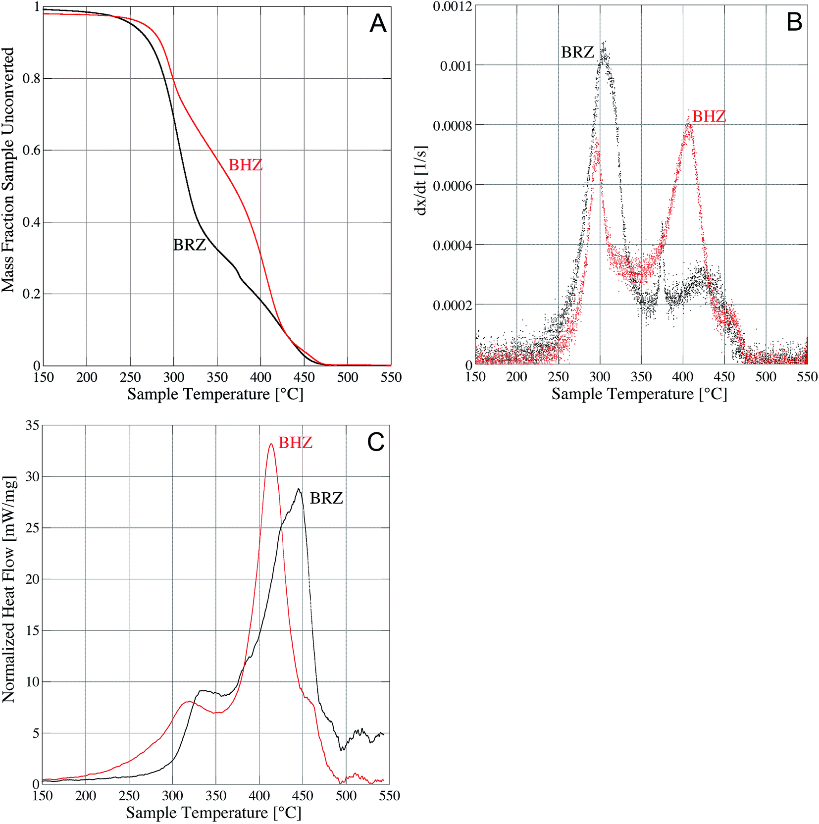 | ||
| Fig. 1 Thermal analysis of impregnated raw banana (BRZ) and hydrochar (BHZ) via (A) mass fraction converted as a function of temperature; (B) derivative thermogravimetric (DTG) curves; (C) exothermic heat flow as a function of temperature. | ||
From Fig. 1B, we note higher reactivity of the BRZ sample as compared to the impregnated hydrochar. Though both impregnated biomasses see a large portion of sample oxidized between 280 and 300 °C, the rate of oxidation for the raw impregnated biomass is considerably higher (more than twice as high) at lower temperatures. The derivative thermogravimetric (DTG) curve for BHZ shows a second peak around 415 °C with slightly higher mass loss rate than the first peak at 280 °C. This behavior corresponds to the changes in slope of the mass fraction conversion plot of Fig. 1A. The higher reactivity of the raw biomass versus the hydrochar can be explained by the carbonization process itself. During hydrothermal treatment, the more volatile compounds are forced out of the raw biomass, concentrating the higher molecular weight, more condensed components in the biomass. Thus, the remaining compounds are more “difficult” to oxidize. The carbonization step, as it is known to do,42 produces an “energy concentrated” biomass. As shown in Fig. 1C, the differential scanning calorimeter data, the peak exotherm of the hydrochar impregnated sample occurs at a lower temperature and higher heat value than the raw biomass.
The mineral content of any plant matter depends upon the composition of the soil in which the plant is grown; given the high water content of the biomass, which is drawn into the plant from the soil, the appearance of minerals throughout the biomass is logical. The ash fractions of a Dwarf Cavendish banana plant harvested in Funchal, Portugal contained between 0.6 and 32.3 (elemental) percent calcium;35 the sandy soil where the present banana stalk was harvested is known to contain elevated amounts of calcium, silicon and sulfur (from groundwater). As such, the presence of both ZnO (from the impregnation) and CaCO3 (naturally present in the biomass) in both the BRZC and BHZC is expected. XRD confirms that both zincite (ZnO) and calcite (CaCO3) phases co-exist in both types of biotemplated BRZC and BHZC materials (Fig. 2), and we note relatively higher concentrations of calcite in the BHZC. These zincite crystalline phases are not present in the raw biomass or hydrochar samples (BRZ, BHZ) prior to calcination. There is a small amount of an unknown crystalline phase detected in BHZ and BRZ that likely comes from the biomass itself. While the overall mineral matter decreased during carbonization (as noted in Section 3.1.), it is possible – given the XRD peak – that a small amount of inorganic minerals initially present crystallized; XRD peaks for BHZ qualitatively suggest that this is a possibility, but the spectral match cannot be confirmed. Zinc acetate, cellulose and lignin were ruled out as possibilities based on lack of spectral match.
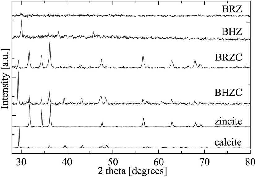 | ||
| Fig. 2 XRD spectra of Zn2+-impregnated banana stalk materials (plotted on left y-axis), zincite (RRUFF ID R060027) and calcite (R050048) (plotted on right y-axis).51 The Background signal from the instrument and Kapton tape was subtracted. | ||
The SEM images of BRZC and BHZC in Fig. 3 and 4 confirm the heterogeneity observed in the XRD results. At least four different particle morphologies are present. Aggregates of finer-grained material are primarily ZnO (e.g. areas 3B, 3D), as porous lattice type structures (e.g. area 4C). These ZnO particles likely form from oxidation of the zinc-impregnated biomasses, but we cannot rule out the possibility of some level of solution growth possible during carbonization, given the many examples of solution-growth crystalline ZnO microtubes43 and nanotubes44 in the literature. Hexagonal plates up to tens of nanometers in diameter are CaCO3 (e.g. areas 3C, 3D, 4A), as are “corrugated” fibers (e.g. areas 3A, 3B). Such hollow CaCO3 structures are known to precipitate in the presence of biomass templates such as starch.45 The ZnO particles generally appear to be coating the CaCO3 particles. CaCO3 is formed during the calcination as calcium naturally taken up by the biomass reacts with organic carbon and oxygen. In addition to forming separate hexagonal plates, the CaCO3 appears to have biotemplated on the banana stalk fibers themselves. However, even the plates were porous and appear to have formed from smaller calcite crystals fused together. Proteins naturally present in the banana plant46 likely promote the growth of CaCO3 crystals.47 The calcite form is more thermodynamically stable than the aragonite or vaterite forms of CaCO3, and given the presence of large organic molecules in both the raw and hydrochar biomass, the calcite form is favored over aragonite.48
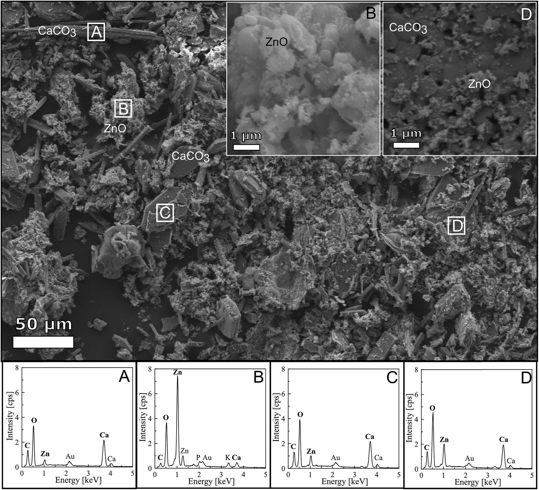 | ||
| Fig. 3 SEM image of raw banana stalk biomass, impregnated with Zn2+ and calcined at 550 °C (BRZC), with approximate area of EDS spectra indicated by letters (A–D). Aggregates of finer-grained particles are ZnO, while hexagonal plates and corrugated fiber structures are CaCO3. Au peak is from the sputter-coated conductive coating. | ||
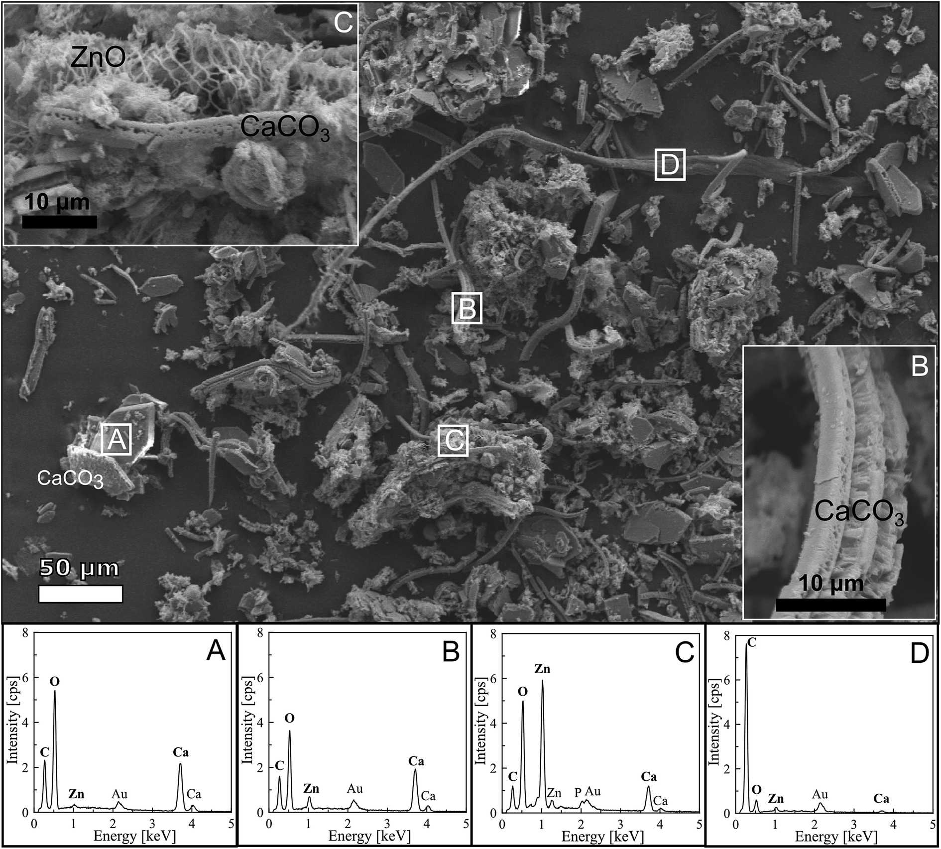 | ||
| Fig. 4 SEM image of banana stalk hydrochar, impregnated with Zn2+ and calcined at 550 °C (BHZC), with approximate area of EDS spectra indicated by letters (A–D). Aggregates of finer-grained particles are ZnO, while hexagonal plates and corrugated fiber structures are CaCO3. Au peak is from the sputter-coated conductive coating. | ||
We note from the SEM images and EDS analysis that few carbon-based fibers from the raw biomass (4D) remain, indicating complete calcination as suggested by the TGA data in Fig. 1. Table 1 shows the surface areas for the resulting ZnO materials. Though hydrothermal processing significantly increased the surface area of the banana stalk biomass, once impregnated and calcined, both BRZC and BHZC materials had similar surface areas, with that of the hydrochar's particles slightly lower than those templated on the raw biomass. Our surface areas are higher than those reported in the literature for ZnO templated on fir wood, which showed a surface area of between 1.28 and 16.09 m2 g−1.29 The surface area results complement the SEM images (Fig. 3 and 4), which show similar composition and degree of heterogeneity of the calcined materials. To determine whether the carbonization has a significant effect on the efficacy of these particles, in addition to the physical characteristics, we turned to photocatalytic water treatment experiments.
3.3. Applicability of biotemplated ZnO–CaCO3 materials to water treatment
Methylene blue dye was used as a model compound to simulate organic water pollutants in a test photocatalytic degradation experiment with BRZC and BHZC. As shown in Table 3, the raw biomass templated ZnO removed almost 50% of the MB from solution, while the hydrochar template biomass removed only 13%. From Fig. 5, we see that the concentration of MB in the vials exposed to UV light and biotemplated materials equilibrates after only 20 minutes.| Removal efficiency | Pseudo first order kinetics kpsf min−1 (over 0–15 min) | R2 | |
|---|---|---|---|
| BRZC | 48.8% | 0.0122 ± 0.0018 | 0.9904 |
| BHZC | 13.0% | 0.0262 ± 0.0018 | 0.9593 |
 | ||
| Fig. 5 Photocatalytic oxidation of methylene blue dye by BRZC and BHZC exposed to UV light or kept in the dark. | ||
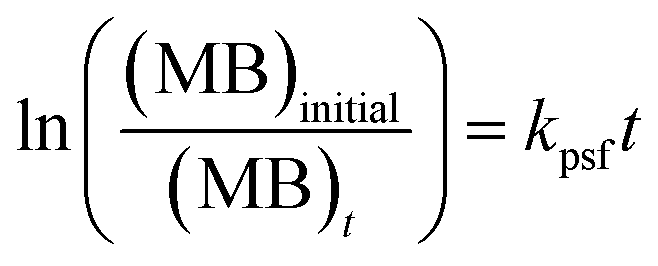
Initially, the dye degradation appears to follow pseudo first order degradation kinetics (with respect to dye concentration), through the data at 15 minutes, such that:
 | (1) |
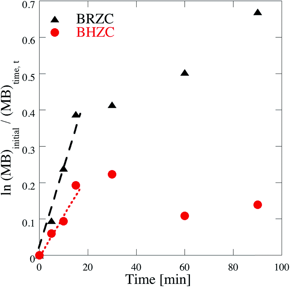 | ||
| Fig. 6 Determination of kinetic rate expressions via pseudo-first order kinetics (linear regression applied to first 15 min of UV exposure). | ||
However, the presence of the CaCO3 nanostructures opens then potential for treatment of complex contaminant mixtures of both organics, that can be degraded photocatalytically via ZnO as demonstrated here, as well as heavy metals such as Cd2+, Pb2+, Cr3+, Fe3+, and Ni2+ that have been shown to be removed from water using stabilized amorphous calcium carbonate nanoparticles.49 Calcium carbonates act as coagulants and buffers for acid mine wastewater treatment and to remove heavy metals from groundwater.50 Further studies are recommended to determine the possibility of removing both organics and inorganics from wastewaters using these heterogeneous materials.
4. Conclusions
This work demonstrates an integrated concept for the conversion of banana stalk, an agricultural waste with little intrinsic value, to liquid bio-fuels and solid biochar via hydrothermal carbonization. Zinc oxide nanostructures biotemplated onto the biomass and biochar demonstrated photocatalytic activity to degrade an aqueous organic dye. Calcium present in the banana plant was converted to calcite during oxidation of the impregnated biomass. This calcite appears to limit the photodegradation degradation capacity of the zincite on the heterogeneous biotemplated material. However, the calcium present may be beneficial to the remediation of water contaminated by both organic and heavy metals. It is recommended that future work explore the capacity of this calcite to treat mixed contaminant systems, as well as the impact of any additional (unidentifiable) mineral matter present on the overall water treatment system.Acknowledgements
The authors thank G. Kriner and his green thumb for cultivating the banana biomass, and A. Krupp for SEM assistance. A portion of this material is based upon work supported by the National Science Foundation under Grant No. NSF CBET-1505718. G. Dou acknowledges support of the China Scholarship Council (CSC) under Grant CSC NO. 201506425012. C. Gopu acknowledges funding from the Boston University STARS Program. A portion of this work was supported by the Boston University Initiative on Cities.References
- United Nations, Water, food and energy nexus, http://www.unwater.org/topics/water-food-and-energy-nexus/en/, accessed 29 August 2016.
- P. T. Anastas and J. B. Zimmerman, Peer reviewed: design through the 12 principles of green engineering, Environ. Sci. Technol., 2003, 37(5), 94A–101A CrossRef PubMed.
- R. Selvakumar, N. Seethalakshmi, P. Thavamani, R. Naidu and M. Megharaj, Recent advances in the synthesis of inorganic nano/microstructures using microbial biotemplates and their applications, RSC Adv., 2014, 4(94), 52156–52169 RSC.
- Z. Chen, J. Zhou, X. Wang, X. Liao, X. Huang and B. Shi, Natural collagen fiber-enabled facile synthesis of carbon@Fe3O4 core–shell nanofiber bundles and their application as ultrahigh-rate anode materials for Li-ion batteries, RSC Adv., 2016, 6(13), 10824–10830 RSC.
- B. Li, J. Zhao, J. Liu, X. Shen, S. Mo and H. Tong, Bio-templated synthesis of hierarchically ordered macro-mesoporous anatase titanium dioxide flakes with high photocatalytic activity, RSC Adv., 2015, 5(20), 15572–15578 RSC.
- A. Taubert and G. Wegner, Formation of uniform and monodisperse zincite crystals in the presence of soluble starch, J. Mater. Chem., 2002, 12(4), 805–807 RSC.
- Q. Dong, H. Su, J. Xu, D. Zhang and R. Wang, Synthesis of biomorphic ZnO interwoven microfibers using eggshell membrane as the biotemplate, Mater. Lett., 2007, 61(13), 2714–2717 CrossRef CAS.
- W. Zhang, D. Zhang, T. Fan, J. Ding, Q. Guo and H. Ogawa, Fabrication of ZnO microtubes with adjustable nanopores on the walls by the templating of butterfly wing scales, Nanotechnology, 2006, 17(3), 840 CrossRef CAS.
- A. Zampieri, G. T. Mabande, T. Selvam, W. Schwieger, A. Rudolph, R. Hermann, H. Sieber and P. Greil, Biotemplating of Luffa cylindrica sponges to self-supporting hierarchical zeolite macrostructures for bio-inspired structured catalytic reactors, Mater. Sci. Eng. C, 2006, 26(1), 130–135 CrossRef CAS.
- M. Sun, T. Li, Z. Zhang, N. Wang, A. Xie, X. Lv, Y. Wang, F. Wu and M. Wang, Natural biological template for ZnO nanoparticle growth and photocatalytic dye degradation under visible light, RSC Adv., 2015, 5(103), 84406–84409 RSC.
- H. Zhou, T. Fan and D. Zhang, Hydrothermal synthesis of ZnO hollow spheres using spherobacterium as biotemplates, Microporous Mesoporous Mater., 2007, 100(1), 322–327 CrossRef CAS.
- S. K. Kansal, M. Singh and D. Sud, Studies on photodegradation of two commercial dyes in aqueous phase using different photocatalysts, J. Hazard. Mater., 2007, 141(3), 581–590 CrossRef CAS PubMed.
- C. Hariharan, Photocatalytic degradation of organic contaminants in water by ZnO nanoparticles: revisited, Appl. Catal., A, 2006, 304, 55–61 CrossRef CAS.
- Y. L. Chen, L. C. Kuo, M. L. Tseng, H. M. Chen, C. K. Chen, H. J. Huang, R. S. Liu and D. P. Tsai, ZnO nanorod optical disk photocatalytic reactor for photodegradation of methyl orange, Opt. Express, 2013, 21(6), 7240–7249 CAS.
- N. Daneshvar, M. H. Rasoulifard, A. R. Khataee and F. Hosseinzadeh, Removal of CI acid orange 7 from aqueous solution by UV irradiation in the presence of ZnO nanopowder, J. Hazard. Mater., 2007, 143(1), 95–101 CrossRef CAS PubMed.
- W. Shen, Z. Li, H. Wang, Y. Liu, Q. Guo and Y. Zhang, Photocatalytic degradation for methylene blue using zinc oxide prepared by codeposition and sol–gel methods, J. Hazard. Mater., 2008, 152(1), 172–175 CrossRef CAS PubMed.
- R. Mallampati and S. Valiyaveettil, Biomimetic metal oxides for the extraction of nanoparticles from water, Nanoscale, 2013, 5(8), 3395–3399 RSC.
- G. Applerot, A. Lipovsky, R. Dror, N. Perkas, Y. Nitzan, R. Lubart and A. Gedanken, Enhanced antibacterial activity of nanocrystalline ZnO due to increased ROS-mediated cell injury, Adv. Funct. Mater., 2009, 19(6), 842–852 CrossRef CAS.
- A. Lipovsky, Y. Nitzan, A. Gedanken and R. Lubart, Antifungal activity of ZnO nanoparticles—the role of ROS mediated cell injury, Nanotechnology, 2011, 22(10), 105101 CrossRef PubMed.
- S. Liang, H. Sheng, Y. Liu, Z. Huo, Y. Lu and H. Shen, ZnO Schottky ultraviolet photodetectors, J. Cryst. Growth, 2001, 225(2), 110–113 CrossRef CAS.
- H. Fatemi, A. A. Khodadadi, A. A. Firooz and Y. Mortazavi, Apple–biomorphic synthesis of porous ZnO nanostructures for glucose direct electrochemical biosensor, Curr. Appl. Phys., 2012, 12(4), 1033–1038 CrossRef.
- Y. Zhou, L. Wang, Z. Ye, M. Zhao, H. Cai and J. Huang, Mango core inner shell membrane template-directed synthesis of porous ZnO films and their application for enzymatic glucose biosensor, Appl. Surf. Sci., 2013, 285, 344–349 CrossRef CAS.
- M. Zhao, Z. Li, Z. Han, K. Wang, Y. Zhou, J. Huang and Z. Ye, Synthesis of mesoporous multiwall ZnO nanotubes by replicating silk and application for enzymatic biosensor, Biosens. Bioelectron., 2013, 49, 318–322 CrossRef CAS PubMed.
- Z. Liu, T. Fan, D. Zhang, X. Gong and J. Xu, Hierarchically porous ZnO with high sensitivity and selectivity to H 2 S derived from biotemplates, Sens. Actuators, B, 2009, 136(2), 499–509 CrossRef CAS.
- J. X. Wang, X. W. Sun, Y. Yang, H. Huang, Y. C. Lee, O. K. Tan and L. Vayssieres, Hydrothermally grown oriented ZnO nanorod arrays for gas sensing applications, Nanotechnology, 2006, 17(19), 4995 CrossRef CAS.
- H. Zhang, C. Xu, P. Sheng, Y. Chen, L. Yu and Q. Li, Synthesis of ZnO hollow spheres through a bacterial template method and their gas sensing properties, Sens. Actuators, B, 2013, 181, 99–103 CrossRef CAS.
- T. Prakash, R. Jayaprakash, D. S. Raj, S. Kumar, N. Donato, D. Spadaro and G. Neri, Sensing properties of ZnO nanoparticles synthesized by using albumen as a biotemplate for acetic acid monitoring in aqueous mixture, Sens. Actuators, B, 2013, 176, 560–568 CrossRef CAS.
- H. Rensmo, K. Keis, H. Lindström, S. Södergren, A. Solbrand, A. Hagfeldt, S. E. Lindquist, L. N. Wang and M. Muhammed, High light-to-energy conversion efficiencies for solar cells based on nanostructured ZnO electrodes, J. Phys. Chem. B, 1997, 101(14), 2598–2601 CrossRef CAS.
- M. Saito and S. Fujihara, Large photocurrent generation in dye-sensitized ZnO solar cells, Energy Environ. Sci., 2008, 1(2), 280–283 CAS.
- D. C. Look, Recent advances in ZnO materials and devices, Mater. Sci. Eng. B, 2001, 80(1), 383–387 CrossRef.
- K. Sato and H. Katayama-Yoshida, Material design for transparent ferromagnets with ZnO-based magnetic semiconductors, Jpn. J. Appl. Phys., 2000, 39(6B), L555 CAS.
- J. Markard, R. Raven and B. Truffer, Sustainability transitions: An emerging field of research and its prospects, Res. Pol., 2012, 41(6), 955–967 CrossRef.
- Food and Agricultural Organization of United Nations (FAOSTAT), Banana Production, 2013, http://faostat.fao.org/site/567/DesktopDefault.aspx#ancor, accessed 29 August 2016.
- S. Elanthikkal, U. Gopalakrishnapanicker, S. Varghese and J. T. Guthrie, Cellulose microfibres produced from banana plant wastes: Isolation and characterization, Carbohydr. Polym., 2010, 80(3), 852–859 CrossRef CAS.
- L. Oliveira, N. Cordeiro, D. V. Evtuguin, I. C. Torres and A. J. Silvestre, Chemical composition of different morphological parts from ‘Dwarf Cavendish’ banana plant and their potential as a non-wood renewable source of natural products, Ind. Crops Prod., 2007, 26(2), 163–172 CrossRef CAS.
- M. M. Rahman, T. Islam, J. Nayeem and M. Jahan, Variation of chemical and morphological properties of different parts of banana plant (Musa paradisica) and their effects on pulping, International Journal of Lignocellulosic Products, 2014, 1(2), 93–103 Search PubMed.
- J. A. Libra, K. S. Ro, C. Kammann, A. Funke, N. D. Berge, Y. Neubauer, M. M. Titirici, C. Fühner, O. Bens, J. Kern and K. H. Emmerich, Hydrothermal carbonization of biomass residuals: a comparative review of the chemistry, processes and applications of wet and dry pyrolysis, Biofuels, 2011, 2(1), 71–106 CrossRef CAS.
- J. G. Lynam, M. T. Reza, V. R. Vasquez and C. J. Coronella, Effect of salt addition on hydrothermal carbonization of lignocellulosic biomass, Fuel, 2012, 99, 271–273 CrossRef CAS.
- K. Tekin, S. Karagöz and S. Bektaş, Hydrothermal liquefaction of beech wood using a natural calcium borate mineral, J. Supercrit. Fluids, 2012, 72, 134–139 CrossRef CAS.
- M. K. Akalın, K. Tekin and S. Karagöz, Hydrothermal liquefaction of cornelian cherry stones for bio-oil production, Bioresour. Technol., 2012, 110, 682–687 CrossRef PubMed.
- H. J. Huang, X. Z. Yuan, G. M. Zeng, Y. Liu, H. Li, J. Yin and X. L. Wang, Thermochemical liquefaction of rice husk for bio-oil production with sub-and supercritical ethanol as solvent, J. Anal. Appl. Pyrolysis, 2013, 102, 60–67 CrossRef CAS.
- S. Kang, X. Li, J. Fan and J. Chang, Characterization of hydrochars produced by hydrothermal carbonization of lignin, cellulose, D-xylose, and wood meal, Ind. Eng. Chem. Res., 2012, 51(26), 9023–9031 CrossRef CAS.
- L. Vayssieres, K. Keis, A. Hagfeldt and S. E. Lindquist, Three-dimensional array of highly oriented crystalline ZnO microtubes, Chem. Mater., 2001, 13(12), 4395–4398 CrossRef CAS.
- L. Vayssieres, Growth of arrayed nanorods and nanowires of ZnO from aqueous solutions, Adv. Mater., 2003, 15(5), 464–466 CrossRef CAS.
- W. Wei, G. H. Ma, G. Hu, D. Yu, T. Mcleish, Z. G. Su and Z. Y. Shen, Preparation of hierarchical hollow CaCO3 particles and the application as anticancer drug carrier, J. Am. Chem. Soc., 2008, 130(47), 15808–15810 CrossRef CAS PubMed.
- S. C. Carpentier, E. Witters, K. Laukens, P. Deckers, R. Swennen and B. Panis, Preparation of protein extracts from recalcitrant plant tissues: An evaluation of different methods for two-dimensional gel electrophoresis analysis, Proteomics, 2005, 5(10), 2497–2507 CrossRef CAS PubMed.
- J. Kanakis and E. Dalas, The crystallization of vaterite on fibrin, J. Cryst. Growth, 2000, 219(3), 277–282 CrossRef CAS.
- G. Falini, S. Albeck, S. Weiner and L. Addadi, Control of aragonite or calcite polymorphism by mollusk shell macromolecules, Science, 1996, 271(5245), 67 Search PubMed.
- G. B. Cai, G. X. Zhao, X. K. Wang and S. H. Yu, Synthesis of polyacrylic acid stabilized amorphous calcium carbonate nanoparticles and their application for removal of toxic heavy metal ions in water, J. Phys. Chem. C, 2010, 114(30), 12948–12954 CAS.
- M. Lee, I. S. Paik, I. Kim, H. Kang and S. Lee, Remediation of heavy metal contaminated groundwater originated from abandoned mine using lime and calcium carbonate, J. Hazard. Mater., 2007, 144(1), 208–214 CrossRef CAS PubMed.
- B. Lafuente, R. T. Downs, H. Yang and N. Stone, The power of databases: the RRUFF project, Highlights in Mineralogical Crystallography, 2015, pp. 1–30 Search PubMed.
Footnote |
| † Electronic supplementary information (ESI) available. See DOI: 10.1039/c6ra21663c |
| This journal is © The Royal Society of Chemistry 2016 |