
Effective construction of a AuNPs–DNA system for the implementation of various advanced logic gates†
Chunyang Zhouabc,
Changtong Wuabc,
Yaqing Liu*abc and
Erkang Wang*abc
aState Key Laboratory of Electroanalytical Chemistry, Changchun Institute of Applied Chemistry, Chinese Academy of Sciences, Changchun, Jilin 130022, China. E-mail: ekwang@ciac.ac.cn; Tel: +86-431-85262003
bKey Laboratory of Food Nutrition and Safety, Tianjin University of Science and Technology, Ministry of Education, Tianjin, 300457, China. E-mail: yaqingliu@tust.edu.cn; Tel: +86-22-60912484
cState Key Laboratory on Integrated Optoelectronics, College of Electronic Science and Engineering, Jilin University, Changchun, China
First published on 24th October 2016
Abstract
In this work, we integrate DNA computing at the nanoscale on the basis of the self-assembly of single stranded polyA-DNA on AuNPs as a precursor solution. As a “lab-on-a-nanoparticle”, the intelligent DNA strand modified AuNPs could be operated to realize a variety of Boolean logic gates including half adder, half subtractor, 2:1 encoder and 4:2 encoder under enzyme-free conditions for the first time. The signal readout methods, FAM fluorescence, G-quadruplex enhanced fluorescence of NMM and the i-motif structure enhanced fluorescence of CV were used to report the final signal in different logic operations, respectively. The mechanisms were further investigated by native polyacrylamide gel electrophoresis. These investigations provide a wider field of vision towards prototypical DNA-based circuits in biological systems and promote the development of advanced logic circuits.
Introduction
Logic gates made of molecules or biomolecules at the “nano-size” level are the potential foundation of molecular-scale computers that receive Boolean inputs to generate corresponding Boolean outputs.1 Up to now, various basic molecular logic gates, such as AND, OR, XOR, NOR, NAND, INHIBIT and advanced molecular logic gates such as half adder, half subtractor, full adder, full subtractor, encoder and decoder and so on have been designed based on biomolecular systems.2 Although numerous chemical logic systems mimicking corresponding electronic computing operations have been developed in recent years, this field is still at an early stage of experimental and design and a great future effort is also expected. Due to its low-cost, carrier of genetic information with predictable structures and feasible synthesis, nucleic acids have been confirmed as the most attractive building block for the synthesis of nanomaterial, construction of elaborate nanostructures and particularly design and engineering of biochemical circuits. Although the instances tackled by DNA-based logic gates are rudimentary compare with those solved by conventional electronic computers, it has shown potential as a new computing paradigm. Recent years, the programmable DNA-based logic circuits are developing fast, performing signal restoration, amplification and feedback and so on.3 To fulfill the requirements of increased computational complexity, great efforts are needed to integrate multiple and combinatorial logic gates on a simple platform.Gold nanoparticles (AuNPs) present great potential applications on the fabrication of biosensors and bioelectronics.4 One of the properties of AuNPs is the colorimetric assays based on the color change of AuNPs aggregation, which is widely used as the read-out signal for AuNPs-based logic operations.4b For instance, Yang and coworkers used the melamina-induced aggregation of AuNPs to design the operation of an INHIBIT logic gate, in which the human serum albumin and melamine acted as inputs and the color change of AuNPs solution as the only one output.5 Also, Yang and coworkers developed several logic gates, such as AND, OR, INHIBIT, sequential logic gates and keypad lock by using the glutathione-facilitated dispersion and aggregation of AuNPs as output.6 While such properties can only realize single output signal based logic gates and limit the development of logic gates based on AuNPs. As well known, AuNPs also present the unique optical properties that it can simultaneously quench various dyes with different emission frequencies from visible to near-IR range.7 By taking advantages of DNA and nanomaterial, the combination of DNA and AuNPs has been widely used in biosensor development, nanotechnology, medical diagnosis and computing systems.5–8 Most of the works used the conjugation of thiolated-DNA and AuNPs through the strong Au–S chemistry to self-assemble the thiolated DNA on the surface of AuNPs. However, such conjugation remains challenging to control the orientation and conformation of surface-tethered oligonucleotides in precisely and accurately regulate the hybridization ability.9 To overcome these problems, it has been reported that single-stranded nucleic acids with poly adenine sequences (polyA-DNA) containing multiple consecutive adenines that can serve as an effective anchoring block for preferential binding to the surface of AuNPs with high affinity and good repeatability in a short time at an environmental of pH 3, even comparable to the affinity of Au–S chemistry with the conformation and orientation in control.9,10 So in this work, the assembly of polyA-DNA on AuNPs driven by dynamic fluorescence modified DNA as inputs was operated to display the advanced DNA computing processes containing half adder, half subtractor, 2:1 encoder and 4:2 encoder.
Experimental
All the DNA sequences were listed in Table S1.† DNA was purchased from Sangon Biotechnology Co., Ltd (Shanghai, China). NMM was purchased from Porphyrin Products (Logan, UT, USA). Other chemicals were of reagent grade and were used without further purification. The oligonucleotide was dissolved in water as stock solution and quantified by UV-Vis absorption spectroscopy with the following extinction coefficients (ε 260 nm, M−1 cm−1): A = 15![[thin space (1/6-em)]](https://www.rsc.org/images/entities/char_2009.gif) 400, G = 11500, C = 7400, T = 8700. The oligonucleotides were dissolved in water and diluted with Tris–HCl buffer (20 mM Tris–HCl, 200 mM KCl, 10 mM MgCl2, pH 8.0) for hybridization in the logic circuits. Before use, the DNA solutions diluted with Tris–HCl buffer were first heated at 90 °C for 10 min and then gradually cooled down to room temperature. The stock solution of NMM (50 mM) was prepared in dimethyl sulfoxide (DMSO) and stored in darkness at −4 °C. The water used in the experiments was purified through a Millipore system. Fluorescence spectra were recorded after added their respective inputs and incubated under room temperature for 30 min to implement the function of various logic operations.
400, G = 11500, C = 7400, T = 8700. The oligonucleotides were dissolved in water and diluted with Tris–HCl buffer (20 mM Tris–HCl, 200 mM KCl, 10 mM MgCl2, pH 8.0) for hybridization in the logic circuits. Before use, the DNA solutions diluted with Tris–HCl buffer were first heated at 90 °C for 10 min and then gradually cooled down to room temperature. The stock solution of NMM (50 mM) was prepared in dimethyl sulfoxide (DMSO) and stored in darkness at −4 °C. The water used in the experiments was purified through a Millipore system. Fluorescence spectra were recorded after added their respective inputs and incubated under room temperature for 30 min to implement the function of various logic operations.
Native polyacrylamide gel electrophoresis (PAGE)
Before use, 2 μM DNA stock solution was diluted with Tris–HCl buffer (20 mM Tris–HCl, 200 mM KCl, 10 mM MgCl2, pH 8.0). The solution was then heated at 90 °C for 10 min and slowly cooled down to room temperature. After that, the desired volume of the platforms and their respective inputs were mixed and incubated for 30 min. The DNA solution was analyzed in 15% native polyacrylamide gel. Electrophoresis was conducted in 1 × TBE (17.8 mM Tris, 17.8 mM boric acid, 2 mM EDTA, pH 8.0) at a constant voltage of 120 V for 1 h. The gels were scanned by a UV transilluminator after staining with gel–dye.Circular dichroism measurements
5 μM DNA stock solutions were diluted with Tris–HCl buffer (20 mM Tris–HCl, 200 mM KCl, 10 mM MgCl2, pH 8.0). The solutions were then heated at 90 °C for 10 min and slowly cooled down to room temperature. After that, the CD spectra were measured on a JASCO J-820 spectropolarimeter (Tokyo, Japan) at room temperature. The spectral range was recorded from 220 to 320 nm in 1 mm path length cuvettes and averaged from three scans.Preparation of AuNPs–DNA conjugates
AuNPs with diameter around 13 nm were prepared as previously reported.11 All glass wares applied in this experiment were completely cleaned in aqua regia (HCl:HNO3 = 3:1), rinsed in doubly distilled water, and dried prior to use. In a 100 mL conical flask, 50 mL of 1 mM HAuCl4 in ultrapure water was heated to boil with vigorous stirring, followed by quickly addition of 5 mL of 39 mM sodium citrate. The solution turned deep blue in a short moment, and the final color changed to wine-red after 60 s. Boiling state lasted for an additional 15 min, the heating source was removed, and the colloid solution was stirred for another 30 min. The resulting AuNPs solution was stored in dark bottles at 4 °C and used to prepare the AuNPs–DNA conjugate.
To achieve the reactive condition of pH = 3, the 0.325 M HCl was added to 100 μL prepared AuNPs. Then, the citrate-stabilized AuNPs were incubated with polyA-DNA for about 3 minutes and then brought to 20 mM citrate/HCl buffer. Afterward, the prepared AuNPs–DNA was washed through centrifugation (12000 rpm, 15 min) to remove excess DNA sequences. Finally, the resulting conjugates were ready for experiments.
Results and discussion
With further development, it is critical to integrate multiple logic gates on a simple platform to further close the gap between bio-molecular computation and electrical circuits. In present investigations, half-adder and half-subtractor were developed with AuNPs/polyA-DNA as platform to implement the arithmetic functions. Half adder is a common used logic gate in information technology and can also be used to assist construct more advanced computing circuits. It implemented the binary addition operation by integration of an XOR gate and an AND gate in parallel to generate a SUM output and a CARRY output, respectively. And a half subtractor could perform a binary subtraction operation, which required the combination of XOR gate and an INHIBIT gate to produce a DIFFERENCE output and a BORROW output, respectively.2g Although their fundamental and practical importance, investigations of molecular half-adder and half-subtractor are still on an early stage. The two distinct fluorescent dyes of 6-carboxyfluorescein (FAM, emission max at 519 nm) and N-methylmesoporphyrin IX (NMM, emission max at 619 nm) were selected as signal reporters for the required logic gates of half adder and half subtractor. To establish optimal conditions, the synthesis of AuNPs and conjugated with polyA-DNA at different pH values, different concentrations of AuNPs and polyA-DNA were explored in Fig. S1–S3 in (ESI†). The optimized concentrations of DNA inputs that operated in half adder and half subtractor were explored in explored in Fig. S4 and S5 in ESI.†To perform the half-adder logic function, the polyA-DNAHA (PHA) is first adsorbed on the surface of AuNPs and then PHA can hybridize with FAM–DNAHA (F–DNAHA), forming the platform of AuNPs/PHA/F–DNAHA complex (Fig. 1A). The quenched or recovered fluorescence of FAM is depending on the distance between the AuNPs and the fluorophore, which is controlled by the two inputs. The fluorescence of NMM acts as another signal indicator for the designed half adder. In the absence of any input, the system remains in the original state (Fig. 1C curve a and e). In the presence of INA, it can hybridize with F–DNAHA to form duplex of INA/F–DNAHA and then release from the AuNPs. The FAM is then far away from the AuNPs, leading to a restored high output signal (Fig. 1C curve b). The interaction does not influence the fluorescence of NMM (Fig. 1C curve f). In the presence of INB, it can also hybridize with F–DNAHA and form duplex of INB/F–DNAHA, restoring the fluorescence of FAM (Fig. 1C curve c) and the fluorescence of NMM still keeps a low output signal (Fig. 1C curve g). However, in the coexistence of INA and INB, the DNA duplex of INA/INB is formed since it presents a higher affinity than either INA/PHA or INB/PHA. The hybridization of INA and INB suppresses desorption of F–DNAHA from AuNPs, producing a low signal (Fig. 1C curve d). It is worth noting that the INA/INB duplex can form the G-quadruplex structure, which can increase the fluorescence of NMM,3b (Fig. 1C curve h). The fluorescent responses of FAM at 519 nm and NMM at 619 nm are normalized and plotted as column bar (Fig. 1D), producing the corresponding truth table (Fig. 1B). Similar as the approach used in electronics, here, the output threshold value with an undefined range is defined as “1” or “0” when the normalized fluorescence intensity is higher than 0.45 or lower than 0.35. All the DNA sequences used in this work were listed in Table S1.†
 | ||
| Fig. 1 (A) Schematic illustration of the Au–P-DNA/FDNA nanostructure based half adder logic gate with the corresponding circuit. (B) The truth table for the half adder logic gate. (C) Fluorescence emission responses of FAM and NMM for the half adder logic gate in the absence of the two inputs (a and e), in the presence of INA (b and f), in the presence of INB (c and g) and in the presence of INA and INB (d and h). (D) The normalized fluorescence intensity of FAM at 519 nm and NMM at 619 nm as functions of various inputs signals. | ||
Native polyacrylamide gel electrophoresis (PAGE) and circular dichroism (CD) experiments are performed to validate the hybridization reaction and the formation of G-quadruplex in half adder logic gate (Fig. 2). From the PAGE experiments results (Fig. 2A), the belts show the individual DNA strands of PHA, F–DNAHA, INA and INB in sequence from lane a to lane d. Compared with the belts of lane a and lane b, the single belt in lane e proves the hybridization between PHA and F–DNAHA and the formation of PHA/F–DNAHA duplex. In the existence of INA or INB, the belts appear at different positions, indicating the formation of the duplexes of INA/F–DNAHA (lane f) and INB/F–DNAHA (lane g). In the coexistence of INA and INB (lane h), a new belt appears and locates at a different position from INA or INB and another belt locates at the position of PHA/F–DNAHA duplex, indicating the formation of the duplexes of INA/INB and PHA/F–DNAHA. The results suggest that INA and INB prefer to hybridize together, leaving PHA/F–DNAHA free in solution. The CD experiments are performed to further confirm the formation of G-quadruplex structure in the implementation of half adder logic operation (Fig. 2B). As shown in Fig. 2B, the CD spectrum of PHA (curve a), F–DNAHA (curve b), INA (curve c) and INB (curve d) are of relatively low-amplitude, indicating the random DNA structures. After mixing the PHA and F–DNAHA, no obvious change is found (curve e), indicating that the mixture does not form G-quadruplex structure. When adding INA or INB into the complex of PHA/F–DNAHA, still no obvious change is found (curve f and curve g). Only in the coexistence of INA and INB, the two obvious peaks, a positive peak at 268 nm and a negative peak at 245 nm, can be found from the CD spectrum (curve h), which shows the characteristic of a parallel G-quadruplex structure.12 The results of PAGE, CD and fluorescence experiments are consistent with each other and successfully demonstrate the operation of a half adder.
 | ||
| Fig. 2 (A) PAGE of the half adder logic gate. Lane a: PHA; lane b: F–DNAHA; lane c: INA; lane d: INB; lane e: PHA + F–DNAHA; lane f: PHA + F–DNAHA + INA; lane g: PHA + F–DNAHA + INB; lane h: PHA + F–DNAHA + INA + INB. (B) The CD of different DNA sequences of curve a: PHA; curve b: F–DNAHA; curve c: INA; curve d: INB; curve e: PHA + F–DNAHA; curve f: PHA + F–DNAHA + INA; curve g: PHA + F–DNAHA + INB; curve h: PHA + F–DNAHA + INA + INB. | ||
Similar as half adder, the half subtractor is also designed according to the platform of Au–PHS/F–DNAHS nanostructure, which is constructed by the adsorption of polyA-DNAHS (PHS) on the surface of AuNPs and then the PHS can hybridize with FAM–DNAHS (F–DNAHS), forming the logic platform of AuNPs/PHS/F–DNAHS (Fig. 3A). Here, the fluorescence signal outputs of the half subtractor are also reported by FAM and NMM. As shown in Fig. 3A, in the absence of any input, the system remains in the original state and reports low fluorescence of FAM and NMM (Fig. 3C curve a and e). When the DNA input of INA′ is added to the system, it can hybridize with F–DNAHS and recover the fluorescence of FAM. At the same time no increase of fluorescence of NMM is found since no G-quadruplex is produced (curve b and f). When the DNA input of INB′ is added to the system, it can not only forming the duplex of INB′/F–DNAHS and recovering the fluorescence of FAM but can also generate the G-quadruplex structure to enhance the fluorescence of NMM (curve c and g). However, in the coexistence of INA′ and INB′, the DNA duplex of INA′/INB′ is generated since it presents a higher affinity than either INA′/F–DNAHS or INB′/F–DNAHS. So the hybridization of INA′ and INB′ suppresses desorption of FAM–DNAHS from AuNPs, producing low signal of FAM and NMM, (curve d and h). The fluorescent responses of FAM and NMM are normalized and plotted as column bar (Fig. 3D), producing the corresponding truth table (Fig. 3B). Here, the output threshold value with an undefined range is defined as “1” or “0” when the normalized fluorescence intensity is higher than 0.45 or lower than 0.35.
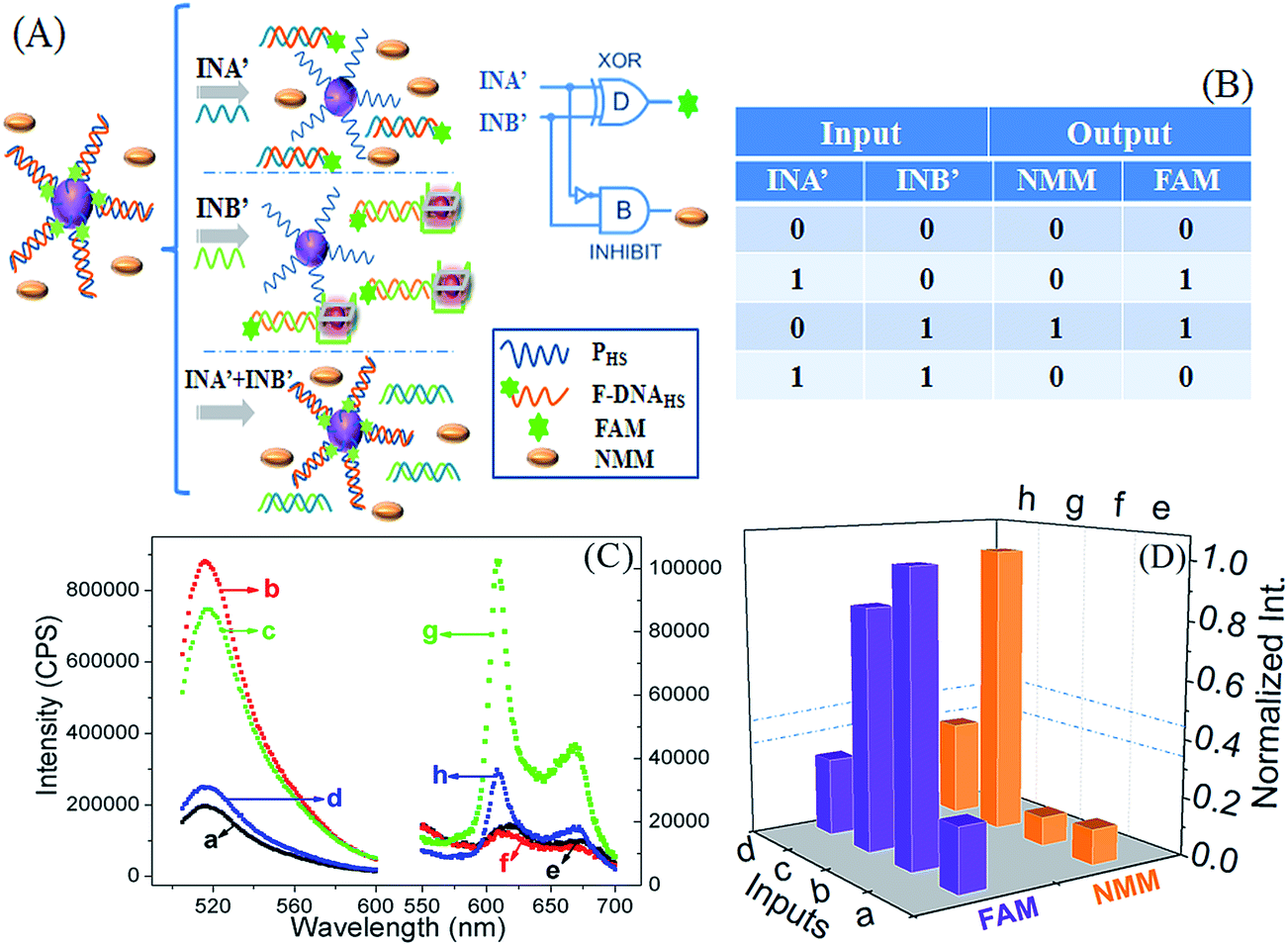 | ||
| Fig. 3 (A) Schematic illustration of the AuNPs/PHS/FAM–DNAHS nanostructure based half subtractor logic gate with the corresponding circuit. (B) The truth table for the half subtractor logic gate. (C) Fluorescence emission responses of FAM and NMM for the half subtractor logic gate in the absence of the two inputs (a and e), in the presence of INA′(b and f), in the presence of INB′(c and g) and in the presence of INA′ and INB′ (d and h). (D) The normalized fluorescence intensity of FAM and NMM as functions of various inputs signals. | ||
The PAGE and CD experiments are also performed to further identify the interactions between DNA strands and the formation of G-quadruplex (Fig. 4). In PAGE experiment shown in Fig. 4A, the DNA strands added in lane a–d are PHS, F–DNAHS, INA′ and INB′ as comparison. The hybridization of PHS with F–DNAHS produces a new belt, lane e. There are two belts in lane f, the above one represents a new belt produced by the hybridization of F–DNAHS and INA′, forming the duplex INA′/F–DNAHS. Another belt is the original position of PHS. The above belt in lane g is the new one produced by the hybridization of F–DNAHS and INB′, forming the duplex of INB′/F–DNAHS and the below belt is the position of PHS. In lane h, the above clear belt is produced by the duplex of INA′/INB′ due to the preferred hybridization of INA′ and INB′. Another belt that under the belt of INA′/INB′ indicates the duplex PHS/F–DNAHS. In CD experiments shown in Fig. 4B, the CD spectrum of PHS (curve a), F–DNAHS (curve b), INA′ (curve c) and INB′ (curve d) are of relatively low-amplitude, indicating the random DNA structures. Only in the coexistence of F–DNAHS and INB′, the two obvious peaks, a positive peak at 265 nm and a negative peak at 243 nm, can be found from the CD spectrum (curve g), which is the characteristic of a parallel G-quadruplex structure. The results of PAGE, CD and fluorescence experiments are consistent with each other and successfully demonstrate the operation of half subtractor.
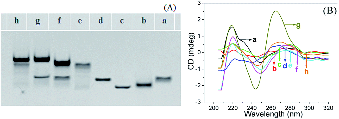 | ||
| Fig. 4 (A) PAGE of the half subtractor logic gate. Lane a: PHS; lane b: F–DNAHS; lane c: INA′; lane d: INB′; lane e: PHS + F–DNAHS; lane f: PHS + F–DNAHS + INA′; lane g: PHS + F–DNAHS + INB′; lane h: PHS + F–DNAHS + INA′ + INB′. (B) The circular dichroism of different DNA sequences of curve a: PHS; curve b: F–DNAHS; curve c: INA′; curve d: INB′; curve e: PHS + F–DNAHS; curve f: PHS + F–DNAHS + INA′; curve g: PHS + F–DNAHS + INB′; curve h: PHS + F–DNAHS + INA′ + INB′. | ||
In order to prove the versatility of the platform of the conjugation of polyA-DNA and AuNPs, we further implement the 2:1 encoder and 4:2 encoder logic gates to further evidence that such platform can implement multiple logic operations at the same time. Encoder is considered to be a non-Boolean operation or device that can convert information from one format to another for the purpose of compression.13 In principle, a single-bit 2:1 encoder can convert two input bits into one output bits. In a similar way, a single-bit 4:2 encoder can convert four input bits into two output bits. The optimized concentrations of DNA inputs that operated in 2:1 encoder and 4:2 encoder were explored in explored in Fig. S6 in ESI.† Herein, a 2:1 encoder is constructed based on the Au–PEN1/F–DNAEN1 nanostructure by the adsorption of polyA-DNAEN1 (PEN1) on the surface of AuNPs and the PEN1 can also hybridize with FAM–DNAEN1 (F–DNAEN1), forming the duplex PEN1/F–DNAEN1 on the surface of AuNPs, which is regarded as the platform for the operation of 2:1 encoder (Fig. 5A). Under this case, the fluorophore of FAM approaches the surface of AuNPs, leading to the quench of fluorescence due to the long-range fluorescence energy resonance transfer. In the addition of IN1, no hybridization is happened and fluorescence of FAM is still quenched by AuNPs (Fig. 5C curve black). However, in the presence of IN2, it can hybridize with F–DNAEN1 and form the duplex of IN2/F–DNAEN1, which can be released from the surface of AuNPs. Thus a restored fluorescence of FAM is monitored due to the long distance of fluorophore from the AuNPs (Fig. 5C curve red). The normalized fluorescent responses of FAM at 519 nm are plotted as column bar (Fig. 5D), producing the corresponding truth table (Fig. 5B). Here, the output threshold value is defined as “1” or “0” when the normalized fluorescence intensity is higher than 0.45 or lower than 0.35. Native polyacrylamide gel electrophoresis (PAGE) experiments were performed to validate the hybridization reaction in 2:1 encoder logic gate in Fig. S7.†
 | ||
| Fig. 5 (A) Schematic illustration of the Au–P-DNA/FDNA nanostructure based 2:1 encoder logic gate with the corresponding circuit. (B) The truth table for the 2:1 logic gate. (C) Fluorescence emission responses of FAM for the 2:1 logic gate in the presence of IN1 (curve black) and in the presence of IN2 (curve red). (D) The normalized fluorescence intensity of FAM at 521 nm as functions of various inputs signals. | ||
Based on the same principle, we further constructed a 4:2 encoder logic gate by the conjugation of polyA-DNAEN2 (PEN2) sequence on the surface of AuNPs and the platform of the Au–PEN2/F–DNAEN2 is constructed via the hybridization between PEN2 and FAM–DNAEN2 (F–DNAEN2) (Fig. 6). Herein, two distinct fluorescent dyes, FAM and crystal violet (CV, emission max at 628 nm), are selected as the two output signal reporters in the developed system. Fluorescence of CV can be significantly enhanced once CV binding on G-quadruplex structure or i-motif structure in acid medium14 (the 5′-CCCCCTTTCCCCCTTT CCCCCTTTCCCCC-3′ sequence of PEN2 can form i-motif structure in acid medium). Redesigned three DNA strands and H+ are used as the four inputs for the logic operation. All the reactions are performed in Tris–HCl buffer (20 mM Tris–HCl, 200 mM KCl, 10 mM MgCl2, pH 8.0). In the addition of a random DNA strand of IN1′ to the platform, no displacement happens and no i-motif structure is formed in pH 8. So the fluorophores of FAM and CV still report low responses (Fig. 6C curve a and e). After the addition of H+ as the IN2′, the solution is modulated to acidic medium of pH 5, then the PEN2 strand can form the i-motif configuration, which can bind CV to enhance the fluorescence of CV (Fig. 6C curve f). While the fluorescence of FAM would be reduced in acidic medium15 (Fig. 6C curve b). In the presence of IN3′, it can hybridize with F–DNAEN2, forming the duplex of IN3′/F–DNAEN2. The formed duplex can be released from AuNPs and the fluorescence of FAM can be restored (Fig. 6C curve c) for the FAM is far away from the quencher of AuNPs. The CV still shows a low fluorescent response (Fig. 6C curve g). In the existence of IN4′, which contains G-quadruplex structure, it can hybridize with F–DNAEN2 and restore the fluorescence of FAM and also can increase the fluorescence of CV due to the formed structure of G-quadruplex (Fig. 6C curve d and h). The fluorescent responses of FAM at 519 nm and CV at 630 nm are normalized and plotted as column bar (Fig. 6D), producing the corresponding truth table (Fig. 6B). The output threshold value is defined as “1” or “0” when the normalized fluorescence intensity is higher than 0.45 or lower than 0.35. We have further conducted PAGE and CD experiments to demonstrate the integrity of the reaction mechanism of the 4:2 encoder logic gate in Fig. S8.†
 | ||
| Fig. 6 (A) Schematic illustration of the Au–P-DNA/FDNA nanostructure based 4:2 encoder logic gate with the corresponding circuit. (B) The truth table for the 4:2 encoder logic gate. (C) Fluorescence emission responses of FAM and CV for the 4:2 encoder logic gate in the presence of IN1′ (a and e), in the presence of IN2′ (b and f), in the presence of IN3′ (c and g) and in the presence of IN4′ (d and h). (D) The normalized fluorescence intensity of FAM at 521 nm and CV at 628 nm as functions of various inputs signals. | ||
Conclusions
In summary, various advanced logic gates including arithmetic and non-arithmetic logic gates, half adder, half subtractor, 2:1 encoder and 4:2 encoder have been successfully demonstrated in proof-of-principle. All the advanced logic gates are implemented on a simple platform by combination of gold nanoparticles and polyA-DNA biomaterials. What more important is that the single-stranded nucleic acids with poly adenine sequences containing multiple consecutive adenines and can serve as an effective anchoring block for preferential binding with AuNPs surface with high affinity in a short time at environmental of pH 3, resulting in great advantages for experiments. Moreover, a constant output threshold value is available for all developed logic gates. The investigations provide a novel prototype for the design and assembly of higher-order circuits on molecular level and develop a new way for constructing multi-component devices on a single biomolecular platform. Though the developed logic gates in our work are implemented in an experimental stage, it is worthy of noting that there is a long road on ahead for practice application.Acknowledgements
This work was supported by the National Natural Science Foundation of China (no. 211900040 and no. 21427811), the State Key Project of Ministry of Science and Technology of China (no. 2016YFA0202000 and no. 2013YQ170585).Notes and references
- (a) J. M. Tour, Acc. Chem. Res., 2000, 33, 791–841 CrossRef CAS PubMed; (b) A. Credi, V. Balzani, S. J. Langford and J. F. Stoddart, J. Am. Chem. Soc., 1997, 119, 2679–2681 CrossRef CAS; (c) T. Gupta and M. Boom, Angew. Chem., Int. Ed., 2008, 47, 5322–5326 CrossRef CAS PubMed; (d) S. E. Cakmak and E. U. Akkaya, Angew. Chem., Int. Ed., 2013, 52, 11364–11368 CrossRef PubMed; (e) R. Yashin, S. Rudchenko and M. N. Stojanovic, J. Am. Chem. Soc., 2007, 129, 15581–15584 CrossRef CAS PubMed; (f) H. L. Li, W. Hong, S. J. Dong, Y. Q. Liu and E. K. Wang, ACS Nano, 2014, 8, 2796–2803 CrossRef CAS PubMed; (g) H. L. Li, J. T. Ren, Y. Q. Liu and E. K. Wang, Chem. Commun., 2014, 50, 704–706 RSC; (h) C. T. Wu, K. Wang, D. Q. Fan, C. Y. Zhou, Y. Q. Liu and E. K. Wang, Chem. Commun., 2015, 51, 15940–15943 RSC; (i) H. L. Li, S. J. Guo, Q. H. Liu, L. D. Qin, S. J. Dong, Y. Q. Liu and E. K. Wang, Adv. Sci., 2015, 2, 1500054–1500058 CrossRef.
- (a) D. Han, Z. Zhu, C. C. Wu, L. Peng, L. J. Zhou, B. Gulbakan, G. Z. Zhu, K. R. Williams and W. H. Tan, J. Am. Chem. Soc., 2012, 134, 20797–20804 CrossRef CAS PubMed; (b) P. Alexander and D. Alexander, Angew. Chem., Int. Ed., 2014, 53, 13192–13195 CrossRef PubMed; (c) J. B. Zhu, L. B. Zhang, Z. X. Zhou, S. J. Dong and E. K. Wang, Chem. Commun., 2014, 50, 3321–3323 RSC.
- (a) C. Y. Zhou, K. Wang, D. Q. Fan, C. T. Wu, D. L. Liu, Y. Q. Liu and E. K. Wang, Chem. Commun., 2015, 51, 10284–10286 RSC; (b) H. L. Li, Y. Q. Liu, S. J. Dong and E. K. Wang, NPG Asia Mater., 2015, 7, e166 CrossRef CAS.
- (a) F. Degliangeli, P. Kshirsagar, V. Brunetti, P. Pompa and R. Fiammengo, J. Am. Chem. Soc., 2014, 136, 2264–2267 CrossRef CAS PubMed; (b) Z. Zhu, C. Wu, H. Liu, Y. Zou, X. Zhang, H. Kang, C. J. Yang and W. H. Tan, Angew. Chem., Int. Ed., 2010, 49, 1052–1056 CrossRef CAS PubMed; (c) L. L. Li, P. W. Wu, K. Hwang and Y. Lu, J. Am. Chem. Soc., 2013, 135, 2411–2414 CrossRef CAS PubMed.
- Z. Z. Huang, H. N. Wang and W. S. Yang, ACS Appl. Mater. Interfaces, 2015, 7, 8990–8998 CAS.
- Z. Z. Huang, H. N. Wang and W. S. Yang, Nanoscale, 2014, 6, 8300–8305 RSC.
- T. J. Song and H. J. Liang, J. Am. Chem. Soc., 2012, 134, 10803–10806 CrossRef CAS PubMed.
- Y. Liu, B. Dong, Z. Wu, W. Fang, G. Zhou, A. Shen, X. D Zhou and J. Hu, Chem. Commun., 2014, 50, 12026–12029 RSC.
- H. Pei, F. Li, Y. Wan, M. Wei, H. Liu, Y. Su, N. Chen, Q. Huang and C. H. Fan, J. Am. Chem. Soc., 2012, 134, 11876–11879 CrossRef CAS PubMed.
- B. Yang, X. B. Zhang, L. P. Kang, Z. M. Huang, G. L. Shen, R. Q. Yu and W. H. Tan, Nanoscale, 2014, 6, 8990–8996 RSC.
- X. Zhang, M. R. Servos and J. W. Liu, J. Am. Chem. Soc., 2012, 134, 7266–7269 CrossRef CAS PubMed.
- (a) S. Čeru, P. Šket, I. Prislan, J. Lah and J. Plavec, Angew. Chem., Int. Ed., 2014, 53, 4881–4886 CrossRef PubMed; (b) P. Tóthová, P. Krafčíková and V. Víglaský, Biochemistry, 2014, 53, 7013–7027 CrossRef PubMed.
- Y. He, Y. Chen, C. Li and H. Cui, Chem. Commun., 2014, 50, 7994–7997 RSC.
- T. Li, D. Ackermann, A. Hall and M. Famulok, J. Am. Chem. Soc., 2012, 134, 3508–3516 CrossRef CAS PubMed.
- U. Schneider, J. Severinsen, I. Géci, L. Okkels, N. Jøhnk, N. Mikkelsen, T. Klinge, E. Pedersen, H. Westh and G. Lisby, BMC Biotechnol., 2010, 10, 4–11 CrossRef PubMed.
Footnote |
| † Electronic supplementary information (ESI) available. See DOI: 10.1039/c6ra21585h |
| This journal is © The Royal Society of Chemistry 2016 |