Water stability of self-assembled peptide nanostructures for sequential formation of two-dimensional interstitial patterns on layered materials

Linhao Sun, Takuma Narimatsu, Shohei Tsuchiya, Tomohiro Tanaka, Peiying Li and Yuhei Hayamizu*
School of Materials Science and Engineering, Tokyo Institute of Technology, Tokyo 152-8550, Japan. E-mail: hayamizu.y.aa@m.titech.ac.jp; Fax: +81 03 57342876; Tel: +81 03 57343651

Received 24th August 2016 , Accepted 4th October 2016

First published on 4th October 2016


Abstract

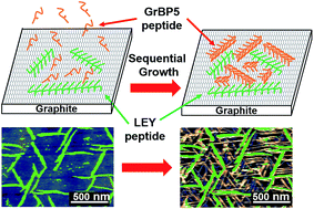
Developing various morphologies and a stable nanostructure of self-assembled peptides on a two-dimensional substrate has played a key role for bioelectronics and biomedical applications. Here, the adsorption and self-assembled characters of two artificial peptides with opposite charges are investigated on graphite and MoS2 surfaces. The ex situ atomic force microscopy (AFM) results show that their morphologies, ordering and stabilities on graphite and MoS2 surfaces are different. The negatively charged peptides self-assembled into an ordered nanostructure on graphite surfaces with six-fold symmetry in a wide range of peptide concentrations. On MoS2 surfaces, the peptide shows a morphology change from randomly orientated nanowires to ordered aligned nanowires as the peptide concentration decreases. On the other hand, the positively charged peptides formed a disordered structure such as wavy structures or aggregates on both substrates. The affinity constants of both peptides on graphite and MoS2 were estimated using concentration-dependence experiments. The stability against water soaking was also examined for both peptides on graphite and MoS2. We found that negatively charged peptides have high affinity constants and stability on both substrates. The results suggest that the stability of self-assembled peptides could be determined by their affinity constants on the substrates. Finally, by using the negatively charged peptides as a stable molecular template, we have demonstrated a sequential self-assembly of two different peptides on a graphite surface. The peptides self-assembled first on the surface show an ability to maintain their nanostructures, and guide the self-assembly of secondary self-assembled peptides. Both of the self-assembled peptides show the same orientations and ordered structures on a graphite surface. These results will open a new door for the development of biosensors with multiple biological probes on a functional surface such as graphite or MoS2.


1. Introduction

Self-assembly of molecules at the solid/liquid interface via a bottom-up approach has been widely studied.1–4 Especially, engineering biomolecules such as proteins and peptides with an ability to self-assemble on solid surfaces into various morphologies and stable nanostructures is significant towards biomedical applications such as biosensors and surface treatments for biocompatibility or anti-fouling.5–8 Generally, the morphology and structure of these self-assembled biomolecules are determined by their amino-acid sequences and surrounding environments.9–14 For example, self-assembly of amyloid β (Aβ) peptides on different substrates has been investigated.10 They formed amorphous structures on mica surfaces, and highly ordered structures on graphite surfaces. Similarly, an artificial GAV-9 peptide has been studied on both substrates as well. The GAV-9 peptides formed ordered structures on both mica and graphite substrates. The results of atomic force microscopy (AFM) indicated that GAV-9 peptides form a standing-up structure on mica surfaces and lying-down structures on graphite surfaces, respectively. It was also suggested that these structural formations result from electrostatic interactions or hydrophobic–hydrophobic interactions, respectively.9 In our earlier work, a genetically selected graphite binding peptide (GrBP5) and its variant peptides have been investigated on highly oriented pyrolytic graphite (HOPG) surfaces.11 The GrBP5 peptides showed highly ordered nanowire structures, probably due to their hydrophobic inter-peptide interactions and π–π interactions with the surface. Ordered nanostructures of biomolecules on atomically flat surfaces have not only been formed by peptides but also by proteins. A de novo designed protein with β-sheet structure demonstrated an ordered nanostructure on HOPG surfaces.15

Some works have shown that electrostatic interactions can efficiently help to adjust the adsorption and self-assembly processes on solid surfaces. The above mentioned GAV-9 peptide showed decreased peptide coverage on a modified mica surface as the electrostatic interaction varied from attractive to repulsive.9 An ionic-complementary peptide EAK16-II formed randomly orientated nanofibers on a mica surface by electrostatic interaction.16 These works clearly showed how the morphology and self-assembled structure of biomolecules can be tuned by their sequences and the substrates. Traditionally, layered materials such as HOPG and mica have been widely chosen as substrates to observe the self-assembly of peptides. Within the last few years, a group of two-dimensional transition metal dichalcogenides (TMD) such as MoS2 (ref. 17–20) have been investigated due to their unique optical and electrical properties.21,22 These materials also have atomically flat surfaces with a large surface area, which can provide an ideal platform for the investigation of peptide self-assembly.

Here, we have designed two different types of peptides based on an antimicrobial LKW peptide and examined their self-assembly on substrates of graphite and MoS2. The LKW peptide is known to have an α-helical structure in solution.23 The formation of the α-helical structure is probably due to the alternating hydrophobic part and hydrophilic part. Our previous work on controlling the self-assembly of GrBP5 and its variant peptides on a graphite surface showed that tyrosine groups can help to form an ordered structure on a graphite surface.11 In this work, we change the original LKW peptide sequence by replacing tryptophan with tyrosine. Also, we introduced glutamic acid (E) residues to make an oppositely charged peptide against LKY with lysine (K). Due to their potential stability in solution, we expect that one of them could have a stable self-assembled structure on a graphite or MoS2 surface resulting from their hydrophobic nature and electrostatic interactions with the surface. The stability of assembled structures of peptides under water is crucial. The stable structures of well-aligned peptides may provide a mean to template the self-assembled structure of another type of peptide by a sequential self-assembly process. This sequential assembly would open a new door for biosensing with multiple probes on a functional surface such as HOPG or MoS2, leading to simultaneous detection of multiple target biomolecules for more sophisticated diagnoses.

In this work, we have examined the self-assembly of two artificial peptides with opposite charges. The morphology on graphite and MoS2 was investigated using AFM. The binding affinity of both peptides on each substrate was obtained from the relationship of coverage versus peptide concentration. Also, the stabilities of both peptides on the graphite and MoS2 surfaces were investigated using a series of water soaking experiments. Finally, the sequential assembly of two peptides on the same surface was demonstrated.

2. Materials and methods

2.1. Raw materials

Natural amino acids with protected groups of 9-fluorenylmethyloxycarbonyl group (Fmoc), O-(benotriazol-1-yl)-N,N,N′,N′-tetramethyluronium hexafluorophosphate (HBTU), dichloromethane (DCM), N,N-dimethylformamide (DMF), piperidine (PPD), 1-hydroxybenzotriazole (HOBT), N,N-diisopropylethylamine (DIEA), divinylbenzene, and polystyrene resin were purchased from Watanabe Chemical Ind., LTD. Trifluoroacetic acid was purchased from Tokyo Chemical Industry CO., LTD. Ethyl ether was purchased from Takahashi pure chemical Co.

2.2. Synthesis and purification process of peptides

Peptides were synthesized by a solid phase peptide synthesis (SPPS) method.24 Polystyrene resin cross-linked with 1% divinylbenzene was generally used as a solid support for peptide synthesis. For GrBP5, polystyrene resins covalently linked to tyrosine with a protecting group (Fmoc) were employed. The resins were swelled with DCM solvent overnight. Then, the resins were washed 3–5 times with DMF solvent. Thirdly, the resins were deprotected using PPD for 10–15 min. After that, the free N-terminal amine of the resins reacted with the carboxyl group of the next N-protected serine amino acid under conditions using HOBT, HBTU and DIEA as coupling reagents. The above operation was repeated by an automated synthesizer several times to obtain the expected peptide sequence, sustaining the side-chain protecting group and resin (PSI-330A, Peptide Scientific Inc.). Finally, the protected peptides were cleaved using trifluoroacetic acid for 1.5 h and extracted with ethyl ether several times. The crude peptides were purified by HPLC (2489UV/Visible detector, 1525 Binary HPLC pump, Waters Corp.) using C18 columns. In the case of LEY and LKY peptides, the polystyrene resins were initially reacted with leucine amino acid containing a protecting group (Fmoc). Then, the same sequential procedure was followed to obtain the final peptide sequence.

2.3. Peptide incubation on surfaces

We utilized flakes of natural graphite and MoS2 substrate (SPI supplies). Freshly exfoliated graphite and MoS2 were deposited on SiO2/Si wafers (the thickness of SiO2 was 270 nm) by the mechanical exfoliation method.25 The concentrated stock solutions of LEY and LKY peptides were diluted into different concentrations (5 μM, 1 μM, 0.5 μM, 0.25 μM and 0.1 μM). The GrBP5 peptides were diluted into 0.25 μM and 0.1 μM concentrations. 120 μL of each peptide solution was dropped onto a graphite or MoS2 surface, and incubated for 1 hour in a humidity-controlled chamber. After that, the samples were dried by gently blowing with nitrogen. The morphology of these samples was characterized using AFM.

2.4. Sequential assembly of LEY and GrBP5 peptides

120 μL of 0.25 μM LEY peptide solution was dropped onto a graphite surface and incubated for 1 hour. Then, the sample was dried using nitrogen gas. After characterizing the morphology of the self-assembled LEY peptides by ex situ AFM, the above sample was further incubated by 0.1 μM GrBP5 peptides (120 μL) for another 1 hour.

2.5. AFM measurement

AFM results were obtained using MFP-3D-SA (Oxford instruments, Asylum Research) in an AC mode. The characterization was carried out using silicon cantilevers with resonance frequencies of 300 (±100) kHz and a spring constant of 26 N m−1. The scanning speed was around 1.5 Hz. The tip radius was 9 ± 2 nm.

2.6. Coverage analysis

Coverage of peptides on graphite and MoS2 surfaces was analized using Igor Pro software. The analysis was based on the height difference of peptides on bare substrates. A height histogram of AFM images was obtained using Gwyddion software. The peaks of the histogram were fitted with the Gauss function. An area corresponding to each fitting peak was obtained. Finally, the ratio between areas covered by peptides and total area equals coverage.

3. Results and discussion

3.1. Self-assembly of two different peptides with opposite charges

The peptides utilized were NH2–LLYELLYEL–COOH (LEY peptide) and NH2–LLYKLLYKL–COOH (LKY peptide). Both peptides have similar sequences with 9 amino acids but different charges as shown in Fig. 1a. LKY has two positive charges, while LEY has two negative charges. The peptide solution was dropped onto graphite and incubated for one hour. The surface was characterized using AFM after drying by gently blowing with nitrogen. Fig. 1b shows AFM results of 0.25 μM LEY peptides on a graphite surface. LEY peptides self-assemble into ordered structures, i.e., nanowires, on the graphite surface. Three preferred orientations can be observed with 60 degree offsets. The Fast Fourier Transform (FFT) image shown in the inset shows a six-fold symmetry, which suggests the templated growth of LEY peptides by the graphite surface. This result is similar to previously reported works of artificial peptides epitaxially-grown on graphite surfaces.9,10 These nanowires range in length from 0.1 μm to 1 μm. The cross-sectional height profile at the location as marked with a red line in Fig. 1b shows that the height of the self-assembled LEY peptides is around 1.5 nm. Since LEY peptides contain tyrosine and hydrophobic amino acids, π–π interactions and hydrophobic interactions may play important roles in the formation of the ordered structure. The AFM result in Fig. 1c gives the morphology of self-assembled LEY peptides (0.25 μM) on an MoS2 surface, which is less hydrophobic than graphite based on the contact angle measurement of water droplets (see Fig. S1). LEY peptides on the MoS2 surface also form ordered nanowire structures. These one-dimensional extended peptide nanowires orient along three preferred orientations. The FFT image in the inset of Fig. 1c exhibits a six-fold symmetry of self-assembled LEY peptides. The lengths of peptide nanowires are distributed from a few hundred nanometers to 1 micrometer. Additionally, some small aggregates of LEY peptides can be observed as well (indicated by a red circle). The height profile in Fig. 1c shows that the height of the self-assembled LEY peptides on MoS2 varies from 0.65 nm to 1.3 nm. The heights of LEY peptides on MoS2 are slightly lower than those of LEY peptides on graphite. This suggests that the substrate can affect the conformation of LEY peptides on their surface resulting from the different interaction between the peptides and substrates. The results of LEY peptides on both substrates may imply that the ordered structure of self-assembled peptides can be formed in a certain range of interaction, which depends on the binding ability of the peptides on the substrates.11,26
image file: c6ra21244a-f1.tif
Fig. 1 Peptide sequences and atomic force microscope (AFM) images. (a) Peptide sequence and charge of LEY and LKY peptides. (b) AFM image of LEY peptides on graphite and a cross-sectional height profile at the location indicated by the red line. (c) LEY peptides on MoS2. (d) LKY peptides on graphite. (e) LKY peptides on MoS2. The insets show fast Fourier transform (FFT) images of each AFM image. The concentrations utilized for both peptides were 0.25 μM, the incubation time was 1 hour.

We also investigate the charge effect using LKY peptides with positive charges. A 0.25 μM solution of LKY peptides was dropped onto graphite and MoS2 surfaces and incubated for one hour. Fig. 1d shows the morphology of LKY peptides on a graphite surface with a randomly dispersed disordered nanostructure including wavy structures and aggregations on the graphite surface. The height profile at the location indicated by the red line shows that LKY peptides have a height of around 1.1 nm, which is lower than the case of LEY peptides on the graphite surface. Similarly, the LKY peptides on the MoS2 surface form aggregations as shown in Fig. 1e. The height profile shows the heights of these aggregates, which widely range from 0.4 nm to 1.2 nm. The above results about LKY peptides on both substrates suggest that the positively charged lysine amino acids prevent the self-assembly of LKY peptides into linear and ordered structures. The different morphologies and heights of LEY and LKY peptides on graphite and MoS2 may arise from their different binding affinities on each substrate and interactions among the peptides. To examine this, we characterized the concentration dependence of both peptides for the self-assembly on graphite and MoS2 surfaces.

3.2. Self-assembly on graphite depending on the peptide concentration

The AFM images shown in Fig. 2a–d exhibit the morphology of LEY peptides on graphite surfaces incubated under different peptide concentrations. The concentrations of peptide solutions range from 0.1 μM to 5 μM. The ordered nanostructures with linear shapes are observed for all the range of concentrations. The inserted FFT images also show the six-fold symmetry structure of self-assembled LEY peptides. The coverage of self-assembled LEY peptides on graphite surfaces gradually reduces with the peptide concentration decreasing. Interestingly, two types of nanostructures can be observed at the peptide concentrations ranging from 0.5 μM to 5 μM. One of them marked with red arrows has wider structures than the others marked with blue arrows. The orientation of these wide nanostructures always has a six-fold symmetry. On the other hand, the orientation of the narrow nanostructures shows a wider distribution rather than a six-fold symmetry. Moreover, the width of the wide nanostructures gradually becomes narrow, and its density decreases as the peptide concentration decreases. Finally, only one type of nanostructure can be observed as shown in Fig. 2d. The height profiles from Fig. 2a–d give the height information of the self-assembled LEY peptides on graphite. The self-assembled peptides have two different heights, which correspond to wide and narrow peptide nanostructures. The wide nanostructures have heights of 1.3 to 1.5 nm, while the narrow ones have a height of 0.8 nm. The different heights suggest that the self-assembled peptides have different conformations on the graphite surfaces. The above observed morphologies could be related to the dynamics of nucleation and growth of peptide self-assembled nanostructures.27,28 At a high concentration, the nucleation and growth rates of peptide nanostructures can be fast. Therefore, wide and dense nanostructures can be formed. On the other hand, at a low concentration, the nucleation and growth rates could be low. This allows peptides to form narrow and sparse nanostructures.
image file: c6ra21244a-f2.tif
Fig. 2 AFM images showing the concentration dependence of peptide self-assembly on graphite. (a)–(d) AFM results and height profiles of self-assembled LEY peptides on graphite. The peptide concentrations are 5 μM, 1 μM, 0.5 μM to 0.1 μM, respectively. (e)–(h) AFM results and height profiles of LKY peptides on graphite. Insets show FFT images of each AFM image. The incubation time was 1 hour for all the samples.

The concentration dependence of LKY peptides on graphite surfaces was done as well. The results are shown in Fig. 2e–h. With peptide concentration varying from 0.1 μM to 5 μM, the LKY peptides mainly form amorphous structures on the graphite surfaces such as wavy structures or aggregations. The coverage of peptides on the graphite surfaces also decreases with reducing peptide concentration. The height profiles show that these disordered LKY peptides have an average height around 0.8 nm on the graphite surfaces. Some aggregations of peptides reach a height of around 1.5 nm.

3.3. Self-assembly on MoS2 depending on the peptide concentration

Fig. 3 shows the AFM results about concentration dependence of LEY and LKY peptides on MoS2 surfaces. In the case of LEY peptides, they self-assemble into nanostructures with a high coverage at the concentrations in the range from 1 μM to 5 μM. However, these nanostructures show less preferred orientations on the MoS2 surfaces (Fig. 3a and b). Actually, these nanostructures are not straight but bent, meaning that the substrate does not induce templated growth of LEY peptides. The height profiles in Fig. 3a and b show that the heights of these nanostructure are around 1 nm. On the other hand, the results at low concentrations (Fig. 3c and d) display ordered nanostructures of self-assembled LEY peptides with three preferred directions as drawn in white lines. The inserted FFT results in Fig. 3c and d also exhibit a six-fold symmetry, which indicates that the underlying substrate templates the growth of LEY peptides at the low concentrations. The height profiles in Fig. 3c and d reveal that the peptides have two different heights of 0.6 nm and 1.3 nm, which can be attributed to the ordered nanowires and aggregations of LEY peptides, respectively. Additionally, in Fig. 3c, many pores can be observed with hexagonal structures, which could be caused by gas bubbles during the incubation.29,30
image file: c6ra21244a-f3.tif
Fig. 3 AFM images showing the concentration dependence of peptide self-assembly on MoS2. (a)–(d) AFM results and height profiles of self-assembled LEY peptides on MoS2. The peptide concentrations are 5 μM, 1 μM, 0.5 μM to 0.1 μM, respectively. (e)–(h) AFM results and height profiles of LKY peptides on MoS2. Insets show FFT images of each AFM image. The incubation time was 1 hour for all the samples.

For LKY peptides on the MoS2 surfaces, the AFM images in Fig. 3e–h show that the morphology of the LKY peptides is disordered for all the range of concentrations. The FFT results also imply their disordered structures. However, in more detail, the morphology varies from a wavy structure (Fig. 3e) to aggregations (Fig. 3f–h) as the peptide concentration decreases. The height of these wavy structures of LKY peptides shown in Fig. 3e is around 0.75 nm, while the height of aggregations in Fig. 3f–h varies from 0.6 nm to 1 nm. In addition, the size of the aggregations in Fig. 3f is less uniform compared with those aggregations in Fig. 3g and h. The coverage of LKY peptides decreases as the peptide concentration decreases.

Based on the above results of the concentration dependence of self-assembled LEY and LKY peptides on both substrates, the relationships between peptide coverage and peptide concentration are plotted in Fig. 4. The affinity constants of both peptides on graphite and MoS2 are obtained by a fitting function with the Langmuir isotherm model. The fitting results show that affinity constants (K) follow the order KLEY,G > KLEY,M > KLKY,G > KLKY,M (G and M represent the affinity constant on graphite and MoS2, respectively), where LEY has the strongest affinity on both substrates over LKY. The weak affinity of LKY might cause the disordered structure on both substrates.


image file: c6ra21244a-f4.tif
Fig. 4 The coverage of LEY and LKY peptides on graphite and MoS2 surfaces depending on the concentration. (a) A plot for graphite. The inset shows the binding affinity constant of LEY peptides (KLEY,G) and LKY peptides (KLKY,G) on graphite surfaces. (b) A plot for MoS2. The inset shows the binding affinity constant of LEY peptides (KLEY,M) and LKY peptides (KLKY,M) on MoS2 surfaces. The dashed lines are fitting curves of the Langmuir isotherm model for the estimation of binding affinity constants.

3.4. Stability of LEY and LKY peptides on substrates

The AFM images shown in Fig. 5a give morphologies of 5 μM LEY peptides on graphite and MoS2 surfaces before and after a water soaking overnight. On the graphite surface, ordered nanowires of LEY peptides transform into a peptide film with small pores after the water soaking (the inset shows a magnified AFM image). Some aggregations of peptides are also found on the top of the film. In the case of the MoS2 surface, the morphology of the LEY peptides changed from less ordered nanostructures to ordered nanostructures. The change in the morphology with constant coverage after the water soaking indicates that rearrangements of peptide nanostructures on the surface occurred via surface diffusion of peptides. The tests for stability of the LKY peptides on both substrates were done as well using the same conditions. The results shown in Fig. 5b exhibit that the LKY peptides retain a wavy structure on both substrates after the water soaking. A quantitative analysis has been done in the bar graph shown in Fig. 5d, which displays the comparison of the peptide coverage before and after the soaking process. The coverage of the LEY peptides on both substrates does not change after the water soaking. This suggests that the self-assembled LEY peptides are stable even under the soaking conditions. The slight increase in the coverage in the case of the graphite could arise from a readsorption of peptides from some aggregates. On the other hand, the coverage of the LKY peptides decreases after the water soaking. For example, the coverage of LKY peptides on the graphite surface decreases from 83% to 56%. These results show a clear difference between LEY and LKY in their stability under the soaking process, where LEY has higher stability.
image file: c6ra21244a-f5.tif
Fig. 5 AFM results showing peptide stability of LEY and LKY peptides on graphite and MoS2 surfaces under water. (a) AFM images of LEY peptides on graphite and MoS2 before and after overnight soaking in water. (b) AFM images of LKY peptides on graphite and MoS2 before and after overnight soaking in water. (c) AFM images of GrBP5 peptides on graphite and MoS2 before and after overnight soaking in water. (d) Comparison of peptide coverage before and after water soaking. G and M indicate graphite and MoS2, respectively.

To further examine the correlation between the stability and ordering of self-assembled peptides, a control experiment has been demonstrated to investigate the stability of graphite-binding peptides (GrBP5) on graphite and MoS2 (Fig. 5c). GrBP5 peptides have been found to self-assemble into ordered nanostructures with a six-fold symmetry on graphite and MoS2 surfaces11 with high coverage. After water soaking overnight, most of the peptides desorb and few small peptide nanostructures and aggregations remain, as shown by AFM images. The bar graph in Fig. 5d shows that the coverage of GrBP5 decreases from 84.4% to 12.6% on the graphite surface. Also, on the MoS2 surface, the coverage decreases from 78.4% to 18%. The above results about GrBP5 demonstrate the significance of designing a peptide sequence for stability under water, and suggest that the peptides with an ability to form uniform and ordered nanostructures do not have water stability.

3.5. Sequential-assembly of two peptides on graphite surfaces

The results on the water soaking in Fig. 5 reveal that LEY peptides have a high stability on graphite surfaces, which provides a possibility for us to perform sequential assembly of peptides on a substrate. Here, we performed a sequential assembly of two peptides, LEY and GrBP5. In the process, a droplet of 0.25 μM LEY peptides was placed on a graphite surface and incubated for one hour. After drying the sample by gently blowing with nitrogen, the surface was observed using AFM. In Fig. 6a, the AFM image shows that self-assembled LEY peptides form linear nanostructures. The inset shows its FFT image with a six-fold symmetry. After the AFM characterization, a solution of 0.1 μM GrBP5 was dropped onto the same sample and incubated for another hour. Fig. 6b shows the formation of ordered nanostructures of GrBP5 peptides. The insets of Fig. 6a and b show a clear six-fold symmetry with the same direction. This indicates that the self-assembled nanostructures of GrBP5 are aligned in the same direction as the LEY nanostructures.
image file: c6ra21244a-f6.tif
Fig. 6 AFM images showing sequential self-assembly of LEY and GrBP5 peptides on graphite. (a) AFM image of LEY peptides on a graphite surface. (b) AFM image of the same surface as (a) after incubating with GrBP5. (c) and (d) AFM images of (a) and (b), respectively, with coloured lines. Red and yellow lines represent self-assembled LEY and GrBP5 nanostructures, respectively. The white dashed lines indicate step edges of graphite. The insets show FFT of each AFM image, which indicate that LEY and GrBP5 peptide nanostructures have a six-fold symmetry aligned in the same directions.

The above AFM observations have been carried out at the same location of the surface. To compare the morphology of LEY and GrBP5 in more detail, Fig. 6c and d respectively show AFM images of Fig. 6a and b with colored lines indicating nanostructures of each peptide. Red and yellow lines indicate the self-assembled LEY and GrBP5 peptides, respectively. First of all, the lines reveal that the location of the self-assembled nanostructures of LEY peptides does not change. A magnified AFM image of Fig. 6a and b (see Fig. S2) shows that the shape of the self-assembled nanostructures of LEY peptides has persisted. This proves the high stability of the self-assembled LEY peptides in the aqueous solution of GrBP5 peptides. Furthermore, GrBP5 peptides self-assemble into ordered nanostructures in such spatially-limited regions among the pre-existing nanostructures of LEY peptides. This suggests that self-assembly of GrBP5 peptides is not disturbed by self-assembled LEY peptides. There is also a possibility that the nanostructures of LEY peptides can provide nucleation sites for facilitating the growth of GrBP5 nanostructures.

We have also performed an additional experiment of the sequential assembly of LEY and LKY peptides on graphite in order to investigate the effect of the pre-formed peptide nanostructures (LEY) on the self-assembly of peptides (LKY) without the ability to form ordered structures alone. Following the same process above, a droplet of 0.25 μM LEY peptides was incubated on a graphite surface for one hour. After drying the sample by gently blowing with nitrogen, the surface was observed using AFM. In Fig. 7a, the AFM image shows that self-assembled LEY peptides form long-range ordered nanostructures. The inset shows its FFT image indicating a six-fold symmetry. After the AFM characterization, a solution of 0.25 μM LKY peptide was dropped onto the same sample and incubated for another one hour. Fig. 7b shows the LKY peptides adsorbed on the edge of LEY peptide nanowires or on the free space of the bare surface without forming ordered structures. This indicates that nanostructures of LEY peptides support the adsorption of LKY peptides on their edges, but do not guide LKY peptides to form ordered structures.


image file: c6ra21244a-f7.tif
Fig. 7 AFM images showing sequential-assembly of LEY and LKY peptides on graphite. (a) AFM image of 0.25 μM LEY peptides incubated on a graphite surface. (b) AFM image of the same surface as (a) after incubating 0.25 μM LKY peptides.

4. Conclusion

In this work, self-assembly and water stability of two artificial peptides on graphite and MoS2 surfaces were investigated. While LKY peptides mainly have disordered structures on both substrates, LEY peptides show ordered nanostructures on graphite and MoS2 surfaces at various peptide concentrations. The analysis of the concentration dependence of the peptide coverage shows affinity constants of both peptides on graphite and MoS2 surfaces, following the order KLEY,G > KLEY,M > KLKY,G > KLKY,M. The water soaking experiments show the high stability of self-assembled LEY peptides on both substrates. Finally, sequential assembly of two peptides on graphite has been demonstrated. Both peptides show ordered structures on the graphite surface with the same specific orientation. The formation of stable nanostructures of self-assembled peptides demonstrates its capability as a molecular scaffold for future applications in biosensors and bioelectronics.31 Furthermore, the result on sequential assembly of two peptides will open a new door for the development of biosensors with multiple biological probes on a functional surface such as graphite or MoS2.

Acknowledgements

This work was supported by KAKENHI No. 25706012 and 16H05973, and the “Program to Promote the Tenure Track System” from MEXT.

Notes and references

  1. A. Ciesielski, C. A. Palma, M. Bonini and P. Samori, Adv. Mater., 2010, 22, 3506–3520 CrossRef CAS PubMed.
  2. L. Piot, R. M. Meudtner, T. El Malah, S. Hecht and P. Samori, Chem.–Eur. J., 2009, 15, 4788–4792 CrossRef CAS PubMed.
  3. S. Lei, M. Surin, K. Tahara, J. Adisoejoso, R. Lazzaroni, Y. Tobe and S. De Feyter, Nano Lett., 2008, 8, 2541–2546 CrossRef CAS PubMed.
  4. G. M. Mustata, Y. H. Kim, J. Zhang, W. F. DeGrado, G. Grigoryan and M. Wanunu, Biophys. J., 2016, 110, 2507–2516 CrossRef CAS PubMed.
  5. D. Khatayevich, T. Page, C. Gresswell, Y. Hayamizu, W. Grady and M. Sarikaya, Small, 2014, 10, 1505–1513 CrossRef CAS PubMed.
  6. P. Thevenot, W. J. Hu and L. P. Tang, Curr. Top. Med. Chem., 2008, 8, 270–280 CrossRef CAS PubMed.
  7. C. J. Wilson, R. E. Clegg, D. I. Leavesley and M. J. Pearcy, Tissue Eng., 2005, 11, 1–18 CrossRef CAS PubMed.
  8. I. Banerjee, R. C. Pangule and R. S. Kane, Adv. Mater., 2011, 23, 690–718 CrossRef CAS PubMed.
  9. F. Zhang, H. N. Du, Z. X. Zhang, L. N. Ji, H. T. Li, L. Tang, H. B. Wang, C. H. Fan, H. J. Xu, Y. Zhang, J. Hu, H. Y. Hu and J. H. He, Angew. Chem., Int. Ed., 2006, 45, 3611–3613 CrossRef CAS PubMed.
  10. T. Kowalewski and D. M. Holtzman, Proc. Natl. Acad. Sci. U. S. A., 1999, 96, 3688–3693 CrossRef CAS.
  11. C. R. So, Y. Hayamizu, H. Yazici, C. Gresswell, D. Khatayevich, C. Tamerler and M. Sarikaya, ACS Nano, 2012, 6, 1648–1656 CrossRef CAS PubMed.
  12. H. Li, F. Zhang, Y. Zhang, J. H. He and J. Hu, Acta Biochim. Biophys. Sin., 2007, 39, 285–289 CrossRef CAS PubMed.
  13. B. Dai, S. G. Kang, T. Huynh, H. Z. Lei, M. Castelli, J. Hu, Y. Zhang and R. H. Zhou, Proc. Natl. Acad. Sci. U. S. A., 2013, 110, 8543–8548 CrossRef CAS PubMed.
  14. C. Whitehouse, J. Y. Fang, A. Aggeli, M. Bell, R. Brydson, C. W. G. Fishwick, J. R. Henderson, C. M. Knobler, R. W. Owens, N. H. Thomson, D. A. Smith and N. Boden, Angew. Chem., Int. Ed., 2005, 44, 1965–1968 CrossRef CAS PubMed.
  15. C. L. Brown, I. A. Aksay, D. A. Saville and M. H. Hecht, J. Am. Chem. Soc., 2002, 124, 6846–6848 CrossRef CAS PubMed.
  16. H. Yang, S. Y. Fung, M. Pritzker and P. Chen, PLoS One, 2007, 2, 1–11 Search PubMed.
  17. A. M. van der Zande, P. Y. Huang, D. A. Chenet, T. C. Berkelbach, Y. M. You, G. H. Lee, T. F. Heinz, D. R. Reichman, D. A. Muller and J. C. Hone, Nat. Mater., 2013, 12, 554–561 CrossRef CAS PubMed.
  18. Y. H. Lee, X. Q. Zhang, W. J. Zhang, M. T. Chang, C. T. Lin, K. D. Chang, Y. C. Yu, J. T. W. Wang, C. S. Chang, L. J. Li and T. W. Lin, Adv. Mater., 2012, 24, 2320–2325 CrossRef CAS PubMed.
  19. X. L. Wang, Y. J. Gong, G. Shi, W. L. Chow, K. Keyshar, G. L. Ye, R. Vajtai, J. Lou, Z. Liu, E. Ringe, B. K. Tay and P. M. Ajayan, ACS Nano, 2014, 8, 5125–5131 CrossRef CAS PubMed.
  20. Y. Zhang, Y. F. Zhang, Q. Q. Ji, J. Ju, H. T. Yuan, J. P. Shi, T. Gao, D. L. Ma, M. X. Liu, Y. B. Chen, X. J. Song, H. Y. Hwang, Y. Cui and Z. F. Liu, ACS Nano, 2013, 7, 8963–8971 CrossRef CAS PubMed.
  21. S. F. Wu, C. M. Huang, G. Aivazian, J. S. Ross, D. H. Cobden and X. D. Xu, ACS Nano, 2013, 7, 2768–2772 CrossRef CAS PubMed.
  22. B. Radisavljevic, A. Radenovic, J. Brivio, V. Giacometti and A. Kis, Nat. Nanotechnol., 2011, 6, 147–150 CrossRef CAS PubMed.
  23. S. H. Lee, S. J. Kim, Y. S. Lee, M. D. Song, I. H. Kim and H. S. Won, Regul. Pept., 2011, 166, 36–41 CrossRef CAS PubMed.
  24. R. B. Merrifield, J. Am. Chem. Soc., 1963, 85, 2149–2154 CrossRef CAS.
  25. K. S. Novoselov, A. K. Geim, S. V. Morozov, D. Jiang, Y. Zhang, S. V. Dubonos, I. V. Grigorieva and A. A. Firsov, Science, 2004, 306, 666–669 CrossRef CAS PubMed.
  26. D. Khatayevich, C. R. So, Y. Hayamizu, C. Gresswell and M. Sarikaya, Langmuir, 2012, 28, 8589–8593 CrossRef CAS PubMed.
  27. J. Garside, V. R. Phillips and M. B. Shah, Ind. Eng. Chem. Fundam., 1976, 15, 230–233 CAS.
  28. J. W. Gibbs, Scientific papers, Dover Publications, New York, 1961 Search PubMed.
  29. S. T. Lou, Z. Q. Ouyang, Y. Zhang, X. J. Li, J. Hu, M. Q. Li and F. J. Yang, J. Vac. Sci. Technol., B: Microelectron. Nanometer Struct., 2000, 18, 2573–2575 CrossRef CAS.
  30. X. H. Zhang, N. Maeda and V. S. J. Craig, Langmuir, 2006, 22, 5025–5035 CrossRef CAS PubMed.
  31. Y. Hayamizu, C. So, S. Dag, T. S. Page, D. Starkebaum and M. Sarikaya, Sci. Rep., 2016, 6, 33778,  DOI:10.1038/srep33778.

Footnote

Electronic supplementary information (ESI) available. See DOI: 10.1039/c6ra21244a

This journal is © The Royal Society of Chemistry 2016
Click here to see how this site uses Cookies. View our privacy policy here.