
Transition from strongly collective to completely isolated ultrafast magnetization dynamics in two-dimensional hexagonal arrays of nanodots with varying inter-dot separation
Sucheta Mondala,
Samiran Choudhurya,
Saswati Barmana,
YoshiChika Otanibc and
Anjan Barman *a
*a
aDepartment of Condensed Matter Physics and Material Sciences, S. N. Bose National Centre for Basic Sciences, Block JD, Sector III, Salt Lake, Kolkata 700106, India. E-mail: abarman@bose.res.in; Fax: +91 33 23353477; Tel: +91 33 23355706 ext. 201 Tel: +91 33 23355707 ext. 201 Tel: +91 33 23355708 ext. 201
bInstitute for Solid State Physics, University of Tokyo, 5 1 5 Kashiwanoha, Kashiwa, Chiba 277-8581, Japan
cRIKEN-CEMS, 2-1 Hirosawa, Wako, Saitama 351-0198, Japan
First published on 14th November 2016
Abstract
A hexagonally arranged array of ferromagnetic nanodots is particularly interesting because it offers the highest areal density of features achievable using modern nanofabrication techniques. They are important for high density magnetic storage, memory, logic, sensors and magnonic crystals. However, understanding the collective static and dynamic magnetic properties by varying the inter-dot separations in such a lattice has yet not been fully explored. Here, we demonstrate transition from a strongly collective to a completely isolated magnetization dynamics via various weakly collective dynamics by systematically varying the inter-dot separation of circular Ni80Fe20 nanodots of 100 nm in diameter (d) arranged in hexagonal lattice. Time-resolved Kerr microscopy has been exploited to study the ultrafast magnetization dynamics of the arrays with varying inter-dot separation (S) between 30 nm and 390 nm. The transition between different collective regimes was identified from sudden change in frequency values and number of modes in the frequency spectra. This was further supported by the bias field variation of the frequency of various spin wave modes and simulated mode profiles. The latter clearly showed the variation in the nature of the spatial distribution of the collective modes in the arrays in different collective regimes. The observations are imperative for selection of correct values of inter-dot separation in hexagonal arrays of nanodots for various applications.
Introduction
Magnonics is a young research field which deals with the generation and manipulation of spin waves (SWs) in periodically modulated ferromagnetic materials. Magnonic Crystals1–6 (MCs) are the magnetic analogue of photonic and phononic crystals, which possess interesting properties arising from the frequency band structure of SWs. The use of SWs may offer high energy efficiency, reconfigurability7 and higher storage density of magnetic storage device.8 Recent advancements in nanoscience have opened up new possibilities to tune the SWs through miniaturization and manipulation of MCs. Various sophisticated techniques for patterning and tailoring of ferromagnetic materials from two-dimensional thin films to zero-dimensional nanocrystals9 and nanomagnets,10 have been nurtured. Ferromagnetic nanodot arrays are potential candidates for magnonic crystals as well as magnetic bit patterned media (BPM).11,12 For construction of BPM, an essential criterion is to eliminate cross-talk or temporal overlapping of information between the bits. For that purpose, the magnetic islands where the bits are supposed to be stored, must be non-interactive. However, in SW filters or logic devices,13 long wavelength collective SWs must be guided through the patterned nanostructures where individual nanoelements maintain constant phase and amplitude relationships with their neighbours. Both of these purposes can be solved by perceptive usage of patterned nanodot arrays with different inter-dot separations. Nanodots are capable of making some uncoupled resonators while one can couple them through magnetostatic interactions by varying their inter-dot separations. In general, in a ferromagnetic material, spins are exchange coupled. But for the nanodot arrays, as the inter-dot separation is much larger compared to the exchange length of the material, the spins near the edges of the nanodots are exchange decoupled. The dots experience magnetic coupling via magnetostatic stray fields and one can observe collective behavior of the SWs through the whole array.14–16 Further increase in separation between the dots will cease the collective behavior and individual dynamics of each dot get exposed. To explore the potentiality of these multitasking nanodots in novel computing devices, studies of their static and dynamic magnetization properties have become extremely popular in the scientific community. A new term ‘photo-magnonics’ related to the excitation and detection of SWs using light, has been disseminated. Static magnetization properties of nanomagnets with different configurations have been studied using static magneto-optical Kerr effect (MOKE) microscopy.17,18 On the other hand study of magnetization dynamics of nanomagnet arrays have also been reported by time-resolved magneto optical Kerr effect microscopy (TRMOKE),6,14,16,19–22 ferromagnetic resonance (FMR)18,23,24 and Brillouin light scattering (BLS)25–27 techniques. Beside these experimental techniques, numerical simulations have been used for the development of nanomagnet based computing.28,29 A detailed study on ferromagnetic nanodot arrays revealed that the response of the nanomagnets to the external pulsed magnetic field becomes non-uniform for the dot size smaller than 220 nm.30 This may cause degradation of signal to noise ratio in future nanomagnet based devices. The quest to overcome the problem of miniaturization of nanomagnet based devices has motivated us to study the precessional dynamics of the systems which consist of circular permalloy (Ni80Fe20) nanodots of 100 nm in diameter, arranged in close packed hexagonal lattice symmetry with varying inter-dot separation. Here, all-optical TRMOKE microscopy is used to excite and detect the magnetization dynamics including SWs. The time-resolved magnetization dynamics contains ultrafast demagnetization followed by two step relaxation processes with damped precession. Though the magnetization dynamics of nanodots having different sizes and shapes arranged in different lattice symmetries have already been reported6,31–33 but the effect of variation of areal density on SW spectra in hexagonal arrays of nanodots has hitherto not been investigated. We observe a clear variation in the SW dynamics associated with a gradual transition from a strongly collective to a completely isolated dynamical behavior with the variation of the inter-dot separation, which is clearly different from what was observed in square lattice symmetry.16,32 Finally, when the inter-dot separation becomes 290 nm or greater, the magnetostatic interaction becomes negligible, which enabled us to probe the magnetization dynamics of a single circular nanodot of 100 nm in diameter by using an all-optical technique. Our experimental findings, along with the numerical analyses, can promote this kind of systems to be promising candidates for construction of magnetic storage, memory and magnonic devices.Experimental details
Two-dimensional Ni80Fe20 (permalloy) dot arrays (each with total area of 10 μm × 10 μm) with dot diameter of 100 nm, dot thickness of 20 nm and with varying lattice constants arranged in hexagonal symmetry were prepared by a combination of electron-beam evaporation and electron-beam lithography. The beam current used during electron-beam lithography is 100 pA for a dose time of 1.0 μs. A bilayer MMA/PMMA (methyl methacrylate/poly methyl methacrylate) resist pattern was first prepared on self-oxidized Si(100) substrate by using electron-beam lithography and permalloy was deposited on the resist pattern by electron-beam evaporation at a base pressure of about 1.3 × 10⁻⁷ Torr. A 5 nm-thick Al2O3 capping layer was deposited on top of the permalloy layer to protect the dots from external contamination in the environment as well as degradation with time. Besides that, the capping layer secures the permalloy surface from exposure to the femtosecond laser during optical pump-probe experiments in air. This was followed by the lifting off of the sacrificial material and oxygen plasma cleaning of the residual resists that remained after the lift-off process. The lattice parameters a and b are varied while γ is kept constant at 120° for the hexagonal lattice as shown in Fig. 1(a) and the unit cell is also marked in that figure. The lattice constant a, is related to the interdot separation S by a = d + S. From the scanning electron micrographs, we obtained the values of (a, b) as (130, 120), (180, 200), (220, 240), (260, 290), (310, 315), (390, 420), and (490, 520) while γ is obtained as 120 ± 2°, as S is varied from 30 to 390 nm. About ±5% deviation in dot diameter and lattice constant is observed in the micrographs. The size of the dot is chosen so that it can accommodate the characteristic edge and centre modes of precession.6,32 Considering the measured values of a, b and γ from the micrographs, we found that the number of unit cells vary from about 72 × 72 for a = 130 nm to about 20 × 20 for a = 490 nm. For convenience, the arrays will be described only by the lattice constant a, from here on.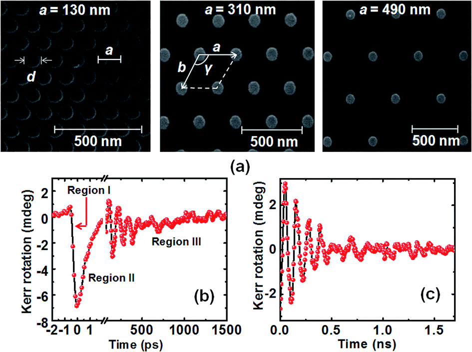 | ||
| Fig. 1 (a) Scanning electron micrographs of the permalloy dot arrays with dot diameter (d) = 100 nm, thickness = 20 nm, arranged in hexagonal lattice with varying lattice constants, a = 130 nm, b = 120 nm; a = 310 nm, b = 315 nm; and a = 490 nm, b = 520 nm. The dot diameter and lattice constants are shown in the micrographs along with the length scales. The unit cell is marked inside the lattice. (b) Typical time-resolved Kerr rotation data for the array with a = 180 nm for H = 1.3 kOe. (c) Background subtracted time-resolved data. | ||
To study the time-resolved magnetization dynamics of these nanodot arrays we used a custom-build TRMOKE microscope.32,34 This technique is based upon a two-colour collinear optical pump-probe geometry. Here, the second harmonic (λb = 400 nm, power = 10 mW, pulse width = 100 fs) of the fundamental laser beam from a mode locked Ti-sapphire laser (Tsunami, Spectra physics) is used to pump the sample by creating a population of hot electrons. This causes a modification of spin population by spin dependent electron scattering process. The fundamental beam (λa = 800 nm, power = 2 mW, pulse width = 80 fs) is used to probe the time varying polar Kerr rotation from the samples. A delay stage situated at the probe path is used to create the necessary time delay with temporal resolution of about 100 fs limited by the cross-correlation of the pump and the probe beams. Finally, both the beams are combined together and focused at the centre of the array by a microscope objective (N.A. = 0.65) in a collinear fashion. The probe beam with spot diameter of about 800 nm is tightly focused and overlapped with the pump having a larger diameter (≈1 μm), at the centre of the array. Under this condition the probe can collect information from the uniformly excited part of the sample. This allows us to measure up to a maximum of 65 elements for the array with smallest lattice constant, whereas a single element was probed for the largest lattice constant. The sample is scanned under the focused pump and probe beams by mounting it on a piezoelectric scanning stage (x–y–z), which gives high stability to the sample in presence of feedback loops. A static magnetic field is applied at a small angle (∼15°) to the sample plane, the in-plane component of which is defined as the bias field H. The time varying polar Kerr rotation is measured at room temperature by using an optical bridge detector and a lock-in amplifier in a phase sensitive manner. The pump beam is modulated at 2 kHz frequency, which is used as the reference frequency of the lock-in amplifier. This detection technique completely isolates the Kerr rotation and reflectivity signals. Fig. 1(b) shows a typical raw data of time-resolved Kerr rotation from the array with a = 180 nm. Here, the ultrafast demagnetization (region l) occurs within first 500 fs from the zero delay, followed by fast (region II) and slow (region III) relaxation processes having relaxation time of 1.3 ps and 700 ps, respectively. The precessional magnetization dynamics is observed as an oscillatory signal on the slowly decaying part of the time-resolved data. Fig. 1(c) shows the time-resolved data after removing the negative delay and ultrafast demagnetization and subtracting a bi-exponential background. Fast Fourier transform (FFT) is performed over this background subtracted data to obtain the power vs. frequency plot. The measurement time window of 1.7 ns is found to be sufficient for resolving the SW spectra in these systems. Although the number of nanodots probed under the focused laser spot varied from about 65 to 1 with increasing areal density, we have improved the sensitivity of our equipment to the extent that even the single dot dynamics is measured with good signal/noise ratio.
Results and discussion
Fig. 2(a) and (b) show the background subtracted experimental time-resolved Kerr rotation data with corresponding FFT power spectra for nanodot arrays with different lattice constants. From the experimental data, a clear variation in the magnetization dynamics is observed with varying lattice constant. Damped nonuniform oscillations consisting of multiple frequency precessional modes are observed for all lattice constants. The corresponding power spectra (Fig. 2(b)) show the presence of three clear SW modes for 130 nm ≤ a ≤ 310 nm. However, there is a sudden downshift in frequencies of all three modes as a changes from 130 to 180 nm, indicating a change in the collective nature of the dynamics. Beyond this, the mode frequencies remain nearly unaltered while the relative intensities and linewidths of the modes change with the increase in lattice constant, indicating a change in the relative powers of the modes. For a ≥ 390 nm, another drastic change occurs and instead of three modes, only two modes are observed. While mode 1 retains its position, mode 2 merges with mode 3 to form a single mode. Consequently, we observe a clear transition from a collective dynamical regime to an isolated regime of dynamics with the variation of lattice constant. | ||
| Fig. 2 (a) Experimental time-resolved Kerr rotation data and (b) corresponding FFT power spectra are shown for the permalloy nanodot lattices arranged in hexagonal symmetry with lattice constant (a) varying from 130 nm to 490 nm at H = 1.3 kOe. (c) FFT power spectra of simulated time-domain magnetization. Mode numbers are shown in both experimental and simulated power spectra. The red and blue dashed vertical lines indicate the positions of edge and centre modes of a single permalloy dot with diameter (d) = 100 nm and thickness = 20 nm. | ||
We further reproduce our experimental data by micromagnetic simulations using OOMMF software.35 The experimental technique is based on optical excitation of magnetization but in the simulation this is achieved by applying a pulsed magnetic field, which reproduces the experimental conditions successfully. The details of simulation can be found elsewhere.21 The samples were discretized into rectangular prisms of dimensions 2 × 2 × 20 nm3 where the lateral cell size is well below the exchange length of permalloy (∼5.2 nm). Material parameters used for permalloy were gyromagnetic ratio γ′ = 18.5 MHz Oe−1, anisotropy field Hk = 0, saturation magnetization Ms = 860 emu cm−3, and exchange stiffness constant A = 1.3 × 10⁻⁶ erg cm−1. The material parameters were extracted by measuring the variation of precessional frequency (f) with bias field for a permalloy thin film and by fitting them using Kittel formula,
 | (1) |
The exchange stiffness constant A is obtained from literature.36 In OOMMF simulation, at first the static magnetic configuration was obtained by applying a large magnetic field to saturate the sample magnetization and then by reducing the magnetic field to bias field value. The system was allowed to reach the equilibrium (maximum torque m × H, where m = M/Ms goes well below 106 A m−1). The magnetization dynamics was simulated by applying a pulsed magnetic field. Calculation of the spatial maps of magnetization at time steps of 10 ps was done for a total duration of 4 ns.
Although the simulation reproduced the main observed features qualitatively (Fig. 2(c)), the quantitative disagreement appears due to some limitations in the simulation such as edge roughness and statistical differences in sizes and shapes between the experimental and simulated nanodots. The average disagreement between the simulated and experimental SW frequencies is maximum (12%) for arrays with higher lattice constants due to the reduction in the amount of magnetic materials, which reduces the signal/noise ratio. Finally, for a ≥ 390 nm, dynamics of a single nanodot of diameter 100 nm is probed. The magnetization in the dot edges gets randomized and affect the stray field distribution around the dot. This leads to the modification of some conventional SW modes in the experimental power spectra and appearance of some other localized modes due to the pinning in magnetization. As a reference, we have also simulated the magnetization dynamics of a single circular nanodot of diameter 100 nm and thickness 20 nm, which reveals two distinct precessional modes. The positions of those two modes are shown by the dotted lines in Fig. 2(c). It is clearly visible from the figure that at higher lattice constants (a ≥ 390 nm) the frequency values of SW modes of the nanodot array are nearly identical to those for a the single circular nanodot of diameter 100 nm.
Fig. 3(a) and (b) show the bias field dependences of SW frequencies extracted from the experimental and simulated results for a = 180 and 390 nm, respectively. The simulated data points corresponding to modes 1, 2 and 3 in Fig. 3(a) are well fitted with Kittel formula. The simulated frequencies are quantitatively slightly different as compared to the experimental frequencies. This is probably due to the unavoidable contribution from the randomized magnetization at the rough edges of the dots, which could not be incorporated in the simulation. The extracted magnetic parameters from the fits are similar to those found earlier for the permalloy thin film except for the saturation magnetization. For the array with a = 180 nm, we obtained Ms values as 700 emu cm−3, 274 emu cm−3 and 153 emu cm−3 for modes 1, 2 and 3, respectively (Fig. 3(a)). As a is increased to 390 nm, we obtain Ms values as 655 emu cm−3 and 226 emu cm−3 for modes 1 and 2, respectively.
 | ||
| Fig. 3 Bias field dependence of precessional frequencies of different SW modes for permalloy nanodot lattices arranged in hexagonal symmetry with (a) a = 180 nm, (b) a = 390 nm and (c) single permalloy dot with diameter = 100 nm and thickness 20 nm (triangular symbols: experimental data, circular symbols: micromagnetic simulation results, solid line: Kittel fit) are plotted as a function of H. | ||
In ref. 6 we already showed that for the array with a = 130 nm, Ms values were obtained as 860 emu cm−3 and 560 emu cm−3 for modes 1 and 2, respectively. For a comparison, we simulated the dynamics of single circular permalloy nanodot with 100 nm diameter and 20 nm thickness and the field dependences of its precession frequencies are shown in Fig. 3(c). The extracted Ms values from the Kittel fit of the field variation of the centre and edge modes of the single nanodot are 672 emu cm−3 and 243 emu cm−3, respectively. From the above results, we understand that when the nanodots are densely packed in an array (a = 130 nm), they show a strongly collective magnetization dynamics and the uniform mode (mode 1) is showing a behavior similar to the precessional mode of a continuous thin film (Ms = 860 emu cm−3). As we increase the inter-dot separation, we enter into weakly collective dynamics and the effective saturation magnetization for the resonant modes reduces due to the reduction in the inter-dot interaction field, and as we increase the inter-dot separation further to a ≥ 390 nm, we enter into a regime of dynamics of isolated dots with effective Ms values similar to that of a single nanodot.
The degradation in precessional signal quality with decrease in areal density introduces line broadening in the FFT spectra. So the simulated data points in Fig. 3(b) qualitatively follow the trend of experimental results but there is quantitative disagreement between the two.
We have further simulated the power and phase maps of various observed SW modes by using a home built code37 as shown in Fig. 4. The spatial profiles of the power and phase information for various modes are obtained by fixing one of the spatial coordinates in the space and time-dependent magnetization and then by performing a discrete Fourier transform with respect to time domain.37 It is well known that for the single circular nanodot the higher and lower frequency modes correspond to the centre and edge modes.6,32 The SW spectra for array with a = 180 nm showed a drastic change from that for a = 130 nm, indicating a change in the collective nature of the mode. This was further confirmed from the bias field dependence of the mode frequencies.
 | ||
| Fig. 4 The power and phase maps for different collective SW modes of permalloy dot lattices arranged in hexagonal symmetry with lattice constants (a) a = 180 nm, (b) a = 310 nm and (c) a = 490 nm at a bias field of H = 1.3 kOe. The colour bars of power and phase maps are shown at the bottom right corner of the image. Here power is given in arbitrary unit, while phase is given in radian. Sizes of the dots are not in scale. | ||
Fig. 4(a) shows although the natures of mode 1 is similar to that for a = 130 nm, there are indeed some changes in modes 2 and 3. Instead of a backward volume-like collective mode of the array, mode 2 here corresponds to a nearly uniform distribution of the edge modes of the dots in the array. On the other hand, the bow-tie like mode (mode 3) as observed in a = 130 nm6 is now transformed to a collective edge mode of the array for a = 180 nm. With increasing lattice constant (a), the strength of collective nature of SW spectra reduces further causing a sudden change in the nature of modes. A new collective SW mode (mode 2) appears with its frequency in between that of the uniform distribution of conventional edge mode (mode 3) and centre mode (mode 1) over the array as shown for a = 310 nm in Fig. 4(b). Here, mode 2 corresponds to a non-uniform collective mode with power of the SW mode concentrated along each alternative column as shown by the dotted boxes. The phase profile shows that the spins precess in phase within the dotted boxes but the spins of two consecutive columns are out of phase with each other. Thus, it forms collective backward volume like standing SW mode directed along the applied bias field. With further increase in lattice constant the dynamics enters into an isolated regime, which is also clear from the SW mode profiles. For the sample with a = 490 nm (Fig. 4(c)) modes 1 and 2 correspond to the centre and edge modes, respectively which are distributed uniformly over entire lattice. These modes have same frequencies and mode profiles as a single nanodot.
We further simulated the SW mode profiles for samples with a = 180 and 310 nm at two different magnetic fields to understand how the collective behavior is affected by the bias field.
Fig. 5(a) and (b) show that for the array with a = 180 nm, the nature of all three modes remain qualitatively same when the bias field is reduced from 1.3 kOe to 0.62 kOe. Only the power in the edges of the dots gets stronger for mode 2 for the lower field value as the contribution from stray field becomes more prominent when the bias field is weaker. Similarly, Fig. 5(c) and (d) show that for a = 390 nm, two distinct modes remain same as centre mode and edge mode of the whole array with the reduction of bias magnetic field. Hence, a variation of bias magnetic field does not cause a transition between different collective regimes but only causes small changes in the relative powers of the modes.
 | ||
| Fig. 5 The power maps of collective SW modes in permalloy nanodot arrays with lattice constants (a), (b) a = 180 nm and (c), (d) a = 390 nm. The applied bias fields are H = 0.78 kOe ((a), (c)), 0.62 kOe (b) and 0.52 kOe (d). The colour bar of power profile is shown at the bottom right corner of the image. Here power is given in arbitrary unit. Sizes of the dots are not in scale. | ||
To gain more insight into the dynamics, we have simulated the magnetostatic field distribution of the arrays by using LLG micromagnetic simulator as shown in Fig. 6. The contour plots of the magnetostatic fields show that dipolar contribution from the magnetostatic stray field dominates for all arrays but the density of the interacting field lines reduces significantly with the increase in lattice constant and nearly vanishes for higher lattice constants (Fig. 6(c)). Consequently, the dynamics of arrays of higher lattice constants are similar to that of a single circular nanodot of diameter 100 nm.
 | ||
| Fig. 6 The contour maps of simulated magnetostatic field distribution (x component) are shown for permalloy nanodot arrays arranged in hexagonal symmetry with lattice constants of (a) a = 180 nm, (b) a = 310 nm and (c) a = 490 nm. The arrows inside the dots represent the magnetization states of the dots and the strength of magnetostatic field is represented by the colour bar given at the bottom right corner of the figure. The red horizontal lines represent the position of the lattice from where the line scans have been taken. Sizes of the dots are not in scale. | ||
To quantify the inter-dot interaction, we took line scans along the solid red lines as shown in Fig. 6(a)–(c) plotted in Fig. 7(a). For a = 130 nm, the magnitude of stray field between two consecutive dots is maximum (∼3500 Oe), which sharply decreases with the increase in the lattice constant and becomes negligibly small for a > 310 nm. The inter-dot interactions across the arrays are modelled simply by considering arrays of nanomagnets behaving like parallely placed magnetic dipoles. Considering the center to center distance (r) between two consecutive nanomagnets (as shown in the inset of Fig. 7(b)) as the lattice constant (a) of the array, the magnetic field (B) of a coupled dipole system orienting in same direction can be expressed as,
 | (2) |
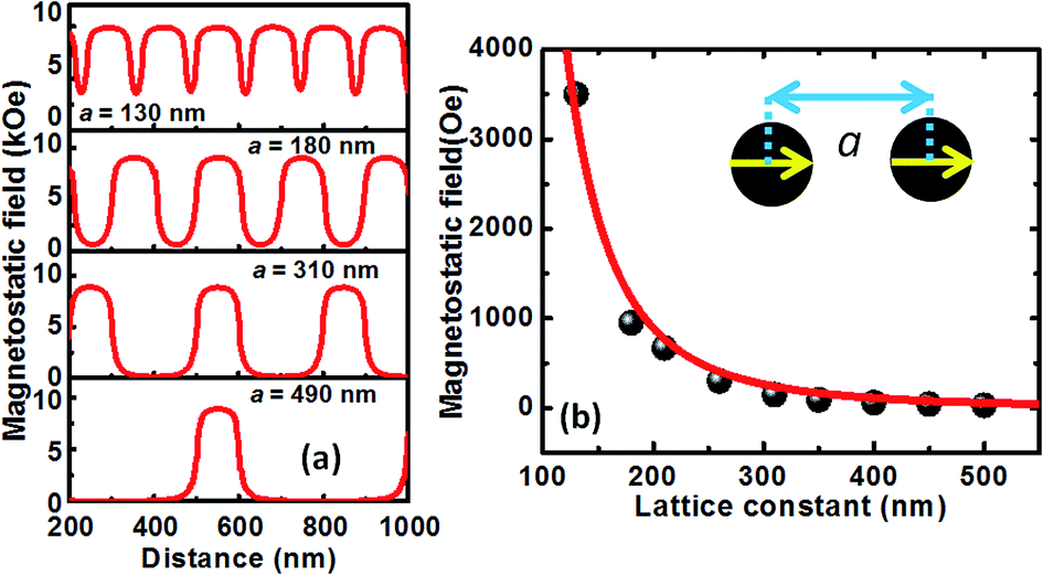 | ||
| Fig. 7 (a) Line scans of simulated magnetostatic fields from the arrays with different lattice constants (a) as obtained from the positions indicated by horizontal solid lines (as shown in Fig. 6). (b) The variation of magnetostatic stray field with the lattice constant (circular symbols: micromagnetic simulation, solid line: fitted curve). The schematic of the dipolar coupled dots is shown in the inset. | ||
Fig. 7(b) shows a good fit of the variation of simulated magnetostatic stray field with lattice constant using eqn (2). This indicates that the inter-dot interaction is of purely dipolar in nature as opposed to square nanodots, which showed quadrupolar interaction to be dominant.21 From Fig. 5(b), it is clear that at a = 130 nm, the system is strongly coupled via magnetostatic interaction which can guide a broad spectrum of long wave length SW to carry and process information. Hence, they can be considered as promising candidate for magnonics applications, whereas for a > 310 nm, the arrays behave like magnetostatically isolated dots, which can be used to store bits, free from any external disturbances.
Conclusion
In essence, we have investigated the variation in collective magnetization dynamics in permalloy circular nanodot arrays arranged in hexagonal symmetry with systematic variation of inter-dot separation, by using time-resolved Kerr microscopy. The experimental results along with the numerical analysis, reveal that the nature of magnetization dynamics gets modified significantly with inter-dot separation. For smaller separation, the dots in the array strongly interact with each other leading towards a strongly collective dynamics with modes different from the modes of the individual nanodots. With the increase in inter-dot separation the dynamics goes through weakly collective regimes with the appearance of new modes in addition to the uniform distribution of the centre and edge modes of the individual dots in the array. At larger separation the system enters into an isolated regime with only the modes of the individual nanodots. Further, the bias field dependence of the SW modes reveals that the modes are robust enough to sustain the variation in external bias field. No qualitative changes are observed with the decreasing bias field values barring a variation in relative powers of the modes. These salient features of dot arrays with hexagonal lattice symmetry can make them viable for construction of various nanomagnet based devices. For smaller separations the arrays can be used as SW band-pass filter in GHz regime whereas for higher lattice constants, reduction in magnetostatic coupling strength makes these nanodot arrays suitable for storing information within them. Besides these, the dots in the arrays, are arranged in hexagonal lattice symmetry which is the most closely packed structure. Consequently, they will be capable of maintaining reasonable signal to noise ratio for much smaller nanodots leading towards their potential applications. The above experimental findings along with circumstantial numerical analyses have a pragmatic approach towards the development of nanomagnonic devices and patterned magnetic media.Acknowledgements
The authors gratefully acknowledge the financial supports from the Department of Science and Technology, Government of India under Grant No. SR/NM/NS-09/2011(G) and S. N. Bose National Centre for Basic Sciences, India (Grant No. SNB/AB/12-13/96). S. M. acknowledges DST for INSPIRE fellowship and S. C. acknowledge S. N. Bose National Centre for Basic Sciences. We also acknowledge Susmita Saha for technical assistances.References
- V. V. Kruglyak, S. O. Demokritov and D. Grundler, J. Phys. D: Appl. Phys., 2010, 43, 264001 CrossRef.
- B. Lenk, H. Ulrichs, F. Grabs and M. Munzenberg, Phys. Rep., 2011, 507, 107 CrossRef.
- S. A. Nikitov, Ph. Tailhades and C. S. Tsai, J. Magn. Magn. Mater., 2001, 236, 320 CrossRef CAS.
- A. V. Chumak, A. A. Segra, B. Hillebrands and M. P. Kostylev, Appl. Phys. Lett., 2008, 93, 022508 CrossRef.
- Z. K. Wang, V. L. Zhang, H. S. Lim, S. C. Ng, M. H. Kuok, S. Jain and A. O. Adeyeye, ACS Nano, 2010, 4, 643 CrossRef CAS PubMed.
- S. Saha, R. Mandal, S. Barman, D. Kumar, B. Rana, Y. Fukuma, S. Sugimoto, Y. Otani and A. Barman, Adv. Funct. Mater., 2013, 23, 2378 CrossRef CAS.
- J. Topp, D. Heitmann, M. P. Kostylev and D. Grundler, Phys. Rev. Lett., 2010, 104, 207205 CrossRef PubMed.
- M. Krawczyk and D. Grundler, J. Phys.: Condens. Matter, 2014, 26, 123202 CrossRef CAS PubMed.
- C. T. Black, C. B. Murray, R. L. Sandstrom and S. Sun, Science, 2000, 290, 1131 CrossRef CAS PubMed.
- J. Shen and J. Krischner, Surf. Sci., 2002, 500, 300 CrossRef CAS.
- M. Todorovic, S. Schultz, J. Wong and A. Scherer, Appl. Phys. Lett., 1999, 74, 2516 CrossRef CAS.
- T. W. McDaniel, J. Appl. Phys., 2012, 112, 093920 CrossRef.
- A. Haldar and A. O. Adeyeye, ACS Nano, 2016, 10, 1690 CrossRef CAS PubMed.
- V. V. Kruglyak, P. S. Keatley, A. Neudert, R. J. Hicken, J. R. Childress and J. A. Katine, Phys. Rev. Lett., 2010, 104, 027201 CrossRef CAS PubMed.
- G. Gubbiotti, M. Madami, S. Tacchi, G. Socino, G. Carlotti and T. Okuno, Surf. Sci., 2006, 600, 4143 CrossRef CAS.
- B. Rana, S. Pal, S. Barman, Y. Fukuma, Y. Otani and A. Barman, Appl. Phys. Express, 2011, 4, 113003 CrossRef.
- R. P. Cowburn, D. K. Koltsov, A. O. Adeyeye, M. E. Welland and D. M. Tricker, Phys. Rev. Lett., 1999, 83, 1042 CrossRef CAS.
- J. Ding and A. O. Adeyeye, Adv. Funct. Mater., 2012, 23, 1684 CrossRef.
- V. V. Kruglyak, A. Barman, R. J. Hicken, J. R. Childress and J. A. Katine, J. Appl. Phys., 2005, 97, 10A706 CrossRef.
- A. Barman, S. Wang, J. D. Maas, A. R. Hawkins, S. Kwon, A. Liddle, J. Bokor and H. Schmidt, Nano Lett., 2006, 6, 2939 CrossRef CAS PubMed.
- B. Rana, D. Kumar, S. Barman, S. Pal, Y. Fukuma, Y. Otani and A. Barman, ACS Nano, 2011, 5, 9559 CrossRef CAS PubMed.
- S. Saha, S. Barman, Y. Otani and A. Barman, Nanoscale, 2015, 7, 18312 RSC.
- S. Jung, B. Watkins, L. DeLong, J. B. Ketterson and V. Chandrasekhar, Phys. Rev. B: Condens. Matter Mater. Phys., 2002, 66, 132401 CrossRef.
- O. N. Martyanov, V. F. Yudanov, R. N. Lee, S. A. Nepijko, H. J. Elmers, C. M. Schneider and G. Schonhense, Appl. Phys. A, 2005, 81, 679 CrossRef CAS.
- G. Gubbiotti, G. Carlotti, T. Okuno, M. Grimsditch, L. Giovannini, F. Montoncello and F. Nizzoli, Phys. Rev. B: Condens. Matter Mater. Phys., 2005, 72, 184419 CrossRef.
- S. Tacchi, M. Madami, G. Gubbiotti, G. Carlotti, H. Tanigawa, T. Ono and M. P. Kostylev, Phys. Rev. B: Condens. Matter Mater. Phys., 2010, 82, 024401 CrossRef.
- A. Haldar, D. Kumar and A. O. Adeyeye, Nat. Nanotechnol., 2016, 11, 437 CrossRef CAS PubMed.
- S. K. Kim, J. Phys. D: Appl. Phys., 2010, 43, 264004 CrossRef.
- D. Kumar, S. Barman and A. Barman, Sci. Rep., 2014, 4, 4108 CAS.
- V. V. Kruglyak, A. Barman, R. J. Hicken, J. R. Childress and J. A. Katine, Phys. Rev. B: Condens. Matter Mater. Phys., 2005, 71, 220409 CrossRef.
- B. K. Mahato, B. Rana, D. Kumar, S. Barman, S. Sugimoto, Y. Otani and A. Barman, Appl. Phys. Lett., 2014, 105, 012406 CrossRef.
- B. Rana and A. Barman, SPIN, 2013, 3, 1330001 CrossRef.
- S. Saha, S. Barman, S. Sugimoto, Y. Otani and A. Barman, RSC. Adv., 2015, 5, 34027 RSC.
- A. Barman and A. Haldar, Solid State Phys., 2014, 65, 1 Search PubMed.
- M. Donahue and D. G. Porter, OOMMF User's guide, Version 1.0, NIST Interagency Report no. 6376, National Institute of Standard and Technology, Gaithersburg, MD, 1999, http://math.nist.gov/oommf Search PubMed.
- K. H. J. Buschow, J.: Handbook of Magnetic Materials, North Holland, Amsterdam, 2009. vol. 18, p. 168 Search PubMed.
- D. Kumar, O. Dmytriiev, S. Ponraj and A. Barman, J. Phys. D: Appl. Phys., 2012, 45, 015001 CrossRef.
| This journal is © The Royal Society of Chemistry 2016 |