Magnetic field induced push–pull motility of liquibots

Sunny Kumara, Md Rashid Ali Faridia, Ashok Kumar Dasmahapatra*ab and Dipankar Bandyopadhyay*ab
aDepartment of Chemical Engineering, Indian Institute of Technology Guwahati, Assam 781039, India. E-mail: akdm@iitg.ernet.in; dipban@iitg.ernet.in
bCentre for Nanotechnology, Indian Institute of Technology Guwahati, Assam 781039, India

Received 20th August 2016 , Accepted 3rd November 2016

First published on 3rd November 2016


Abstract

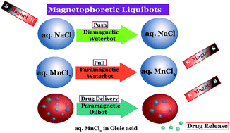
Liquid droplets loaded with paramagnetic or diamagnetic salts, namely liquibots, showed controlled migration inside a fluid medium and on slippery solid surfaces under remote magnetic guidance. The water or oilbots of size ranging from a millimetre to a few microns showed facile attraction, repulsion, division, and coalescence when guided by a magnetic field. The speed of the liquibots could be tuned by varying the size, salt-loading, and magnetic field strength. While the paramagnetic liquibot migrated towards a magnet with a velocity as high as ∼8 body length per s, the diamagnetic one migrated away from the field with a maximum velocity of ∼1 body length per s. The liquibots transported and delivered commercially available drugs to targeted locations showing their potential as drug-delivery vehicles. Remarkably, the experiments showed the utility of the liquibots in digital microfluidics because they moved easily on slippery solid surfaces. For example, a waterbot was split into many droplets on an oil coated solid surface before forming the patterns resembling polygons under magnetic guidance. Further, the liquibot based Packman™ game could also be played with the help of magnetic guidance. The extent of control demonstrated on the motions of the remotely guided liquibots could be useful in diverse futuristic applications including drug-transport, digital-microfluidics, and droplet-electronics.


Introduction

In recent times droplet microfluidics has shown significant potential in staging important paradigm shifts in technologies related to biomolecule detection, drug-delivery, 3-D printing, therapeutics, diagnostics, and digital electronics.1–5 In particular, self-motile microscale digitized objects guided by various in situ or external stimulants have shown their remarkable ability as next generation transporters of essential vitamins, antibiotics, minerals, anti-cancer agents, or enzymes to targeted locations.6–8 A number of pioneering works have shown that these futuristic devices can be propelled by the Marangoni effect,9,10 chemiosmotic force,11 an electric field,12 photonic13 or acoustic14 excitations, and electromagnetic fields.15,16 These studies also unveiled pathways that include the specialties of nanoscale particles, rods, tubes, or fibres on the solid or soft-solid objects targeting various cutting-edge applications.17–19 In comparison, the liquid micro-propellers having efficacies very similar to or better than their nano-enabled solid or soft-solid counterparts have started making appearance in the academic arena only in the recent years.11,20 However, integrating the specialties of these nanoscale materials into the liquid droplets has largely been limited by the conditional agglomeration of solids from the bulk, which has somewhat clipped the flight of the applications associated with the droplet based locomotions.21,22

In this direction, one of the major challenges has been to infuse magnetic properties to the micro or nanoscopic liquid droplets.23 A droplet suspended with the magnetic micro or nanoscale particles or fibres often encounter the problem of phase-separation of the nanoparticles or nanofibres due to rapid nucleation followed by settling.24 The problem becomes more severe in presence of an external magnetic field where the nanoscopic objects are preferentially attracted towards the magnet causing a faster solid–liquid separation from a suspension.25 Previous studies suggest that the use of surfactant coated nanoparticles can be one of the temporary solutions to stabilize solid–liquid suspensions at the microscale26 and delay the phase separation.27 Alternatively, use of paramagnetic ionic liquids and ferrofluids has also been suggested to resolve this problem.28–31 However, most of these solutions are either temporary or costly or toxic. Further, infusing different types of magnetic properties to the microdroplets composed of commonly available materials such as water or oil has been an open challenge so far.

Fig. 1 schematically shows that liquid droplets composed of aqueous solutions of the paramagnetic or diamagnetic salts could engender interesting magnetic field induced motions. The transparent to translucent to opaque salt-laden droplets, namely, the ‘liquibots’ could show facile pull, push, veer, halt, or reverse motions under the remote guidance of an external magnet. For example, a ‘waterbot’ loaded with paramagnetic salt could be pulled while the same loaded with diamagnetic salt could be pushed with the help of a magnetic guidance.


image file: c6ra20948c-f1.tif
Fig. 1 Schematic illustration of liquibots undergoing magnetic field induced motion. A diamagnetic waterbot (aq. NaCl soln) showing a push motion, a paramagnetic waterbot (aq. MnCl2 soln) showing a pull motion, and a paramagnetic oilbot (droplets of aq. MnCl2 soln suspended in oleic acid) showing pull and delivery motions.

The speed of the liquibots could be modulated by altering the size, magnetic field strength, and salt-loading. In addition, the proposed liquibots could be split into parts or joined or arranged in an ordered manner on a slippery solid surface, which was previously possible only through ferrofluids.31 The magnetic field guided actuation and migration shown by the liquibots indicated that they might be suitable for a number of recently proposed droplet microfluidic applications based on electric field.32,33 Importantly, we have also shown a recipe to synthesize water-in-oil liquibots, which could fluoresce, transport as well as release drugs to the targeted locations through remote magnetic guidance. The reported prototype is expected to improve the efficiency of many recently reported droplets based sensors,34,35 drug transporters,36–38 environmental remediates,39 and microreactors.40–43

Fig. 2(a–d) and ESI video S1 show that a transparent paramagnetic waterbot composed of 0.5 M aqueous MnCl2 was ‘pulled’ by an electromagnet having field strength of 148 mT on a chloroform bath at a maximum speed of 3.44 mm s−1. In comparison, Fig. 2(e–h) and ESI video S1 show that a transparent diamagnetic waterbot composed of aqueous solution of 1 M NaCl could be ‘pushed’ by a permanent magnet of strength 120 mT at a maximum speed of ∼1.4 mm s−1 on the same bath. Control experiments suggested that a de-ionized (DI) waterbot was unable to show such motions on the same chloroform bath. Both the diamagnetic and paramagnetic waterbots moved at the air–chloroform interface because of the higher density of chloroform bath than the waterbots. ESI video S2 show a contrasting pull and push (attractive and repulsive) reciprocating motion of the 10 μL transparent paramagnetic (right side) and diamagnetic (left side) waterbots when placed near a permanent magnet of strength 240 mT. In the ESI video S3, we have shown the motion of a miniaturized version of the paramagnetic waterbot of diameter of 40 μm moving at a speed of ∼2 body lengths per second when placed near a permanent magnet of strength 80 mT.


image file: c6ra20948c-f2.tif
Fig. 2 Images (a–d) show the magnetic field induced ‘pull’ motion of a 5 μL paramagnetic waterbot loaded with 0.5 M MnCl2 at 0 s, 0.8 s, 2.08 s, and 3.2 s, respectively, on a Petri dish filled with chloroform and under the influence of an electromagnet of strength 148 mT. Images (e–h) show the magnetic field induced ‘push’ motion of 3 μL diamagnetic waterbot loaded with 1 M NaCl at 0 s, 2.48 s, 6.32 s, and 10 s, respectively, on chloroform filled Petri dish and under the influence of a permanent magnet of strength 120 mT. The minimum box dimension of graph was 1 mm × 1 mm.

Fig. 3(a–d) and ESI video S4 show that an opaque paramagnetic waterbot loaded with paracetamol drug (0.5 M) was pulled by a magnet on a chloroform bath at a maximum speed of 2 mm s−1. In such a situation, the fluorescent tracer provided an optical indication of the local position of the motile droplet, as shown in the Fig. 3(e–h) and the ESI video S5. Fig. 1–3 and the ESI videos 1–5 together showed diverse pull–push migrations of the waterbots, which can be of importance for in vivo applications of the proposed liquibots.


image file: c6ra20948c-f3.tif
Fig. 3 Images (a–d) show the magnetic field induced motion of a drug loaded paramagnetic waterbot at 0 s, 3 s, 5 s, and 7 s, respectively, Petri dish filled with chloroform and under the influence of an electromagnet of strength 148 mT. The minimum box dimension of graph was 1 mm × 1 mm. Images (e–h) show the motion of a paramagnetic waterbot tagged with a fluorescent probe under the influence of a permanent magnet of strength 80 mT. The scale bar in these images represents 1 mm.

The magnetic force exerted on the waterbot could be evaluated from the expression, Fm = Vχ/μ)B·∇B,44,45 in which ∇ was the gradient operator, B was the magnetic field vector, V was the volume of the droplet, Δχ = χpMC + χwV was the difference in the magnetic susceptibility where χpM and χwV are the molar and volume susceptibilities of salt and water, C was the concentration of the salt, and μ was the magnetic susceptibility of vacuum (4π × 10−7 N A−2). The theoretical average velocity of the droplet could be obtained by applying the Newton's second law of motion (Fm + Fd = ma) in which the drag force on the droplet was evaluated by the Stokes formula, Fd = 6πηrv.46 Where m and a were the mass and acceleration of the droplet, η was the viscosity of the bounding medium, r was the radius of the droplet, and v was the average velocity of the droplet.

We measured the time to traverse 1 cm distance to obtain the experimental average velocity of the liquibots. Further, to evaluate the theoretical average velocity we enforced Newton's 2nd law of motion in the Stokes flow limit, Fm + Fd = 0, which provided the expression for theoretical velocity of the droplet as, v = [(2r2Δχ)/(9η)μ]B·∇B. The expression could further simplify to, v = [(2r2Δχ)/(9ημ)]Bx(∂Bx/∂x), for unidirectional magnetic field where x is the direction of the movement. Plots (a)–(d) in the Fig. 4 show a comparison between the average velocities (Vm) obtained from the theoretical calculations (solid line) and experiments (symbols) with the variations in volume of the paramagnetic waterbot (ϕ), salt concentration (CMn), magnetic field strength (B), and volume of the diamagnetic droplet (ϕ). Table 1 shows the typical properties employed for the calculations in the present manuscript.


image file: c6ra20948c-f4.tif
Fig. 4 Plot (a) shows the variation in the average velocity (Vm) of paramagnetic waterbot with its volume (ϕ) when the concentration of MnCl2 was CMn = 0.5 M, magnetic field strength was B = 148 mT, and viscosity of the waterbot and medium was, η = 0.001 Pa s and ηm respectively. Plot (b) shows the variation of Vm with CMn when B = 148 mT, η = 0.001 Pa s, and ϕ = 5 μL. Plot (c) shows the variation in Vm with B when CMn = 0.5 M, ϕ = 5 μL, and η = 0.001 Pa s. Plot (d) shows the variation in Vm of diamagnetic waterbot with ϕ when CNa = 1 M, B = 240 mT, and η = 0.001 Pa. Plot (e) shows the variation in Vm of paramagnetic waterbot with the viscosity of the medium (η) when CMn = 0.5 M, B = 148 mT, and ϕ = 5 μL. The symbols (solid lines) represent experiments (theoretical prediction).
Table 1 Physical properties of fluid in the experiments47–50
η (Pa s) ρ (kg m−3) Mw (MnCl2·4H2O) χMnM (m3 mol−1) χNaM (m3 mol−1) χwV
0.001 1000 198 1.8 × 10⁻⁴ 30 × 10−6 9 × 10⁻⁶


The plots (a)–(c) suggest that Vm monotonically increased with the increase in ϕ, CMn, and B. Interestingly, the proposed theoretical model could not only predict the trend of the variations in Vm with ϕ, CMn, and B but also quantitatively corroborated the experimentally measured values. The Vm attained by a 2 μL paramagnetic waterbot was ∼2.19 × 10⁻³ ms−1, which increased to ∼6.34 × 10⁻³ ms−1 for a ∼9 μL waterbot. Further, a waterbot containing 2 M MnCl2 showed a speed as high as 11.9 × 10⁻³ ms−1 when the applied field strength was 148 mT. Increase in volume, salt loading, and magnetic field strength led to the manifestation of larger amount of magnetic body force on the waterbot. Plot (d) shows that even for a diamagnetic waterbot Vm monotonically increased with ϕ. We also observed (not reported here) that Vm increased with the NaCl loading (CNa) inside the droplet. The Vm attained by the liquid motor was ∼1.36 × 10⁻³ ms−1 when CNa ∼1 M and B was 240 mT. However, with the increase in CNa to ∼5 M, Vm increased ∼1.95 × 10⁻³ ms−1. Further, with increase in ϕ, Vm increased to ∼2.66 × 10⁻³ ms−1 for a ∼10 μL waterbot. The plot (d) showed that diamagnetic waterbots could also be pushed strongly with the help of remote magnetic guidance inside a fluidic environment, which was also in well accordance with the theoretically predicted average velocity.

Plot (e) show the variation in Vm with the change in the viscosity of the bounding medium (η). The theoretical formula indicated the Vm versus η plots to be a rectangular hyperbola, as depicted in the figure by the broken (η = √ηwηm) and the solid lines (η = ηm). Here the subscripts ‘m’ and ‘w’ indicate water and bounding medium, respectively. The symbols in the plot show the experimentally obtained values. The plots suggest that the theoretical and experimental values matched reasonably well in the limits when the medium viscosity was either high or low. At the intermediate values they deviated significantly. We observed that when the exact experimental values were used for the theoretical calculations (solid line) the deviation was significant in the intermediate domain. Instead, when the geometric mean of the viscosity of the waterbot and the medium (broken line) was employed for the calculations of the Vm, the deviation was significantly less. Further, with a little change in the radius of the waterbots, v ∞ (1/r2), the differences could also be bridged or widened significantly. Thus, it was apparent that the use of Stokes law for a flow past circular solid object was not very appropriate to predict exactly the velocities of the proposed waterbots. The theoretical model employed was able to predict the velocities rather qualitatively and needed additional considerations such as, (i) the liquibot was floating on the chloroform bath having a deformable water–air interface at the top and another deformable water–chloroform interface at the bottom; (ii) liquidbot was strongly slipping on the water–chloroform interface; and (iii) the exact shape of the liquibot was not exactly a sphere, among others. The difference in the magnitude of the values in the experiments and the theoretical predictions could be bridged through a more rigorous analysis of the force balance equations, which was kept as a future scope of research work.

Apart from the waterbots, we also prepared emulsion based oilbots where a 1 M aqueous MnCl2 solution was mixed with commercially available paracetamol before shaken vigorously with oleic acid at different volumetric proportions. The drug and the paramagnetic salt were initially loaded in the water phase and then the drug loaded waterbots were dispersed through shaking inside oleic acid to prepare the oilbots. Fig. 5(a) schematically shows the transport and release of the drug loaded waterbots from the oilbots under magnetic guidance. The optical image shows the dispersion of waterbots in the oil matrix. Images 5 (b–e) and ESI video 6 show motion of the drug-loaded oilbot with the help of permanent magnet (80 mT) in a water bath. The images and video show the transport of the oilbot loaded with the drug embedded waterbots under the remote magnetic guidance before the release of the drug ∼16.9 s in the bath. The plot (f) shows that the average velocity (Vm) of these oilbots could be modulated by tuning the salt loading in the waterbots. The plot (g) suggests that the time for drug release (tr) could also be controlled by tuning the water to oil loading in the oilbot. In particular, dispersing lower amount of water into the oil-phase showed a larger time of release under these conditions. Concisely, Fig. 5 and ESI video 6 suggest that an emulsion of paramagnetic waterbots in oil can be employed for drug transport and delivery under magnetic guidance. The drug loaded paramagnetic waterbots having dimensions of few microns to few hundred microns dispersed inside the oilbots could act as in vivo transporters of drugs in diverse body fluids51,52 such as blood stream, cerebrospinal fluid, mucus, lymph, bile, saliva, tear, or extracellular fluids.


image file: c6ra20948c-f5.tif
Fig. 5 Image (a) shows the schematic representation of transport, delivery, and release of an oilbot having dispersed paramagnetic waterbots trapped in a continuous oil phase. The optical micrograph shows the typical dispersion of microscale waterbots in the oil matrix. Images (b–e) show the magnetic field induced motion of an oilbot loaded with paracetamol drug at 0 s, 3 s, 5.8 s, and 16.9 s, respectively, and subsequent release of the drug when ϕ = 10 μL and B = 80 mT. The scale bar in the image represents 2.5 mm. Plot (f) shows the variation in Vm of the oilbot with the salt loading in the water phase (CM) when ϕ = 5 μL and B = 240 mT. Plot (g) shows the time of release of the waterbots (tr) for different water to oil ratios, respectively.

A typical phase separation experiment of oleic acid and water shown in the Fig. 6 supported the observations of the release in the Fig. 5. In the beginning of this experiment, different water-in-oil emulsions were prepared by mixing 1 M aqueous MnCl2 with oleic acid through adequate shaking at proportions (v/v), 1[thin space (1/6-em)]:[thin space (1/6-em)]1, 1[thin space (1/6-em)]:[thin space (1/6-em)]2, 1[thin space (1/6-em)]:[thin space (1/6-em)]3 and 1[thin space (1/6-em)]:[thin space (1/6-em)]5, as shown in the rows (a) and (b). The images (c) show that the 1[thin space (1/6-em)]:[thin space (1/6-em)]1, 1[thin space (1/6-em)]:[thin space (1/6-em)]2, 1[thin space (1/6-em)]:[thin space (1/6-em)]3 and 1[thin space (1/6-em)]:[thin space (1/6-em)]5 emulsions phase separated at different time intervals 0.67, 3.12, 4.37, and 9.12 min, respectively. It may be noted here that the use of surfactants or surface active agents in these experiments could further help in modulating the time for mixing and phase separation. The experiment indicated that the release mechanisms of the oilbots could efficiently be designed by tuning the tr of these emulsions to ensure targeted release at stipulated time.


image file: c6ra20948c-f6.tif
Fig. 6 Preparation of water-in-oil (W/O) emulsions (v/v) of water (transparent) and oleic acid (pale yellow). Row (a) showing the images of different initial volumes in the vials having W/O ratios of, 1[thin space (1/6-em)]:[thin space (1/6-em)]1, 1[thin space (1/6-em)]:[thin space (1/6-em)]2, 1[thin space (1/6-em)]:[thin space (1/6-em)]3, and 1[thin space (1/6-em)]:[thin space (1/6-em)]5, respectively. Row (b) shows the situation of the same vials after vigorously shaking for 5 min. Row (c) shows the vials after, 0.67, 3.12, 4.37 and 9.12 min when complete phase separation took place.

Remarkably, apart from the facile movement inside the liquid mediums, the waterbots could also show controlled migration on the solid surfaces coated with thin oil layer, as shown in the Fig. 7a and ESI video 7. In this case, initially dialling patterns were drawn on a white paper with the help of a pen and then covered by a transparent plastic sheet. Following this, the transparent sheet was coated with a thin layer of oleic acid to reduce the surface friction before dispensing FeCl3 loaded paramagnetic waterbots. The camera recorded the motion from the top while the magnetic control was offered from the bottom of the paper surface. The magnet was not visible in the figures or video because it was masked by the opaque paper surface. The broken line on the image shows the migration path of the waterbot with the remote magnetic guidance on the solid surface. Using a similar setup, we performed the experiments shown in the Fig. 7(b and c) and ESI video 8, which show that the smaller waterbots from different corners could be merged and coalesced into a bigger one emulating the popular ‘Packman’™ game on a solid surface.


image file: c6ra20948c-f7.tif
Fig. 7 Controlled migration of FeCl3 loaded paramagnetic waterbots. Image (a) shows the intricate movement represented by the broken line representing telephone dialling when, ϕ = 5 μL, CFe = 2 M, B = 80 mT, and η = 0.001 Pa s. The scale bar in the image represents 5 mm. Images (b) and (c) show magnetic field induced merging of waterbots emulating the ‘Packman’™ game, when, CFe = 2 M, B = 80 mT, and η = 0.001 Pa s.

Fig. 8 and ESI video 9 together show that a paramagnetic waterbot (CFe, 2 M aqueous FeCl3) could be split into multiple droplets with the remote guidance of the magnet. Again, in this experiment, a paper was initially coated with a transparent sheet before a thin layer of oleic acid was coated on it to reduce the friction of the waterbot dispensed on it. The magnet was placed underneath the surface while the camera recorded the motion from the top. The movement of the magnet pulled the paramagnetic waterbot towards it against the surface friction on the slippery surface, which caused the splitting of the waterbot into parts.


image file: c6ra20948c-f8.tif
Fig. 8 Droplet splitting phenomena. Image (a) shows a schematic diagram of breaking a single waterbot into many and simultaneously ordering them. Image (b–g) show the stage wise splitting of a single FeCl3 waterbot into 2–7 droplets and then ordering them in the shape of a line, triangle, pentagon, hexagon, and heptagon, respectively. Here CFe = 2 M, B = 80 mT, and η = 0.001 Pa s. The scale bar shown here is 5 mm.

Image (b–g) show the splitting of a parent waterbot into 2–7 daughters and then ordering them in the shape of a line, triangle, pentagon, hexagon, and heptagon, respectively. A very recent work had shown similar happenings with a droplet composed of ferrofluid.31 Here we could achieve analogous findings employing a much simpler, non-toxic, and transparent system in place. In the present case, the paramagnetic waterbots behaved like a magnet under the influence of magnetic field, which underwent cohesive failure due to viscous stress at the droplet-oil interface53 to cause splitting while moving under the magnetic guidance on a slippery solid surface coated with a thin oil layer.

The magnetically guided droplet actuation shown in the Fig. 7 and 8 had various advantages in the domain of droplet microfluidics over the previously reported methodologies, which employ the electric field as the driving force.32,33 For example, (a) the electrically modulated migrations required high intensity AC or DC fields while the proposed methodology suggested the use of magnets for similar operations; (b) the embedded electrical circuits for electrically actuated drop migration were prone to intense heat generation, which could be avoided employing the proposed methodology; (c) the embedded electrical circuit for the electric field induced motions required complex surface patterning of electrodes employing costly fabrications methodologies. In comparison, the proposed methodology was simple to operate, devoid of the problems associated with heat generation, independent of any circuitry, and did not demand any costly fabrication technique for preparation.

Conclusions

We demonstrated attractive (repulsive) motions of paramagnetic (diamagnetic) salt laden liquibots under remote magnetic guidance.

(1) The non-toxic oilbots or waterbots were prepared employing simple methodologies in which either the salt was dissolved in water or the salt laden water medium was emulsified in oil. This is in stark contrast to the preparation of the previously reported nano-enabled solid or soft-solid self-propellers, which required the involvement of costly and complex fabrication mythologies.54

(2) The proposed waterbot was devoid of any phase-separation during its migration whereas the oilbots showed a tuneable phase-separation suitable for in vitro delivery of commercial drugs such as paracetamol, doxorubicin, lopressor, or efavirenz for cancer treatment, heart problem, or HIV antiviral, respectively.55–57

(3) The paramagnetic (diamagnetic) waterbot could migrate, ∼8 body length per s (∼1 body length per s) under remote magnetic guidance, which could further be increased by enhancing the strength of the external magnetic field. Facile controls on the direction and speed of the motions were shown by varying the size and salt loading. The drug loaded water or oilbots could be envisioned as future in vivo transporters of drugs inside diverse body fluids such as blood stream, cerebrospinal fluid, mucus, lymph, bile, tear, mucus, or extracellular fluids.51,52

(4) The motion of the waterbots could be qualitatively explained by balancing the magnetic and viscous forces acting on the waterbots.

(5) The waterbot was split into many smaller parts on an oil coated solid surface and then arranged in the shapes of polygons. A liquibot based ‘Packman’™ game was also demonstrated where the bots moved on the slippery solid surfaces under the magnetic guidance. These experiments suggested the extent of remote control that can be achieved on the liquibots using the present methodology.

The experiments pointed the suitability of the proposed liquibots in diverse droplet microfluidic applications.

Experimental section

Materials

The chemicals, chloroform (CHCl3), silicone oil (C6H18OSi2), oleic acid (C18H34O2), manganese(II) chloride tetrahydrate (MnCl2·4H2O) and sodium chloride (NaCl) was purchased from Merck (India) ltd. Fluorescein sodium salt dye was purchased from Sisco Research Laboratories Pvt. Ltd. Ferrous chloride (FeCl3) was purchased from the Sigma-Aldrich chemicals. The Milli-Q grade deionized water was used for cleaning and to prepare the solutions. The drug paracetamol (P-500, Apex India) was purchased from local medical store.

Methods

Synthesis and motions of waterbots and oilbots. The waterbots were prepared by mixing a known amount of paramagnetic manganese (MnCl2·4H2O) or iron (FeCl3) salts in DI water. The manganese salt in water dissolved fully (limiting solubility 4.5 M) to give a transparent waterbot and the iron salt dissolved in water led to a greenish yellow waterbot. The oilbots were prepared by emulsifying water loaded salt into the oil, which were rather opaque. A 40 mm Petri dish filled with 7 mL of either chloroform (for a waterbot) or water (for an oilbot) was the in vitro system to perform the experiments on the motion of the liquibots. The Petri dish was cleaned after every experiment and the external vibrations and disturbances were avoided during the experiments. In order to show the motion of a paramagnetic waterbot, a droplet of aqueous manganese chloride (volume 1–10 μL) or ferrous chloride (volume 1–50 μL) was put on the surface of chloroform bath and then pulled by either an electromagnet or a permanent magnet. The motion of the diamagnetic waterbot was shown by putting a droplet of aqueous sodium chloride on the surface of chloroform bath and then pushed by a permanent magnet. The 1 mL oilbot was prepared from an emulsion using 1[thin space (1/6-em)]:[thin space (1/6-em)]1 W/O ratio of aqueous MnCl2 solution and oleic acid with 5 mg paracetamol drug. The emulsion was found to be stable after 5 min vigorous shaking. The oilbot could also be made paramagnetic based on the choice of the salt for emulsification and was moved on a water surface with the help the permanent magnet. Paracetamol and fluorescent particles could be mixed with all types of reported liquibots and was transported and delivered to the targeted locations. The motions were recorded with a camcorder for further analysis.
Velocity measurements. In order to measure the velocities of the paramagnetic waterbots, the Petri dish filled with chloroform was placed in between two poles of an electromagnet. The waterbot was placed carefully on the chloroform bath with the help of suitable micropipette, near one of the poles of the magnet. A constant magnetic field was applied with the help of electromagnet or permanent magnet while the liquibot migrated. In order to measure the velocities of the diamagnetic waterbots, the permanent magnet was placed underneath a Petri dish filled with chloroform before the waterbot was dispensed at the air–chloroform interface of the bath. A graph paper with 1 mm scale bar was placed underneath to measure the distance travelled by the liquibots. The migration towards (away) the poles of the paramagnetic (diamagnetic) liquibots was recorded with a camcorder to measure the time for displacements. Instantaneous velocity (Vi) was calculated for every 1 mm displacement per unit time and then the average velocity (Vm) was calculated for 1 cm displacement per unit time. The experiments were repeated thrice in order to confirm the repeatability of the reported velocities. Droplet splitting experiments were performed by initially attaching a transparent sheet on a paper surface, then coating the open surface of the transparent sheet with a very thin layer of oleic acid, and thereafter placing a FeCl3 loaded waterbot on the surface coated with oleic acid. A permanent magnet was employed to stretch the waterbots for splitting into parts and then join them.
Characterizations. The magnetization was determined by vibrating sample magnetometer (VSM, Lakeshore GMW magnetic systems 3474-140). The motion of the droplet was recorded with a Sony HD Camcorder (model: HDR-XR160E). The electromagnet model EMU-50V with a constant current power supply unit (DPS-50) was employed to apply magnetic field and the field strength was measured by a Digital Gaussmeter Model DGM-102 (SES Instruments, India). The migrations of the microscopic and fluorescent liquibots were observed under the Leica DM2500 upright microscope. The motions of the microemulsion oilbot image were recorder by the Leica DM2500 upright microscope.

The magnetization curve for the paramagnetic and diamagnetic liquibots were obtained from the VSM data at 25 °C by varying the magnetic field from −15 to 15 kG. The magnetization curves in ESI Fig. S1a show that the liquibot loaded with manganese(II) chloride was highly paramagnetic. The magnetization curves in ESI Fig. S1b show that liquibot loaded with sodium chloride was diamagnetic in nature. The other paramagnetic salt FeCl3 also showed paramagnetic behaviour when loaded inside the liquibot as shown by Fig. S1c.

Conflict of interest

Sunny Kumar, Md. Rashid Ali Faridi, Ashok Kumar Dasmahapatra, and Dipankar Bandyopadhyay designed all the experiments. The experiments were performed by Sunny Kumar and Md. Rashid Ali Faridi. All the authors have given approval to the final version of the manuscript and declare no conflict of interest.

Acknowledgements

We thank DST Nano-Mission program, Grant no. SR/NM/NS-1109/2012(G), DeitY – grant no. 5(9)/2012-NANO, and the DST-FIST-grant SR/FST/ETII-028/2010, Government of India, for financial support. We also thank the support from CIF, IIT Guwahati for characterization facilities.

References

  1. V. Taly, D. Pekin, A. E. Abed and P. Laurent-Puig, Trends Mol. Med., 2012, 18, 405–416 CrossRef CAS PubMed .
  2. K. Pataky, T. Braschler, A. Negro, P. Renaud, M. P. Lutolf and J. Brugger, Adv. Mater., 2012, 24, 391–396 CrossRef CAS PubMed .
  3. S. Juul, C. J. F. Nielsen, R. Labouriau, A. Roy, C. Tesauro, P. W. Jensen, C. Harmsen, E. L. Kristoffersen, Y.-L. Chiu, R. Frøhlich, P. Fiorani, J. Cox-Singh, D. Tordrup, J. Koch, A.-L. Bienvenu, A. Desideri, S. Picot, E. Petersen, K. W. Leong, Y.-P. Ho, M. Stougaard and B. R. Knudsen, ACS Nano, 2012, 6, 10676–10683 CrossRef CAS PubMed .
  4. R. Dong, J. Li, I. Rozen, B. Ezhilan, T. Xu, C. Christianson, W. Gao, D. Saintillan, B. Ren and J. Wang, Sci. Rep., 2015, 5, 13226 CrossRef CAS PubMed .
  5. L. Florea, K. Wagner, P. Wagner, G. G. Wallace, F. Benito-Lopez, D. L. Officer and D. Diamond, Adv. Mater., 2014, 26, 7339–7345 CrossRef CAS PubMed .
  6. K. K. Dey, X. Zhao, B. M. Tansi, W. J. Méndez-Ortiz, U. M. Córdova-Figueroa, R. Golestanian and A. Sen, Nano Lett., 2015, 15, 8311–8315 CrossRef CAS PubMed .
  7. M. Kiristi, V. V. Singh, B. Esteban-Fernández de Ávila, M. Uygun, F. Soto, D. Aktaş Uygun and J. Wang, ACS Nano, 2015, 9, 9252–9259 CrossRef CAS PubMed .
  8. S. Sengupta, D. Patra, I. Ortiz-Rivera, A. Agrawal, S. Shklyaev, K. K. Dey, U. Córdova-Figueroa, T. E. Mallouk and A. Sen, Nat. Chem., 2014, 6, 415–422 CrossRef CAS PubMed .
  9. M. Manjare, F. Yang, R. Qiao and Y. Zhao, J. Phys. Chem. C, 2015, 119, 28361–28367 CAS .
  10. M. K. Chaudhury and G. M. Whitesides, Science, 1992, 256, 1539–1541 CAS .
  11. J. Čejková, M. Novák, F. Štěpánek and M. M. Hanczyc, Langmuir, 2014, 30, 11937–11944 CrossRef PubMed .
  12. N. Miljkovic, D. J. Preston, R. Enright and E. N. Wang, Nat. Commun., 2013, 4, 1–9 Search PubMed .
  13. A. Diguet, R.-M. Guillermic, N. Magome, A. Saint-Jalmes, Y. Chen, K. Yoshikawa and D. Baigl, Angew. Chem., Int. Ed., 2009, 48, 9281–9284 CrossRef CAS PubMed .
  14. Y. N. Cheung, N. T. Nguyen and T. N. Wong, Soft Matter, 2014, 10, 8122–8132 RSC .
  15. R. S. M. Rikken, R. J. M. Nolte, J. C. Maan, J. C. M. van Hest, D. A. Wilson and P. C. M. Christianen, Soft Matter, 2014, 10, 1295–1308 RSC .
  16. A. K. Singh, K. K. Dey, A. Chattopadhyay, T. K. Mandal and D. Bandyopadhyay, Nanoscale, 2014, 6, 1398–1405 RSC .
  17. H. Wang and M. Pumera, Chem. Rev., 2015, 115, 8704–8735 CrossRef CAS PubMed .
  18. R. Laocharoensuk, J. Burdick and J. Wang, ACS Nano, 2008, 2, 1069–1075 CrossRef CAS PubMed .
  19. S. Palagi, A. G. Mark, S. Y. Reigh, K. Melde, T. Qiu, H. Zeng, C. Parmeggiani, D. Martella, A. Sanchez-Castillo, N. Kapernaum, F. Giesselmann, D. S. Wiersma, E. Lauga and P. Fischer, Nat. Mater., 2016, 15, 647–654 CrossRef CAS PubMed .
  20. O. D. Velev, B. G. Prevo and K. H. Bhatt, Nature, 2003, 426, 515–516 CrossRef CAS PubMed .
  21. J. Yin, X. Li, J. Yu, Z. Zhang, J. Zhou and W. Guo, Nat. Nanotechnol., 2014, 9, 378–383 CrossRef CAS PubMed .
  22. G. Katsikis, J. S. Cybulski and M. Prakash, Nat. Phys., 2015, 11, 588–596 CrossRef CAS .
  23. C. T. Yavuz, J. T. Mayo, W. W. Yu, A. Prakash, J. C. Falkner, S. Yean, L. Cong, H. J. Shipley, A. Kan, M. Tomson, D. Natelson and V. L. Colvin, Science, 2006, 314, 964–967 CrossRef PubMed .
  24. D. Heinrich, A. R. Goni, T. M. Osan, L. M. C. Cerioni, A. Smessaert, S. H. L. Klapp, J. Faraudo, D. J. Pusiol and C. Thomsen, Soft Matter, 2015, 11, 7606–7616 RSC .
  25. Y. Zhang, D. J. Shin and T.-H. Wang, Lab Chip, 2013, 13, 4827–4831 RSC .
  26. S. Laurent, D. Forge, M. Port, A. Roch, C. Robic, L. Vander Elst and R. N. Muller, Chem. Rev., 2008, 108, 2064–2110 CrossRef CAS PubMed .
  27. J. Jeong, E. Um, J.-K. Park and M. W. Kim, RSC Adv., 2013, 3, 11801–11806 RSC .
  28. K. A. Mirica, S. T. Phillips, C. R. Mace and G. M. Whitesides, J. Agric. Food Chem., 2010, 58, 6565–6569 CrossRef CAS PubMed .
  29. K. S. Khalil, S. R. Mahmoudi, N. Abu-dheir and K. K. Varanasi, Appl. Phys. Lett., 2014, 105, 041604 Search PubMed .
  30. D. K. Bwambok, M. M. Thuo, M. B. J. Atkinson, K. A. Mirica, N. D. Shapiro and G. M. Whitesides, Anal. Chem., 2013, 85, 8442–8447 CrossRef CAS PubMed .
  31. J. V. I. Timonen, M. Latikka, L. Leibler, R. H. A. Ras and O. Ikkala, Science, 2013, 341, 253–257 CrossRef CAS PubMed .
  32. R. Dey, U. U. Ghosh, S. Chakraborty and S. DasGupta, Langmuir, 2015, 31, 11269–11278 CrossRef CAS PubMed .
  33. A. R. Wheeler, Science, 2008, 322, 539–540 CrossRef CAS PubMed .
  34. B. Nie, S. Xing, J. D. Brandt and T. Pan, Lab Chip, 2012, 12, 1110–1118 RSC .
  35. M. Bhattacharjee, V. Pasumarthi, J. Chaudhuri, A. K. Singh, H. Nemade and D. Bandyopadhyay, Nanoscale, 2016, 8, 6118–6128 RSC .
  36. S. Sattayasamitsathit, H. Kou, W. Gao, W. Thavarajah, K. Kaufmann, L. Zhang and J. Wang, Small, 2014, 10, 2830–2833 CrossRef CAS PubMed .
  37. Y. Wu, X. Lin, Z. Wu, H. Möhwald and Q. He, ACS Appl. Mater. Interfaces, 2014, 6, 10476–10481 CAS .
  38. S. S. Banerjee, A. Jalota-Badhwar, K. R. Zope, K. J. Todkar, R. R. Mascarenhas, G. P. Chate, G. V. Khutale, A. Bharde, M. Calderon and J. J. Khandare, Nanoscale, 2015, 7, 8684–8688 RSC .
  39. T. H. Seah, G. Zhao and M. Pumera, ChemPlusChem, 2013, 78, 395–397 CrossRef CAS .
  40. P. Dubois, G. Marchand, Y. Fouillet, J. Berthier, T. Douki, F. Hassine, S. Gmouh and M. Vaultier, Anal. Chem., 2006, 78, 4909–4917 CrossRef CAS PubMed .
  41. L.-H. Hung, K. M. Choi, W.-Y. Tseng, Y.-C. Tan, K. J. Shea and A. P. Lee, Lab Chip, 2006, 6, 174–178 RSC .
  42. Y. Sheng, G. Sun, J. Wu, G. Ma and T. Ngai, Angew. Chem., 2015, 127, 7118–7123 CrossRef .
  43. S. C. C. Shih, N. S. Mufti, M. D. Chamberlain, J. Kim and A. R. Wheeler, Energy Environ. Sci., 2014, 7, 2366–2375 CAS .
  44. S. S. Shevkoplyas, A. C. Siegel, R. M. Westervelt, M. G. Prentiss and G. M. Whitesides, Lab Chip, 2007, 7, 1294–1302 RSC .
  45. N. Pamme, Lab Chip, 2006, 6, 24–38 RSC .
  46. S. Kumar, A. K. Singh, A. K. Dasmahapatra, T. K. Mandal and D. Bandyopadhyay, Carbon, 2015, 89, 31–40 CrossRef CAS .
  47. L. Börnstein, Numerical Data and Functional Relationships in Science and Technology, New Series, II/16, Diamagnetic Susceptibility, Springer-Verlag, Heidelberg, 1986 Search PubMed .
  48. L. Börnstein, Numerical Data and Functional Relationships in Science and Technology, New Series, III/19, Subvolumes a to i2, Magnetic Properties of Metals, Springer-Verlag, Heidelberg, 1986-1992 Search PubMed .
  49. L. Börnstein, Numerical Data and Functional Relationships in Science and Technology, New Series, II/2, II/8, II/10, II/11, and II/12a, Coordination and Organometallic Transition Metal Compounds, Springer-Verlag, Heidelberg, 1966-1984 Search PubMed .
  50. G. Foex, C. J. Gorter and L. J. Smits, Tables de Constantes et Données Numérique, Relaxation Paramagnetique, Masson, Paris, 1957, vol. 7 Search PubMed.
  51. J. R. Baylis, J. H. Yeon, M. H. Thomson, A. Kazerooni, X. Wang, A. E. St. John, E. B. Lim, D. Chien, A. Lee, J. Q. Zhang, J. M. Piret, L. S. Machan, T. F. Burke, N. J. White and C. J. Kastrup, Sci. Adv., 2015, 1, 1–8 Search PubMed .
  52. G. Zhao, M. Viehrig and M. Pumera, Lab Chip, 2013, 13, 1930–1936 RSC .
  53. Z. Li, A. M. Leshansky, L. M. Pismen and P. Tabeling, Lab Chip, 2015, 15, 1023–1031 RSC .
  54. Y. Xia and G. M. Whitesides, Angew. Chem., Int. Ed., 1998, 37, 550–575 CrossRef CAS .
  55. S. Kalepu, M. Manthina and V. Padavala, Acta Pharm. Sin. B, 2013, 3, 361–372 CrossRef .
  56. W. L. Lee, P. Wee, C. Nugraha and S. C. J. Loo, J. Mater. Chem. B, 2013, 1, 1090–1095 RSC .
  57. C. W. Pouton, Eur. J. Pharm. Sci., 2006, 29, 278–287 CrossRef CAS PubMed .

Footnote

Electronic supplementary information (ESI) available. See DOI: 10.1039/c6ra20948c

This journal is © The Royal Society of Chemistry 2016
Click here to see how this site uses Cookies. View our privacy policy here.