Degradation of the active species in the catalytic system Pd(OAc)2/NEt3

Jesús Sanmartín-Matalobos*a, Concepcion González-Bellob, Lucía Briones-Miguénsa, Matilde Fondoa and Ana M. García-Deibea
aDepartamento de Química Inorgánica, Facultad de Química, Universidad de Santiago de Compostela, Avda. de las ciencias s/n, 15782-Santiago de Compostela, Spain. E-mail: jesus.sanmartin@usc.es; Fax: +34 981 528073
bCentro Singular de Investigación en Química Biolóxica e Materiais Moleculares (CIQUS), Departamento de Química Orgánica, Universidade de Santiago de Compostela, calle Jenaro de la Fuente s/n, 15782 Santiago de Compostela, Spain

Received 19th August 2016 , Accepted 22nd October 2016

First published on 25th October 2016


Abstract

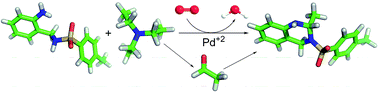
The degradation under ambient humidity and room temperature of Pd(OAc)2(NEt3), which is an efficient catalyst for the aerobic oxidation of alcohols, to diethylamine and acetaldehyde derivatives is disclosed. Evolved diethylamine reacted with Pd(OAc)2 to give Pd(OAc)2(HNEt2), which is in dynamic equilibrium with Pd(OAc)2(HNEt2)2. The evolved acetaldehyde generated during degradation process was trapped with a nucleophile to form 2-methyl-3-tosyl-1,2,3,4-tetrahydroquinazoline. NMR spectroscopy and single crystal X-ray diffraction studies have been used to characterize the reaction products of the reaction system.


Introduction

The use of the catalytic system Pd(OAc)2/NEt3 has proven to be a convenient synthetic strategy for the aerobic oxidation of alcohols to carbonyl compounds, at both high and room temperatures.1,2 The role of NEt3 is crucial in the ligand-accelerated catalysis due to the reduction of the activation energy barrier in the rate-determining β-hydride elimination step (Scheme 1).3 Using a combination of experimental and theoretical studies, it was found that at room temperature the active species is Pd(OAc)2(NEt3), which is in dynamic equilibrium with Pd(OAc)2(NEt3)2.3 As the concentration of NEt3 is increased, the equilibrium favours Pd(OAc)2(NEt3)2 and an inhibitory effect on the oxidation rate is observed.
image file: c6ra20886j-s1.tif
Scheme 1 Proposed mechanism for the aerobic oxidation of a benzyl alcohol to the corresponding aldehyde catalysed by Pd(OAc)2/NEt3 at room temperature.

Besides the displacement of the chemical equilibrium towards Pd(OAc)2(NEt3)2, other processes could contribute to the degradation of Pd(OAc)2(NEt3). In this way, Taqui Khan4,5 and Abbas6 have reported that NEt3 can be transformed into HNEt2 and acetaldehyde, under mild conditions, mediated by Ru3+ and Fe3+, respectively. These findings led us to think that Pd2+ could give similar results. In fact, although under severe conditions (at 200 °C for 40 h) and using palladium(0), the metal-catalysed oxidative hydrolysis of some tertiary amines to secondary amines and carbonyl compounds has been achieved.7

Since Pd(OAc)2(NEt3) is an efficient catalyst for aerobic oxidation of alcohols to carbonyl compounds, it is expected that its degradation into diethylamine and acetaldehyde derivatives could lead to an inhibitory effect on the oxidation rate. Therefore, we have undertaken studies towards the Pd-mediated hydrolysis of triethylamine under ambient humidity and room/reflux temperature. We plan to use NMR spectroscopy to study the composition of the reaction system.

Results and discussion

We have spectroscopically investigated the composition of a solution of the catalytic system Pd(OAc)2/NEt3 without added substrates, and also in the presence of methanol and ethanol.

Diethylamine derivatives

NMR monitoring of the reaction progress shown that, a THF solution of Pd(OAc)2/NEt3 after 30 minutes in absence of air gave rise to Pd(OAc)2(NEt3) with traces of Pd(OAc)2(NEt3)2 (Fig. 1). After 5 h exposure to air (room temperature and ambient humidity) the presence of Pd(OAc)2(HNEt2) can be clearly observed. As time passes concentration of Pd(OAc)2(HNEt2), Pd(OAc)2(HNEt2)2 and HNEt2 increases, while concentration of Pd(OAc)2(NEt3) decreases. The formation of these diethylamine derivatives suggests that an acid-catalyzed hydrolysis is involved in the process of degradation of Pd(OAc)2(NEt3). Since HNEt2 was not found when the reaction takes place in the absence of Pd(OAc)2, the formation of Pd(OAc)2(HNEt2)2 is an evidence of the palladium-mediated hydrolysis of triethylamine. We have performed an experiment in the presence of O2 and absence of humidity, but we have not observed the formation of diethylamine derivatives. Besides, hydrolysis reaction rate is increased when, in the presence of water, air is replaced by oxygen, but not significantly. We have observed that when water in 50[thin space (1/6-em)]:[thin space (1/6-em)]1 molar ratio (NEt3[thin space (1/6-em)]:[thin space (1/6-em)]H2O) is added the time required to obtain the same rate of hydrolysis is shortened in several hours, but the effect is much more marked when the reaction takes place at 60 °C instead of at room temperature. Besides, we have observed that as the concentration of NEt3 is increased, the equilibrium favours Pd(OAc)2(HNEt2)2, leading to the maximum yield (85%) the 1[thin space (1/6-em)]:[thin space (1/6-em)]8 molar ratio at 60 °C. After 63 h of exposure to air at room temperature and ambient humidity, the constituents of the mixture were separated and spectroscopically characterized.
image file: c6ra20886j-f1.tif
Fig. 1 1H NMR spectra in dmso-d6 showing the evolution of system Pd(OAc)2/NEt3 (1[thin space (1/6-em)]:[thin space (1/6-em)]8 molar ratio) at room temperature and ambient humidity. A colour code was used for characteristic resonances of each species.

The 1D-spectrum of Pd(OAc)2(HNEt2) shows only five proton signals at about 1.6, 1.7, 2.1, 2.4 and 5.5 ppm corresponding to HNEt2–methyl, acetate–methyl, HNEt2–methylene (two signals), and amino groups, respectively. The diastereotopic nature of the geminal protons explains the observation of two signals as multiplets for methylene groups (1 and 1′).

Fig. 2 shows the H–H NMR correlation spectrum of Pd(OAc)2(HNEt2), which is very similar to the spectra of Pd(OAc)2(HNEt2)2 and HNEt2 (see ESI). H–H couplings between amino and methylene groups as well as between methyl and methylene groups are nicely observed in these 2D-spectra. The most characteristic feature in these spectra is a broad triplet signal, which corresponds to the amino proton, at about 6.1, 5.5 and 5.2 ppm in Pd(OAc)2(HNEt2), Pd(OAc)2(HNEt2)2 and HNEt2, respectively. These chemical shifts are coherent with the palladium complexation by acetate in Pd(OAc)2(HNEt2) and Pd(OAc)2(HNEt2)2, since it causes a decrease of the electron density around the amino proton. This decrease in shielding (i.e. deshielding) displaces the NMR signal toward a lower field in Pd(OAc)2(HNEt2), which shows a more significant decrease of the electron density around the amino proton. Likewise, exhaustive NMR studies (see ESI) demonstrate that Pd(OAc)2(HNEt2)2 in solution has virtually the same structure as that observed in the solid state by X-ray diffraction.8,9


image file: c6ra20886j-f2.tif
Fig. 2 Two-dimensional proton–proton NMR correlation spectrum of Pd(OAc)2(HNEt2). A colour code was used to highlight the observed H–H couplings.

Acetaldehyde derivatives

We have used the formation10 of 2-methyl-3-tosyl-1,2,3,4-tetrahydroquinazoline from the nucleophile 2-tosylaminomethylaniline11 (Scheme 2) as a tool to make acetaldehyde easier to monitor by 1H NMR spectroscopy, since it is a volatile compound (bp 21 °C). We have chosen 2-tosylaminomethylaniline (HATs) because it has already been used successfully for verifying the Pd-mediated aerobic oxidation of methanol to formaldehyde.12
image file: c6ra20886j-s2.tif
Scheme 2 Pd-mediated hydrolysis of triethylamine in the presence of 2-tosylaminomethylaniline (HATs) to yield 2-methyl-3-tosyl-1,2,3,4-tetrahydroquinazoline and di(acetato)bis(diethylamine)palladium(II).

We have investigated the evolution of the chemical composition of a solution of Pd(OAc)2/NEt3 to which 2-tosylaminomethylaniline has been added. After heating at about 60 °C for 5 h, NMR spectroscopy revealed the presence of Pd(OAc)2(HNEt2)2 and 2-methyl-3-tosyl-1,2,3,4-tetrahydroquinazoline (Fig. 3). Since Pd(OAc)2(HNEt2) results from Pd(OAc)2(NEt3), which is the catalytic species on the oxidation of alcohols, it is expected an inhibitory effect on the oxidation rate of alcohols. In fact we have not detected oxidation of methanol to formaldehyde, even though we have found possible oxidation of methanol under not very different reaction conditions.12 In view that 2-substituted-1,2,3,4-tetrahydroquinazolines10 are accessible in almost quantitative yield via nucleophilic addition of 2-aminobenzylamine derivatives to aldehydes, the presence of 2-methyl-3-tosyl-1,2,3,4-tetrahydroquinazoline in the mixture is a sign of the formation of acetaldehyde. Besides diethylamine and acetaldehyde derivatives, there are other by-products in the reaction mixture, such as Pd(OAc)(ATs)(NEt3) and Pd(ATs)2 (see ESI). As time passes, concentration of Pd(OAc)2(HNEt2)2 and 2-methyl-3-tosyl-1,2,3,4-tetrahydroquinazoline increases, while concentration of Pd(OAc)(ATs)(NEt3) and Pd(ATs)2 decreases. After 8 h, the main constituents of the mixture were separated and spectroscopically characterized.


image file: c6ra20886j-f3.tif
Fig. 3 1H NMR spectra in dmso-d6 showing the evolution of the reaction system Pd(OAc)2/NEt3 in the presence of HATs at about 60 °C. A colour code was used for the characteristic resonances of each species.

Two-dimensional correlation NMR spectroscopy in combination with a selective NOE provides insight into the molecular structure of 2-methyl-3-tosyl-1,2,3,4-tetrahydroquinazoline. The deduced spatial arrangement of this molecule in solution is in good agreement with that displayed by the calculated most stable conformer of 2-methyl-3-tosyl-1,2,3,4-tetrahydroquinazoline,13 which displays an anti-disposition of methyl and tosyl groups (see ESI).

An isotope labelling experiment using NEt3-d15 as starting material has demonstrated that acetaldehyde derived from triethylamine. Thus, a 1H NMR spectrum of tetradeuterated 2-methyl-3-tosyl-1,2,3,4-tetrahydroquinazoline obtained from a reaction mixture containing Pd(OAc)2/NEt3-d15 and 2-tosylaminomethylaniline evidenced total deuteration at 2-position, leading to CD3 and CD groups (Fig. 4). As a consequence of the replacement of the CH by the CD group the original multiplicity of the NH proton changed from a doublet to a singlet.


image file: c6ra20886j-f4.tif
Fig. 4 Two-dimensional proton–proton NMR correlation spectrum of 2-methyl-3-tosyl-1,2,3,4-tetrahydroquinazoline in acetone-d6. A colour code was used to highlight the observed H–H couplings.

2-Tosylaminomethylaniline derivatives

Although we have used 2-tosylaminomethylaniline (HATs) to trap the acetaldehyde generated during degradation of NEt3, the mentioned nucleophile interacts with Pd(OAc)2 resulting in Pd(ATs)2. We have observed that in the absence of Pd(OAc)2, Pd(ATs)2 acts as a source of Pd2+ and ATs− allowing the hydrolysis of NEt3 and the formation of 2-methyl-3-tosyl-1,2,3,4-tetrahydroquinazoline. Therefore Pd(ATs)2 plays a role in the degradation of NEt3. A two-dimensional carbon–proton NMR correlation spectrum (HMBC) of Pd(ATs)2 with fully assigned 1H and 13C spectra is shown in Fig. 5. From the NMR study it has been deduced that Pd(ATs)2 adopts a square-planar geometry.
image file: c6ra20886j-f5.tif
Fig. 5 Correlation spectrum (HMBC) of Pd(ATs)2 (in dmso-d6). The most important C–H couplings were highlighted.

The electronic spectra of HAT and Pd(ATs)2 have been recorded in acetonitrile as solvent. Three absorptions, in the ultraviolet region, at about 205, 230 and 293 nm, are assignable to intra-ligand orbital transitions in the mentioned compounds. In the visible region of square-planar palladium complexes, three spin-allowed d–d transitions and three spin forbidden singlet–triplet d–d transitions from the three lower lying d levels to the empty dx2y2 orbitals are predicted.14 Spin-allowed d–d transitions were expected to correspond to the transitions 1A1g1A2g, 1A1g1B1g and 1A1g1Eg. However, strong charge transfer transition for Pd(ATs)2 interfere and prevent the observation of all the expected bands in the visible region.

The molecular structure of Pd(ATs)2, which is represented as an ORTEP diagram in Fig. 6, was determined using single crystal X-ray diffraction techniques.15–18 The asymmetric unit only contains half molecule of the neutral complex, and the complete molecule is generated by a symmetry operation based on an inversion centre located at the centre of the metal ion (#1 −x + 1, −y, −z). This inversion implies both a trans-disposition of the donor atoms, and an anti-conformation of the tosyl groups respect to the chromophore, which is absolutely plane. Thus, the four donor N-atoms and the palladium ion are exactly in the same plane, so the four N–Pd–N angles sum 360°. The main angles and bond distances are collected in Table 1.


image file: c6ra20886j-f6.tif
Fig. 6 Molecular structure of Pd(ATs)2. Ellipsoids have been represented at 50% probability level.
Table 1 Main bond lengths, bond and torsion angles, and H bonding scheme for Pd(ATs)2
Atomsa Distancesb Atoms Anglesc
a #1 = −x + 1, −y, −z; #2 = −#1 = x + 1, y, z.b In Å.c In degrees.
Pd1–N2 2.029(6) N2–Pd1–N2#1 180.0
Pd1–N2#1 2.029(6) N2–Pd1–N1 92.1(2)
Pd1–N1 2.044(6) N2#1–Pd1–N1 87.9(2)
Pd1–N1#1 2.043(6) N1–Pd1–N1#1 180.0
N1–C1 1.460(10) C7–N3–S1 117.4(5)
N2–C7 1.482(9) N2–S1–C8 109.7(4)
N2–S1 1.598(7)    
    N1–C1–C6–C7 6.3(11)
    C1–C6–C7–N2 50.5(10)
    C7–N2–S1–C8 −80.5(6)

D–H⋯Aa D–Hb H⋯Ab D⋯Ab D–H⋯Ac
N1–H1B⋯O1#1 1.02 2.21 2.909 (8) 124
N1–H1A⋯O2#2 0.76 2.17 2.929 (8) 173


Pd(ATs)2 crystallizes in the monoclinic space group P21/c. A polymorph of this compound, which crystallized in the triclinic space group P[1 with combining macron] has been already reported by us.19 Despite of the subtle differences between the molecules of the two polymorphs (Fig. 7 top), their packing scheme are sensibly different. Thus, the triclinic crystal displays double chains of molecules that are connected through intermolecular H bonds between parallel single chains. By contrast, the monoclinic polymorph only displays single chains, without connecting to neighboring chains (Fig. 7 bottom). Mutual intermolecular N–H⋯O interactions between coordinated amino groups and tosyl groups of contiguous molecules lead to the formation of a ring of 12 elements based on two donors and two acceptors R2,2(12) according to graph sets.20 As Fig. 7 shows, intramolecular π-stacking takes place between the relatively electron deficient tosyl group and the electron rich aminomethyl aniline moiety. As a consequence, the centroids of these two aromatic rings are at ca. 3.64 Å, while they are forming an angle of only 10.8°.


image file: c6ra20886j-f7.tif
Fig. 7 Top: Comparison of monoclinic (yellow) and triclinic (green) polmorrphs of Pd(ATs)2. Bottom: Intermolecular N–H⋯O bonds that give rise to a single-chain architecture.

Conclusions

The results reported here showed that in air (and ambient humidity, at room or reflux temperature), occurs degradation of Pd(OAc)2(NEt3) into HNEt2 and acetaldehyde. Evolved diethylamine reacted with Pd(OAc)2 to give Pd(OAc)2(HNEt2), which is in dynamic equilibrium with Pd(OAc)2(HNEt2)2. The evolved acetaldehyde generated during degradation process was trapped with a nucleophile to form 2-methyl-3-tosyl-1,2,3,4-tetrahydroquinazoline.

Experimental

Materials and methods

Excluded 2-tosylaminomethylaniline11 all starting materials and reagents were commercially available and were used without further purfication. 1H NMR spectra (500 MHz) and 13C NMR spectra (125 MHz) were measured in deuterated solvents. J values are given in hertz. NMR assignments were carried out by a combination of COSY, NOESY, HSQC and HMBC experiments. Infrared spectra were recorded as KBr pellets on a Jasco FT/IR-410 spectrophotometer in the range 4000–600 cm−1. Elemental analyses were performed on a Carlo Erba EA 1108 analyzer. Electrospray mass spectra were recorded on a Bruker Microtof spectrometer. MALDI-TOF mass spectra were recorded on a Bruker Ultraflex III TOF/TOF using methanol as solvent and DCTB as matrix.

Crystal structure analysis data

Diffraction data for Pd(ATs)2 were collected from a thin crystal needle at 100(2) K, using graphite-monochromated Mo-Kα radiation (λ = 0.71073 Å) from a fine focus sealed tube. Some significant crystal parameters and refinement data are summarized in Table 2.
Table 2 Diffraction data for Pd(ATs)2
Formula C28H30N4O4PdS2
Mr 657.08
Crystal system Monoclinic
Space group P21/c (no. 14)
Unit cell a = 6.8995(15) Å
  b = 8.5875(19) Å
  c = 22.797(4) Å
  α = 90°
  β = 98.652(7) °
  γ = 90°
Volume (Å3) 1335.4(5)
Z 2
Dc (g cm−3) 1.634
μ (mm−1) 0.90
F(000) 672
θ range (°) 1.8 to 24.7
Ref. col./ref. ind 9929/2264
Rint 0.0743
Data/restr./param. 2264/0/175
R1, wR2 [I > 2σ(I)] R1 = 0.0692, wR2 = 0.1561
R1, wR2 (all data) R1 = 0.1066, wR2 = 0.1794
Residuals (e Å−3) 1.388, −1.466


Data were processed and corrected for Lorentz and polarization effects. Multi-scan absorption corrections were performed using the SADABS routine.15,16 The structure was solved by standard direct methods.17 Some problems related to the thin nature of the crystals were observed during the refinement by full matrix least squares on F2.18 Thus, non-hydrogen atoms when were anisotropically refined, but some of them were oblate, and even prolate. Since U restraints were not effective in this case, one of them, which is sited on an aromatic ring, was isotropically refined. Hydrogen atoms were included in the structure factor calculation in geometrically idealized positions, with thermal parameters depending of the parent atom, by using a riding model. H atoms of the coordinated amine group where localized in Fourier maps and then refined with a thermal parameter depending of the parent N atom.

CCDC 991659 contains the supplementary crystallographic data for this paper.

NMR monitoring of the system Pd(OAc)2/NEt3 at r. t.

A THF solution (8 mL) of Pd(OAc)2 (40.4 mg, 0.18 mmol) and NEt3 (0.2 mL, 1.44 mmol) was stirred for 30 min under anhydrous conditions. The filtrate was concentrated to dryness at room temperature under reduced pressure and a sample of the resulting yellow solid was studied by 1H NMR spectroscopy. Then, the isolated powdery mixture was dissolved in THF and exposed to air, and after 5 h a small amount of Pd(OAc)2(HNEt2) was formed. After 63 h of exposure to air, Pd(OAc)2(HNEt2)2 (main), PdH(OAc)(NEt3) and HNEt2, were also formed.

NMR monitoring of the system Pd(OAc)2/NEt3 at 60 °C

A THF solution (8 mL) of NEt3 (0.2 mL, 0.8 mmol) was charged with Pd(OAc)2 (40 mg, 0.2 mmol) and HATs (24 mg, 0.08 mmol) and then was heated under reflux for 8 h. Samples of the solution were concentrated to dryness under vacuum and the resulting solids were studied by 1H NMR spectroscopy.

Degradation of Pd(OAc)2(NEt3) into Pd(OAc)2(HNEt2)2

Without added substrates. A THF solution (2 mL) of Pd(OAc)2 (10.0 mg, 0.045 mmol) and NEt3 in various molar ratios (5.6 μL, 0.040 mmol; 11.1 μL, 0.08 mmol; 16.7 μL, 0.12 mmol; 44.6 μL, 0.32 mmol and 55.7 μL, 0.40 mmol) was heated under reflux for 30 min. The resulting black powdery solid was filtered off. The filtrate was concentrated to dryness under reduced pressure and the resulting yellow solid was solved in diethyl ether. After recrystallization (4 h evaporation at room temperature) yellow prismatic crystals of Pd(OAc)2(HNEt2)2 suitable for X-ray diffraction studies were obtained.

Degradation of Pd(OAc)2(NEt3) into 2-methyl-3-tosyl-1,2,3,4-tetrahydroquinazoline

Without added substrates. A solution of Pd(OAc)2 (10 mg, 0.045 mmol), HATs (6 mg, 0.02 mmol) and NEt3 (28 μL, 0.20 mmol) in THF (2 mL) was heated under reflux (about 60 °C) for 8 h. The resulting black powdery solid was filtered off. The filtrate was concentrated to dryness under vacuum at about 45 °C. The experiment was reproduced by using 5 times the amount of reagents and the resulting mixture was separated by chromatography with ethyl acetate[thin space (1/6-em)]:[thin space (1/6-em)]hexane (50[thin space (1/6-em)]:[thin space (1/6-em)]50). The eluting solution was then concentrated to dryness under vacuum resulting in a white solid.
In the presence of methanol. A tetrahydrofuran solution (2 mL) of NEt3 (14 μL, 0.10 mmol) was charged with Pd(OAc)2 (10 mg, 0.05 mmol), HATs (6 mg, 0.02 mmol) and methanol (40 μL). Then, the solution was heated under reflux for 8 h. The resulting black powdery solid was filtered off. The filtrate was concentrated to dryness under vacuum to remove tetrahydrofuran as well as excess of methanol and NEt3. Yield = 40%.

Pd(OAc)2(HNEt2)2

Yields depending on the used molar ratio Pd(OAc)2/NEt3 (in parenthesis): 39% (1[thin space (1/6-em)]:[thin space (1/6-em)]1), 50% (1[thin space (1/6-em)]:[thin space (1/6-em)]2), 64% (1[thin space (1/6-em)]:[thin space (1/6-em)]3), 84% (1[thin space (1/6-em)]:[thin space (1/6-em)]7) and 58% (1[thin space (1/6-em)]:[thin space (1/6-em)]9). 1H NMR (500 MHz, dmso-d6): δ/ppm 5.55 (t, J = 9.5 Hz, 2H, 2 × NH), 2.44 (m, 4H, 4 × H-1), 2.10 (m, 4H, 4 × H-1′), 1.72 (s, 6H, 2 × COCH3) and 1.54 (t, J = 7.1 Hz, 12H, 12 × H-2). 13C NMR (125 MHz, dmso-d6): δ/ppm 178.0 (2 × CO), 46.5 (4 × CH2-1), 24.1 (2 × COCH3) and 14.4 (4 × CH3-2). IR (KBr, cm−1): 3206 ν(NH), 1587 νa(COO) and 1404 νs(COO). Elemental analysis (found): C 38.9, H 7.8, N 7.8%. Calc. for C12H28N2O4Pd: C, 38.7; H, 7.6; N, 7.6%.

2-Methyl-3-tosyl-1,2,3,4-tetrahydroquinazoline

Yield = 12.3 mg (41%). 1H NMR (400 MHz, dmso-d6): δ/ppm 7.56 (d, J = 8.2 Hz, 2H, 2 × H-2′), 7.16 (d, J = 8.1 Hz, 2H, 2 × H-3′), 6.83 (m, 2H, H-5 + H-7), 6.46 (t, J = 7.1, 1H, H-6), 6.25 (d, J = 8.1 Hz, 1H, H-8), 6.09 (d, J = 3.4 Hz, 1H, NH), 5.22 (m, 1H, H-2), 4.54 (d, J = 17.2 Hz, 1H, CHH-4) and 4.36 (d, J = 17.2 Hz, 1H, CHH-4), 2.25 (s, 3H, CH3-4′) and 1.22 (d, 3H, J = 6.3 Hz, CH3-2). 1H NMR (250 MHz, CDCl3): δ/ppm 7.59 (d, J = 8.3 Hz, 2H, 2 × H-2′), 7.06 (d, J = 8.3 Hz, 2H, 2 × H-3′), 6.90 (t, 1H, H-7), 6.86 (d, 1H, H-5), 6.67 (dt, J = 7.5 and 1.1 Hz, 1H, H-6), 6.29 (d, J = 8.1 Hz, 1H, H-8), 5.36 (dq, J = 6.4 and 1.0 Hz, 1H, H-2), 4.70 (d, J = 17.4 Hz, 1H, CH2-4), 4.47 (d, J = 17.4 Hz, 1H, CH2-4), 2.29 (s, 3H, CH3) and 1.40 (d, J = 6.4 Hz, 3H, CH3). 13C NMR (62.5 MHz, CDCl3): δ/ppm 143.2 (C4′), 139.7 (C8a), 136.2 (C1′), 129.0 (2 × C3′), 127.6 (C5), 127.3 (2 × C2′), 126.4 (C7), 118.8 (C6), 116.9 (C4a), 116.4 (C8), 61.4 (CH), 41.8 (CH2), 21.5 (CH3) and 21.4 (CH3). IR (KBr, cm−1): 3387(s) ν(NH) cm−1, 1326(s) νas(SO2), 1158(vs) νs(SO2). MS (ESI) m/z = 325 (MNa+). HRMS calcd for C16H18N2NaO2S (MNa+): 325.0981; found, 325.0967. Elemental analysis (found): C 63.5, H 5.8, N 9.1; S, 10.5%. Calc. for C16H18N2O2S: C, 63.6; H, 6.0; N, 9.3; S, 10.6%.

Pd(ATs)2

1H NMR (400 MHz, dmso-d6): δ/ppm 7.55 (d, J = 8.1 Hz, 4H, 4 × H-2′), 7.04 (t, J = 7.3 Hz, 2H, 2 × H-5), 7.03 (d, J = 8.0 Hz, 4H, 4 × H-3′), 7.02 (s, 4H, 2 × NH2), 6.90 (d, J = 7.9 Hz, 2H, 2 × H-3), 6.89 (t, J = 7.9 Hz, 2H, 2 × H-4), 6.83 (d, J = 7.7 Hz, 2H, 2 × H-6), 4.07 (s, 4H, 2 × CH2), 2.27 (s, 6H, 2 × CH3). 13C NMR (100 MHz, dmso-d6): δ/ppm 141.3 (2 × C-1′), 140.2 (2 × C-4′), 137.4 (2 × C-1), 133.9 (2 × C-2), 129.1 (2 × C-3), 128.4 (4 × C-3′), 128.1 (2 × C-5), 125.9 (4 × C-2′), 124.6 (2 × C-4), 120.4 (2 × C-6), 49.2 (2 × CH2) and 20.7 (2 × CH3). IR (KBr, cm−1): 3442 ν(OH), 3248 and 3180 ν(N–H), 1272 νas(SO2) and 1127(s) νs(SO2). MS (MALDI-TOF, DCTB): m/z (%): 655.9 (100) [M + H]+. Elemental analysis (found): C, 51.1; H, 4.5; N, 8.3; S, 9.4%. Calc. for C28H30N4O4S2Pd: C, 51.2; H, 4.6; N, 8.5; S, 9.8%.

Acknowledgements

Financial support from the Spanish Ministry of Economy and Competitiveness (SAF2013-42899-R), Xunta de Galicia (GRC2013-041) and the European Regional Development Fund (ERDF) is gratefully acknowledged. We are also grateful to the Centro de Supercomputación de Galicia (CESGA) for use of the SVG supercomputer.

Notes and references

  1. M. J. Schultz, C. C. Park and M. S. Sigman, Chem. Commun., 2002, 3034 RSC .
  2. M. J. Schultz, S. S. Hamilton, D. R. Jensen and M. S. Sigman, J. Org. Chem., 2005, 70, 3343 CrossRef CAS PubMed .
  3. M. J. Schultz, R. S. Adler, W. Zierkiewicz, T. Privalov and M. S. Sigman, J. Am. Chem. Soc., 2005, 127, 8499 CrossRef CAS PubMed .
  4. M. M. Taqui Khan, S. A. Mirza and H. C. Bajaj, J. Mol. Catal., 1986, 37, 253 CrossRef .
  5. M. M. Taqui Khan, S. A. Mirza and H. C. Bajaj, React. Kinet. Catal. Lett., 1987, 33, 67 CrossRef .
  6. K. Abbas and D. Marji, Z. Naturforsch., 2005, 60a, 667 Search PubMed .
  7. H. W. Walker, C. T. Kresge, P. C. Ford and R. G. Pearson, J. Am. Chem. Soc., 1979, 101, 7429 CrossRef .
  8. N. N. Lyalina, S. V. Dargina, A. N. Sobolev, T. M. Buslaeva and I. P. Romm, Koord. Khim., 1993, 19, 57 CAS .
  9. S. V. Kravtsova, I. P. Romm, A. I. Stash and V. K. Belsky, Acta Crystallogr., Sect. C: Cryst. Struct. Commun., 1996, 52, 2201 CrossRef .
  10. W. H. Correa, S. Papadopoulos, P. Radnidge, B. A. Roberts and J. L. Scott, Green Chem., 2002, 4, 245 RSC .
  11. J. Sanmartín, F. Novio, A. M. Garcia Deibe, M. Fondo and M. R. Bermejo, New J. Chem., 2007, 31, 1605 RSC .
  12. (a) J. Sanmartín-Matalobos, A. M. García-Deibe, L. Briones-Miguéns, C. González-Bello, C. Portela-García and M. Fondo, Dalton Trans., 2014, 43, 722 RSC ; (b) J. Sanmartín-Matalobos, A. M. García-Deibe, L. Briones-Miguéns, E. Lence, C. González-Bello, C. Portela-García and M. Fondo, New J. Chem., 2013, 37, 3043 RSC .
  13. M. J. Frisch, G. W. Trucks, H. B. Schlegel, G. E. Scuseria, M. A. Robb, J. R. Cheeseman, G. Scalmani, V. Barone, B. Mennucci, G. A. Petersson, H. Nakatsuji, M. Caricato, X. Li, H. P. Hratchian, A. F. Izmaylov, J. Bloino, G. Zheng, J. L. Sonnenberg, M. Hada, M. Ehara, K. Toyota, R. Fukuda, J. Hasegawa, M. Ishida, T. Nakajima, Y. Honda, O. Kitao, H. Nakai, T. Vreven, J. A. Montgomery Jr, J. E. Peralta, F. Ogliaro, M. Bearpark, J. J. Heyd, E. Brothers, K. N. Kudin, V. N. Staroverov, R. Kobayashi, J. Normand, K. Raghavachari, A. Rendell, J. C. Burant, S. S. Iyengar, J. Tomasi, M. Cossi, N. Rega, N. J. Millam, M. Klene, J. E. Knox, J. B. Cross, V. Bakken, C. Adamo, J. Jaramillo, R. Gomperts, R. E. Stratmann, O. Yazyev, A. J. Austin, R. Cammi, C. Pomelli, J. W. Ochterski, R. L. Martin, K. Morokuma, V. G. Zakrzewski, G. A. Voth, P. Salvador, J. J. Dannenberg, S. Dapprich, A. D. Daniels, Ö. Farkas, J. B. Foresman, J. V. Ortiz, J. Cioslowski and D. J. Fox, Gaussian 09, Revision A.2, Gaussian, Inc., Wallingford CT, 2009 Search PubMed .
  14. S. Das and S. Pal, J. Organomet. Chem., 2004, 689, 352 CrossRef CAS .
  15. SADABS-BrukerAXS area detector scaling and absorption correction. Version 2008/1, University of Göttingen, Germany 2008 Search PubMed .
  16. R. H. Blesing, Acta Crystallogr., Sect. A: Found. Crystallogr., 1995, 51, 33 CrossRef .
  17. M. C. Burla, R. Caliandro, M. Camalli, B. Carrozzini, G. L. Cascarano, L. De Caro, C. Giacovazzo, G. Polidori and R. Spagna, J. Appl. Crystallogr., 2005, 38, 381 CrossRef CAS .
  18. G. M. Sheldrick, Acta Crystallogr., Sect. A: Found. Crystallogr., 2008, 64, 112 CrossRef CAS PubMed .
  19. M. Fondo, C. Portela-García, A. H. Baleeiro-Tadiotto, A. M. García-Deibe and J. Sanmartín-Matalobos, Eur. J. Inorg. Chem., 2015, 2744 CrossRef CAS .
  20. M. C. Etter, J. C. MacDonald and J. Bernstein, Acta Crystallogr., Sect. B: Struct. Sci., 1990, 46, 256 CrossRef .

Footnote

Electronic supplementary information (ESI) available: Spectroscopic characterization of the compounds. CCDC 991659. For ESI and crystallographic data in CIF or other electronic format see DOI: 10.1039/c6ra20886j

This journal is © The Royal Society of Chemistry 2016