
Preparation and characterization of a chitin/platelet-poor plasma composite as a hemostatic material
Lingmei Lv†
ab,
Fengling Tang†ab and
Guangqian Lan*ab
aCollege of Textile and Garments, Southwest University, Chongqing 400715, China. E-mail: j070218@swu.edu.cn
bChongqing Engineering Research Center of Biomaterial Fiber and Modern Textile, Chongqing 400715, China
First published on 27th September 2016
Abstract
The development of life-saving hemostatic materials for emergencies can reduce death caused by uncontrolled hemorrhaging. In this study, we developed a composite hemostatic material based on interactions between chitin and platelet-poor plasma (PPP) that promote in situ thrombin attachment to the chitin surface. Optimal conditions for preparing the chitin/PPP composite hemostatic material (CP), including reaction time, temperature, inducing ion, and thrombin concentration, were determined; thrombin activity and PPP clotting time were then measured under these conditions. We found that chitin and 2-fold-diluted PPP induced with 0.2 mol L−1 CaCl2 and reacted in a 35 °C water bath for 1 h yielded the fastest clotting time in vitro (75.0 ± 3.0 s). Furthermore, CP showed a more potent hemostatic effect than chitin in a rabbit model of arterial and hepatic bleeding, which was attributable to the strong adsorption of thrombin. The activated partial thromboplastin time revealed that CP promoted blood coagulation via activation of the endogenous coagulation pathway. CP caused no obvious cytotoxicity in L929 cells, and histopathological examination indicated that CP had no immunogenicity, and accelerated wound healing. These results highlight the potential for using CP as a hemostatic agent in clinical settings.
Introduction
Uncontrolled hemorrhaging is a major cause of death following trauma in military and other settings.1–5 Approximately half of such deaths occur during the prehospital period.2 Significant blood loss can lead to hypothermia, clotting disorders, acidosis, infection, and multiple organ failure.6 Therefore, controlling hemorrhage is a priority in emergency medical treatment. The development of emergency life-saving hemostatic materials that function within a short period can reduce the risk of death due to bleeding. An ideal hemostatic agent should not only stop hemorrhage from actively bleeding arteries and veins, but should also demonstrate good biocompatibility, durable effects, and long shelf life; be risk free, lightweight, and inexpensive;7,8 and should not require on-scene mixing or preparation before use.9Chitin is a crystalline, high-molecular weight linear polymer composed of N-acetyl-2-amido-2-deoxy-D-glucose units linked by β (1–4) bonds; it is the second most abundant polysaccharide after cellulose,10,11 and is present in the exoskeletons of arthropods (e.g., crabs and shrimp) as well as the cell walls of fungi and yeast.12 Chitin can also be derived from by-products of brewing such as organic acids, antibiotics, and enzymes. Chitin was recently detected within skeletal formations of sponges and can be isolated from marine sponges. The isolated chitin-based scaffolds was a micro-structured network made up of cross-linked chitin fibers approximately 40–100 nm in diameter, and the shape of this network was similar to bandages.13–16 Chitin has many useful properties—including non-toxicity, low immunogenicity, biocompatibility, biodegradability, and antimicrobial activity17—and therefore a wide range of potential applications in the biomedical field. Chitin induces blood coagulation by adhering to platelets, forming a chitin/platelet complex that accelerates the polymerization of the fibrin monomer forming blood clot.18,19 It can also induce red blood cell aggregation via electrostatic forces, stimulate blood vessel contraction, form thrombi, and seal wounds.20 Chitin was shown to reduce whole blood clotting time,21 and dried chitin suspension spray and highly crystalline chitin nanofibres applied directly to the wound significantly reduced the amount of bleeding.22 In the cancellous bone of a canine hemorrhage model, deacetylation of microcrystalline chitin hydrochloride had a hemostatic effect that was more potent than that of traditional hemostatic agents.23 Compared with chitosan, chitin shows more blood compatible property, polymer with acetylation degrees of about 75% degrades faster than chitosan, and chitin also activates fewer macrophages.24
Thrombin is a highly specific serine protease extracted from human or animal blood that plays a major role in the blood coagulation cascade. It can induce platelet activation and aggregation by activating G protein-coupled protease-activated receptors on the platelet surface,25,26 and promote the conversion of fibrinogen into fibrin to accelerate hemostasis. Thrombin is widely used to treat digestive tract bleeding and surgical hemostasis. Commercial thrombin is expensive, requires storage below −20 °C, and must be dissolved at a certain concentration and combined with other materials to exert a hemostatic effect, which greatly restricts its applicability.
Many studies have investigated methods of adsorbing proteins onto solid surfaces.27,28 A recent study showed that fine granular calcium zeolite can be induced by calcium ion exchange technology to form a layer with ultra-high procoagulant activity and thrombin-containing protein corona upon contact with blood, which can greatly shorten clotting time.29 However, zeolite hemostasis dressing causes an exothermic reaction with temperatures reaching up to 100 °C, resulting in tissue damage.30,31 When Quick-Clot was used to stem severe liver trauma hemorrhage, the resultant exothermic reaction resulted in burns to local liver tissue upon completion of hemostasis.31 Zeolite can also cause severe inflammatory reactions and residual material in tissue can lead to abscess formation;32 it must therefore be removed from the wound immediately after completion of hemostasis, which can lead to secondary hemorrhage. Chitin can directly interact with proteins in body fluids, including bovine blood proteins33 and serum albumin.34,35
Platelet-poor plasma (PPP) is the layer of plasma that can be isolated from whole blood. PPP contains an abundance of fibrinogen, fibronectin, coagulation factor VIII, and prothrombin; the latter can be activated to thrombin by Ca2+ to participate in hemostasis and coagulation. Hemostasis has been successfully achieved in iliac crest donor sites, using calcium chloride/thrombin-activated PPP.36 Based on above reports, we combined chitin with PPP to produce a chitin/PPP composite hemostatic material (CP) and used CaCl2 to induce the adhesion of thrombin to the surface of chitin in situ. The purpose of this work was to obtain a new type of hemostatic agent that is easy to use, economical, with a dual hemostatic effect of chitin and thrombin. Furthermore, the hemostatic effect, tissue compatibility, and cytotoxicity properties of CP were also systematically evaluated.
Materials and methods
Reagents and animals
Alpha chitin (203 kDa) was obtained from Aladdin Biochem Technology Co. (Shanghai, China). New Zealand white rabbits (3 months old) were purchased from the Animal Laboratory Center of Third Military Medical University. All animal experiments and care were approved by the National Center of Animal Science Experimental Teaching (ASET) at the College of Animal Science and Technology (CAST) in the Southwest University of China and were in accordance with the “Guide for the Care and Use of Laboratory Animals”. Chromogenic substrate for thrombin (S-2238) was obtained from Ed Hauke Instrument Technology Co. (Beijing, China). Bovine thrombin (40–300 U mg−1) was purchased from Yuanye Biotechnology Co. (Shanghai, China).PPP collection
Blood was collected from rabbits by the heart blood sampling method37 in tubes containing 3.8% sodium citrate and centrifuged at 4000 rpm for 10 min. The upper pale yellow liquid layer (PPP) was stored at −20 °C until use. The PPP was thawed at 37 °C and maintained at a constant temperature in a water bath, and was not re-frozen after thawing.Preparation of CP powder
PPP was diluted with distilled water at a 1![[thin space (1/6-em)]](https://www.rsc.org/images/entities/char_2009.gif) :1 ratio and 2 mL were combined with 0.3 g chitin in a 5 mL centrifuge tube, followed by shaking for 2 min to ensure complete mixture. Excess PPP was removed by pipetting, and 2 mL of CaCl2 (0.01, 0.05, 0.1, 0.15, 0.2, and 0.25 mol L−1) were added. The reaction was allowed to proceed for 2 h at 35 °C in a water bath. The samples were vacuum-filtered (Zi Hua Instrument Co., Henan, China) and then placed in an electrically heated, constant-temperature air-blast drying box (Qi Xin Scientific Instrument Co., Shanghai, China) for 20 h at 37 °C to obtain CP, which was disinfected with epoxyethane alexipharmic ark (Red Sun Medical Instruments Co. Henan, China) and stored in a clean microcentrifuge tube at room temperature for further use. The above steps were repeated at reaction temperatures of 29 °C, 31 °C, 33 °C, 37 °C, and 39 °C; reaction times of 0.5, 1, 1.5, 2.5, and 3 h; and using ions NaCl, KCl, AlCl3, ZnCl2, CuCl2, and FeCl3 for induction.
:1 ratio and 2 mL were combined with 0.3 g chitin in a 5 mL centrifuge tube, followed by shaking for 2 min to ensure complete mixture. Excess PPP was removed by pipetting, and 2 mL of CaCl2 (0.01, 0.05, 0.1, 0.15, 0.2, and 0.25 mol L−1) were added. The reaction was allowed to proceed for 2 h at 35 °C in a water bath. The samples were vacuum-filtered (Zi Hua Instrument Co., Henan, China) and then placed in an electrically heated, constant-temperature air-blast drying box (Qi Xin Scientific Instrument Co., Shanghai, China) for 20 h at 37 °C to obtain CP, which was disinfected with epoxyethane alexipharmic ark (Red Sun Medical Instruments Co. Henan, China) and stored in a clean microcentrifuge tube at room temperature for further use. The above steps were repeated at reaction temperatures of 29 °C, 31 °C, 33 °C, 37 °C, and 39 °C; reaction times of 0.5, 1, 1.5, 2.5, and 3 h; and using ions NaCl, KCl, AlCl3, ZnCl2, CuCl2, and FeCl3 for induction.
Characterization of CP
CP and chitin samples (untreated or in contact with blood) were sprayed with metal at 15 kV to increase conductivity, and morphology and particle size were characterized by scanning electron microscopy (S600; JEOL, Tokyo, Japan). The molecular structures of samples were determined by Fourier transform infrared spectroscopy (FTIR; Bruker Alpha; Bruker, Bremen, Germany). Briefly, samples were ground to a powder and tableted with KBr, then scanned between 4000–500 nm cm−1. The thermal stability of the samples was assessed by thermogravimetric analysis (TGA; TG 209 F3 Tarsus, Netzsch, Selb, Germany).In vitro evaluation of the hemostatic properties of CP
To prepare thrombin chromogenic substrate, 0.1 g CP was added to a 5 mL centrifuge tube containing 1.5 mL of 4 mmol L−1 thrombin substrate solution in a water bath at 37 °C for 3 min. The reaction was terminated by transferring the tube to a 100 °C water bath for 2 min. After cooling to room temperature, the sample was centrifuged at 3000 rpm for 10 min and 200 μL of the supernatant were added to 96-well plates. Absorbance was measured at 405 nm. Results are presented as the mean of six replicate samples.For the in vitro blood coagulation experiment, 0.1 g CP was added to a 5 mL plastic centrifuge tube along with 2 mL PPP and 60 μL of 0.2 mol L−1 CaCl2. The natural clotting time was about 10 min. The mixture was mixed by shaking and placed in a constant-temperature water bath at 37 °C. The tubes were inverted until the plasma was no longer flowing and the time from addition of CaCl2 until PPP coagulation was recorded. The experiment was performed six times and mean values are presented.
Amino acid content analysis
The amino acid content of the samples was determined with an automatic amino acid analyser (L-8800; Hitachi, Tokyo, Japan). Thrombin (0.0057 g) and CP (0.0155 g) were added to a 15 × 150 mm hydrolysis tube along with 3 mL of 6 mol L−1 hydrochloric acid. The sample was mixed by oscillation for 3 min. The solution volume was reduced from a point that was 1/3 of the distance from the mouth of the tube to a height of 4–6 mm using an alcohol burner; the tube was sealed after 10 min of vacuum pumping and placed in a 110 °C ± 1 °C constant-temperature oven with a sand bath for 22 h. The sample was cooled to room temperature, and after shaking and filtering, 1 mL of the filtrate was transferred to a 50 mL beaker and dried in a constant-temperature water bath at 60 °C. For the last step of sample pretreatment, 0.02 M hydrochloric acid was added to dilute the dried filtrate 2–4 fold followed by passage through a 0.22 μm membrane filter, and the amino acid content of the diluted solution was analysed.In vitro blood plasma coagulation assay
To determine whether CP activates plasma coagulation, activated partial thromboplastin time (APTT) and pro-thrombin time (PT) were determined in vitro. Samples were prepared in duplicate and measurements were made by the same experienced hematology technician using an automated coagulation analyser (Sysmex CA-1500; Siemens, Tokyo, Japan) at the Ninth People's Hospital of Chongqing, China.For APTT measurements, 0.02 mol L−1 CaCl2 solution and 100 μL APTT reagent were separately pre-warmed at 37 °C for 5 min; 0.1 g CP was then added to 2 mL citrated plasma, and 100 μL PPP was incubated for 1 min at 37 °C in a cuvette. A 100 μL volume of pre-warmed APTT reagent was added to the pre-warmed plasma supernatant, followed by incubation for 3 min at 37 °C, and 100 μL of pre-warmed 0.2 mol L−1 CaCl2 was added to induce intrinsic clotting; the clotting time was measured. For the PT test, 100 μL of pre-warmed PT reagent was added to 100 μL of citrated plasma supernatant followed by incubation for 1 min at 37 °C and the PT was measured immediately afterwards. Results represent the mean of six independent experiments.
Evaluation of hemostasis in animal models
Hemostatic efficacy was assessed in ear arterial wound and liver injury models. Hemostatic material composed of 3 g CP and gauze or 3 g chitin and gauze were used for the experimental group, whereas the control group was untreated.Evaluation of chitin stability on thrombin
To assess the stabilizing effect of chitin on thrombin, washing and time stability tests were carried out. To test washing stability, 1.5 g of CP sample prepared under optimal conditions were placed in a 10 mL plastic centrifuge tube with 5 mL distilled water. The contents of the tube were mixed by inversion and allowed to stand for 2 min, which was considered as one wash. The sample was washed with distilled water up to six times, and blood coagulation activity was evaluated as described above.For the temporal stability test, the CP prepared under optimal conditions was stored in a vacuum drying oven at room temperature for 1, 5, 10, 20, 60, 120, or 180 days, and blood coagulation activity was evaluated as above.
Cell proliferation assay
The sample (0.1 g) was sterilized by ultraviolet irradiation for 30 min and immersed in Roswell Park Memorial Institute (RPMI)-1640 medium at 4 °C for 24 h, then centrifuged at 3000 rpm for 5 min. The supernatant constituted the soaking liquid of the sample (SLS). L929 cells were cultured in RPMI-1640 medium supplemented with fetal bovine serum (FBS) and 2% antibiotics (200 mg mL−1 penicillin and 200 mg mL−1 streptomycin) for 48–72 h until attachment; the medium was then removed and the cells were washed with 1× phosphate-buffered saline PBS warmed to 37 °C in a water bath. The cells were then trypsinized by adding 1 mL of 0.05% trypsin at 37 °C for 30–60 s, the digestion was terminated by adding 1 mL high-glucose Dulbecco's Modified Eagle's Medium (DMEM) containing 50% FBS. The supernatant was removed and 1 mL high-glucose DMEM with 10% FBS was added followed by gentle mixing to obtain single cell suspensions. These were diluted to 1 × 10⁴ cells per mL with RPMI 1640 medium containing 8% FBS. Cell viability was determined with the trypan blue assay according to standard protocols. A 1 mL volume of SLS and 1 mL cell suspension were mixed and added to each well of a 24-well plate, which was incubated at 37 °C and 5% CO2 for 24, 48, or 72 h. A negative control (with no hemostatic material) was included for each time point. Cell proliferation was evaluated with the 3-(4,5-dimethylthiazol-2-yl)-2,5-diphenyltetrazolium bromide assay and optical density (OD) was measured with a microplate reader (iMark; Bio-Rad, Hercules, CA, USA) at a wavelength of 490 nm. The experiment was repeated at least six times and results represent the means relative to the negative control.Statistical analysis
Data are presented as mean ± standard deviation. Differences between means were evaluated for statistical significance with the Student's t test or by one-way analysis of variance. P values < 0.05 were considered significant.Results
Optimization of reaction conditions for CP preparation
 | ||
| Fig. 1 (A and B) Effect of CaCl2 concentration on thrombin amount, as revealed by OD (A) and clotting time (B). The temperature and reaction time were 35 °C and 2 h, respectively (n = 6). (C and D) Effect of reaction temperature on thrombin amount, as revealed by OD (A) and clotting time (D). CaCl2 concentration and reaction time were 0.2 mol L−1 and 2 h, respectively (n = 6). | ||
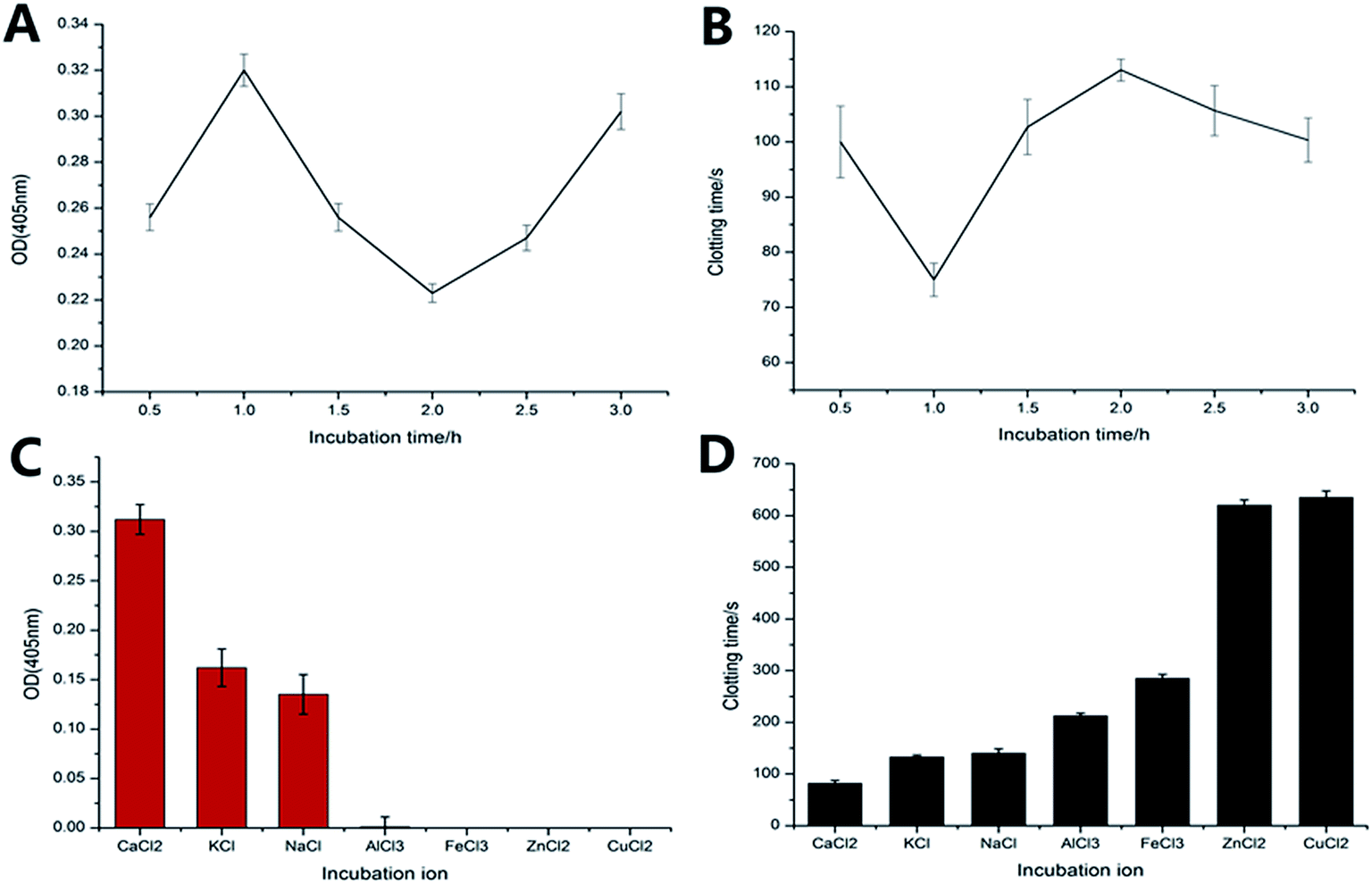 | ||
| Fig. 2 (A and B) Effect of incubation time on thrombin amount, as revealed by OD (A) and clotting time (B). The temperature and CaCl2 concentration were 35 °C and 0.2 mol L−1, respectively (n = 6). (C and D) Effect of metal ions on thrombin amount, as revealed by OD (C) and clotting time (D). CaCl2 concentration and reaction temperature and time were 0.2 mol L−1, 35 °C, and 1 h, respectively (n = 6). | ||
Characterization
 | ||
| Fig. 3 Scanning electron micrographs of (A) chitin and (B) CP. (C) Contact between CP and blood at 500× magnification. | ||
![[double bond, length as m-dash]](https://www.rsc.org/images/entities/char_e001.gif) O stretching was shifted to 1072 cm−1 in CP, indicating that after chitin came in contact with PPP, thrombin was adsorbed onto the chitin surface. There was no apparent chemical bonding between CP and chitin, indicating that the interaction between chitin and thrombin involved physical adsorption.
O stretching was shifted to 1072 cm−1 in CP, indicating that after chitin came in contact with PPP, thrombin was adsorbed onto the chitin surface. There was no apparent chemical bonding between CP and chitin, indicating that the interaction between chitin and thrombin involved physical adsorption.
 | ||
| Fig. 4 (A) Infrared spectra of (a) chitin, (b) CP, and (c) thrombin. (B) TG profiles of (a) thrombin, (b) CP, and (c) chitin. (C) PT and APTT for PPP with chitin and CP. **P < 0.01. (D) TG profiles of (a) CP, (b) chitin, and (c) thrombin. | ||
When temperature was increased to 800 °C, chitin underwent the most extensive decomposition, with a residue rate of 24.57% as compared to 43.68% for thrombin and 33.26% for CP (Fig. 4B). The intermediate value for CP may be explained by the attachment of thrombin. The decomposition occurred in two steps. The first peak for CP, chitin, and thrombin was at 86.9 °C, 88.9 °C and 83.4 °C, respectively, and was attributed to the evaporation of moisture (Fig. 4D). The second peak of CP was at 367.12 °C and the chitin peak emerged at 383.6 °C, but thermal decomposition of amino acids reached a maximum value of 2.96 at 271.83 °C and 5.18 at 363.35 °C. For chitin and CP, this could be explained by the degradation of the polysaccharide structure, while for thrombin it was likely due to thermal decomposition of amino acids. The higher pyrolysis temperatures for CP and chitin than for thrombin indicated that CP had greater thermal stability than thrombin. On the other hand, the peak of CP was between those of thrombin and chitin; this slight shift was attributable to the attachment of thrombin. These results indicate that thrombin generation and adhesion occurred in the reaction.
Amino acid analysis
The amino acid contents of CP and thrombin were similar (Table 1). Thrombin is composed of two A and B chains connected by disulfide bonds (Cys1–Cys12).40,41 The proportion of cysteine in CP (0.64%) was comparable to that of thrombin (0.63%), implying that thrombin was attached to CP.| Serial number | Amino acid | Thrombin | CP |
|---|---|---|---|
| 1 | Aspartate | 4.362 | 0.567 |
| 2 | Threonine | 2.371 | 0.291 |
| 3 | Serine | 2.445 | 0.269 |
| 4 | Glutamate | 5.780 | 0.773 |
| 5 | Glycine | 1.890 | 0.186 |
| 6 | Alanine | 1.883 | 0.262 |
| 7 | Cysteine | 0.278 | 0.042 |
| 8 | Valine | 2.609 | 0.403 |
| 9 | Methionine | 0.526 | 0.073 |
| 10 | Isoleucine | 2.036 | 0.284 |
| 11 | Leucine | 3.622 | 0.614 |
| 12 | Tyrosine | 1.958 | 0.245 |
| 13 | Phenylalanine | 2.724 | 0.399 |
| 14 | Lysine | 3.000 | 0.802 |
| 15 | Histidine | 1.005 | 0.068 |
| 16 | Arginine | 3.307 | 0.221 |
| 17 | Proline | 3.187 | 0.197 |
| 18 | Total | 43.511 | 7.148 |
PT and APTT measurement
The APTT and PT of CP were 8.0 ± 1.2 s and 6.4 ± 1.0 s, respectively, as compared to 29.0 ± 3.0 s and 12.6 ± 1.7 s, respectively, for chitin (Fig. 4C). These values were lower than for the negative control (45.2 ± 2 s and 13.0 ± 1.5 s, respectively). The APTT value of CP was shorter than that of chitin, whereas PT was shorter for CP but not for chitin relative to the negative control.Animal hemostasis model
The effectiveness of chitin and CP as hemostatic materials was evaluated in rabbit models of ear and liver injury by measuring bleeding time and blood loss (Fig. 5B and C). In the ear arterial injury model, bleeding ceased after 44.8 ± 10.2 s with CP as compared to 129.0 ± 6.8 s with chitin, while blood loss was also lower when CP was used (3.11 ± 0.28 g vs. 5.78 ± 0.31 g with chitin). In the rabbit liver injury model, the hemostasis time with CP was 34.2 ± 5.4 s, which was 245% shorter than with chitin (118.0 ± 5.6 s). In addition, blood loss was reduced with CP relative to chitin (0.30 ± 0.01 g vs. 1.64 ± 0.17 g). These results indicate that CP has a more portent hemostatic effect than chitin.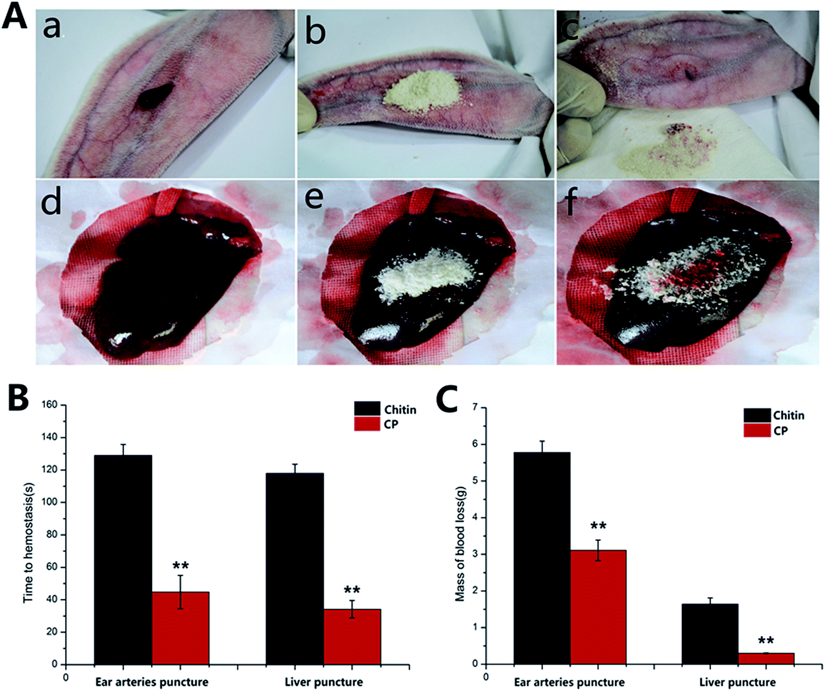 | ||
| Fig. 5 (A) CP applied to rabbit ear arterial bleeding and liver injury models. (a) Bleeding of freshly cut ear artery; (b) CP applied to the bleeding ear artery; (c) stemming of blood flow from ear artery by CP. (d) Bleeding from the liver wound; (e) CP applied to the bleeding liver; (f) stemming of blood flow from the liver by CP. **P < 0.01 (n = 6). (B and C) Time to hemostasis (B) and blood loss (C) in rabbit models of ear arterial bleeding and liver injury (n = 6). | ||
Washing and temporal stability of CP
The adhesion stability of thrombin on the CP surface was investigated by examining washing and temporal stability (Fig. 6). After one wash, the coagulation time of CP (93.0 ± 5.0 s) increased by 17.7% as compared to without washing (79.0 ± 3.0 s); after seven washes, the coagulation time (104.0 ± 5.0 s) increased by only 31.6% and was stable. After repeated washes, the positive effect of CP on PPP coagulation time did not decrease significantly, indicating that thrombin was strongly adhered to the CP surface. There was little change in the coagulation time of CP after storage at room temperature for 3 months (77.0 ± 0.5 s), and it retained good coagulation activity (81.0 ± 0.6 s) after 6 months (Fig. 6B). Thus, CP is stable when stored at room temperature for a long period. | ||
| Fig. 6 (A) Washing stability of CP. (B) Temporal stability of CP (n = 6). (C) Biocompatibility of CP and chitin, as revealed by L929 cell growth at 24, 48, and 72 h. **P < 0.01. | ||
Cytotoxicity
L929 fibroblasts were cultured in the presence of SLS for 24, 48, and 72 h (Fig. 6C). Absorbance values under all conditions exceeded 80%, indicating that SLS was not cytotoxic. Cell growth was induced by 97.6% ± 18.1% at 24 h, 102.6% ± 16.2% at 48 h, and 140.8% ± 10.1% at 72 h in the presence of CP as compared to 93.9% ± 12.2% at 24 h, 96.0% ± 27.3% at 48 h, and 129.0% ± 13.1% at 72 h with chitin. Although the difference between the two materials was relatively small after 24 and 48 h, the absorbance of cells cultured with CP exceeded 140% after 72 h, which was significantly higher than in the chitin group. These results indicate that CP is biocompatible.In vivo tissue biocompatibility
Histological examination of the wounds on different days revealed a clear wound in all groups after 2 h; the control and chitin groups showed a small amount of local bleeding while no bleeding was observed in the CP group (Fig. 7B). On day 3, the wounds were surrounded by inflammatory cells and showed granulation and there were no obvious differences among groups except for some residual bleeding in the chitin group (Fig. 6A). On day 7, the epidermis had partially healed in the control and chitin groups, while in the CP group, skin repair was complete: the scab had fallen off, and the skin at the wound site had regenerated, showing that wound healing was accelerated. In addition, there was no obvious inflammatory reaction in the CP as compared to the control and chitin groups. These results indicate that CP is a non-immunogenic hemostatic material. | ||
| Fig. 7 Effect of hemostatic materials on wound healing. (A) Images of wounds in rabbits treated with different hemostatic materials for 2 h or 3 or 7 days. (B) Hematoxylin and eosin-stained sections of tissue treated with different hemostatic materials for 2 h or 3 or 7 days. | ||
Discussion
During blood coagulation, soluble fibrinogen transforms into an insoluble fibrin clot upon catalysis by thrombin.42–46 In this study, we used Ca2+ ions to induce the conversion of plasma prothrombin into thrombin that attached to the surface of chitin, yielding a chitin/PPP compound hemostatic material. This circumvented the cost associated with commercially available thrombin but also the harsh preservation conditions and requirement for a solution prior to use while exploiting the hemostatic properties of chitin. Structural studies of chitin have suggested that it can readily induce red blood cell adhesion and aggregation,47 which we confirmed by scanning electron microscopy analysis. Moreover, thrombin attached to the surface of chitin had a more potent hemostatic effect than either molecule alone.Ca2+ is a key factor in thrombin activation; it activates blood coagulation factors to form a prothrombin complex on the surface of the phospholipid membranes, which can induce the conversion of prothrombin to thrombin.48,49 In this study, the optimal Ca2+ concentration for thrombin activation was 0.2 mol L−1 at 35 °C for 1 h. Biological materials can non-specifically interact with proteins as a result of a static electric field, forming a protein corona in a process that is normally irreversible.50,51 Chitin has a positive and thrombin a negative charge, leading to a strong interaction between the two molecules.52 Indeed, we found that even after repeated washing and 6 months of storage, CP maintained a potent hemostatic effect, indicating strong adsorption of thrombin on the chitin surface; this was confirmed by FTIR and amino acid analyses.
In cases of severe hemorrhaging, it is important that the applied hemostatic material stop bleeding within a short period of time. In our rabbit models of arterial bleeding and liver injury, CP stopped the bleeding after 44.8 ± 10.2 s and 34.2 ± 5.4 s, respectively; this was shorter than chitin and was attributed to the presence of thrombin. We also found that CP induced blood coagulation via the intrinsic coagulation pathway by promoting the activation of blood factor XII, which in turn activated factors XI, IX, VII, and X, transforming the latter into a factor Xa. In the presence of factor IV and PF3, factor Xa forms the prothrombin complex, which stimulates the Ca2+-mediated conversion of prothrombin to thrombin.53
Conclusion
A new hemostatic composite, CP, was produced by attaching thrombin to the surface of chitin. The optimal conditions for preparing CP with the most greatest hemostatic effect were a CaCl2 concentration of 0.20 mol L−1, reaction time of 1 h, and reaction temperature of 35 °C. CP showed higher clotting activity and stability than chitin and accelerated wound healing in rabbit arterial bleeding and liver injury models. Moreover, it induced higher cell proliferation than chitin, with no obvious cytotoxicity and superior biocompatibility. These results suggest that CP can be used as a safe and effective hemostatic material in clinical settings that is a lower-cost alternative to commercially available thrombin.Disclosure of conflicts of interest
The authors declare that they have no conflicts of interest related to this work.Acknowledgements
This work was supported by the Fundamental Research Funds for the Central Universities (XDJK2014B004, XDJK2013A021), National Under-graduate Training Programs for Innovation and Entrepreneurship (201510635048). This work was also funded by Hi-Tech Research and Development 863 Program of China Grant (No. 2013AA102507).References
- H. R. Champion, R. F. Bellamy, C. P. Roberts and A. Leppaniem, A profile of combat injury, Trauma, 2003, 54, S13–S19 Search PubMed.
- H. Khoshmohabat, B. Dalfardi and A. Dehghanian, et al., The effect of CoolClot hemostatic agent on skin wound healing in rats, J. Surg. Res., 2016, 200(2), 732–737 CrossRef CAS PubMed.
- L. R. Burnett, J. G. Richter and M. B. Rahmany, et al., Novel keratin (KeraStat™) and polyurethane (Nanosan®-Sorb) biomaterials are hemostatic in a porcine lethal extremity hemorrhage model, J. Biomater. Appl., 2014, 28(6), 869–879 CrossRef PubMed.
- H. B. Alam, D. Burris and J. A. DaCorta, et al., Hemorrhage control in the battlefield: role of new hemostatic agents, Mil. Med., 2005, 170(1), 63–69 CrossRef PubMed.
- A. E. Pusateri, S. J. McCarthy and K. W. Gregory, et al., Effect of a chitosan-based hemostatic dressing on blood loss and survival in a model of severe venous hemorrhage and hepatic injury in swine, J. Trauma: Inj., Infect., Crit. Care, 2003, 54(1), 177–182 CrossRef.
- K. R. Ward, M. H. Tiba and W. H. Holbert, et al., Comparison of a new hemostatic agent to current combat hemostatic agents in a swine model of lethal extremity arterial hemorrhage, J. Trauma: Inj., Infect., Crit. Care, 2007, 63(2), 276–284 CrossRef PubMed.
- R. Jayakumar, N. Nwe and S. Tokura, et al., Sulfated chitin and chitosan as novel biomaterials, Int. J. Biol. Macromol., 2007, 40(3), 175–181 CrossRef CAS PubMed.
- K. Azuma, M. Nishihara and H. Shimizu, et al., Biological adhesive based on carboxymethyl chitin derivatives and chitin nanofibers, Biomaterials, 2015, 42, 20–29 CrossRef CAS PubMed.
- J. Granville-Chapman, N. Jacobs and M. J. Midwinter, Pre-hospital hemostatic dressings: a systematic review, Injury, 2011, 42(5), 447–459 CrossRef CAS PubMed.
- R. A. A. Muzzarelli, Chitin Nanostructures in Living Organisms, Top. Geobiol., 2011, 34, 1–34 Search PubMed.
- A. M. Salaberria, J. Labidi and S. C. M. Fernandes, Different routes to turn chitin into stunning nano-objects, Eur. Polym. J., 2015, 68, 503–515 CrossRef CAS.
- K. Azuma, S. Ifuku and T. Osaki, et al., Preparation and biomedical applications of chitin and chitosan nanofibers, J. Biomed. Nanotechnol., 2014, 10(10), 2891–2920 CrossRef CAS PubMed.
- A. Anitha, S. Sowmya and P. T. S. Kumar, et al., Chitin and chitosan in selected biomedical applications, Prog. Polym. Sci., 2014, 39(9), 1644–1667 CrossRef CAS.
- F. Thomas, M. Rivelino and S. K. Hui, et al., Chitin-based tubes for tissue engineering in the nervous system, Biomaterials, 2005, 26(26), 4624–4632 Search PubMed.
- M. Wysokowski, I. Petrenko and A. L. Stelling, et al., Poriferan Chitin as a Versatile Template for Extreme Biomimetics, Polymers, 2015, 7(2), 235–265 CrossRef CAS.
- K. Y. Lee, S. H. Wan and W. H. Park, Blood compatibility and biodegradability of partially N-acylated chitosan derivatives, Biomaterials, 1995, 16(16), 1211–1216 CrossRef CAS PubMed.
- C. Peniche, W. Argüelles-Monal and F. M. Goycoolea, Chitin and chitosan: major sources, properties and applications, Monomers, Polym. Compos. Renewable Resour., 2008, 1, 517–542 Search PubMed.
- H. S. Thatte, S. Zagarins and S. F. Khuri, et al., Mechanisms of Poly-N-Acetyl Glucosamine Polymer–Mediated Hemostasis: Platelet Interactions, J. Trauma: Inj., Infect., Crit. Care, 2004, 57(1), S13–S21 CrossRef CAS.
- T. H. ischer, H. S. Thatte and T. C. Nichols, et al., Synergistic platelet integrin signaling and factor XII activation in poly-N-acetyl glucosamine fiber-mediated hemostasis, Biomaterials, 2005, 26(27), 5433–5443 CrossRef PubMed.
- C. R. Valeri, R. Srey and D. Tilahun, et al., In vitro effects of poly-N-acetyl glucosamine on the activation of platelets in platelet-rich plasma with and without red blood cells, J. Trauma: Inj., Infect., Crit. Care, 2004, 57(1), S22–S25 CrossRef CAS.
- W. Janvikul, P. Uppanan and B. Thavornyutikarn, et al., In vitro comparative hemostatic studies of chitin, chitosan, and their derivatives, J. Appl. Polym. Sci., 2006, 102(1), 445–451 CrossRef CAS.
- R. A. A. Muzzarelli, P. Morganti and G. Morganti, et al., Chitin nanofibrils/chitosan glycolate composites as wound medicaments, Carbohydr. Polym., 2007, 70(3), 274–284 CrossRef CAS.
- T. Sugamori, H. Iwase and M. Maeda, et al., Local hemostatic effects of microcrystalline partially deacetylated chitin hydrochloride, J. Biomed. Mater. Res., 2000, 49(2), 225–232 CrossRef CAS PubMed.
- E. Brunner, H. Ehrlich and P. Schupp, et al., Chitin-based scaffolds are an integral part of the skeleton of the marine demosponge Ianthella basta, J. Struct. Biol., 2009, 168(3), 539–547 CrossRef CAS PubMed.
- S. R. Coughlin, Thrombin signalling and protease-activated receptors, Nature, 2000, 407(6801), 258–264 CrossRef CAS PubMed.
- L. F. Brass, Thrombin and platelet activation, Chest, 2003, 124(3), 18S–25S CrossRef CAS PubMed.
- R. J. Chen, Y. Zhang and D. Wang, et al., Noncovalent sidewall functionalization of single-walled carbon nanotubes for protein immobilization, J. Am. Chem. Soc., 2001, 123(16), 3838–3839 CrossRef CAS PubMed.
- E. Pâslaru, M. C. Baican and E. G. Hitruc, et al., Immunoglobulin G immobilization on PVDF surface, Colloids Surf., B, 2014, 115, 139–149 CrossRef PubMed.
- Y. Li, X. Liao and X. Zhang, et al., In situ generated thrombin in the protein corona of zeolites: relevance of the functional proteins to its biological impact, Nano Res., 2014, 7(10), 1457–1465 CrossRef CAS.
- T. Gerlach, J. K. Grayson and K. O. Pichakron, et al., Preliminary study of the effects of smectite granules (WoundStat) on vascular repair and wound healing in a swine survival model, J. Trauma: Inj., Infect., Crit. Care, 2010, 69(5), 1203–1209 CrossRef CAS PubMed.
- J. McManus, T. Hurtado and A. Pusateri, et al., A case series describing thermal injury resulting from zeolite use for hemorrhage control in combat operations, Prehosp. Emerg. Care, 2007, 11(1), 67–71 CrossRef PubMed.
- J. K. Wright, J. Kalns and E. A. Wolf, et al., Thermal injury resulting from application of a granular mineral hemostatic agent, J. Trauma: Inj., Infect., Crit. Care, 2004, 57(2), 224–230 CrossRef.
- S. I. Nishimura, Y. Ikeuchi and S. Tokura, The adsorption of bovine blood proteins onto the surface of O-(carboxymethyl) chitin, Carbohydr. Res., 1984, 134(2), 305–312 CrossRef CAS PubMed.
- A. I. Ledesma-Osuna, G. Ramos-Clamont and A. M. Guzman-Partida, et al., Conjugates of bovine serum albumin with chitin oligosaccharides prepared through the Maillard reaction, J. Agric. Food Chem., 2010, 58(22), 12000–12005 CrossRef CAS PubMed.
- M. Kaya, I. Sargin and V. Aylanc, et al., Comparison of bovine serum albumin adsorption capacities of α-chitin isolated from an insect and β-chitin from cuttlebone, J. Ind. Eng. Chem., 2016, 38, 146–156 CrossRef CAS.
- W. S. Pietrzak, Y. H. An and Q. K. Kang, et al., Platelet-rich and platelet-poor plasma: development of an animal model to evaluate hemostatic efficacy, J Craniofac. Surg., 2007, 18(3), 559–567 CrossRef PubMed.
- G. Lan, B. Lu and T. Wang, et al., Chitosan/gelatin composite sponge is an absorbable surgical hemostatic agent, Colloids Surf., B, 2015, 136, 1026–1034 CrossRef CAS PubMed.
- A. J. Varma and V. B. Chavan, Calcium complexation by low molecular weight dicarboxycellulose in aqueous solution, Carbohydr. Polym., 2001, 45(1), 101–103 CrossRef CAS.
- R. Gu, W. Sun and H. Zhou, et al., The performance of a fly-larva shell-derived chitosan sponge as an absorbable surgical hemostatic agent, Biomaterials, 2010, 31(6), 1270–1277 CrossRef CAS PubMed.
- R. R. Singh and J. Y. Chang, Structural stability of human α-thrombin studied by disulfide reduction and scrambling, Biochim. Biophys. Acta, Proteins Proteomics, 2003, 1651(1), 85–92 CrossRef.
- R. J. Butkowski, J. Elion and M. R. Downing, et al., Primary Structure of Human Prethrombin 2 and cw-Thrombin, J. Biol. Chem., 1977, 252, 4942–4957 CAS.
- L. R. Paborsky, S. N. McCurdy and L. C. Griffin, et al., The single-stranded DNA aptamer-binding site of human thrombin, J. Biol. Chem., 1993, 268(28), 20808–20811 CAS.
- C. A. Holland, A. T. Henry and H. C. Whinna, et al., Effect of oligodeoxynucleotide thrombin aptamer on thrombin inhibition by heparin cofactor II and antithrombin, FEBS Lett., 2000, 484(2), 87–91 CrossRef CAS PubMed.
- M. T. Stubbs and W. Bode, The clot thickens: clues provided by thrombin structure, Trends Biochem. Sci., 1995, 20(1), 23–28 CrossRef CAS PubMed.
- B. Deng, Y. Lin and C. Wang, et al., Aptamer binding assays for proteins: the thrombin example—a review, Anal. Chim. Acta, 2014, 837, 1–15 CrossRef CAS PubMed.
- E. Cera, Thrombin as procoagulant and anticoagulant, Journal of Thrombosis and Hemostasis, 2007, 5(s1), 196–202 CrossRef PubMed.
- H. S. Thatte, S. E. Zagarins and M. Amiji, et al., Poly-N-Acetyl Glucosamine-Mediated Red Blood Cell Interactions, J. Trauma: Inj., Infect., Crit. Care, 2004, 57(1), S7–S12 CrossRef CAS.
- A. D. Korczyn, The amyloid cascade hypothesis, Alzheimer's Dementia, 2008, 4(3), 176–178 CrossRef CAS PubMed.
- R. L. Edwards and F. R. Rickles, Macrophage procoagulants, Prog. Hemostasis Thromb., 1983, 7, 183–209 Search PubMed.
- J. Talbot, G. Tarjus and P. R. Van Tassel, et al., From car parking to protein adsorption: an overview of sequential adsorption processes, Colloids Surf., A, 2000, 165(1), 287–324 CrossRef CAS.
- P. Kingshott and H. J. Griesser, Surfaces that resist bioadhesion, Curr. Opin. Solid State Mater. Sci., 1999, 4(4), 403–412 CrossRef CAS.
- S. Mondal, C. Li and K. Wang, Bovine Serum Albumin Adsorption on Gluteraldehyde Cross-Linked Chitosan Hydrogels, J. Chem. Eng. Data, 2015, 60(8), 2356–2362 CrossRef CAS.
- B. Sterud and S. I. Rapaport, Activation of factor IX by the reaction product of tissue factor and factor VII: additional pathway for initiating blood coagulation, Proc. Natl. Acad. Sci. U. S. A., 1977, 74(12), 5260–5264 CrossRef.
Footnote |
| † Equally contribute. |
| This journal is © The Royal Society of Chemistry 2016 |