Aqueous synthesis and photochromic study of Mo/W oxide hollow microspheres

Yuehong Song, Jingzhe Zhao*, Yan Zhao and Zhifang Huang
College of Chemistry and Chemical Engineering, Hunan University, Changsha 410082, P. R. China. E-mail: zhaojz@hnu.edu.cn; Tel: +86-731-82548686

Received 16th August 2016 , Accepted 14th October 2016

First published on 14th October 2016


Abstract

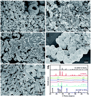
Mo/W oxide hollow microspheres with MoO3 as matrix material were fabricated via a polyethylene glycol (PEG) induced aqueous route. The hierarchical Mo/W oxide samples are constructed by elementary fusiform particles, which were characterized by X-ray powder diffraction (XRD) and scanning electron microscopy (SEM) methods. Initial formed h-WO3 crystallites acted as nuclei for subsequent formation of MoO3 precipitates, thus Mo/W oxide samples formed through epitaxial growth strategy show weak crystallization because of the lattice mismatch between h-MoO3 and h-WO3 crystals. The MoO3 based Mo/W oxide samples exhibit good photochromic performances compared to unitary h-MoO3 and h-WO3 crystals.


1. Introduction

Transition metal oxides, especially photochromic materials such as WO3 and MoO3 have attracted significant attention because of their widespread applications in solar cells, gas sensors, catalysis, rechargeable batteries, electrochromic, photochromic and thermochromic.1–10 Compared to unary metal oxide constituents, binary oxide materials are more promising due to the additional opportunity for component control, morphological control, physical/chemical property modulation, as well as improved performances in the applications due to the expected “synergistic effect” in the composites.11–15 Therefore, considerable efforts have been made towards the preparation and property investigation of MoO3 based binary metal oxides in recent years. Illyaskutty16 reported drastic variation on morphology by incorporating ZnO with MoO3 via RF magnetron sputtering and oxidation method, the ZnO-incorporated MoO3 nanostructures showed enhanced photoluminescence. Bai17 reported that, compared to pure MoO3, metal (Ag, Au, Pt, and Pd)/MoO3 nanobelts exhibited excellent visible-light photocatalytic activity and high stability for the degradation of azo dyes and for photocatalytic synthesis of benzyl compounds. Miyazaki18 fabricated MoO3 based composite films, whose photochromic property (such as bleaching speed) was controlled by copper addition. Yao19 prepared Au–MoO3 composite film by depositing gold nanoparticles onto the surface of MoO3 film, which showed improved coloration performance under UV-light irradiation. Wang20 have prepared α-MoO3/In2O3 core–shell nanorods via hydrothermal method, it as anode of LIBs exhibit enhanced lithium storage properties in terms of high reversible capacity, good cycling stability and excellent rate capability. Such good performance can be ascribed to the synergistic effect between α-MoO3 and In2O3.

Although various MoO3 or WO3 nanostructures have been synthesized,21–24 the diversity of binary Mo/W oxide nanostructures is relatively limited. Hierarchical structures with hollow interior show promising characteristics such as large internal void, high specific surface area, low density and high loading capacity, which are beneficial to the applications.25 According to literatures, synthetic methods for Mo/W oxide materials include liquid phase deposition technique,26 thermal evaporation,27 RF sputtering,28 electrochemical technique,29 spray pyrolysis,30 sol–gel method,31,32 and hydrothermal reactions.33–37 However, it still remains a challenge to develop facile aqueous methods for the preparation of hollow micro/nanostructures under mild conditions.

In this study, Mo/W oxide hollow microspheres with MoO3 as matrix material were prepared by a facile aqueous method at 90 °C. As a crucial factor, PEG played a key role for the successful preparation of hollow architectures. The sphere-like hierarchical nanostructures were assembled by fusiform particles. The hollow Mo/W oxide microspheres exhibited good photochromic property under visible-light stimulation.

2. Experimental section

2.1. Synthesis of Mo/W oxide hollow microspheres

All of the chemicals were used as purchase without further purification. Mixed molybdenum/tungsten (Mo/W) oxides were synthesized via a facile aqueous route. In a typical procedure, 45 mL of 0.2 mol L−1 Na2MoO4·2H2O and 5 mL of 0.2 mol L−1 Na2WO4·2H2O were mixed together in a flask at 90 °C to get a transparent solution with Mo/W molar ratio of 9[thin space (1/6-em)]:[thin space (1/6-em)]1. 0.0432 g of PEG 20k in 10 mL of deionized water was subsequently added into the above solution under vigorous stirring. Then, a certain amount of HCl solution (2 mol L−1) was added into the solution to reach a pH value of 1.0. After the reaction continued for 3 h, the precipitates were collected by vacuum filtration and washed with deionized water and ethanol for several times to remove the residual reactants and byproducts. Finally, the precipitates were dried in an air oven of 60 °C for several hours to get final Mo/W oxide samples.

2.2. Characterization

Crystalline structures of samples were characterized by X-ray powder diffraction (XRD) on a Shimadzu X-ray diffractometer 6100. The morphologies of samples were examined on a Hitachi S-4800 scanning electron microscope (FESEM). The optical properties of samples were investigated by a UV-Vis spectrophotometer (Shimadzu UV-2600).

2.3. Photochromic activity

Sample powders were put into a round groove with a diameter of 10 mm and a thickness of about 1 mm and pressed tightly for the photochromic activity test. Thereafter, visible-light irradiation was performed for 3 min on samples with a 500 W iodine tungsten lamp. The bleaching process was performed under dark.

3. Results and discussion

3.1. Crystalline structure and morphology

SEM and XRD analyses were used to characterize the phase composition and morphology of Mo/W oxide samples. Three MoO3 based samples with Mo/W molar ratios of 9[thin space (1/6-em)]:[thin space (1/6-em)]1, 8[thin space (1/6-em)]:[thin space (1/6-em)]2 and 7[thin space (1/6-em)]:[thin space (1/6-em)]3 were chosen for investigation, which are respectively assigned as samples S1–S3 for clear explanation. Pure h-MoO3 and h-WO3 crystals prepared under parallel reaction parameters were included for comparison. From XRD patterns of the five samples in Fig. 1f, we know that samples S1–S3 of Mo/W oxides extend weak crystallization, while samples of pure materials have good crystallinity. The results reveal that the Mo/W oxides are composite materials with interaction instead of simple mixing. Fig. 1a–e give SEM images of the five samples. Mo/W oxide samples (S1–S3) were constructed by elementary fusiform particles, as revealed from the insets of Fig. 1a–c. The 1D structure of the elementary particles in samples S1–S3 is similar to that of h-WO3 sample (inset in Fig. 1d), but is obviously different from that of h-MoO3 microrods (Fig. 1e). The smooth surfaces of h-MoO3 microrods (Fig. 1e) and compact aggregations of h-WO3 (Fig. 1d) demonstrate well crystallization of the samples, which accord with XRD results in Fig. 1f. Only one prominent peak at 22.8° is present in the XRD patterns of samples S1–S3, which was exclusively index to (001) peak of h-WO3 crystals (JCPDS card no. 33-1387). As revealed from energy dispersive X-ray (EDX) result of sample S1 (Fig. S1, ESI), the sample is composed of Mo, W and O in an atom ratio of 18.5[thin space (1/6-em)]:[thin space (1/6-em)]1[thin space (1/6-em)]:[thin space (1/6-em)]49, Mo oxide is the major composition for sample S1. No intensive peaks of MoO3 crystals in XRD patterns of samples S1–S3 suggest that MoO3 in the samples would exist as amorphous structure. On experimental experiences, we knew that the formation speed of h-WO3 crystals was faster than that of h-MoO3 crystals in separate reactions under parallel reaction conditions, and limited introduction of W to MoO3 reaction system also can accelerate the formation speed of oxide precipitates. Thus in our work, h-WO3 crystallites are assumed to form first in the mixed system of W and Mo oxides, and acted as nuclei for the succeeding formation of MoO3 particles. As reported in literatures,38,39 h-WO3 crystallites frequently grow along (001) direction. MoO3 precipitates evolved and grew on h-WO3 crystallites, the crystalline mismatch of h-WO3 and h-MoO3 is the reason for the formed MoO3 particles with worse crystallization.
image file: c6ra20665d-f1.tif
Fig. 1 SEM images of three Mo/W oxide samples with different Mo/W molar ratios, pure h-MoO3 and h-WO3 samples are given for comparison: (a) sample S1, Mo/W = 9[thin space (1/6-em)]:[thin space (1/6-em)]1; (b) sample S2, Mo/W = 8[thin space (1/6-em)]:[thin space (1/6-em)]2; (c) sample S3, Mo/W = 7[thin space (1/6-em)]:[thin space (1/6-em)]3; (d) h-WO3; (e) h-MoO3. (f) XRD patterns of the five samples. Reaction parameters for typical experiments: reaction temperature of 90 °C, reaction time of 3 h, PEG 20k introduction: 3 wt%.

The obvious difference for samples S1–S3 is the hierarchical structure of sample S1, which contains the least amount of W introduction (Mo/W ratio of 9[thin space (1/6-em)]:[thin space (1/6-em)]1) among the three samples. Hollow interior of the assembled microspheres can be seen clearly in the inset of Fig. 1a. The results demonstrate the introducing amount of W element influence the assembly of final samples, less introduction of W is of benefit to the formation of hollow microspheres.

Temperature-dependent experiments were performed on the basis of sample S1 to examine the influences. The reaction temperature for sample S1 is 90 °C. Fig. 2 gives SEM and XRD results of two Mo/W oxide samples (Mo/W = 9[thin space (1/6-em)]:[thin space (1/6-em)]1) prepared at 80 °C (sample S4) and 100 °C (sample S5). Sample S4 prepared at 80 °C exhibits microsphere morphology (Fig. 2a) with similar crystallinity as that of sample S1 (90 °C). Sample S5 at higher reaction temperature (100 °C) changed a lot both in morphology and crystallinity (Fig. 2b and c) compared to that of sample S1. Distributed short nanobelts are in the image of Fig. 2b with 100 nm in width. From the XRD pattern of sample S5 in Fig. 2c, we can see that the crystallization is better than sample S1, some diffraction peaks of h-MoO3 are also present in the pattern except the peak of h-WO3. The results reveal that high reaction temperature facilitates the crystallization of Mo/W oxides, but it is not beneficial to the formation of assembly structure.


image file: c6ra20665d-f2.tif
Fig. 2 SEM images of samples S4 and S5 obtained at different reaction temperatures on the basis of sample S1: (a) 80 °C, (b) 100 °C, (c) XRD patterns of samples S4 and S5.

To understand the assembly mechanism of Mo/W oxide microspheres, three samples were prepared at different reaction time (0.5, 1 and 2 h) under the reaction parameters parallel to sample S1 (reaction time of 3 h). Fig. 3 gives the results on the morphology and crystalline structure of the samples. XRD patterns in Fig. 3d combined with the pattern of sample S1 in Fig. 1f reveal the crystallization of Mo/W oxides merely completed at the reaction time of 0.5 h, and the crystallinity changed a little from 0.5 to 3 h. We can observe the amount of precipitates increasing in solution with reaction time. From the SEM images in Fig. 3a–c, it can be seen that the samples of 0.5, 1 and 2 h have similar hierarchical structures as that of sample S1 (Fig. 1a), the sizes of microspheres increase a little with the reaction time. The time-dependent experiments demonstrate that the introduction of W to MoO3 system resulted in fast precipitation of the system. Epitaxial growth of MoO3 precipitates on h-WO3 crystallites can explain the phenomenon.40,41 A sample with prolonged reaction time of 6 h was also done, the results are given in Fig. S2. SEM images of the sample (Fig. S2a) also show spherical assembled structure with prominent elementary units. XRD pattern in Fig. S2b demonstrates enhanced crystallization of the sample compared to sample S1, intensive diffraction peaks of h-MoO3 existed in the pattern besides that of (001) peak of h-WO3. The results reveal that prolonged reaction time led to better crystallization of MoO3 base material, but limited influence on the morphology. Thus we can get Mo/W oxide microspheres with different crystallinity of MoO3 by tuning the reaction time.


image file: c6ra20665d-f3.tif
Fig. 3 SEM images of samples obtained at different reaction time: (a) 0.5 h, (b) 1 h and (c) 2 h. (d) Corresponding XRD patterns of the three samples.

As reported in literatures, organic molecules with long chain would play essential role for the formation and growth of crystals. In our strategy, PEG 20k was introduced in the reaction system for the synthesis of Mo/W oxide samples. Some samples with different amount of PEG 20k introduction were prepared for the examination, SEM images of the samples are shown in Fig. 4, the sample without PEG introduction is also given in Fig. 4a for comparison. The samples with PEG 20k amount of 6 and 9 wt% preserve identical hierarchical morphologies as that of samples S1 (PEG 20k of 3 wt%). And some hollow interiors are exposed in the view of the images (insets of Fig. 4c and d). The samples without PEG and with 1 wt% of PEG 20k introduction exhibit irregular assemblies in morphology (Fig. 4a and b). Thus the results show that Mo/W oxide microspheres with hollow interior can be achieved with adequate amount of PEG introduction.


image file: c6ra20665d-f4.tif
Fig. 4 SEM images of samples with different weight percent of PEG 20k: (a) 0 wt%, (b) 1 wt%, (c) 6 wt%, (d) 9 wt%.

The influences of other surfactants were also studied. Sodium dodecyl sulfate (SDS), cetyltrimethylammonium bromide (CTAB) and polyvinylpyrrolidone (PVP) were chosen for parallel preparations. The results are shown in Fig. S3. Substitution of PEG 20k with other surfactants resulted in the disappearance of microsphere structures in the images.

Based on the above experimental results, it revealed that there exist interactions between MoO42−/WO42− and PEG molecules, and the interaction had non-negligible impact on the morphology of the hollow Mo/W oxide microspheres. In this mechanism, the amount of surfactants and other variations (such as different Mo/W molar ratio, different surfactants, temperature and reaction time etc.) might impose influences on the morphology as well as the crystalline structure of the samples. The hydroxyl radicals at the ends of PEG molecule and ether bonds among the molecule chain can interact with the initial formed WO3 crystallites, the curled PEG/WO3 chains acted as the templates for the formation of hollow Mo/W oxide microspheres. The proposed formation mechanism is illustrated in Scheme 1. In the process, the fast formed WO3 crystallites as nuclei resulted in the epitaxial growth of MoO3 particles. The induced formation of MoO3 particles on the surface of WO3 crystallites led to the composites with weak crystallinity.


image file: c6ra20665d-s1.tif
Scheme 1 Formation process of Mo/W oxide hollow microspheres.

3.2. Optical and photochromic properties of Mo/W oxide samples

The optical absorption properties of materials are important and informative in evaluating their application activities, in our case they are highly relevant to the photochromic properties of Mo/W oxide samples. UV-vis diffuse reflectance spectroscopy (UV-vis DRS) was made on samples S1–S3, the results are shown in Fig. 5. Pure h-MoO3 and h-WO3 crystals were also detected for comparison. The results indicate that all the five samples have optical absorption from UV to visible region. Pure h-MoO3 and h-WO3 crystals have strong absorption in UV region and weak absorption in visible region. Samples S1–S3 with different Mo/W molar ratios (9[thin space (1/6-em)]:[thin space (1/6-em)]1, 8[thin space (1/6-em)]:[thin space (1/6-em)]1 and 7[thin space (1/6-em)]:[thin space (1/6-em)]3) show increased absorption in visible region compared to those samples with single composition (h-MoO3 or h-WO3). And sample S1 with microsphere structure has stronger absorption in visible region than samples S2 and S3 of fusiform particles. The absorption transitions can be attributed to charge transfer between the oxygen and molybdenum/tungsten atoms, in which oxygen 2p electrons go into the empty 4d orbitals of molybdenum or 5d orbitals of tungsten. The above optical absorption results demonstrate that photochemical properties of the Mo/W oxide samples would be superior to that of pure h-MoO3 or h-WO3 crystals.
image file: c6ra20665d-f5.tif
Fig. 5 UV-vis DRS spectra of samples S1, S2 and S3 with h-MoO3 and h-WO3 crystals for comparison.

The photochromic responses of the five samples (S1, S2, S3, h-MoO3 and h-WO3) were tested subjecting to visible-light irradiation. The photochromic experiments were performed by first putting the sample powders into a round groove and pressing into sample discs with a diameter of 10 mm and a thickness of about 1 mm. Thereafter, visible-light irradiation was done on disc samples for 3 min under a 500 W iodine tungsten lamp. Fig. 6 gives the pictures of the disc samples before and after coloration. Before irradiation, the Mo/W oxide samples (S1–S3) are faint yellow in color, whereas pure h-MoO3 sample is white, and h-WO3 sample is yellow. After irradiation for 3 min under visible-light, the three faint yellow powders (S1–S3) showed apparent coloration to blue, h-MoO3 and h-WO3 samples showed weak coloration. It is obvious that Mo/W oxide samples exhibit an enhanced photochromic property compared to that of pure h-MoO3 and h-WO3 samples. The photochromic performances of Mo/W oxide samples are also dependent on the constituent composition of the samples. Sample S1 with less W introduction in MoO3 has relatively good photochromic property. The reversible coloration of the samples was also examined. The blue colored samples were bleached under dark for 14 h. After bleaching, the irradiation was repeated, and the Mo/W oxide samples showed good cycled coloration for eight runs (Fig. 6).


image file: c6ra20665d-f6.tif
Fig. 6 Optical photographs of Mo/W oxide samples (S1–S3) and pure h-MoO3, h-WO3 samples before and after coloration.

The good photochromic performances of Mo/W oxide samples would be attributed to two factors on comparison to pure h-MoO3 or h-WO3 crystals: one is their weak crystallization, the other is intervalence-charge transfer among Mo, W and O atoms.42 Inorganic materials with weak crystallization preserve more crystalline defects, which is benefit to the generation of electron–hole pairs in the photochromic process. According to the model of double insertion–extraction of ions and electrons,43 the high number of protons and photo-generated electron–hole pairs has a large effect on the photochromic properties of the samples. When h-MoO3 or h-WO3 crystals were subjected to light source, the coloration of powders was caused by a simultaneous injection of protons and electrons to form hydrogen molybdenum bronze or hydrogen tungsten bronze. As for Mo/W oxide samples, the intervalence-charge transfer from Mo5+ to W6+ is also assumed to take place in the complex samples. Thus photochromic property of Mo/W oxide samples is better than samples with single composition.

Optical absorption detection was also made on Mo/W oxide samples (S1, S4 and S5) with different morphologies, the results are summarized in Fig. 7a. The three samples have strong absorptions in UV region, and samples S1 and S4 with microsphere structure have wider absorption in visible-light region compared to sample S5 of short nanobelts. The photochromic properties of the three Mo/W oxide samples S1, S4 and S5 are given in Fig. 7b. After 2 min's irradiation under visible-light, the three sample powders (S1, S4 and S5) showed apparent coloration from faint yellow to blue. It is obvious that samples S1 and S4 exhibit better photochromic property than that of sample S5. Thus the photochromic performances of Mo/W oxide samples are also dependent on the morphology of the samples, the samples with hierarchical structure exhibit good photochromic properties. The reason would be that hollow structures of Mo/W oxide samples possess more exposed surface area, which contributes large absorption to the excitation light and results in larger quantity of protons and photo-generated electron–hole pairs in the photochromic processes.


image file: c6ra20665d-f7.tif
Fig. 7 (a) UV-vis absorption spectra of Mo/W oxide samples S1, S4 and S5, (b) optical photographs of the corresponding Mo/W oxide samples (S1, S4 and S5) before and after coloration under visible-light irradiation.

The optical and photochromic properties of Mo/W oxide samples with different weight percent of PEG 20k (0, 1, 3, 6 and 9 wt%) were also studied. The five samples all exhibited intense absorptions from UV to visible region in Fig. S4a. The threshold of curves shows slightly red shift with an increase of PEG 20k amount in the samples. The coloration results are given in Fig. S4b. It is obvious that samples with more PEG 20k introduction exhibited better photochromic property. Thus the photochromic performances of Mo/W samples are also dependent on the amount of PEG 20k introduction.

4. Conclusion

Hierarchical Mo/W oxide microspheres with hollow interior were prepared by a PEG-induced aqueous method. Mo/W molar ratio and PEG molecules have strong contributions to the formation of hollow microspheres. Reaction temperature and reaction time were also found to play a key role in morphology-controlled synthesis. The initially formed h-WO3 crystallites along (001) direction accelerated the succeeding formation of h-MoO3 crystals in solution, thus resulted in Mo/W oxide hierarchical structures with weak crystallization. The Mo/W oxide samples exhibited enhanced photochromic properties compared to h-MoO3 or h-WO3 crystals with single composition.

Acknowledgements

This work was supported by the National Natural Science Foundation of China (Grant No. 21571057, J1210040), the Fundamental Research Funds for the Central Universities, and Science and Technology Project of Changsha City (Grant No. k1403007-11).

References

  1. A. Chithambararaj, N. R. Yogamalar and A. C. Bose, Cryst. Growth Des., 2016, 16, 1984–1995 CAS.
  2. Y. C. Xia, C. S. Wu, N. Y. Zhao and H. Zhang, Mater. Lett., 2016, 171, 117–120 CrossRef CAS.
  3. Y. Shen, L. Pan, Z. Y. Ren, Y. L. Yang, Y. L. Xiao and Z. Li, J. Alloys Compd., 2016, 657, 450–456 CrossRef CAS.
  4. S. Yang, Y. L. Liu, W. Chen, W. Jin, J. Zhou, H. Zhang and G. S. Zakharova, Sens. Actuators, B, 2016, 226, 478–485 CrossRef CAS.
  5. R. Nadimicherla, R. H. Zha, L. Wei and X. Guo, J. Alloys Compd., 2016, 687, 79–86 CrossRef CAS.
  6. D. S. Jiang, Y. Wang, W. Wei, F. Li, Y. J. Li, L. H. Zhu, C. H. Feng, C. X. Liu and S. P. Ruan, RSC Adv., 2015, 5, 18655–18659 RSC.
  7. J. B. Zhou, N. Lin, L. B. Wang, K. L. Zhang, Y. V. Zhu and Y. T. Qian, J. Mater. Chem. A, 2015, 3, 7463–7468 CAS.
  8. M. Jafarpour, M. Ghahramaninezhad and A. Rezaeifard, RSC Adv., 2014, 4, 1601–1608 RSC.
  9. H. Najafi-Ashtiani, A. Bahari and S. Ghasemi, J. Electroanal. Chem., 2016, 774, 14–21 CrossRef CAS.
  10. Y. Liu, J. Li, H. Tang, W. Z. Li, Y. H. Yang, Y. M. Li and Q. Y. Chen, Electrochem. Commun., 2016, 68, 81–85 CrossRef CAS.
  11. C. Wang, L. X. Wu, H. Wang, W. H. Zuo, Y. Y. Li and J. P. Liu, Adv. Funct. Mater., 2015, 25, 3524–3533 CrossRef CAS.
  12. M. Yang, L. Zhang, B. W. Jin, L. Huang and Y. Gan, Appl. Surf. Sci., 2016, 364, 410–415 CrossRef CAS.
  13. H. Y. Yu, Y. J. Li, L. Zhao, G. M. Li, J. W. Li, H. Rong and Z. F. Liu, Mater. Lett., 2016, 169, 65–68 CrossRef CAS.
  14. M. E. Navgire, P. Gogoi, B. Mallesham, A. Rangaswamy, B. M. Reddy and M. K. Lande, RSC Adv., 2016, 6, 28679–28687 RSC.
  15. N. Li, Y. M. Li, W. J. Li, S. D. Ji and P. Jin, J. Phys. Chem. C, 2016, 120, 3341–3349 CAS.
  16. N. Illyaskutty, S. Sreedhar, H. Kohler, R. Philip, V. Rajan and V. P. M. Pillai, J. Phys. Chem. C, 2013, 117, 7818–7829 CAS.
  17. H. Bai, F. Ye, Q. Lv, G. C. Xi, J. F. Li, H. F. Yang and C. Q. Wan, J. Mater. Chem. A, 2015, 3, 14550–14555 CAS.
  18. H. Miyazaki, H. Ichioka, H. Suzuki and T. Ota, J. Ceram. Soc. Jpn., 2014, 1421, 83–85 CrossRef.
  19. T. He, Y. Ma, Y. A. Cao, P. Jiang, X. T. Zhang, W. S. Yang and J. N. Yao, Langmuir, 2001, 17, 8024–8027 CrossRef CAS.
  20. Q. Wang, J. Sun, Q. Wang, D. A. Zhang, L. L. Xing and X. Y. Xue, J. Mater. Chem. A, 2015, 3, 5083–5091 CAS.
  21. L. N. Wang, X. Zhang, Y. Ma, M. Yang and Y. X. Qi, Mater. Lett., 2016, 164, 623–626 CrossRef CAS.
  22. S. H. Wei, G. Y. Zhao, W. M. Du and Q. Q. Tian, Vacuum, 2016, 124, 32–39 CrossRef CAS.
  23. N. Lu, X. Q. Gao, C. Yang, F. Xiao, J. D. Wang and X. T. Su, Sens. Actuators, B, 2016, 223, 743–749 CrossRef CAS.
  24. W. Z. Xua, C. C. Li, Y. X. Hu, L. Liu, Y. Hu and P. C. Wang, Polym. Adv. Technol., 2016, 27, 964–972 CrossRef.
  25. S. H. Wei, Y. Xing, Y. H. Li, Y. Zhao, W. M. Du and M. H. Zhou, Vacuum, 2016, 129, 13–19 CrossRef CAS.
  26. H. M. Farveez Ahmed and N. S. Begum, Bull. Mater. Sci., 2013, 1, 45–49 CrossRef.
  27. E. E. Khawaja, S. M. A. Durrani and M. A. Daous, J. Phys.: Condens. Matter, 1997, 9, 9381–9392 CrossRef CAS.
  28. S. Morandi, G. Ghiotti, A. Chiorino, B. Bonelli, E. Comini and G. Sberveglieri, Sens. Actuators, B, 2005, 111–112, 28–35 CrossRef CAS.
  29. X. C. Song, X. Wang, Y. F. Zheng and Y. H. Yin, Curr. Nanosci., 2013, 3, 330–334 CrossRef.
  30. P. R. Patil and P. S. Patil, Thin Solid Films, 2001, 382, 13–22 CrossRef CAS.
  31. J. C. Shi, G. M. Wu, J. Shen, G. H. Gao, T. A. Liang and B. Zhou, Rare Met. Mater. Eng., 2010, 39, 40–43 Search PubMed.
  32. R. R. Kharade, S. S. Mali, S. S. Mohite, V. V. Kondalkar, P. S. Patil and P. N. Bhosale, Electroanalysis, 2014, 26, 2388–2397 CrossRef CAS.
  33. A. Michailovski, F. Krumeich and G. R. Patzke, Chem. Mater., 2004, 16, 1433–1440 CrossRef CAS.
  34. R. Kiebach, N. Pienack, W. Bensch, J. D. Grunwaldt, A. Michailovski, A. Baiker, T. Fox, Y. Zhou and G. R. Patzke, Chem. Mater., 2008, 20, 3022–3033 CrossRef CAS.
  35. L. Zhou, J. Zhu, M. H. Yu, X. D. Huang, Z. Li, Y. H. Wang and C. Z. Yu, J. Phys. Chem. C, 2010, 114, 20947–20954 CAS.
  36. Y. Zhou, K. B. Zheng, J. D. Grunwaldt, T. Fox, L. L. Gu, X. L. Mo, G. R. Chen and G. R. Patzke, J. Phys. Chem. C, 2011, 115, 1134–1142 CAS.
  37. A. Arzola-Rubio, J. Camarillo-Cisneros, L. Fuentes-Cobas, V. Collins-Martínez, L. D. L. Torre-Sáenz and F. Paraguay-Delgado, Superlattices Microstruct., 2015, 81, 175–184 CrossRef CAS.
  38. V. V. Kondalkar, S. S. Mali, R. R. Kharade, K. V. Khot, P. B. Patil, R. M. Mane, S. Choudhury, P. S. Patil, C. K. Hong, J. H. Kime and P. N. Bhosale, Dalton Trans., 2015, 44, 2788–2800 RSC.
  39. W. J. Mu, M. Li, X. L. Li, Z. P. Ma, R. Zhang, Q. H. Yu, K. Lv, X. Xie, J. H. He, H. Y. Wei and Y. Jian, Dalton Trans., 2015, 44, 7419–7427 RSC.
  40. E. Lee and Y. Kwon, RSC Adv., 2016, 6, 47468–47473 RSC.
  41. C. J. Liu, J. Li, Y. M. Li, W. Z. Li, Y. H. Yang and Q. Y. Chen, RSC Adv., 2015, 5, 71692–71698 RSC.
  42. T. He and J. N. Yao, J. Photochem. Photobiol., C, 2003, 4, 125–143 CrossRef CAS.
  43. Y. Yang, Y. Cao, B. Loo and J. N. Yao, J. Phys. Chem. B, 1998, 102, 9392–9396 CrossRef CAS.

Footnote

Electronic supplementary information (ESI) available. See DOI: 10.1039/c6ra20665d

This journal is © The Royal Society of Chemistry 2016
Click here to see how this site uses Cookies. View our privacy policy here.