Advances in Fe-based biodegradable metallic materials

Jin He, Feng-Li He, Da-Wei Li, Ya-Li Liu, Yang-Yang Liu, Ya-Jing Ye and Da-Chuan Yin*
Institute of Special Environmental Biophysics, Key Laboratory for Space Bioscience and Biotechnology, School of Life Sciences, Northwestern Polytechnical University, Xi'an 710072, PR China. E-mail: yindc@nwpu.edu.cn

Received 15th August 2016 , Accepted 22nd November 2016

First published on 22nd November 2016


Abstract

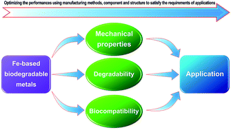
In recent years, biodegradable metallic materials, such as Mg-, Fe-, Zn- and W-based materials, have been the focus of many studies. As one of the two most studied types of biodegradable metallic materials, Fe-based materials have aroused a great deal of interest because of their outstanding mechanical properties, which are similar to stainless steel. The processing methods can directly affect the microstructure of the material and influence the mechanical and degradation properties of the material. Furthermore, biocompatibility is directly affected by the degradation properties. Therefore, the processing methods, mechanical properties, degradability and biocompatibility are several of the main concerns in the study of biodegradable Fe-based materials. Here, we systematically summarize recent studies on Fe-based materials and discuss these findings in terms of their processing methods, degradability and biocompatibility.


1. Introduction

The use of metallic materials in the medical field has a long history. Traditional biomedical metallic materials, such as Ti (Ti alloy), Co alloy and stainless steel,1–3 have excellent biocompatibility and mechanical properties. Thus, they occupy a dominant position in medical implant materials. Table 1 gives a summary of the major merits and shortcomings of some frequently studied biomedical metallic materials. Among these materials, Ti (Ti alloy), Co alloy and stainless steel have already been applied commercially. However, these materials are non-degradable and permanently exist in the body as a foreign object, which can cause negative effects.
Table 1 A summary of the main merits and shortcomings of biomedical metallic materials1–3,5,6,8–10
Biomaterials Typical samples Merits Shortcomings
Fe-Based materials Pure Fe, Fe–Mn, Fe–Fe2O3 Good mechanical properties, appropriate biocompatibility, various processing methods, biodegradable Low degradation rates, ferromagnetism
Mg/Mg alloys Pure Mg, Mg–Zn, Mg–Zn–Ca Excellent biocompatibility, biodegradable, similar mechanical properties with bone Too fast degradation rates, hydrogen evolution
Ti/Ti alloys Pure Ti, Ti–6Al–4V Good mechanical properties, excellent biocompatibility, high specific strength, good corrosion resistance Non-degradable, toxicity elements (e.g., V)
Stainless steel 316L Excellent corrosion resistance, high mechanical properties, appropriate biocompatibility Non-degradable, toxicity elements (e.g., Ni), too high modulus, poor plastic workability
Co alloys Co–Cr–W–Ni, Co–Cr–Mo Excellent wear resistance, appropriate biocompatibility, high corrosion resistance Too high modulus, poor cold workability, toxicity elements (e.g., Ni), non-degradable


In recent years, biodegradable metals have aroused a great deal of interest because of their attractive properties, such as the excellent mechanical properties that are similar to traditional biomedical metallic materials, superior biocompatibility and degradability.4–11 Zheng et al.9 defined biodegradable metals: biodegradable metals are metals expected to corrode gradually in vivo, with an appropriate host response elicited by released corrosion products, then dissolve completely upon fulfilling the mission to assist with tissue healing with no implant residues. Over the years, many types of biodegradable metals have been studied, including Mg-,5,6 Fe-,8–10 W-12–15 and Zn-based materials.16–19 Currently, the study of biodegradable metals mainly focuses on two categories: Fe-based and Mg-based materials. There is relatively less research conducted on other biodegradable metals. Fe is a traditional engineering material that has many advantages, such as excellent mechanical properties, superior machinability and low price. These inherent advantages make Fe-based materials a potentially exceptional source of biodegradable metallic materials for the medical field.

2. Application of biodegradable Fe-based materials

According to reported studies, the applications of biodegradable Fe-based metallic materials mainly consist of three categories: intravascular stents, implant materials for bone surgery and scaffolds for bone tissue engineering.

2.1 Stents

Stents have been widely used for the treatment of cardiovascular disease. However, their applications can produce undesirable complications.20 Restenosis can occur within the stent because of inflammation and neointimal proliferation. Furthermore, stents implanted in children result in a fixed vessel diameter, and subsequent restenosis occurs due to vessel growth at the stent site. Degradable stents can overcome these shortcomings.20 This type of stent can eliminate the continuous interference process between the stent and the vessel wall, and its use as a carrier for drugs can avoid re-dilatation surgery. Another potential advantage of degradable stents is the avoidance of subsequent surgeries on vessels with implanted stents.

An inherent advantage of iron is its excellent mechanical properties, which make it an interesting candidate for use in biodegradable stents.8 For example, the higher elastic modulus of iron can produce a high radial strength of the stent, which is beneficial when producing stents with thinner struts. In addition, iron has high ductility, which can be useful when the stent is plastically deformed during the implantation procedure.3 Therefore, the development of degradable stents is one of the main uses for degradable Fe-based materials. Fig. 1 shows examples of Fe and Fe–Mn alloy stents.


image file: c6ra20594a-f1.tif
Fig. 1 Photographs of a Fe stent (a) and Fe–35Mn alloy stents (b) (reproduced with permission of Elsevier from ref. 20 and 21).

2.2 Implants for bone surgery

The non-degradable metal implants used in bone surgery, such as screws22 and plates,23 can lead to adverse responses, such as a toxic response to specific ions released from the materials due to their long-term presence in the body. Although the implants can be removed as a second operation, it can result in adverse consequences, such as a second defect at the implantation site, pain, and increased treatment costs. Fe-Based materials are a good candidate to address these shortcomings. First, Fe-based materials have excellent mechanical properties that are similar to those of stainless steel and can satisfy the requirement of high mechanical properties for bone repair. In addition, Fe-based materials have good biocompatibility. Above all, Fe corrodes in the body environment and is expected to be completely dissolved during the lifetime.

2.3 Scaffolds for bone tissue engineering

Scaffolds play an important role in tissue engineering and provide essential conditions for tissue repair. For example, scaffolds provide a surface and space that allows for cell growth and new tissue formation, thus aiding in the remodelling of damaged tissue.24 An ideal scaffold must have reliable degradation and resorption rates that are consistent with the rate of tissue regeneration. Moreover, scaffolds must have appropriate mechanical properties that match those of normal tissues and organs.25 Because metallic materials have excellent mechanical properties, the application of biodegradable metal porous scaffolds is primarily used for the reparation of hard tissue, such as bone. Thus, these novel scaffolds avoid the shortcomings of traditional non-degradable metal scaffolds and overcome the weaknesses of polymer scaffolds.

3. Manufacturing methods and mechanical properties

Many methods, including the manufacturing methods for raw materials and post-treatment methods (Fig. 2), have been used to manufacture biodegradable metallic materials; notably, these methods are similar to those used with traditional metals. The properties of the material, such as mechanical properties and degradability, can be affected by the manufacturing method. Indeed, mechanical properties are most vulnerable. Table 2 summarizes the mechanical properties of the materials after processing with different methods. According to published studies, the methods can be divided into five types: casting, sintering, electrodeposition, surface modification and other methods. Except for surface modification, the other methods refer to the manufacturing or processing of raw materials.
image file: c6ra20594a-f2.tif
Fig. 2 Diagram of the different manufacturing methods. Initial methods refer to the production methods that transform raw materials into materials that can be used for device manufacturing. Post-processing refers to further treatments after the production of raw materials. Note: surface modification is not an initial method, but it is one of the most studied manufacturing methods.
Table 2 Summary of the manufacturing methods and the mechanical properties of different Fe-based materialsa
Processing methods Components Mechanical properties Ref.
Tensile Compress Hardness
Ultimate strength (MPa) Yield strength (MPa) Elasticity modulus (GPa) Elongation at break (%) Yield strength (MPa) Ultimate strength (MPa) Elasticity modulus (GPa)
a Note: pure Fe that served as a control is not listed in this table. The error values in the original data from the references are not shown. The values of the same materials manufactured using different processes were expressed as a range.
Casting + forging Fe–(0.5–6.9)Mn 353–1041 295–814 11.5–31.3 38
Casting + forging + heat treatment Fe–10Mn 1300–1400 650–800 14 374–428 (HV10) 32
Fe–10Mn–1Pb 1450–1550 850–950 2.0–11.0 376–437 (HV10)  
Casting + forging + heat treatment Fe–21Mn–0.7C(–1Pd) 1255 725 38 33
(Armco ingot Fe) rolling + annealing Fe-UD75% 600 593 3.5 35
Fe-BD75% 517 507 4.0
Fe-UD + annealing 248–283 108–246 34.8–48.3
Fe-BD + annealing 232–274 97–229 40.2–51.7
Casting + forging Fe–30Mn 632 242 94 413 175 (HBW) 39
Casting + forging Fe–30Mn 530 15 175 (HV 5) 40
SPS Fe–5Pd 445 754 41
Fe–5Pt 503 785
Sintering Porous Fe 16.1–67.7 0.6–4 42
Sintering Fe–35Mn 20.2–30.1 0.74–0.95 13.58–27.72 (HRH) 43
Sintering + cold rolling + sintering Fe–35Mn 550 235 31 38 (Rockwell A) 44
Sintering + cold rolling Fe–(20–35)Mn 428–723 234–421 4.8–32 38–59 (Rockwell A) 45
Sintering Fe–HA 325 717 46
Fe–TCP 312 708
Fe–BCP 312 696
Sintering Fe–TCP 696 47
Electroforming + annealing Fe 169–292 130–270 18.4–32.3 48
Electrodeposition Fe/Fe–W alloy scaffold 4.01 49
3D-printing + sintering Fe–30Mn 115.53 106.07 32.47 0.73 50
ECAP Fe 470 444 kgf mm−2 51
Nitriding Fe 614.35 287.06 (HV) 52
Magnetron sputtering Fe 343–638 267–612 1.3–20 53
Vacuum infiltration and dipping PLGA-infiltrated porous Fe 0.20–0.38 0.24–0.42 4.17–8.78 MPa 54
PLGA–coated porous Fe 0.27–0.65 0.30–0.71 6.15–14.22 MPa


3.1 Casting

As a traditional manufacturing method of metals, the advantage of casting is the convenience of adjustments to the alloy components. A certain proportion of raw materials (powders or blocks) are melted in a furnace and then cast to obtain alloys. Many researchers prefer this method of preparing biodegradable iron alloys. However, the poor properties of as-cast alloys are induced by defects (such as segregation, blowholes and shrinkages). Therefore, as-cast alloys must be further processed by forging, rolling and/or heat treatment.

Liu et al.26 casted a Fe30Mn6Si ternary alloy with a shape-memory function. The alloy consisted of martensite and austenite and had a smaller grain size and higher ultimate strength when compared with pure Fe. The recovery of this alloy reached 53.7% (casting + solution treated). The authors also produced a series of binary Fe alloys (Fe–Mn/Co/Al/W/Sn/B/C/S) by casting and rolling.27 Here, the addition of Mn, Co, Al, and W had no obvious effect on the grain size of the Fe alloys; however, B notably decreased the grain size. Sn sharply decreased the mechanical properties of the alloy. Mn, Co, W, B, C and S increased the yield and ultimate strength of Fe in the as-rolled alloys; furthermore, these elements increased the magnitude of the difference between the yield strength and ultimate strength of Fe.

Except for casting followed by hot working, more complex technology is typically needed to obtain alloys with specific properties. A Fe–10Mn–1Pd alloy was produced by casting, swaging, solution-heat-treatment and quenching, and the hardening kinetics of a solution-heat-treatment were studied by Moszner et al.28 The authors suggested that the pronounced strengthening was due to the thermally activated formation of coherent plate-like Pd-rich precipitates on 100 matrix planes. The new alloy was somewhat similar to maraging steel, e.g., the initial microstructure, aging behaviour and precipitation; thus, it represents a new high-strength, Ni-free maraging steel. To demonstrate the composition of the Fe–Mn–Pd alloy precipitates, Fe–Mn–Pd alloys containing 10 wt% Mn with varying Pd concentrations (1, 3, and 6 wt%) were created by casting, solution-heat-treatment, quenching, isothermal aging and quenching.29 The authors found that the precipitate was mainly a face-centred tetragonal β1-MnPd phase that consisted of Mn and Pd. The alloys demonstrated remarkable age-hardening after isothermal aging at 500 °C. The mechanism of reverse transformation from martensite to austenite in the Fe–Mn–Pd alloys and the influence of prior precipitation on this process were studied.30 The atomic-scale microstructures of prior austenite grain boundaries in Fe–Mn–Pd alloys were characterized by site-specific atom probe tomography, and the mechanism of embrittlement and de-embrittlement have been discussed.31

Schinhammer et al.32 discussed the design strategy of biodegradable Fe-based alloys. The authors suggested that noble intermetallic phases improved the degradation rate and increased the strength of the Fe matrix. For example, the availability of Mn and Pd has been proven. The degradation resistance of the newly developed Fe–Mn–Pd alloys is one order of magnitude less than that of pure iron. Furthermore, both the choice of alloying elements and changes in heat treatment can be used to adjust mechanical properties.

The interaction of recrystallization and precipitation in a biodegradable twinning-induced plasticity (TWIP) steel (Fe–Mn–C–Pd) was studied by Schinhammer et al.33 The authors found that the formation of a Pd-rich precipitate dramatically impeded recrystallization during the annealing treatment. The impedance was caused by grain boundary pinning by the Pd-rich precipitates and reduced dislocation mobility due to a solute drag effect from the Pd-enrichment of dislocation cores. These alloys showed high strength and superior ductility that exceeded other TWIP steels and the typical alloys used in biomedical implants.

The mechanical properties and corrosion rate of alloys can be affected by the average grain size; thus, these parameters can be optimized by altering the grain size. Obayi et al.34 found that thermo-mechanically treated (cold-rolled and annealed ingot) pure iron had an optimum microstructure with an average grain size of 20–25 μm. The authors also determined that the rolling direction affects the grain size and mechanical properties of pure Fe.35 The grain orientation of unidirectional rolled pure Fe was obvious; however, this orientation was irregular for bi-directional rolled pure Fe. Moreover, the mean grain size and the grain size distribution in bi-directional samples were larger and broader, respectively, when compared with unidirectional samples after annealing. The yield strength and tensile strength of unidirectional samples was larger than bi-directional samples. After annealing, the yield strength and tensile strength were markedly decreased.

Cast Fe–33Mn alloys were further processed with varying methods, including heavy cold-rolling, large-strain machining (LSM) and annealing.36 The grain shape of the alloy using LSM resembled dendrite bands and was approximately 16 dendrite band diameters in length, which is less than that of samples processed with other methods. In order to improve the interface compatibility of implants with bone and orthopaedic soft tissues, a cast Fe–30Mn alloy was further processed by a four-step dealloying process to create a nanoporous surface.37 This surface was designed by the selective leaching of the less noble components (Mn and Zn) from the outer surface layer.

After casting and hot forging, the phase of FeMn30 was mainly composed of γ-austenite and ε-martensite. Its mechanical properties were better than that of iron and 316L stainless steel.39 Manganese remains dissolved in γ-Fe, thus stabilising the austenitic structure at room temperature and causing significant solid-solution hardening and strengthening.40 Fe–Mn alloys with lower Mn concentrations (0.5, 2.7, and 6.9 wt% Mn) that were produced by casting and forging exhibited superior mechanical properties.38

3.2 Sintering

Spark plasma sintering (SPS)55 is the most advanced sintering technique. SPS has obvious advantages over other sintering techniques, such as lower sintering temperatures and shorter holding times. The technique enables the sintering of nanosized powders and can obtain a nearly full-densification of materials with minimum grain. Cheng et al.56 reported that the grain size of the pure iron processed by SPS was far smaller than that of as-cast pure iron. W and CNT, which served as the second phase, were added into the Fe matrix and clearly diminished the grain size; furthermore, the decrease in grain size correlated with increasing second phase fractions. The ultimate compressive strength (UCS) of the Fe matrix composite is greater than pure Fe, but the enhancement of yield strength (YS) for the composite was not significantly different from pure Fe. The YS and UCS of the SPS of pure Fe may be greater than as-cast pure Fe due to the smaller grain size of the former. Similarly, a Fe–Fe2O3 composite prepared by SPS57 has a fast corrosion rate, good mechanical properties and excellent biocompatibility. Huang et al.41 prepared Fe-5 wt% Pd and Fe-5 wt% Pt composites using SPS. Because of the second phase strengthening effects, the mechanical properties of the composites, including yield strengths, ultimate compressive strength, rigidity and microhardness, were much higher than that of pure Fe. Furthermore, small grain size improves the mechanical properties of composites. The low ductility of the composites may be due to their relatively low density. Fe–Au and Fe–Ag were also prepared by SPS.58 Fe-5 wt% Ag, Fe-2 wt% Au, and Fe-10 wt% Au composites showed improved mechanical properties. This improvement was attributed to the decrease in grain size and second-phase strengthening effects. The enhanced strength of the Fe–Au composite may be due to the ability of Au to partly dissolve into iron and result in solid solution strengthening.

The sintering method is typically used for the manufacturing of porous materials. A polyurethane sponge acted as a template, and the replication method based on powder metallurgy was used to produce an iron matrix composite (Fe–CNTs, Fe–Mg) with open-cell foam structure.59,60 The pore size of the foam was 35–50 PPI with an apparent density of approximately 24 kg m−3. However, there were many closed pores in the foam, which is a disadvantage for tissue penetration. Ammonium bicarbonate served as a space-holder, and porous iron was prepared by vacuum sintering.42,61 The highest porosity of the samples was 82 vol%. Increased compression pressure decreased porosity, while the use of a finer iron powder and more space-holder material in the initial mixture led to an increase in porosity. Increasing the porosity can decrease the mechanical properties of the sample; however, the use of finer powder increased the porosity and mechanical properties simultaneously. Furthermore, a higher compacting pressure enhanced flexural and compressive properties. The flexural and compressive performance of the sample was similar to that of human bone. A porous Fe–35Mn alloy with an open-cell porosity of 25–31% was prepared by sintering with ammonium bicarbonate as a porogen.43 The mechanical properties were decreased by increasing the amount of porogen used.

The mechanical properties of sintered metals are typically lower; however, they can be improved by rolling. Hermawan et al.44 reported that the porosity of a Fe–35Mn alloy prepared by cold-rolling and sintering cycles decreased by 0.3%, and the mechanical properties of this allow were similar to those of 316L stainless steel. This method can also change MnO particles into much smaller particles, which are then distributed in the rolling direction. Furthermore, Fe–Mn alloys with varying Mn content (20, 25, 30, and 35 wt%) were prepared using this method.45 The authors reported that alloys with lower Mn content existed mainly in the γ phase with some appearance of the ε phase. The yield strengths were between 234 MPa and 421 MPa, and the elongations were between 7.5% and 32%. Next, the authors processed Fe–35Mn alloy bulk materials produced from a similar method into minitubes with a 1.80 mm outside diameter, 0.15 mm wall thickness and 40 mm length using wire-cut electrical discharge machining and a programmable conventional turning machine.21 The minitubes were transformed into stents using laser cutting techniques. The mechanical properties of the stents, including expansion performance and radial force, were satisfactory. However, magnetic stents and those with too rough of a surface were unfavourable.

Using sintering, other elements have also been added into the iron matrix to form alloys. Wegener et al.62 prepared Fe–(C, P, B, and Ag) alloys using powder metallurgy. The addition of B and P had a notable effect on the microstructure of the alloy. The addition of P significantly decreased microporosity, while the addition of Ag and B increased microporosity.

Bioceramics have excellent biocompatibility. Therefore, a Fe/bioceramic composite is an interesting biodegradable metallic material. Ulum et al.46 prepared Fe/bioceramic (hydroxyapatite (HA), tricalcium phosphate (TCP), and biphasic calcium phosphate (BCP)) composites using sintering. The yield and compressive strength was slightly more than pure Fe. Reindl et al.47 prepared a Fe/β-TCP composite with 50 vol% β-TCP using powder injection moulding combined with sintering; here, the maximum compressive yield strength of the composite was 696 MPa, which is markedly greater than the 361 MPa compressive yield strength of pure Fe.

3.3 Electrodeposition

Electrodeposition is a film growth process that consists of the formation of a metallic coating onto a base material via the electrochemical reduction of metal ions in electrolytes.63 Electroforming is an application of electrodeposition that is used for the reproduction of moulds to directly form objects in their final shape.63 The electroforming technique is simple and does not require complex equipment or a large amount of energy consumption. Moravej et al.48 reported that electroformed iron had a very fine texture and excellent mechanical properties. Heat treatment decreased the strength of the electroformed iron, whereas elongation was improved. The texture, grain size and grain shape of electrodeposited iron were markedly affected by the current density. The microstructure of the iron film was altered by annealing at 550 °C. This result was due to the recrystallization induced during the annealing process.64 Electrodeposition cannot be restricted by shape of substrate, and can even occur on complex surfaces. He et al.49 prepared a porous scaffold with a double-layer structured skeleton using a two-step electrodeposition. The structure of the scaffold was similar to that of cancellous bone, and the tensile strength of the scaffold was identical to that of cancellous bone.

3.4 Surface modification

Surface modification is a method typically used to improve the biocompatibility or degradability of biodegradable metal. To prevent the premature loss of mechanical stability of Fe stents by corrosion, oxide or nitride layers were prepared on the surface of pure iron using different methods. For example, a Fe–O thin film was prepared using plasma immersion ion implantation,65 a La2O3 film was prepared with a metal vapour vacuum arc,66 and a nitride layer was created on the surface of pure Fe using plasma nitriding.67 Feng et al.52 modified a pure Fe stent with plasma nitriding. The Fe4N particles generated by nitriding were uniformly dispersed in the Fe matrix, which enhanced its mechanical properties, including tensile strength, microhardness, radial strength and stiffness. In addition, metal ions can be implanted in the pure Fe matrix. For example, Ag ions were implanted into the surface of pure iron using a metal vapour vacuum arc (MEVVA) to form an approximately 60 nm gradient layer that consisted of Ag2O in the outermost layer and Ag atoms in the inner layer of the Fe matrix.68 Zn was also implanted into the surface of pure Fe by MEVVA, and the implantation depth reached to about 60 nm. In the outer layer, Zn element existed as ZnO, and Zn existed as Fe–Zn solid solution beneath the ZnO layer.69 Some micro-patterned noble metals arrays, such as Au70 and Pt,71 were coated on the surface of pure Fe to improve its degradation performance. Lin et al.72 designed a novel iron-based drug-eluting stent, which was created by electroplating Zn and coating sirolimus-carrying PDLLA on the surface of the stents. Nitride iron materials have outstanding comprehensive mechanical properties, thus stents made with this material show excellent device performance. The Zn barrier layer of the stent allows for ultrathin struts, and the thick PDLLA coating maintains adequate scaffolding at 3 months after implantation. A calcium phosphate/chitosan coating was prepared on the surface of iron foam using electrophoretic deposition.73 After immersion in PBS and m-SBF, apatite coatings were formed on the composite surface. Plasma polymerisation was employed to deposit an allylamine coating onto pure Fe samples, and the results showed that an ultra-thin, pinhole-free, polymer-like, and amine-rich layer with a thickness of ∼250 nm was completely and uniformly coated on the samples.74

3.5 Other methods

Three dimensional (3D)-printing technology has been used to produce versatile scaffolds with complex shapes that are capable of homogeneous cell distribution; in addition, this technique shows high precision and accuracy for creating intricately-detailed biomimetic 3D structures.75 Fe–30Mn scaffolds were processed using inkjet 3D-printing combined with sintering.50 The scaffolds had an open porosity of 36.3%. The tensile mechanical properties were very similar to those of natural bone. Equal channel angular pressing (ECAP) is a processing method for nanocrystalline (NC) metallic materials, and it was used to obtain NC pure Fe.51,76 After eight passes, the mean grain size decreased from approximately 50 μm to 80–200 nm. Accordingly, the micro-hardness and tensile strength greatly increased, and these results conform to the Hall–Petch rule. Furthermore, the contact angle of the NC pure Fe was smaller than that of microcrystalline (MC) pure Fe. This result shows that NC pure Fe has a lower surface energy relative to MC pure Fe. The surface roughness of NC-Fe is larger than MC-Fe, which may be beneficial for protein adsorption, cell spread and attachment. Magnetron sputtering was used for produce pure iron foils.53 The foils showed a preferential orientation, a fine-grained structure and an enhanced mechanical strength when compared with cast Fe. A series of biodegradable wires composed of Fe, Mn, Mg and Zn were manufactured by cold drawing technology.77 Because of the fine microstructures and fibre textures produced during cold drawing, these wires demonstrated excellent mechanical properties. Among them, an Fe35Mn–Mg composite wire showed more than a 7% increase in elasticity, a similar fatigue strength, an ultimate strength of more than 1.4 GPa and a toughness exceeding 35 mJ mm−3 when compared with 316L stainless steel. Vacuum infiltration and dipping methods were employed to prepare PLGA–porous pure iron composites, including PLGA-infiltrated and coated pure porous iron.54 The compressive strength of PLGA–coated porous pure iron was higher than PLGA-infiltrated porous pure iron and similar to that of cancellous bone.

4. Degradation properties

The corrosion rates of different Fe-based materials are summarized in Table 3. An ideal biodegradable metal device should have an appropriate degradation rate that matches with the repair rate at the implant site. For stents, a complete degradation is expected to occur after the vessel remodelling phase, which typically takes 90–120 days.9 During this phase, a very low degeneration rate provides sufficient mechanical support to the injured vessel. Depending on the clinical conditions, the mechanical support from bone implants should be sustained for 12–24 weeks, as shown in Fig. 3.9 For biodegradable Fe materials, a relatively low degradation rate is currently a major problem. For example, pure iron stents were implanted in blood vessels, and although there were signs of degradation after 1 year, a large portion of the stent remained intact, which can produce negative effects similar to those caused by permanent stents, such as restenosis.20 Thus, improving the degradation rate of Fe is a problem that needs to be resolved by further research into biodegradable Fe-based materials. A variety of methods have attempted to improve the degradation performance of Fe, such as alloying, adding a second phase, surface modification and other processing methods. Fig. 4 summarizes the previous reports on the regulation methods used to control the corrosion rate of Fe-based materials.
Table 3 Summary of the corrosion rates of different Fe-based materialsa
Materials Corrosion environment Corrosion rates from different methods Ref.
Electrochemical Static immersion Dynamic immersion In vivo
a Note: pure Fe that served as a control is not listed in this table. The error values in the original data from the references are not shown. The following units were changed to g m−2 d−1 or mm per year: a, μg m−2 h−1; b, mg cm−2 d−1; c, mg m−2 d−1; d, g m−2 h−1; e, μm per year; and f, μg cm−2 h−1.
Fe (rolling) Modified Hank's solution 0.172–0.244 mm per year 0.120–0.146 mm per year 34
Fe (rolling) Modified Hank's solution 0.209–0.243 mm per year 0.115–0.144 mm per year 35
Electroforming Fe Modified Hank's solution 0.85 mm per year 48
Electroforming Fe, annealed   0.51 mm per year
Nanocrystalline Fe Hank's solution a1.896 g m−2 d−1 51
Nanocrystalline Fe Gas-flow physiological saline 0.009–0.100 mm per year 76
Microcrystalline Fe   0.039–0.127 mm per year
Magnetron sputtered Fe Modified HBSS 0.06–0.10 mm per year 53
As-Electroformed Fe Hank's solution 0.4 mm per year 83
E-Fe annealed   0.25 mm per year
CTT-Fe annealed   0.14 mm per year
Fe SBF f4.896 g m−2 d−1 84
Fe Hank's solution 0.105 mm per year 85
Fe–Mn/Co/Al/W/B/C/S Hank's solution 1.863–3.991 g m−2 d−1 0.028–0.361 g m−2 d−1 0.678–3.456 g m−2 d−1 27
Fe–30Mn HBSS 0.73 mm per year 50
Fe–33Mn (LSM) Osteogenic media 0.8354 mm per year 36
Hot forged Fe–30Mn SBF b18.91 g m−2 d−1 0.028 mm per year 39
Fe–35Mn Modified Hank's solution 0.44 mm per year 44
Fe–(20–35)Mn quenched Modified Hank's solution 0.4–1.3 mm per year 45
Cold rolled   0.5–0.7 mm per year
Fe–35Mn 5% NaCl and SBF 3.72–8.28 mm per year 43
Fe–20Mn (casting) Osteogenic media 0.9427 mm per year 81
Fe–20Mn (cold rolling)   0.5397 mm per year
Fe–35Mn SBF 0.51 mm per year 82
Fe–21Mn–0.7C–1Pd SBF 0.21 mm per year 86
Fe–W Hank's solution 1.604–3.025 g m−2 d−1 0.560–0.663 g m−2 d−1 56
Fe–CNT   2.108–2.419 g m−2 d−1 0.884–1.028 g m−2 d−1
Fe–(2–50)Fe2O3 Hank's solution 0.107–2.407 g m−2 d−1 0.273–0.668 g m−2 d−1 57
Fe–CNTs Hank's solution c0.009058 g m−2 d−1 59
Fe–Mg   c0.02362 g m−2 d−1
Fe–HA SBF d4.776 g m−2 d−1 d1.008 g m−2 d−1 46
Fe–BCP   d4.608 g m−2 d−1 d1.488 g m−2 d−1
Fe–TCP   d4.344 g m−2 d−1 d2.16 g m−2 d−1
Fe–TCP 0.9% NaCl e0.196 mm per year 47
HA/PCL–Fe SBF 0.002 mm per year 87
HA–Fe   0.003 mm per year
Nitrided Fe stent PBS 0.225 mm per year 52
Ag ion implanted Fe Hank's solution b1.01 g m−2 d−1 b0.55 g m−2 d−1 68
Micro-patterned Au arrays coated Fe Hank's solution 2.338–3.174 g m−2 d−1 1.134–1.417 g m−2 d−1 70
Pt disc patterned Fe Hank's solution b4.4285–4.7927 g m−2 d−1 b3.4565–3.8324 g m−2 d−1 71
Fe/Fe-W alloy scaffolds Hank's solution 0.149–0.264 g m−2 d−1 49
PLGA–porous Fe composite PBS 0.42–0.72 mm per year 0.76–6.42 mm per year 54
Fe–PPAam PBS 0.0386 mm per year 74
Fe stent Descending aorta of minipigs After 1 year, a large portion of the stent remains complete 20
Fe–10Mn–1Pd Fe–21Mn–0.7C–1Pd Femoral mid-diaphyseal region of male Sprague-Dawley rat No statistically significant loss in volume 88
Fe–(0.5–6.9)Mn Underneath the subcutis of mice No significant corrosion after 9 months 38
Fe wires Wall and luminal implant in abdominal aorta of male Sprague-Dawley rats Substantial biocorrosion by 22 days in wall and minimal biocorrosion at 9 months 89


image file: c6ra20594a-f3.tif
Fig. 3 The schematic diagram of degradation behaviour and changes in the mechanical integrity of biodegradable metal implants during the healing process: (a), vascular; and (b), bone (reproduced with permission of Elsevier from ref. 9).

image file: c6ra20594a-f4.tif
Fig. 4 Summary diagram of the regulation methods for the corrosion rates of Fe-based materials. Blue boxes represent regulation methods, green boxes represent specific measures, and purple boxes represent the role of the specific measures.

4.1 Alloying

Alloying with other elements is a preferred method to improve the corrosion rate of Fe. There are many alloying elements in industrial steels; however, the effects of these elements on the corrosion resistance of Fe can differ. To determine which alloying elements could be used to produce biodegradable Fe alloys, Liu et al.27 studied the feasibility of numerous elements, including Mn, Co, Al, W, Sn, B, C and S. The authors reported that the corrosion rates of pure Fe and its corresponding Fe–X binary alloys were of the same order of magnitude. With regards to mechanical properties, corrosion rates and biocompatibility, the authors suggested that Co, W, C and S are feasible alloying elements for biodegradable Fe alloys.

The standard potential of reaction for Mn → Mn2+ + 2e is −1.18 V, which is lower than the standard potential of reaction for Fe → Fe2+ + 2e, which is −0.440 V.78 Because Fe and Mn can form a solid solution that exists as a less noble state, the standard potential of the Fe–Mn alloy was decreased by increasing Mn content.32 Moreover, Mn is an essential trace element that plays an important role in the growth, development and maintenance of healthy bones.79 Therefore, Fe–Mn alloy aroused a great deal of interest, and Mn was added into the Fe matrix as a preferred alloying element. The corrosion properties of Fe–Mn alloys with varying Mn content, including Fe20Mn,45,80,81 Fe25Mn,45,80 Fe30Mn,39,45,50,80 Fe33Mn,36 Fe35Mn43–45,80,82 and lower Mn concentration Fe–Mn alloys (0.5, 2.7, and 6.9 wt% Mn),38 have been extensively studied.

Hermawan et al.45,80 found that the average corrosion rate of Fe–Mn alloys was approximately 520 μm per year, which is two times the rate of pure iron. In these alloys, the corrosion rate of the double-phase (γ + ε) alloys was higher than that of the single phase (γ) alloys. A similar result was reported for a 3D printed Fe–30Mn alloy consisting of two phases (martensite and austenite).50 After cold-rolling, the corrosion rate of Fe30Mn and Fe35Mn slightly increased, whereas the rate for Fe20Mn and Fe25Mn declined.45 The degradation products mainly consist of Ca/P compounds and iron hydroxides. Because the degradation products are not completely soluble, the release of iron and manganese ions into solution was limited.80

Čapek et al.39 found that the free corrosion potential of the hot-forged FeMn30 alloy was lower than that of iron, whereas the corrosion rate was faster than that of iron. However, the corrosion rate derived from a semi-static immersion test was remarkably lower than that of iron. Heiden et al.81 reported that the oxidation layer formed on the surface of the Fe–Mn alloy strongly inhibited the corrosion rate. Machining affected the morphology of oxidation layer on the surface of the samples, and the structure and morphology of oxidation layer impacted the diffusion of metal ions and the corrosion rate of the alloy. Compared with cast Fe–20Mn, the instantaneous corrosion rate of cold-rolled Fe–20Mn was lower. After quenching, cast samples have a finer grain and larger grain boundary, which promotes corrosion. After large-strain machining (LSM), the corrosion rate of the cast Fe–33Mn alloy showed a 140% increase when compared with the cast alloy without LSM.36

Zhang et al.43 reported that the degradation rate of Fe–35Mn prepared by sintering was remarkably increased when compared with compact samples. The degradation rate of approximately 2–8 mm per year meets the required timeframe for degradation. The degradation mechanisms include uniform corrosion and crevice corrosion.43 A Fe–35Mn alloy prepared by sintering-cold rolling-sintering included more dispersed MnO fine particles and micropores, which provided more microsites with a different potential and larger surface; thus, the corrosion rate was greatly increased for this alloy.44 Because of the surface alterations caused by the stent fabrication process, the degradation rate of stent prototypes produced from a Fe35Mn alloy was higher than that of the original alloy. The expanded stents showed a lower polarisation resistance when compared with nonexpanded stents, which highlights the effect of the expansion procedure on degradation.82

Fe–Mn alloys with lower Mn content (0.5–6.9 wt%) showed suitable in vitro corrosion properties. However, no remarkable corrosion was observed during in vivo tests, which may be due to the inhibition of corrosion by phosphate passivation layers.38 Moreover, different manganese concentrations had no detectable influence on the biodegradation rate.

The addition of Si increased the content of austenite. The electrical resistivity of martensitic is higher than that of austenite; therefore, the Fe30Mn6Si alloy showed a higher corrosion rate in electrochemical tests.26 However, immersion tests revealed that the corrosion rate of Fe30Mn6Si was lower than pure Fe because of the influence of the corrosion products on the surface. An in vivo study90 of 1.5Fe–Mn–Si implants showed generalized corrosion, and the corrosion products consisted of hydroxide layers on the surface. Except for implants with basic elements, P, C, Ca, S and K compounds were formed on the surface of subcutaneous implants, whereas P, C, Ca and Na but not Mn compounds were found in tibia implants.90

Small amounts of noble alloying elements added into the Fe matrix can enhance its corrosion susceptibility. Moreover, the addition of noble alloying elements can produce small and finely dispersed intermetallic phases, which serve as cathodic sites in the Fe matrix and cause microgalvanic corrosion.32 Therefore, noble elements, such as Pd32,86,88 and Ag,62 were added to the Fe matrix to improve its corrosion rate. Schinhammer et al.32 reported that the addition of Pd decreased the polarisation resistance and increased the degradation rate of the alloy. Furthermore, heat treatment generated homogeneously distributed intermetallic phases, which facilitates macroscopically homogeneous degradation behaviour in Fe–Mn–Pd alloys. The authors also suggested that Pd deposited on the metal surface and formed a macroscopic, short-circuited primary cell that accelerated metal degradation. In vitro tests showed that the addition of only 1 wt% of Pd to Fe–21Mn–0.7C effectively improved the degradation rate.86 In vivo tests88 did not produce similar results. Cylindrical Fe, Fe–10Mn–1Pd and Fe–21Mn–0.7C–1Pd pins were implanted in the femurs of Sprague-Dawley rats. The results showed that during the implantation phase of 52 weeks and after, the volume and surface of all of the sample pins showed no obvious statistical changes. The samples showed a slight, non-significant decrease in weight.88 A lower oxygen concentration at and near the implant site produces a very slow degradation rate of the alloy. In addition, degradation products can hinder oxygen diffusion to the sample surface and limit further degradation. In Fe alloys fabricated with powder metallurgy,62 the addition of C, Ag, P and B increased the corrosion rate relative to pure Fe. However, the C and P content as well as P and B content had no significant effect on corrosion rates.

4.2 Adding a second phase

Adding a second phase into the Fe matrix is another method used to improve the corrosion rate of Fe composites. Cheng et al.56,57 found that the addition of second phases led to a decrease in corrosion potential; however, the corrosion current densities increased. An immersion test showed that W did not affect the long-term corrosion rate of the composite, whereas the addition of CNTs and Fe2O3 markedly increased the corrosion rate. A more uniform corrosion mode occurred on the composite. Another study showed that the degradation rate of Fe-based composite scaffolds decreased after the addition of CNTs and immersion in Hank's solution for 8 weeks, whereas the addition of Mg greatly increased the degradation rate of the scaffolds.59 A similar result for electrochemical measurements was reported.60

Huang et al.41,58 selected noble metal elements (Pd, Pt, Ag and Au) to serve as second phases in the Fe matrix. In Hank's solution, Fe-5 wt% Pd and Fe-5 wt% Pt composites showed uniform corrosion.41 Pd- and Pt-rich areas act as cathodes and have a very high corrosion potential. The iron acted as an anode and abundant small galvanic cells were formed, thus, markedly enhancing the corrosion rate of the iron matrix. The corrosion rate of Fe–Ag and Fe–Au composites was improved because pure Ag and Fe–Au solid solutions, which have relatively high corrosion potential, act as a cathode and the iron substrate acts as an anode to form galvanic cells on the surface of the material when immersed in Hank's solution.58

Bioceramics are frequently used for second phases. Bioceramics (e.g., HA, TCP, and BCP) are soluble; therefore, the addition of bioceramics to the Fe matrix increased the corrosion rate and enhanced the biocompatibility of the Fe matrix. Ulum et al.46 reported that the corrosion rate of the Fe/bioceramic (HA, TCP, BCP) composite tested with an electrochemistry method was lower than pure Fe; however, the immersion test yielded contrasting results. The authors suggested that the time required for the electrochemistry test procedure was shorter than the immersion test, and the bioceramics did not have enough time to go into solution. However, during the long immersion period, bioceramics can enter the solution and form a degradation layer. Immersion medium can also affect the corrosion rate;47 for instance, the corrosion rate of the Fe/β-TCP composite in a 0.9% NaCl solution was faster than that of SBF. The corrosion rate of the Fe/β-TCP composite containing 40 vol% of β-TCP was 196 μm per year, which was 28% more than pure Fe.

4.3 Surface modification

Surface modification can improve degradability and biocompatibility of implants during the initial implantation phase.

The oxide film deposited on the surface remarkably improved the corrosion resistance of pure Fe in SBF,65,66 and a nitride layer improved the corrosion resistance of pure Fe in a 0.9% NaCl aqueous solution.67 These layers may prevent the premature loss of mechanical stability in Fe stents due to corrosion. Feng et al.52 reported that the effect of nitriding on corrosion potential is limited, but it markedly enhanced the corrosion current density of pure Fe stents. Twelve months after implantation, the nitrided pure Fe stent showed severe corrosion with a visible bulk decline (Fig. 5), and the strut thickness of the stent decreased from 120 μm prior to implantation to approximately 60 μm. Lin et al.72 reported that nitrided iron-based drug-eluting coronary stents had remarkably shortened degradation periods, almost completely corroded by 13 months after implantation, and produced a small amount of degradation products, as shown in Fig. 6.


image file: c6ra20594a-f5.tif
Fig. 5 Histological sections of nitrided iron stents at 3 (a), 6 (b), and 12 (c) months after implantation (reproduced with permission of Springer from ref. 52).

image file: c6ra20594a-f6.tif
Fig. 6 The images of the nitrided iron-based drug-eluting coronary stent after (a) 3 days, (b) 3 months, (c) 6 months, and (d) 13 months after implantation in the rabbit abdominal aorta (reproduced with permission of Elsevier from ref. 72).

Noble metal elements improved the degradability of the Fe matrix when used as alloying elements or second phases. Similar results were obtained when these elements were used for surface modification. Pure iron implanted with Ag ions showed faster and more uniform corrosion.68 Micro-patterned noble metal arrays coated on the surface of pure Fe modulated the corrosion behaviour of pure Fe. Cheng et al.70 reported that depositing a micro-patterned Au disc film on the surface of pure iron effectively increased the corrosion rate. Au served as an anode and the iron acted as the cathode, thus micro-galvanic corrosion occurred and accelerated the corrosion of the iron matrix. Another advantage of the Au disc coating was macroscopically uniform corrosion. This was due to the wide and uniform distribution of Au which formed numerous active sites for corrosion reactions on the surface of the matrix. Similar results were reported with the coating of the pure Fe surface with Pt disc arrays.71

PLGA–porous Fe composites showed a faster degradation because of a stronger interfacial interaction between PLGA and the surface of porous Fe.54 The hydrolysis of PLGA accelerated the degradation of composites. Daud et al.87 reported that the degradation rate of porous iron scaffold coated with HA and HA/PCL was approximately 10 times lower than samples without a coating. The coating may have inhibited the degradation of the samples. Farack et al.91 reported that unmodified iron foams demonstrated the fastest corrosion rate and were accompanied by high concentrations of H2O2. The iron foam coated with a brushite layer was highly protected and showed almost no corrosion. HA-coated iron foams showed significant iron release, hydrogen peroxide formation and oxygen depletion. The brushite coating was a remarkably more effective means of corrosion control than the HA coating. Furthermore, the corrosion behaviour was affected by the type of the incubation medium. DMEM induced a more powerful corrosion when compared with McCoy's medium.

A plasma allylamine polymerized coating remarkably decreased the corrosion rate and the formation of biodegradable Fe products.74 Polypyrrole film was added to the surface of an iron electrode using electrochemical polymerisation. This film increased the corrosion resistance of the iron. The redox and electrical properties of the film can be tailored by applying appropriate potentials during synthesis.92 A phosphate coating (CaZn2(PO4)2·2H2O) on the Fe surface effectively improved the corrosion resistance of pure iron and provided effective protection during the initial implantation stages.93 Because of the inhibition of corrosion products, the corrosion rates of Fe/Fe–W alloy double-layer scaffolds gradually decreased with increasing immersion time, and the final corrosion rates of different samples were approximated during static immersion.49

4.4 Degradation rate of pure Fe

The degradation rates of pure Fe fabricated by different methods showed obvious differences. Typically, different fabrication methods produce materials with different microstructures. Thus, the corrosion performance is changed.

Obayi et al.34 reported that the corrosion rates of cold-rolled and annealed pure Fe slightly declined with a decrease of grain size and grain size distribution. However, the authors also reported that the effect of mean grain size on the corrosion rates was not significant.35 Annealing may reduce the corrosion rate by reducing defect density. The corrosion rates decreased with rising annealing temperatures due to more heterogeneity and a higher dislocation density at lower annealing temperatures. The corrosion rate of UD samples was slightly faster than BD samples due to more residual stress. UD samples exhibited more grain boundary corrosion than BD samples. This result shows that the corrosion of cross-rolled samples is more uniform.

Electroformed Fe showed uniform degradation with a moderate rate,83 and the corrosion rate of this composite in Hank's solution was faster than that of iron produced by casting and thermomechanical treatment.48 The current density can affect the corrosion rate of electrodeposited Fe film.64 Because of larger grains size and texture, electrodeposited Fe film at 2 A dm−2 presented with the lowest corrosion rate during electrochemical tests. Furthermore, the degradation morphology of this film showed uniform corrosion during a static immersion test because of its strong texture. A degradation layer that decreased the degradation rate was formed on the sample surface; therefore, static degradation tests demonstrated lower corrosion rates for iron. Microscopic pits were found for Fe film with an electrodeposition at 1 A dm−2, 5 A dm−2 and 10 A dm−2.

Nie et al.51 reported that the corrosion rate of pure Fe gradually decreased as ECAP times increased. Corresponding to the decreased corrosion rate, the corrosion mode became milder, as evidenced by the change of larger and deeper holes to only small dimples on the corroded surfaces. Thus, the NC distinctly reduced rigorous corrosion. This effect benefits the design of degradable stents. To simulate the corrosion of stents in the body, electrochemical measurements for NC and MC pure Fe were completed in physiological saline solutions with varying dissolved oxygen concentrations.76 NC-Fe exhibited higher corrosion resistance when compared with MC-Fe, and the corrosion resistance was improved by decreasing the oxygen concentration in the physiological saline. In an oxygen-free solution, corrosion was attenuated by a hydrogen adsorption mechanism. This mechanism was dominated by an oxygen-consuming mechanism in oxygen-containing solutions.

The degradation properties of other pure Fe composites were studied. Magnetron-sputtered pure iron foils showed a comparable low degradation rate that could be increased by annealing and grain coarsening.53 The in vivo corrosion behaviour of an iron wire was assessed by Pierson et al.89 The iron wire wrapped in the extracellular matrix of the arterial wall underwent substantial corrosion by 22 days, but a large corrosion product remained in the vessel wall for 9 months. However, iron wire implanted in the artery lumen showed minimal corrosion. This phenomenon was caused by a metallic surface that was passivated in ion-rich blood and an inhibited corrosion reaction; however, the wires placed in the artery wall may not be protected due to minor levels of passivation. Zhu et al.84 reported that pure iron showed uniform corrosion and no obvious pitting corrosion during a dynamic corrosion test. The mean corrosion rate was 20.4 μg (cm−2 h−1).

5. Biocompatibilities

5.1 In vitro

Table 4 shows the results of in vitro tests of Fe-based materials. In general, the results of these in vitro tests were directly affected by the ion concentration in the culture medium; furthermore, the ion concentration correlated with the corrosion rate and composition of the material.
Table 4 Summary of the results of in vitro biocompatibility studies on different Fe-based materials
Materials Methods Cells Results Ref.
a Alloy powders.b Disc specimens.
Fe Extraction medium culture L929, ECV304 No signs of cytotoxicity 85
Nanocrystalline pure Fe Extraction medium culture L929, ECV304, VSMC Inhibited growth of VSMCs but promoted growth of ECs and improved cytocompatibility with L929 cells 51
Electroformed Fe Indirect contactb SMC No inhibition of metabolic activity or cell proliferation 83
Ag-implanted pure Fe Extraction medium culture L929, VSMC, EA.hy926 Viabilities were decreased slightly 68
Phosphating pure Fe Extraction medium culture MSC Relative growth rate was higher than 90% and exhibited no toxicity to cells 93
Nitride iron stent Extraction medium culture/direct cell culture L929, SMC, EC Iron ions in high concentrations show no cytotoxicity to L929 cells 94
Fe–O film Direct cell culture HUVEC Good adhesion and proliferation of HUVECs 65
Fe–30Mn Extraction medium culture/ direct cell culture MC3T3-E1 Direct culture did not show reduced cell viability, and high live cell attachment; indirect cultures show good cytocompatibility 50
Fe–Mn Indirect contacta 3T3 mouse fibroblast cell line Low inhibition of fibroblasts' metabolic activities 80
Fe-30Mn Extraction medium culture L929 Metabolic activity is lower than iron but higher than 70% limit 39
Fe–Mn/Co/Al/W/Sn/B/C/S alloy Extraction medium culture L929, ECV304, VSMC Except for the Fe–Mn alloy, no remarkable cytotoxicity to ECV304 cells but reduced the viability of L929 cells and VSMCs 27
Fe–21Mn–0.7C(–1Pd) Extraction medium culture HUVEC Cytocompatibility was related to degradation rate 96
Fe–(0.5–6.9)Mn Direct cell culture SMC, EC No significant inhibition zones after 24 and 72 h; inhibition zones around the discs were observed for all tested discs in both cell types after 144 and 240 h 38
Fe–(C/P/B/Ag) Direct cell culture Fibroblast Monolayer cell culture test revealed cell death after 24 h; dynamic perfusion chamber revealed cytotoxicity was greatly reduced 62
Fe-Matrix wires Direct cell culture/metal chloride solutions EC, SMC Significant EC attachment, good EC coverage and proliferation; SMC migration differed depending on ion species (Fe2+, Fe3+, Mn2+ and Mg2+) 97
Fe/Fe-W alloy porous scaffolds Extraction medium culture MC3T3-E1 A higher corrosion rate led to lower cell viability 49
Fe–(2–50)Fe2O3 Extraction medium culture L929, VSMC, ECV304 After culturing for 4 days, the cell viabilities were 80–90% for L929 cells, decreased for VSMCs, and approximately 90% for ECV304 cells 57
Fe–5Pd/Pt Extraction medium culture L929, VSMC, ECV304 Slight cytotoxicity to L929 and ECV304 cells and inhibited VSMCs 41
Fe–Ag/Au Extraction medium culture L929, VSMC, HUVEC No cytotoxicity to HUVECs or L929 cells; suppressed cell viability of VSMCs 58
Fe–W/CNTs Extraction medium culture L929, ECV304, VSMC Fe–W showed no significant cytotoxicity to L929 and ECV304 cells; all composites showed mild cytotoxicity to VSMCs 56
Fe–CNTs/Mg Direct cell culture Fibroblast, MC3T3 Fibroblasts died after 24 h of incubation; nuclei morphology in MC3T3 cells showed significant changes and cell viability was inhibited 59 and 60
Fe–HA/BCP/TCP Direct cell culture/ extraction medium culture RSMC Cellular activity increased compared to pure iron 46
(Fe, Fe–HA, Fe–brushite) foams Direct cell culture SaOs-2, hMSC Perfusion cell culture enhanced proliferation and osteogenic differentiation of hMSCs 91
Fe–HA, Fe/HA–PCL Direct cell culture HSF1184, hMSC Cell viability was high; cells preferably attached and grew actively 87
Pt disc patterned pure Fe Extraction medium culture EA.hy926, VSMC No toxicity to human umbilical vein endothelial cells; inhibited the proliferation of VSMCs 71
PLGA–porous Fe composite Direct cell culture HSF1184 Accelerated degradation enhanced cell viability during an early degradation period of up to 72 h 54
Plasma polymeric allylamine coated Fe Direct cell culture HUVEC Enhanced EC attachment, spreading, and proliferation 74
Heparin modified Fe Extraction medium culture/direct cell culture ECV304, VSMC Increased the viability and number of adhered VSMCs and ECV304 cells 98


Pure Fe consists of only one element; therefore, its biocompatibility is only affected by Fe2+, Fe3+, Fe hydroxide and Fe particles. These products have been used in medical applications because of their advantageous properties, such as drug loading and disease diagnoses.9 In vitro studies of pure Fe showed excellent biocompatibility. Electroformed pure Fe did not inhibit the metabolic activity of primary rat SMCs. When used as a cardiovascular stent, electroformed Fe produced a beneficial inhibition of cell proliferation.83 Cheng et al.85 reported that industrial pure Fe showed no signs of cytotoxicity on L929 or ECV304 cells; in addition, the haemolysis percentage was less than 5%, and platelets showed a normal round shape. These results suggest that industrial pure Fe has excellent biocompatibility and haemocompatibility. Eighth pass ECAPed bulk nanocrystalline pure Fe distinctly inhibited the growth of VSMCs, markedly enhanced the growth of ECs and the cytocompatibility of L929 cells, and resulted in a haemolysis percentage of less than 5%; thus, this material showed superior haemocompatibility and biocompatibility.51 The in vitro cytotoxicity of nitrided iron stents was studied by Lin et al.94 These authors suggested that the extraction medium and available oxygen are very important during the processes of extraction and incubation. Extracts with high iron ion concentrations showed no cytotoxicity to L929 cells. The in vitro cytotoxicity was produced by the size effect of corrosion particles rather than the material itself.

The biocompatibility of Fe alloys can be affected to varying degrees due to the addition of alloying elements. A study on the filtering of alloying elements used to create Fe alloys showed that, except for the Fe–Mn alloy, the extracts of Fe–Co/Al/W/Sn/B/C/S binary alloys showed no remarkable cytotoxicity to ECV304 cells; however, these extracts reduced the cell viabilities of L929 cells and VSMCs. The haemolysis percentage of the alloys was less than 5%.27 A decrease in the metabolic activity of L929 cells was caused by a hot-forged FeMn30 alloy; however, the decrease was less than the tolerable 70% limit.39 The higher release of ions from the Fe30Mn6Si alloy extracts decreased cell viability.26 However, the viability of ECV304 cells began to increase after two days, and a visible inhibition of VSMCs was observed. Moreover, the haemolysis percentage of the Fe30Mn6Si alloy was less than 2%, thus showing no haemolysis.

Excess manganese can produce intoxication and neurotoxicity;95 therefore, the biocompatibility of Fe–Mn alloys has been extensively studied. Compared to pure manganese, the Fe–Mn alloy showed a low inhibition effect on the metabolic activities of 3T3 fibroblast cells. Increasing the alloy concentration in the cellular medium increased the inhibition effect. A 50% inhibition effect was reported at a concentration of 6 mg ml−1, and a 100% inhibition effect was reached with concentrations of 16 mg ml−1 or greater.80 In vitro cell cultures incubated with an inkjet 3D printed Fe–30Mn extract showed a cell viability that was 80% of control after 3 days (cultured in cell culture media). After direct culturing of the samples for 3 days, the attachment level of live cells and the cell density was higher than culture day 1. This result demonstrates that the material had good in vitro cytocompatibility and that the cells permeated through the pores.50 SMCs and ECs were cultured on Fe–(0.5–6.9)Mn alloy samples and showed excellent biocompatibility.38 After 24 and 72 h, no significant inhibition zones were observed. After 144 and 240 h, inhibition zones were observed around the sample discs for all of the tested samples in both cell types. For SMCs, the inhibition zones decreased with increasing amounts of manganese in the alloys, whereas this produced larger inhibition zones for ECs. Multicomponent Fe alloys were also studied. Schinhammer et al.96 investigated the cytocompatibility of the extract media of an Fe–Mn–C(–Pd) alloy with HUVECs. The authors showed that cell viability and metabolic activity were closely related to the concentration of the extract media. The addition of Pd did not impact the cytocompatibility of the alloy. The authors suggested that as long as the release rate of ions is within tolerance level, Fe-based alloys will be cytocompatible. The release rate of ions depends on the degradation rate; therefore, the degradation rate of Fe-based alloys can influence cytocompatibility. Cell viability closely correlated with the corrosion rate of a Fe–W alloy.49 A higher corrosion rate led to lower cell viability. In a monolayer cell culture test,62 cells died after 24 h. An in vitro study of Fe alloys performed cytotoxic tests in a dynamic perfusion chamber and found a marked decrease in cytotoxicity. This effect was likely due to the continuous removal of cumulative cytotoxic agents within the dynamic perfusion chamber. P in the Fe alloy did not significantly influence cytotoxicity. A large number of ECs attached to Fe-matrix wires, and all Fe-matrix wires showed good EC coverage and proliferation.97 The migration of SMCs showed different tendencies depending on the ion species (Fe2+, Fe3+, Mn2+ and Mg2+).

Fe matrix composites prepared by SPS, including Fe–W, Fe–CNT,56 Fe–Fe2O3,57 Fe–Pd and Fe–Pt,41 had no obvious cytotoxicity on L929 or ECV304 cells, except for Fe–Pd and Fe–Pt. This effect may be due to the faster corrosion rate of these two composites, which led to a higher ion concentration in the extract media.41 Pd showed mild cytotoxicity in eukaryotic cells, and Pt showed a relatively higher cytotoxicity. However, their content was very low in the extract media, which suggests they had a negligible impact. All of the composites showed inhibition of VSMCs. The haemolysis percentage for all of the composites was less than 5% and showed good haemocompatibility. These results suggest that the composites are promising candidates for degradable stents. Fe–Ag and Fe–Au composites showed almost no cytotoxicity to EA.hy926 and L929 cells; however, the cell viability of VSMCs was remarkably suppressed.58 The haemolysis percentages of these composites were lower than 5%, and a similar number of round platelets adhered on the surface of the samples. Oriňák et al.59 studied in vitro static cell cultures with porous Fe-based scaffolds and reported that the fibroblasts died after 24 h; furthermore, the nuclei of MC3T3 cells showed significant changes in morphology, and cell viability was inhibited.60 However, the scaffolds showed good blood compatibility. The authors suggest that under the conditions of the static culture, the degradation products of the scaffold inhibited the activity of the cell. Therefore, the cytotoxicity assessment should be performed in a dynamic system to avoid excessive accumulation of corrosion products. Ulum46 et al. reported that Fe/bioceramic composite increased the cell viability of RSMCs when compared with pure Fe. The fast degradation of PLGA–porous pure Fe and its interaction with PLGA resulted in good fibroblast cell viability during the early and most active period of degradation.54

In general, biocompatibility at the beginning phases of implantation can be improved by surface modification. This is very important for the initial phase of implantation, which requires good biocompatibility and slow corrosion rates. HA- or HA/PCL-coated samples showed high viability for HSF cells and hMSCs, which indicates that HA improves the cytocompatibility of the surface.87 In comparison to uncoated specimens, plasma polymeric allylamine-coated Fe resulted in higher cell viability, as shown by the enhancement of EC attachment, spreading, and proliferation.74 The oxide films deposited on the surface of pure iron reduced the number of adhered platelets and inhibited the aggregation and activation of platelets;65,66 in addition, these composites showed good adhesion and proliferation of HUVECs.65 The anti-haemolysis properties and cell compatibility of pure iron coated with phosphate were notably enhanced, whereas the anti-coagulant properties slightly decreased.93 After modification with heparin, the hydrophilicity of iron markedly increased. The blood clotting coagulation time was extended; therefore, the risk of thrombosis was reduced. VSMC proliferation was reduced, whereas ECV304 cells were not dramatically affected.98 Ag ions implanted into pure Fe slightly decreased the viabilities of L929 cells, EA.hy926 cells and VSMCs. The haemolysis percentage was lower than 2% and demonstrated good haemocompatibility; however, an increased the risk of thrombosis was revealed by platelets adhesion tests.68 Pt patterned discs of pure iron showed almost no toxicity to HUVECs, but exhibited a remarkable inhibition of VSMC proliferation. The haemolysis percentage was lower than 1%. The number of adhered platelets on the samples was less than that of the uncoated samples.71 Farack et al.91 suggested that the continuous supply of fresh medium and removal of cytotoxic corrosion products in perfusion cell cultures enhanced the proliferation and osteogenic differentiation of hMSCs on calcium iron foams coated with phosphate.

5.2 In vivo

In vivo testing is the most straightforward method to estimate the biocompatibility and degradability of Fe-based materials. Table 5 summarizes the results of in vivo tests of Fe-based materials. Degradable stents are one of the most important applications of Fe-based materials; thus, in vivo studies of stents were the first to be completed. The first in vivo testing of pure iron stents was published by Peuster et al.99 The authors implanted pure iron stents into the native descending aorta of New Zealand white rabbits. During the follow-up of 6 to 18 months, no thromboembolic complications or adverse events occurred. No remarkable neointimal proliferation, inflammatory response or systemic toxicity was observed (Fig. 7). Next, the authors implanted pure iron stents into the descending aorta of minipigs for 1–360 days. There was no occurrence of Fe overload or Fe-related organ toxicity; in addition, there was no local toxicity caused by corrosion products, as shown in Fig. 8.20
Table 5 Summary of the results of in vivo tests of different Fe-based materials
Implants Animals Site of implantation Results Ref.
Fe stent Minipigs Descending aorta No local or systemic toxicity 20
Fe stent New Zealand white rabbit Native descending aorta No thromboembolic complications or adverse events 99
Fe stent Juvenile domestic pigs Coronary arteries No stent particle embolisation or thrombosis; no traces of excess inflammation or fibrin deposition 100
Fe stent New Zealand white rabbit Abdominal aorta No adverse events or adverse effects to local tissues 72
Fe stent Juvenile pigs Iliac arteries Mild luminal loss and relative stenosis after 12 months 52
Fe stent Mini-swine Coronary arteries Mild intimal hyperplasia and intimal coverage appears more complete after 28 days; did not cause inflammation or other adverse reactions 101
Fe wire Wistar rats Subcutaneous loose connective tissue in the dorsal thoracic region The Fe implant was encapsulated with granulation tissue; emigration of macrophages, neutrophils and multinucleated giant cells; dilation of blood vessels 102
Fe–21Mn(–0.7C)–1Pd (cylindrical pins) Sprague-Dawley rats Femoral mid-diaphyseal region All animals showed good general health; the implants were well-integrated and sheathed by a narrow capsule of connective tissue; no signs of inflammation or local toxicity 88
Fe–(0.5–6.9)Mn (cylindrical plate) NMRI mice Underneath the subcutis resting on the fascia of the gluteal muscle No adverse effects or signs of infection 38
1.5Fe–Mn–Si (small piece) Wistar rats Subcutaneous/tibia crest Subcutaneous implant showed very good biocompatibility; no local reaction of any kind; adverse biological reactions were not induced by bone implants 90
Fe–HA/BCP/TCP (small slice) Indonesian thin tailed sheep Below the radius periosteum membrane of radial forelegs on medio proximal region Positive tissue response for up to 70 days 46
Fe–HA/BCP/TCP (small slice) Indonesian thin tailed sheep Radial bones of leg Minimal tissue response; normal dynamic change in blood cellular response; no stress effects; lower inflammatory giant cell counts 103


image file: c6ra20594a-f7.tif
Fig. 7 Histological sections of degradable iron stent 18 months after implantation in rabbit aorta. A stent strut is covered by neointima (N). There is moderate infiltration of macrophages along the adventitial side (arrows) (reproduced with permission of MBJ Publishing Group Ltd from ref. 99).

image file: c6ra20594a-f8.tif
Fig. 8 Histological sections of iron and 316-L (control) stent struts during the course of follow-up (reproduced with permission of Elsevier from ref. 20).

In a short-term study, Fe stents were implanted into juvenile pig coronary arteries for 28 days to assess safety and efficacy.100 No particle embolisation or thrombosis was observed, and histomorphometry revealed no excess inflammation or fibrin deposition at 28 days after implantation. The tissue in close proximity to the stent turned a brown colour, which was caused by the absorption of the ferric salts generated during stent degradation by adjacent tissue. In a similar study, nitrided Fe stents were implanted into mini-swine coronary arteries.101 Twenty-eight days after implantation, the Fe stent produced intimal hyperplasia similar to that of the control Co–Cr alloy stent, whereas the intimal coverage appeared more complete than the control. Furthermore, the degradation of the stent did not lead to an inflammatory response of the surrounding tissue or remote organs. No other negative effects were observed, suggesting that the Fe stent was safe and effective.

Feng et al.52 implanted nitrided Fe stents into the iliac arteries of juvenile pigs. One month after implantation, the stent was covered by endothelial cells, and the inflammatory response had decreased. After 12 months, intimal hyperplasia led to mild luminal loss; in addition, the piglets' growth resulted in stenosis of the vessel at the implanted stent site. Thrombosis or local tissue necrosis was not observed. The nitrided Fe stent showed acceptable biocompatibility. Next, the nitrided iron stents were underwent Zn electroplating and were coated with a PDLLA carrying sirolimus.72 The drug-eluting coronary iron stents were implanted into New Zealand white rabbits. No adverse events or adverse effects on the local tissue were observed. After implantation for up to 13 months, there were no identified biological problems.

Kraus et al.88 implanted Fe, Fe–10Mn–1Pd, and Fe–21Mn–0.7C–1Pd pins into the femur of Sprague-Dawley rats. Over the course of one year, the authors found that the animals showed no severe pathological characteristics. After surgery, the wound showed slight swelling for 1–2 days, which conforms with clinical features, then showed no signs of inflammation and healed at the expected rate. All animals were healthy after surgery. The histological results showed that the Fe degradation products in the tissue near the implants were mainly Fe3+ and a relatively low amount of Fe2+. No inflammation or local toxicity was observed, and the degradation process did not harm the tissue adjacent to the implant, as shown in Fig. 9.


image file: c6ra20594a-f9.tif
Fig. 9 Detection of (a) Fe3+/Fe2+, (b) Fe2+ and (c) Fe3+ in consecutive sections of pure Fe samples after 36 weeks (reproduced with permission of Elsevier from ref. 88).

Fe–(0.5–6.9)Mn cylindrical plates were implanted underneath the subcutis on the fascia of the gluteal muscle in NMRI mice. During implantation, no adverse effects or signs of infection were observed. All animals survived the implantation procedure and reached the assigned follow-up period.38 The 1.5Fe–Mn–Si implants showed excellent biocompatibility, and no local reactions were observed. Adverse biological reactions were not induced by the bone implants.90

In soft tissue implantation studies,102 Fe showed the least toxicity when compared with the same concentration of Ni or Cu. The damage to surrounding tissue caused by the Fe implant was relatively minimal, and the implant was encapsulated by granulation tissue.

Ulum et al.103 implanted Fe–bioceramic composites into the medio-proximal region of the radial leg bones of male Indonesian thin tailed sheep to thoroughly investigate their bioactivity. Inflammation disappeared 35 days after implantation. B-Mode ultrasonography showed minimal tissue response during the wound healing process. A normal dynamic change in blood cellular response and no effects of stress were observed. The number of inflammatory giant cells was lower than that of SS316L. The composites showed enhanced bioactivity and were beneficial to wound healing. X-ray radiography revealed that the composite showed a consistent degradation progress and good tissue response.46

6. Concluding remarks

As one of the main types of biodegradable metallic materials, the Fe-based materials are remarkable because of their excellent mechanical properties and biocompatibility. Previous researches already proved that the Fe-based biomedical materials can be successfully manufactured by many techniques. The biocompatibility studies, both in vitro and in vivo, together with other studies like mechanical research, showed that the Fe-based materials are potentially useful for applications in stents, orthopedics and bone tissue engineering.

Nevertheless, on the way to practical applications, there are still some problems to be solved. First, the degradation rate of Fe-based materials and its effect on biocompatibility require more researches. The degradation rate is relatively slow and should be improved and controlled. The degradation rate can impact the biocompatibility of the material. A lower degradation rate yields better biocompatibility and vice versa. The excessive degradation products produced under higher degradation rates can lead to a decline in biocompatibility. Therefore, fast degradation rates are not necessarily better. The ideal degradation rate is the one with sufficient biocompatibility and complete degradation by an expected time. Effort to make the degradation rate controllable should be an important research direction for these materials.

Second, corrosion mode is another important issue that directly relates to safety and implant efficacy. Uniform corrosion is an ideal corrosion mode. Non-uniform corrosion (such as point corrosion, intergranular corrosion, crevice corrosion, and so on) can lead to the premature failure of implants. Corrosion mode depends not only on the microstructure of the materials, but also on the implantation environment. The two factors need to be considered in the control of the corrosion mode. For the Fe-based materials, a homogeneous phase and fine microstructure are conductive for the corrosion to occur uniformly. Some reports32,35,64 have shown that relatively uniform corrosion can be attained by controlling microstructure of the Fe-based materials. The composition and the distribution of the body fluid can also affect the corrosion mode of the implant, because they are different at different implant positions. So far the effects of implantation position on corrosion mode of Fe-based materials have not been reported. Therefore, a systematic research needs to be considered in the future.

The above two problems, i.e., controlling the degradation rate and corrosion mode, can be solved via controlling the composition and structure of the materials. To realize the controllability, the composition and structure of the materials should be designed according to the specific requirements in the applications. For example, the corrosion rate of the implant should not be constant. On the contrary, it should be varying against the time (as shown in Fig. 3). In the beginning after implantation, the corrosion rate should be slow, and the slow degradation rate should remain for a period of time during which remodelling or repair can happen. After that the materials should be degraded at an increasing rate so that a complete degradation will finish within an expected period of time. A multilayer structure may be a feasible solution to this requirement. In the outer layer, a material with low corrosion rate can be used to satisfy the requirement in the remodelling or repair phase. In the inner layer, a material with high corrosion rate can be used to make sure that the implant will be degraded completely within expected time. More advanced materials for different purposes can be similarly designed and manufactured.

In summary, biodegradable Fe-based materials are very promising and further researches on controlling the degradation process are key to promote the applicability.

Acknowledgements

This work was supported by the National Basic Research Program of China (973 Program) (Grant No. 2011CB710905), the National Natural Science Foundation of China (Grant No. 31170816, 31200551), the Fundamental Research Funds for the Central Universities (Grant No. 3102014KYJD019, 3102016ZY039, 3102015ZY083), China Postdoctoral Science Foundation (Grant No. 2013T60890), and the Natural Science Foundation of Shannxi Province, China (Grant No. 2016JM3012).

Notes and references

  1. M. Niinomi, M. Nakai and J. Hieda, Acta Biomater., 2012, 8, 3888–3903 CrossRef CAS PubMed.
  2. M. Geetha, A. K. Singh, R. Asokamani and A. K. Gogia, Prog. Mater. Sci., 2009, 54, 397–425 CrossRef CAS.
  3. G. Mani, M. D. Feldman, D. Patel and C. M. Agrawal, Biomaterials, 2007, 28, 1689–1710 CrossRef CAS PubMed.
  4. M. Peuster, P. Beerbaum, F.-W. Bach and H. Hauser, Cardiol Young, 2006, 16, 107–116 CrossRef PubMed.
  5. M. P. Staiger, A. M. Pietak, J. Huadmai and G. Dias, Biomaterials, 2006, 27, 1728–1734 CrossRef CAS PubMed.
  6. F. Witte, N. Hort, C. Vogt, S. Cohen, K. U. Kainer, R. Willumeit and F. Feyerabend, Curr. Opin. Solid State Mater. Sci., 2008, 12, 63–72 CrossRef CAS.
  7. A. Purnama, H. Hermawan, J. Couet and D. Mantovani, Acta Biomater., 2010, 6, 1800–1807 CrossRef CAS PubMed.
  8. M. Moravej and D. Mantovani, Int. J. Mol. Sci., 2011, 12, 4250–4270 CrossRef CAS PubMed.
  9. Y. F. Zheng, X. N. Gu and F. Witte, Mater. Sci. Eng., R, 2014, 77, 1–34 CrossRef.
  10. A. Francis, Y. Yang, S. Virtanen and A. R. Boccaccini, J. Mater. Sci.: Mater. Med., 2015, 26, 138 CrossRef CAS PubMed.
  11. J.-M. Seitz, M. Durisin, J. Goldman and J. W. Drelich, Adv. Healthcare Mater., 2015, 4, 1915–1936 CrossRef CAS PubMed.
  12. T. J. Butler, R. W. Jackson, J. Y. Robson, R. J. Owen, H. T. Delves, C. E. Sieniawska and J. D. Rose, Br. J. Radiol., 2000, 73, 601–603 CrossRef CAS PubMed.
  13. M. Peuster, C. Fink, C. von Schnakenburg and G. Hausdorf, Cardiol Young, 2002, 12, 229–235 CrossRef PubMed.
  14. M. Peuster, C. Fink, P. Wohlsein, M. Bruegmann, A. Günther, V. Kaese, M. Niemeyer, H. Haferkamp and C. v. Schnakenburg, Biomaterials, 2003, 24, 393–399 CrossRef CAS PubMed.
  15. M. Peuster, C. Fink and C. von Schnakenburg, Biomaterials, 2003, 24, 4057–4061 CrossRef CAS PubMed.
  16. H. Gong, K. Wang, R. Strich and J. G. Zhou, J. Biomed. Mater. Res., Part B, 2015, 103, 1632–1640 CrossRef CAS PubMed.
  17. J. Kubásek, D. Vojtěch, E. Jablonská, I. Pospíšilová, J. Lipov and T. Ruml, Mater. Sci. Eng., C, 2016, 58, 24–35 CrossRef PubMed.
  18. H. F. Li, X. H. Xie, Y. F. Zheng, Y. Cong, F. Y. Zhou, K. J. Qiu, X. Wang, S. H. Chen, L. Huang, L. Tian and L. Qin, Sci. Rep., 2015, 5, 10719 CrossRef CAS PubMed.
  19. X. Liu, J. Sun, K. Qiu, Y. Yang, Z. Pu, L. Li and Y. Zheng, J. Alloys Compd., 2016, 664, 444–452 CrossRef CAS.
  20. M. Peuster, C. Hesse, T. Schloo, C. Fink, P. Beerbaum and C. von Schnakenburg, Biomaterials, 2006, 27, 4955–4962 CrossRef CAS PubMed.
  21. H. Hermawan and D. Mantovani, Acta Biomater., 2013, 9, 8585–8592 CrossRef CAS PubMed.
  22. P. Tengvall, B. Skoglund, A. Askendal and P. Aspenberg, Biomaterials, 2004, 25, 2133–2138 CrossRef CAS PubMed.
  23. S. Radin and P. Ducheyne, Biomaterials, 2007, 28, 1721–1729 CrossRef CAS PubMed.
  24. G. F. Muschler, C. Nakamoto and L. G. Griffith, J. Bone Jt. Surg., Am. Vol., 2004, 86, 1541–1558 CrossRef.
  25. D. L. Shi, Introduction to Biomaterials, Tsinghua University Press, World Scientific Publishing Co. Pte. Ltd., Beijing, Singapore, 2006 Search PubMed.
  26. B. Liu, Y. F. Zheng and L. Q. Ruan, Mater. Lett., 2011, 65, 540–543 CrossRef CAS.
  27. B. Liu and Y. F. Zheng, Acta Biomater., 2011, 7, 1407–1420 CrossRef CAS PubMed.
  28. F. Moszner, A. S. Sologubenko, M. Schinhammer, C. Lerchbacher, A. C. Hänzi, H. Leitner, P. J. Uggowitzer and J. F. Löffler, Acta Mater., 2011, 59, 981–991 CrossRef CAS.
  29. F. Moszner, S. S. A. Gerstl, P. J. Uggowitzer and J. F. Löffler, J. Mater. Res., 2014, 29, 1069–1076 CrossRef CAS.
  30. F. Moszner, E. Povoden-Karadeniz, S. Pogatscher, P. J. Uggowitzer, Y. Estrin, S. S. A. Gerstl, E. Kozeschnik and J. F. Löffler, Acta Mater., 2014, 72, 99–109 CrossRef CAS.
  31. F. Moszner, S. S. A. Gerstl, P. J. Uggowitzer and J. F. Löffler, Acta Mater., 2014, 73, 215–226 CrossRef CAS.
  32. M. Schinhammer, A. C. Hänzi, J. F. Löffler and P. J. Uggowitzer, Acta Biomater., 2010, 6, 1705–1713 CrossRef CAS PubMed.
  33. M. Schinhammer, C. M. Pecnik, F. Rechberger, A. C. Hänzi, J. F. Löffler and P. J. Uggowitzer, Acta Mater., 2012, 60, 2746–2756 CrossRef CAS.
  34. C. S. Obayi, R. Tolouei, A. Mostavan, C. Paternoster, S. Turgeon, B. A. Okorie, D. O. Obikwelu and D. Mantovani, Biomatter, 2014, 6, e959874 CrossRef PubMed.
  35. C. S. Obayi, R. Tolouei, C. Paternoster, S. Turgeon, B. A. Okorie, D. O. Obikwelu, G. Cassar, J. Buhagiar and D. Mantovani, Acta Biomater., 2015, 17, 68–77 CrossRef CAS PubMed.
  36. M. Heiden, A. Kustas, K. Chaput, E. Nauman, D. Johnson and L. Stanciu, J. Biomed. Mater. Res., Part A, 2015, 103, 738–745 CrossRef PubMed.
  37. M. Heiden, D. Johnson and L. Stanciu, Acta Mater., 2016, 103, 115–127 CrossRef CAS.
  38. A. Drynda, T. Hassel, F. W. Bach and M. Peuster, J. Biomed. Mater. Res., Part B, 2015, 103, 649–660 CrossRef PubMed.
  39. J. Čapek, J. Kubásek, D. Vojtěch, E. Jablonská, J. Lipov and T. Ruml, Mater. Sci. Eng., C, 2016, 58, 900–908 CrossRef PubMed.
  40. D. Vojtěch, J. Kubásek, J. Čapek and I. Pospíšilová, Mater. Tehnol., 2015, 49, 877–882 CrossRef.
  41. T. Huang, J. Cheng and Y. F. Zheng, Mater. Sci. Eng., C, 2014, 35, 43–53 CrossRef CAS PubMed.
  42. J. Čapek and D. Vojtěch, Mater. Sci. Eng., C, 2014, 43, 494–501 CrossRef PubMed.
  43. Q. Zhang and P. Cao, Mater. Chem. Phys., 2015, 163, 394–401 CrossRef CAS.
  44. H. Hermawan, H. Alamdari, D. Mantovani and D. Dubé, Powder Metall., 2008, 51, 38–45 CrossRef CAS.
  45. H. Hermawan, D. Dubé and D. Mantovani, J. Biomed. Mater. Res., Part A, 2010, 93, 1–11 Search PubMed.
  46. M. F. Ulum, A. Arafat, D. Noviana, A. H. Yusop, A. K. Nasution, M. R. A. Kadir and H. Hermawan, Mater. Sci. Eng., C, 2014, 36, 336–344 CrossRef CAS PubMed.
  47. A. Reindl, R. Borowsky, S. Hein, J. Geis-Gerstorfer, P. Imgrund and F. Petzoldt, J. Mater. Sci., 2014, 49, 8234–8243 CrossRef CAS.
  48. M. Moravej, F. Prima, M. Fiset and D. Mantovani, Acta Biomater., 2010, 6, 1726–1735 CrossRef CAS PubMed.
  49. J. He, F.-L. He, D.-W. Li, Y.-L. Liu and D.-C. Yin, Colloids Surf., B, 2016, 142, 325–333 CrossRef CAS PubMed.
  50. D.-T. Chou, D. Wells, D. Hong, B. Lee, H. Kuhn and P. N. Kumta, Acta Biomater., 2013, 9, 8593–8603 CrossRef CAS PubMed.
  51. F. L. Nie, Y. F. Zheng, S. C. Wei, C. Hu and G. Yang, Biomed. Mater., 2010, 5, 065015 CrossRef CAS PubMed.
  52. Q. Feng, D. Zhang, C. Xin, X. Liu, W. Lin, W. Zhang, S. Chen and K. Sun, J. Mater. Sci.: Mater. Med., 2013, 24, 713–724 CrossRef CAS PubMed.
  53. T. Jurgeleit, E. Quandt and C. Zamponi, Adv. Mater. Sci. Eng., 2015, 2015, 294686 Search PubMed.
  54. A. H. M. Yusop, N. M. Daud, H. Nur, M. R. A. Kadir and H. Hermawan, Sci. Rep., 2015, 5, 11194 CrossRef PubMed.
  55. L. J. Wang, J. F. Zhang and W. Jiang, Int. J. Refract. Met. Hard Mater., 2013, 39, 103–112 CrossRef CAS.
  56. J. Cheng and Y. F. Zheng, J. Biomed. Mater. Res., Part B, 2013, 101, 485–497 CrossRef CAS PubMed.
  57. J. Cheng, T. Huang and Y. F. Zheng, J. Biomed. Mater. Res., Part A, 2014, 102, 2277–2287 CrossRef CAS PubMed.
  58. T. Huang, J. Cheng, D. Bian and Y. F. Zheng, J. Biomed. Mater. Res., Part B, 2016, 104, 225–240 CrossRef CAS PubMed.
  59. A. Oriňák, R. Oriňáková, Z. Králová, A. Turoňová, M. Kupková, M. Hrubovčáková, J. Radoňák and R. Džunda, J. Porous Mater., 2014, 21, 131–140 CrossRef.
  60. R. Orinakova, A. Orinak, L. M. Buckova, M. Giretova, L. Medvecky, E. Labbanczova, M. Kupkova, M. Hrubovcakova and K. Koval, Int. J. Electrochem. Sci., 2013, 8, 12451–12465 CAS.
  61. J. Čapek, D. Vojtěch and A. Oborná, Mater. Des., 2015, 83, 468–482 CrossRef.
  62. B. Wegener, B. Sievers, S. Utzschneider, P. Muller, V. Jansson, S. Rossler, B. Nies, G. Stephani, B. Kieback and P. Quadbeck, Mater. Sci. Eng., B, 2011, 176, 1789–1796 CrossRef CAS.
  63. Y. D. Gamburg and G. Zangari, Theory and Practice of Metal Electrodeposition, Springer, New York, 2011 Search PubMed.
  64. M. Moravej, S. Amira, F. Prima, A. Rahem, M. Fiset and D. Mantovani, Mater. Sci. Eng., B, 2011, 176, 1812–1822 CrossRef CAS.
  65. S. Zhu, N. Huang, L. Xu, Y. Zhang, H. Liu, Y. Lei, H. Sun and Y. Yao, Surf. Coat. Technol., 2009, 203, 1523–1529 CrossRef CAS.
  66. S. Zhu, N. Huang, H. Shu, Y. Wu and L. Xu, Appl. Surf. Sci., 2009, 256, 99–104 CrossRef CAS.
  67. C. Z. Chen, X. H. Shi, P. C. Zhang, B. Bai, Y. X. Leng and H. Nan, Solid State Ionics, 2008, 179, 971–974 CrossRef CAS.
  68. T. Huang, Y. Cheng and Y. Zheng, Colloids Surf., B, 2016, 142, 20–29 CrossRef CAS PubMed.
  69. T. Huang, Y. Zheng and Y. Han, Regener. Biomater., 2016, 205–215 CrossRef PubMed.
  70. J. Cheng, T. Huang and Y. F. Zheng, Mater. Sci. Eng., C, 2015, 48, 679–687 CrossRef CAS PubMed.
  71. T. Huang and Y. Zheng, Sci. Rep., 2016, 6, 23627 CrossRef CAS PubMed.
  72. W.-J. Lin, D.-Y. Zhang, G. Zhang, H.-T. Sun, H.-P. Qi, L.-P. Chen, Z.-Q. Liu, R.-L. Gao and W. Zheng, Mater. Des., 2016, 91, 72–79 CrossRef CAS.
  73. Z. H. Wen, L. M. Zhang, C. Chen, Y. B. Liu, C. J. Wu and C. S. Dai, Mater. Sci. Eng., C, 2013, 33, 1022–1031 CrossRef CAS PubMed.
  74. P. Qi, Y. Yang, S. Zhao, J. Wang, X. Li, Q. Tu, Z. Yang and N. Huang, Mater. Des., 2016, 96, 341–349 CrossRef CAS.
  75. A.-V. Do, B. Khorsand, S. M. Geary and A. K. Salem, Adv. Healthcare Mater., 2015, 4, 1742–1762 CrossRef CAS PubMed.
  76. F. L. Nie and Y. F. Zheng, J. Biomed. Mater. Res., Part B, 2012, 100, 1404–1410 CrossRef CAS PubMed.
  77. J. E. Schaffer, E. A. Nauman and L. A. Stanciu, Metall. Mater. Trans. B, 2012, 43, 984–994 CrossRef CAS.
  78. R. W. Revie and H. H. Uhlig, Corrosion and Corrosion Control: An Introduction to Corrosion Science and Engineering, 4th edn, John Wiley & Sons, Inc., Hoboken, 2008 Search PubMed.
  79. J. H. Beattie and A. Avenell, Nutr. Res. Rev., 1992, 5, 167–188 CrossRef CAS PubMed.
  80. H. Hermawan, A. Purnama, D. Dube, J. Couet and D. Mantovani, Acta Biomater., 2010, 6, 1852–1860 CrossRef CAS PubMed.
  81. M. Heiden, E. Walker, E. Nauman and L. Stanciu, J. Biomed. Mater. Res., Part A, 2015, 103, 185–193 CrossRef PubMed.
  82. N. B. Sing, A. Mostavan, E. Hamzah, D. Mantovani and H. Hermawan, J. Biomed. Mater. Res., Part B, 2015, 103, 572–577 CrossRef CAS PubMed.
  83. M. Moravej, A. Purnama, M. Fiset, J. Couet and D. Mantovani, Acta Biomater., 2010, 6, 1843–1851 CrossRef CAS PubMed.
  84. S. F. Zhu, N. Huang, L. Xu, Y. Zhang, H. Q. Liu, H. Sun and Y. X. Leng, Mater. Sci. Eng., C, 2009, 29, 1589–1592 CrossRef CAS.
  85. J. Cheng, B. Liu, Y. H. Wu and Y. F. Zheng, J. Mater. Sci. Technol., 2013, 29, 619–627 CAS.
  86. M. Schinhammer, P. Steiger, F. Moszner, J. F. Löffler and P. J. Uggowitzer, Mater. Sci. Eng., C, 2013, 33, 1882–1893 CrossRef CAS PubMed.
  87. N. Mohd Daud, N. B. Sing, A. H. Yusop, F. A. Abdul Majid and H. Hermawan, J. Orthop. Transl., 2014, 2, 177–184 CrossRef.
  88. T. Kraus, F. Moszner, S. Fischerauer, M. Fiedler, E. Martinelli, J. Eichler, F. Witte, E. Willbold, M. Schinhammer, M. Meischel, P. J. Uggowitzer, J. F. Loeffler and A. Weinberg, Acta Biomater., 2014, 10, 3346–3353 CrossRef CAS PubMed.
  89. D. Pierson, J. Edick, A. Tauscher, E. Pokorney, P. Bowen, J. Gelbaugh, J. Stinson, H. Getty, C. H. Lee, J. Drelich and J. Goldman, J. Biomed. Mater. Res., Part B, 2012, 100, 58–67 CrossRef PubMed.
  90. M. Fântânariu, L. C. Trincă, C. Solcan, A. Trofin, Ş. Strungaru, E. V. Şindilar, G. Plăvan and S. Stanciu, Appl. Surf. Sci., 2015, 352, 129–139 CrossRef.
  91. J. Farack, C. Wolf-Brandstetter, S. Glorius, B. Nies, G. Standke, P. Quadbeck, H. Worch and D. Scharnweber, Mater. Sci. Eng., B, 2011, 176, 1767–1772 CrossRef CAS.
  92. K. Cysewska, S. Virtanen and P. Jasinski, J. Electrochem. Soc., 2015, 162, E307–E313 CrossRef CAS.
  93. H. Chen, E. Zhang and K. Yang, Mater. Sci. Eng., C, 2014, 34, 201–206 CrossRef CAS PubMed.
  94. W. J. Lin, G. Zhang, P. Cao, D. Y. Zhang, Y. F. Zheng, R. X. Wu, L. Qin, G. Q. Wang and T. Y. Wen, J. Biomed. Mater. Res., Part B, 2015, 103, 764–776 CrossRef PubMed.
  95. M. Aschner, T. R. Guilarte, J. S. Schneider and W. Zheng, Toxicol. Appl. Pharmacol., 2007, 221, 131–147 CrossRef CAS PubMed.
  96. M. Schinhammer, I. Gerber, A. C. Hänzi and P. J. Uggowitzer, Mater. Sci. Eng., C, 2013, 33, 782–789 CrossRef CAS PubMed.
  97. J. E. Schaffer, E. A. Nauman and L. A. Stanciu, Acta Biomater., 2013, 9, 8574–8584 CrossRef CAS PubMed.
  98. X. Xu, M. Li, Q. Liu, Z. Jia, Y. Shi, Y. Cheng, Y. Zheng and L. Q. Ruan, Prog. Nat. Sci.: Mater., 2014, 24, 458–465 CrossRef CAS.
  99. M. Peuster, P. Wohlsein, M. Brugmann, M. Ehlerding, K. Seidler, C. Fink, H. Brauer, A. Fischer and G. Hausdorf, Heart, 2001, 86, 563–569 CrossRef CAS PubMed.
  100. R. Waksman, R. Pakala, R. Baffour, R. Seabron, D. Hellinga and F. Tio, J. Intervent. Cardiol., 2008, 21, 15–20 CrossRef PubMed.
  101. C. Wu, H. Qiu, X. Y. Hu, Y. M. Ruan, Y. Tian, Y. Chu, X. L. Xu, L. Xu, Y. Tang and R. L. Gao, Chin. Med. J., 2013, 126, 4752–4757 Search PubMed.
  102. M. Uo, F. Watari, A. Yokoyama, H. Matsuno and T. Kawasaki, Biomaterials, 2001, 22, 677–685 CrossRef CAS PubMed.
  103. M. F. Ulum, A. K. Nasution, A. H. Yusop, A. Arafat, M. R. A. Kadir, V. Juniantito, D. Noviana and H. Hermawan, J. Biomed. Mater. Res., Part B, 2015, 103, 1354–1365 CrossRef CAS PubMed.

This journal is © The Royal Society of Chemistry 2016