Lan-Rong Zou,
Gui-Fang Huang*,
Dong-Feng Li,
Jin-Hua Liu,
An-Lian Pan and
Wei-Qing Huang*
Department of Applied Physics, School of Physics and Electronics, Hunan University, Changsha 410082, China. E-mail: gfhuang@hnu.edu.cn; wqhuang@hnu.edu.cn
First published on 6th September 2016
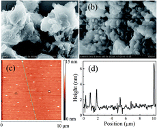
A graphitic carbon nitride (g-C3N4) nanosheet and its nanocomposites have recently attracted increasing interest due to their massive potentials in applications ranging from fluorescence imaging to solar energy conversion. An economical mass-production method for the synthesis of g-C3N4 nanosheets is urgently needed for the application of these intriguing nanomaterials. Here we develop a facile and rapid route to synthesize g-C3N4 nanosheets by using chemical exfoliation followed by extraction and thermal treatment. The feature of this approach lies in its rapid speed with exfoliation time of only about one minute and facile operation free of long-time ultrasonication or stirring, filtration and repeated washing processes to remove the residual acid. Moreover, the method is high-yield and easily upscalable. Meanwhile, the exfoliated g-C3N4 nanosheets exhibit high adsorption capability and photocatalytic activity due to the synergistic effects of large surface area, decreased recombination probability of photoinduced electron–hole pairs and enlarged band gap. This simple and rapid route enables the possibility of large-scale synthesis of g-C3N4 nanosheets with high yield, thus promoting their application in environmental purification and solar energy conversion.
Motivated by the intriguing properties of nanosheets and the analogous graphite structure of g-C3N4, great efforts have been made to exfoliate bulk g-C3N4 into 2D nanosheets in an attempt to achieve excellent performance. So far, various methods, such as thermal oxidation,13 chemical exfoliation,14 and liquid exfoliation,15–17 have been developed for the synthesis of g-C3N4 nanosheets (Table S1†). Thermal oxidation etching is in general performed at raised annealing temperature (500 °C) in an open container or prolonged time (four hour) of thermal treatment, whereas, its low yield (about 6%) owing to thermal etching and decomposition restricts the large-scale production of g-C3N4 nanosheets.13,18 Liquid phase exfoliation is carried out in a variety of solvents (water,19 methanol,20 N-methyl-pyrrolidone,21 N-dimethylformamide,22 and so on) by virtue of ultrasonic energy, however, the long time of ultrasonic treatment (usually more than 10 hours) and nonuniform sizes are far from satisfactory. Chemical exfoliation is a frequently-used approach to produce g-C3N4 nanosheets10,23–35 by using strong acid, alkali and/or oxidant. For example, the exfoliated g-C3N4 nanosheets are obtained by stirring the mixture solution of bulk g-C3N4 and concentrated sulfuric acid for a long time (7–9 h), pouring into deionized water under ultrasonic irradiation, and washing thoroughly with water to remove the residual acid.36,37 The g-C3N4 exfoliation procedures in Wang's work include the stirring for 2–3 h at 140 or 170 °C, cooling and injecting into 800 mL of deionized water, stirring at 70 °C for 2 h after the addition of 85.58 g NH4Cl, stirring in ice bath for 1.5 h, filtration and washing.27 However, the stirring process and post-treatments in these works are time-consuming, cumbersome and complicated, greatly limiting its large-scale application. Recently, a modified acid exfoliation method to produce g-C3N4 nanosheets with faster rate and relatively high-yield production has been reported.38 Unfortunately, the post-treatments of this route, such as filtration, repeated washing process to remove the residual acid, evaporation under reduced pressure and dry in vacuum, is still time-consuming. Therefore, it is urgently required to explore simple and efficient methods to exfoliate bulk g-C3N4 into nanosheets with high yield to satisfy its large-scale production and application.
Herein, we develop a facile and rapid route to exfoliate bulk g-C3N4 into g-C3N4 nanosheets with yield up to about 30%. Besides the rapid speed with exfoliation time of only about one minute, this route features facile operation without the filtration and repeated washing process to remove the residual acid. Importantly, the method is reliable and easily upscalable. Comparing with bulk g-C3N4, the synthesized g-C3N4 nanosheets display slight blue shift of the adsorption edge, enhanced adsorption capability and photocatalytic activity.
The morphological details of the prepared samples are probed by an S-4800 field emission scanning electron microscopy (FESEM). Atomic force microscopy (AFM) images are acquired in tapping mode in air using a Digital Instrument Shimadzu SPM-9500. The crystal structure of the samples is characterized by power X-ray diffraction (XRD, Siemens D-5000 diffractometer with Cu Kα irradiation). The Fourier transform infrared spectra (FTIR) of synthesized samples are recorded on an IR Affinity-1 FTIR spectrometer using conventional KBr pellets in the 400–4000 cm−1 range. The UV-vis diffuse reflectance spectra (DRS) are obtained on UV-Vis spectrometer (UV-2450, Shimadzu). An F-2500 fluorescence spectrometer with pulsed xenon discharge lamps upon excitation by incident light of 320 nm is used to measure the photoluminescence (PL) spectra at room temperature.
The photocatalytic behavior of g-C3N4 is evaluated by photodegradation of MB in aqueous solution with an initial concentration of 10 mg L−1 under the radiation of 300 W UV or visible light lamp. The photodegradation experiments are carried out with 80 mL MB solution and 30 mg g-C3N4 samples under vigorous stirring. The suspensions are magnetically stirred in the dark to obtain absorption–desorption equilibrium. Under illumination, 5 mL of the suspensions are taken and centrifuged to remove the photocatalyst at certain time intervals. The concentrations of MB are analyzed by measuring the absorbance of supernatant at 664 nm on a TU-1910 UV-vis spectrophotometer. The degradation efficiency can be evaluated by the function Ct/C0 × 100%, where C0 is the initial concentration of MB and Ct is the concentration after degradation.
In order to analyze the surface morphology of the products, the typical SEM image of g-C3N4 is depicted in Fig. 1. It can be seen that the bulk g-C3N4 tends to agglomerate into irregular thick block with a lateral size of several micrometers as shown in Fig. 1(a). Compared to bulk g-C3N4, g-C3N4 nanosheets (Fig. 1(b)) show laminar morphology, which appear as random and loose agglomerates, and tend to curl as a result of the minimizing surface energy.14 The size distribution (Fig. S1†) is estimated by measuring 40 flakes from SEM images for g-C3N4 nanosheets. It can be found that the lateral sizes of g-C3N4 nanosheets are centered at 75 nm. An average size about 73 nm is calculated as shown in Table S1.† Furthermore, to gain insight into the lateral size and thickness of exfoliated g-C3N4 nanosheets, AFM image is recorded. Fig. 1(c) displays the typical AFM image of the exfoliated g-C3N4 nanosheets. As can be observed from Fig. 1(c), the lateral size of g-C3N4 nanosheets is mainly ranging from 30 nm to 140 nm, and the average lateral size is about 75 nm based on statistical analysis. These results are consistent to those observed from SEM images (Fig. 1(b) and S1†). As shown in Fig. 1(d), the randomly measured nanosheets show thickness less than 4 nm, indicating that bulk g-C3N4 is successfully exfoliated into ultrathin nanosheets. In addition, there are a few nanosheets with lateral size of about 220 nm and thickness of about 5.7 nm are observed in Fig. 1(c) and (d), which presumably corresponds to the overlapping of g-C3N4 nanosheets and wrinkles on the nanosheets.38 The nearly transparent feature of the resulting solution after exfoliation, the apparent Tyndall effect of g-C3N4 nanosheet aqueous solution (Fig. S2†), the SEM and AFM images jointly confirm the ultra-small thickness of the exfoliated product.
![]() | ||
| Fig. 1 Typical SEM images of as-prepared (a) bulk g-C3N4 and (b) g-C3N4 nanosheets. AFM image of g-C3N4 nanosheets (c) and the corresponding height profiles of randomly chosen sections (d). | ||
The crystal structure of the products is investigated using XRD. As illustrated in Fig. 2(a), two feature diffraction peaks at about 27.6° and 13.2° can be observed in bulk g-C3N4 and g-C3N4 nanosheets, which confirm the basic g-C3N4 crystalline structure are well preserved in g-C3N4 nanosheets. The strong XRD peak at 27.6°, originated from the (002) interlayer diffraction of g-C3N4, sharply decreases for g-C3N4 nanosheets in comparison with that of bulk g-C3N4, suggesting the appearance of the few-layered g-C3N4 nanosheets with successful exfoliation.16 Further observation indicates that the (002) peak of g-C3N4 nanosheets shifts toward lower diffraction angle from 27.6° to 26.8°, corresponding to an increase in the interplanar stacking distance from 0.323 to 0.332 nm. This feature could be interpreted as the reunification of exfoliated g-C3N4 nanosheets.40 The intensity decrease of diffraction peak at 13.2°, which is derived from the in-planar repeated tri-s-triazine units, is also observed for the synthesized g-C3N4 nanosheets. This indicates the simultaneously decrease of planar size for the g-C3N4 layers after exfoliation, which is consistent to the SEM observation. Moreover, no other peaks are detected in XRD patterns, indicating the formation of pure g-C3N4 phase. Furthermore, the mean thickness of g-C3N4 nanosheets is determined to be about 4 nm according to the full width at half maximum of XRD peak using Scherrer's equation, which is consistent with AFM measurement.
Fig. 2(b) shows the FTIR spectra of bulk and exfoliated g-C3N4 to further demonstrate their graphitic structures. The band in the 1200–1700 cm−1 region corresponds to the typical stretching modes of C–N/C
N heterocycles.31,41 Peaks at 1632, 1560, 1467, and 1416 cm−1 can be attributed to the stretching vibrations of heptazinederived repeating units, well consistent with the FTIR analysis of the polymeric melon and g-C3N4. Peaks at 1324 and 1252 cm−1 reflect the out-of-plane bending vibrations characteristic of heptazine rings. The absorption band at 880 cm−1 and approximately 808 cm−1 is considered the deformation mode of N–H resulting from the incomplete condensation of amino groups and the typical breathing mode of tri-s-triazine units. The FTIR observation is in good accordance with the previous reports of g-C3N4 material.10,40 All peaks observed in bulk g-C3N4 are retained in the g-C3N4 nanosheets, suggesting that the synthesized g-C3N4 nanosheets have similar chemical structure as their parent bulk g-C3N4. However, the peak intensity of g-C3N4 nanosheets is slightly changed with respect to that of bulk g-C3N4. The changes would be relevant to the protonation of g-C3N4 with H2SO4.31
Fig. 3(a) shows the UV-vis absorption spectra of bulk g-C3N4 and g-C3N4 nanosheets. As illustrated in Fig. 3(a), the UV-vis spectrum of bulk g-C3N4 shows an absorption band at about 452 nm, the rapid increase of absorbance below 452 nm is due to the absorption of light caused by the excitation of electrons from the valence band to the conduction band of g-C3N4. While the absorption edge of g-C3N4 nanosheets shows a slight blue shift from 452 nm to 443 nm in comparison with that of bulk g-C3N4, corresponding to an increase in the bandgap from 2.74 eV of bulk g-C3N4 to 2.84 eV of g-C3N4 nanosheets (Fig. 3(a)). This larger bandgap may be associated with the quantum confinement effect with conduction and valence bands shifting in opposite directions.14 Moreover, it can be observed that g-C3N4 nanosheets exhibits optical absorption capabilities in the whole range of 300–800 nm in comparison of bulk g-C3N4, leading to the enhancement of the absorbance of light. The enhanced photoresponsivity of g-C3N4 nanosheets with respect to the bulk g-C3N4 is in accordance with that reported in the literature.19 The PL spectra of bulk g-C3N4 and g-C3N4 nanosheets are illustrated in Fig. 3(b). Fig. 3(b) demonstrates that the emission peak of bulk g-C3N4 locates at about 462 nm, whereas, the position of emission peak shows a blue shift to 433 nm for g-C3N4 nanosheets, which is consistent to that observed in the UV-vis absorption spectra. Furthermore, it is obvious that the PL emission intensity of the g-C3N4 nanosheets is lower than that of bulk g-C3N4, which implies that g-C3N4 nanosheets have a lower recombination rate of electrons and holes and may show better photocatalytic performance under light irradiation. The experimental observation, as well as SEM and XRD further reveal that the bulk g-C3N4 is successfully exfoliated into ultrathin g-C3N4 nanosheets.
![]() | ||
| Fig. 3 (a) UV-vis DRS spectra, band gaps (inset) and (b) PL spectra of the bulk g-C3N4 and g-C3N4 nanosheets. | ||
Based on the above analysis, a possible formation process of g-C3N4 nanosheets from the bulk counterpart is proposed in Fig. 4. First, the bulk g-C3N4, which is obtained by direct pyrolysis of guanidine hydrochloride, could be partial intercalated and protonated when it is mixed with concentrated H2SO4. Then, the rapid exothermal effect as adding water into the mixture will lead to the exfoliation of g-C3N4 by the dissociation of weak hydrogen-bonds bridging, which in turn accelerate the further intercalation of H2SO4 into the interlayer space of g-C3N4, and subsequently the –NH– linking group is cleaved and underwent oxidation owing to the heating effect and strong oxidizing medium, and then g-C3N4 is further exfoliated into smaller-sized fragments. Finally, g-C3N4 nanosheets could be extracted from the mixing solution using ethanol combined with a subsequent thermal process. The adopting of thermal treatment in a low temperature may easily remove the residue sulfur acid molecules on g-C3N4 nanosheets free of the filtration and repeated washing process.
To elucidate the adsorption capacity and photocatalytic activity of the prepared bulk g-C3N4 and g-C3N4 nanosheets, MB, a typical organic dye, is used as the probe contaminant to test the adsorption and photocatalytic performance of g-C3N4 products. For comparison, the blank test without the addition of any product is also performed in dark and under the identical conditions as displayed in Fig. 5. Fig. 5(a) shows that the decrease in the concentration of MB without photocatalyst is negligible in dark, whereas, 13% and 29.2% of MB are removed after the absorption–desorption equilibrium from the solution in the presence of bulk g-C3N4 and g-C3N4 nanosheets, respectively, confirming that g-C3N4 nanosheets exhibit high adsorption capability. Furthermore, 91% of MB is totally degraded under UV light irradiation for 15 min in the presence of g-C3N4 nanosheets. The apparent reaction-rate constant of g-C3N4 nanosheets (0.116 min−1) is more than three times higher than that of bulk g-C3N4 (0.037 min−1) as shown in the inset of Fig. 5(a). The photocatalytic activity in the presence of bulk g-C3N4 or g-C3N4 nanosheets are also investigated under visible-light irradiation. Fig. 5(b) shows the relative MB concentration change versus time for prepared samples under visible light irradiation. It can be observed that the relative concentration of MB decreases continuously with an increase of irradiation time due to the degradation of MB. Moreover, g-C3N4 nanosheets show a significant improvement over bulk g-C3N4. The obviously enhanced adsorption capacity and photocatalytic activity of g-C3N4 nanosheets can be explained as the cooperative effect of the distinct characteristic of 2D nanosheets and unique photo-physical property of the charge carries on the surface. Firstly, the character of 2D g-C3N4 nanosheets, derived from bulk g-C3N4 through intercalation and exfoliation, enables them to disperse well in the reaction system. Secondly, the resulted expanding specific surface area of g-C3N4 nanosheets is larger than that of bulk g-C3N4,27,36,39 which may provide more active sites for adsorbing MB molecules to participate in the subsequent photocatalytic degradation reaction. Thirdly, thin nanosheet structure can reduce the distance for the photogenerated electron–hole pairs to transfer from the site generated to the solid–liquid interface and the bulk recombination probability of charge carriers may be largely decreased.13,38 In addition, the increase of band-gap as confirmed by UV-vis absorption spectra and PL spectra increases the redox ability of charge carriers generated in nanosheets.14 All these favorable factors conjointly lead to the high adsorption capability and enhanced photocatalytic activities of the synthesized g-C3N4 nanosheets.
The stability is also an important issue for the practical application of photocatalyst, so the repeatability experiments of MB degradation over of g-C3N4 nanosheets are carried out under identical conditions. After each adsorption and photodegradation cycle, the photocatalyst is collected by centrifugation and reused in the following run. Fig. 6 illustrates the adsorption and degradation curves of MB in five consecutive applications with g-C3N4 nanosheets. As can be seen from Fig. 6, no obvious decrease in adsorption capacity and photocatalytic activity is found after five runs. At the fifth run, the adsorption capacity and degradation rate of MB under UV light irradiation for 15 min is still up to 27.9% and 87% over g-C3N4 nanosheets, which is similar to those in the first run. The results confirm that g-C3N4 nanosheets exhibit good stability for the adsorption and photocatalytic degradation of MB. In addition, the morphology and microstructure before and after photocatalysis are investigated via SEM images. From Fig. S3,† it's clear that the morphology and microstructure of g-C3N4 nanosheets before and after photocatalysis reaction are analogous, no obvious change in the morphology and microstructure of g-C3N4 nanosheets indicates that the nature of g-C3N4 nanosheets is stable.
![]() | ||
| Fig. 6 The cycling runs of g-C3N4 nanosheets in the adsorption and photodegradation of MB under UV light irradiation. | ||
Footnote |
| † Electronic supplementary information (ESI) available. See DOI: 10.1039/c6ra20514c |
| This journal is © The Royal Society of Chemistry 2016 |