DOI:
10.1039/C6RA20401E
(Paper)
RSC Adv., 2016,
6, 89578-89584
Stereoselective innovative synthesis and biological evaluation of new real carba analogues of minimal epitope Manα(1,2)Man as DC-SIGN inhibitors†
Received
12th August 2016
, Accepted 14th September 2016
First published on 14th September 2016
Abstract
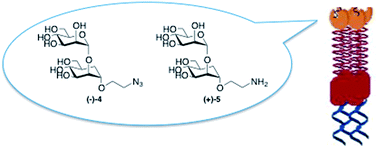
Antagonists of the C-type lectin DC-SIGN are promising therapeutic agents against viruses and bacteria. The development of glycomimetic ligands for DC-SIGN has so far proved to be challenging, since this membrane-protein presents four carbohydrate-binding domains (CRD) that specifically recognize mannose and fucose. In the recent past, we were able to develop inhibitors mimicking the minimal natural epitope Manα(1,2)Man using a mannoside with conformationally restricted dimethyl cycloexandicarboxylate-based aglycons designed to exploit the high enzymatic stability and to generate multivalent or solid supported systems as potent lectin ligands. Herein we describe the innovative synthesis of a different class of pseudodisaccharides, mimics of the natural Manα(1,2)Man moiety, characterized by the presence of a real D-carbamannose unit instead of a simpler mimic structure. Their chemical synthesis and biological activity using an SPR inhibition assay are reported. These pseudodisaccharides display inhibition values similar to those of the natural disaccharide Manα(1,2)Man, with a good affinity for DC-SIGN and can be considered as possible candidates for further structural modifications towards improved inhibitors.
Introduction
A broad range of biological processes including cellular adhesion, signalling, migration and infection are mediated by carbohydrate–lectin interactions.1–4 In this framework, DC-SIGN (Dendritic Cell-Specific Intercellular adhesion molecule-3 (ICAM-3)-Grabbing Non-integrin) is a C-type lectin involved in the recognition of viruses and pathogens at the mucosal level. In particular, the interaction between gp120 on the surface of HIV-1 and DC-SIGN allows the delivery of the virus to tissues rich in CD4+ T cells, such as lymphoid organs, playing therefore a crucial role in the infection process at the mucosal level.5,6 The main carbohydrate ligand present on gp120 and recognized by the DC-SIGN carbohydrate-binding domains (CRDs) is the high mannose glycan Man9(GlcNAc)2 (Man9, 1, Fig. 1). Therefore, derived glycomimetic structures can inhibit DC-SIGN action and are of high interest against HIV infections as antiviral agents.7–17 Notably, the 1,2-mannobioside disaccharide unit, Manα(1,2)Man 2 (Fig. 1), contained in all three arms D1, D2 and D3 of Man9, is one of the minimal epitopes for binding to DC-SIGN. Its incorporation into gold glyconanoparticles (Au-GNPs) increases the inhibitory activity by more than four orders of magnitude compared to that of the monovalent disaccharide,18 underlining the efficiency of this epitope and the importance of polyvalent systems. We previously reported that disaccharide mimetics 3a–c (Fig. 1) possess a three-dimensional structure and a conformational behaviour comparable to those of Manα(1,2)Man 2 and display a micromolar activity against DC-SIGN mediated Ebola viral infection, demonstrating for the first time that DC-SIGN antagonism by small molecules might be used to inhibit viral transfection mediated by DC-SIGN.10,17,19
 |
| Fig. 1 High mannose glycan Man9(GlcNAc)2 1, minimal epitope Manα(1,2)Man 2 and chemical structure of disaccharide mimetics 3a–c. | |
Herein, we describe the innovative synthesis of pseudodisaccharides 4 and 5 (Fig. 2), real carba analogues of the natural disaccharide epitope 2, and their biological evaluation as prospective DC-SIGN ligands. In our approach, the non reducing portion of the natural disaccharide Manα(1,2)Man is replaced by a carbasugar, in view of an improved drug-like character of the resulting glycomimetic compared to the natural carbohydrate. Indeed, sugar mimics are generally more soluble and membrane penetrant, less hydrophilic and less metabolically labile than the sugars themselves.20
 |
| Fig. 2 Chemical structures of pseudodisaccharides 4 and 5 real carba analogues of epitope 2. | |
Results and discussion
Synthesis of pseudodisaccharides 4 and 5
The stereoselective synthesis of pseudodisaccharides 4 and 5 proceeds through the construction of skeleton 8 (Scheme 1) by glycosylation of the new carbamannose glycosyl acceptor 7 with tetrabenzoyl mannose trichloroacetimidate 6 as typical glycosyl donor.21
 |
| Scheme 1 Stereoselective synthesis of skeleton 8. | |
Two carbamannose units 15 and 16 (Scheme 2), carrying either acetyl or benzyl protecting groups, were prepared and studied in this reaction. Both were prepared starting from the pivotal carba epoxy diol 11 obtained by m-CPBA epoxidation of unsaturated diol 10, in turn prepared from tri-O-acetyl-D-glucal 9 (ref. 22 and 23) as shown in Scheme 2.
 |
| Scheme 2 Stereoselective synthesis of carbamannose glycosyl acceptor 15 and 16. | |
Allyl alcohol 10 is stereoselectively epoxidized by m-CPBA, which reacts with the double bond face syn to the allyl hydroxy group affording β epoxide 11 as the only reaction product.24,25 Key epoxide 11 was then separately elaborated to give tri-O-benzyl-5a-carba-β-epoxide (+)-14 (ref. 25) upon treatment with NaH/BnBr in DMF, or debenzylated at C(6) under reductive conditions with H2/(10%)Pd–C, to afford β-epoxy triol 12, which was in situ acetylated (Ac2O/Py) to give tri-O-acetyl-5a-carba-β-epoxide (+)-13 (ref. 26) (Scheme 2). Epoxides (+)-13 and (+)-14 represent the carbaglycosylating agents which were subjected to ring opening reaction with freshly prepared 1.3 M 2-azido-1-ethanol in CH2Cl2,27 in the presence of a catalytic amount of Cu(OTf)2 as the Lewis acid catalyst.28 In accordance with reports by Ogawa and Cumpstey29,30 on the behaviour of 1,2-epoxides with β-manno configuration, such as (+)-13 and (+)-14, nucleophilic attack exclusively occurs in a trans-diaxial fashion, at the less sterically hindered, and more electronically favoured, oxirane C(1) carbon. This way, the corresponding (2-azidoethoxy)-cyclohexanol derivatives (+)-15 and (+)-16 are obtained through a completely C(1)-regio- and anti-stereoselective ring opening process from (+)-13 and (+)-14, respectively.
The glycosylation reaction of glycosyl acceptors (+)-15 and (+)-16 by the typical mannosyl donor 6 was successfully carried out in standard conditions (TMSOTf in CH2Cl2, −20 °C, 2 h), to afford glycoconjugates (−)-17 and (−)-18 in a completely α-stereoselective fashion. Catalytic hydrogenolysis of (−)-18 reduced the azido functionality and removed the benzyl protecting groups to afford pseudodisaccharide (−)-19. Subsequent Zemplén deprotection provided the final pseudodisaccharide (+)-5 with a 2-aminoethoxy chain at the pseudoanomeric C(1) in 69% yield after recrystallization from i-Pr2O. On the other hand, Zemplén deprotection of glycoconjugate (−)-17, afforded the 2-azidoethoxy pseudodisaccharide (−)-4 in 88% yield (Scheme 3).
 |
| Scheme 3 Stereoselective synthesis of glycoconjugates (−)-4 and (+)-5. | |
Preparation of pseudodisaccharides (−)-4 and (+)-5 represents the first example of stereoselective synthesis of real carba analogues of the minimal natural epitope Manα(1,2)Man. Their activity as DC-SIGN antagonists was thus evaluated using an inhibition assay based on Surface Plasmon Resonance (SPR) technique.
DC-SIGN inhibition assays
The affinity of the newly synthesized sugar conjugates 4 and 5 for DC-SIGN was tested by competition experiments using surface plasmon resonance (SPR), as previously described.11 A CM4 sensorchip was functionalized with mannosylated Bovine Serum Albumin (BSA-mannotriose) and a fixed amount of the extracellular domain of DC-SIGN was injected over the surface with increasing concentrations of ligands. The natural 1,2-mannobioside 2 and its mimic 3 were used as control. From the inhibition curves (Fig. 3) and according to a 4-parameter model, an IC50 of about 1 mM was estimated for all the compounds.
 |
| Fig. 3 Inhibition of DC-SIGN extracellular domain (DC-SIGN ECD) binding to immobilized mannosylated-bovine serum albumin (BSA) using surface plasmon resonance (SPR). (A and B) Sensorgrams of DC-SIGN ECD inhibition by increasing concentrations of compound 4 and 5, respectively, from 0 μM to 5000 μM. (C) Comparison of the inhibitory potency of 2 (○), 3 (□), 4 (●), and 5 (■) toward the DC-SIGN ECD mannosylated-BSA interaction determined by competition assays. | |
These data show that the new pseudodisaccharides (+)-5 and (−)-4 have inhibitory potency similar to the natural disaccharide Manα1-2Man. The IC50 values extracted from the inhibition curves are 964 ± 14.6 μM for 2, 941 ± 20.2 μM for 3, 1467 ± 32.1 μM for 4 and 1453 ± 30.6 μM for 5. Taking into account their enzymatic stability, (+)-5 and (−)-4 could be therefore considered as possible candidates to prepare multivalent systems to be used as inhibitors of viral infection.
Conclusions
We have successfully planned and achieved the innovative stereoselective synthesis of the two glycoconjugates (−)-4 and (+)-5, which were intended to represent mimics of the natural Manα(1,2)Man moiety, characterized by the presence of a real D-carbamannose unit. These glycoconjugates were designed to mime the minimal natural epitope Manα(1,2)Man of the high mannose glycan Man9(GlcNAc)2, which is the main carbohydrate ligand recognized by DC-SIGN. The biological activity was evaluated using an SPR inhibition binding assay and encouraging IC50 values, similar to the natural disaccharide Manα1-2Man 2 and to the previously reported mimic 3, were obtained for the new pseudodisaccharides (−)-4 and (+)-5. Compounds 2 and 3 have different binding modes in the DC-SIGN binding site19,31 but very similar binding affinity. Binding inhibition data alone do not allow for the time being to speculate on which of these modes is adopted by the new carbasugar derivatives 4 and 5 and thus to interpret their activity on a structural basis. These data, however, confirm the validity of the DC-SIGN antagonism by small molecules approach and pave the way to more extensive studies of enzymatically stable glycoconjugates to be used for the preparation of multivalent systems as DC-SIGN inhibitors.
Experimental
Materials and methods
All solvents and chemicals were used as purchased without further purification. Chromatographic separations were performed on silica gel columns by flash (Kieselgel 40, 0.040–0.063 mm; Merck) or by automated chromatography with Isolera® Biotage. Reactions were followed by thin-layer chromatography (TLC) on Merck aluminum silica gel (60 F254) sheets that were visualized under a UV lamp. Evaporation was performed in vacuo (rotating evaporator). Sodium sulfate was always used as the drying agent. Melting points were determined with a Kofler hot-stage apparatus and are uncorrected. Optical rotations were measured with a ATAGO AP-300 Automatic Polarimeter at 25 °C. Proton (1H) and carbon (13C) NMR spectra were obtained with a Bruker Avance 250 MHz spectrometer using the indicated deuterated solvents. Chemical shifts are given in parts per million (ppm) (δ relative to residual solvent peak for 1H and 13C). FTIR spectra were obtained with an IR Cary 600 FTIR (Agilent Tecnologies). Yields refer to isolated and purified products. High-resolution mass spectrometry (HRMS) analysis were performed using a Waters Xevo® G2-S QTof at Mass Spectrometry facility, PCN-ICMG of Grenoble. Compounds are dissolved in 1
:
1 acetonitrile/water (concentration 1 × 10⁻⁵ M).
Synthetic procedures
6-O-Benzyl-1,2-anhydro-5a-carba-β-D-mannopyranose (11). MCPBA (0.627 g, 3.64 mmol, 1.2 equiv.) was added to a solution of trans diol 10 (ref. 22) (0.709 g, 3.03 mol, 1.0 equiv.) in anhydrous CH2Cl2 (17 mL) at 0 °C and the reaction mixture was stirred at room temperature. After 18 h, the reaction was diluted with CH2Cl2, the organic phase was washed with 10% aqueous Na2S2O3, saturated aqueous NaHCO3 and brine, dried (Na2SO4) and concentrated to afford a crude reaction product (0.585 g), which was subjected to a flash chromatography. Elution with 3
:
7 hexane/AcOEt mixture afforded β epoxide 11 (0.438 g, 59% yield), pure as a white solid: mp 86–88 °C. Rf = 0.13 (3
:
7 hexane/AcOEt). [α]20D +8.03 (c 0.61, CHCl3). FTIR (neat) ν 3360.2, 3255.3, 3026.3, 2920.5, 2852.6, 1453.5, 1362.3, 1257.7, 1112.6, 1057.9, 1015.3, 933.3, 731.7, 695.4 cm−1. 1H NMR (250 MHz; CDCl3) δ 7.20–7.43 (m, 5H; Ar), 4.54 (d, J = 12.0 Hz, 1H; benzylic), 4.48 (d, J = 12.0 Hz, 1H; benzylic), 3.84 (dd, J = 8.2, 1.7 Hz, 1H; H3), 3.61 (dd, J = 10.2, 8.3 Hz, 1H; H4), 3.52 (dd, J = 9.2, 4.8 Hz, 1H; H6), 3.47 (dd, J = 9.2, 4.8 Hz, 1H; H6′), 3.37 (ddd, J = 4.0, 1.7, 0.6 Hz, 1H; H2), 3.27 (t, J = 4.3 Hz, 1H; H1), 2.00–2.12 (m, 1H; H5aα), 1.79–1.95 (m, 1H; H5), 1.73 (dd, J = 14.2, 11.2 Hz, 1H; H5aβ).13C NMR (62.5 MHz; CDCl3) δ 137.8, 128.6, 128.0, 127.8, 74.6, 73.5, 73.1, 72.2, 56.6, 53.3, 38.9, 26.4.
3,4,6-Tri-O-benzyl-1,2-anhydro-5a-carba-β-D-mannopyranose (14)25,32. A 60% dispersion of NaH (0.374 g, 9.36 mmol, 4.0 equiv.) was added to a solution of β epoxide 11 (0.584 g, 2.34 mmol, 1.0 equiv.) in anhydrous DMF (8 mL) at 0 °C and the reaction mixture was stirred at the same temperature for 30 minutes and then at room temperature for 30 minutes. After cooling at 0 °C, BnBr (0.70 mL, 5.86 mmol, 2.5 equiv.) was added dropwise and the solution was stirred 2 h at room temperature. Dilution with Et2O and ice and evaporation of the washed (brine) and dried (Na2SO4) organic solution afforded a crude reaction mixture (0.920 g), which was subjected to a flash chromatography. Elution with an 8
:
2 hexane/AcOEt mixture afforded tri-O-benzyl β epoxide 14 (0.740 g, 73% yield), pure as a white solid. Mp 53–55 °C. Rf = 0.24 (8
:
2 hexane/AcOEt). [α]20D +32.7 (c 0.81, CHCl3). FTIR (neat) ν 3025.0, 2913.9, 2852.0, 1946.5, 1873.9, 1744.0, 1453.3, 1366.2, 1102.3, 1081.5, 1023.5, 930.9, 737.6, 695.5 cm−1. 1H NMR (250 MHz, CDCl3) δ 7.27–7.45 (m, 14H; Ar), 7.20–7.25 (m, 1H; Ar), 4.84 (d, J = 10.4 Hz, 1H; benzylic), 4.82 (s, 2H; benzylic), 4.52 (d, J = 10.4 Hz, 1H; benzylic), 4.46 (s, 2H; benzylic), 3.83 (dd, J = 8.1, 1.9 Hz, 1H; H3), 3.65 (dd, J = 10.9, 8.1 Hz, 1H; H4), 3.45–3.55 (m, 2H; H6 + H6′), 3.34 (dd, J = 4.1, 1.8 Hz, 1H; H2), 3.26 (t, J = 3.9 Hz, 1H; H1), 1.97–2.20 (m, 2H; H5aα + H5aβ), 1.90–1.63 (m, 1H; H5).
(2-Azidoethyl)-3,4,6-tri-O-benzyl-5a-carba-α-D-mannopyranoside (16). Tri-O-benzyl β epoxide 14 (0.370 g, 0.859 mmol, 1 equiv.) was dissolved in a solution 1.3 M 2-azido-1-ethanol in CH2Cl2 (2.65 mL, 3.44 mmol, 4.0 equiv.) freshly prepared27 and Cu(OTf)2 (0.124 g, 0.344 mmol, 0.4 equiv.) was added. The reaction mixture was stirred for 18 h at room temperature. Dilution with CH2Cl2 and evaporation of the washed (1
:
1 NaHCO3/NH4Cl mixture and brine) and dried (Na2SO4) organic solution afforded a crude reaction mixture (0.363 g) which was subjected to a flash chromatography. Elution with 7
:
3 hexane/AcOEt mixture afforded product 16 (0.304 g, 68% yield), pure as a pale yellow liquid. Rf = 0.50 (7.3 hexane/AcOEt); [α]20D +22.34 (c 0.67, CHCl3). FTIR (neat) ν 3449.6, 2927.2, 2871.8, 2103.9, 1737.6, 1284.4, 1242.6, 1109.5, 1048.3, 993.4, 731.3, 696.8 cm−1. 1H NMR (250 MHz; CDCl3) δ 7.20–7.40 (m, 15H; Ar), 4.83 (d, J = 10.9 Hz, 1H; benzylic), 4.72 (d, J = 11.5 Hz, 1H; benzylic), 4.66 (d, J = 11.5 Hz, 1H; benzylic), 4.51 (d, J = 10.9 Hz, 1H; benzylic), 4.47 (s, 2H; benzylic), 4.13 (t, J = 3.4 Hz, 1H; H1), 3.80 (dd, J = 9.0, 3.0 Hz, 1H; H2), 3.45–3.75 (m, 6H), 3.18–3.39 (m, 2H; H6 + H6′), 1.95–2.14 (m, 1H; H5), 1.85–1.92 (m, 2H; H5aα + H5aβ).13C NMR (62.5 MHz, CDCl3) δ 139.1, 138.9, 138.5, 128.7, 128.7, 128.5, 128.5, 128.1, 128.1, 128.0, 127.7, 127.6, 127.5, 127.2, 82.4, 77.9, 77.1, 75.1, 73.2, 72.8, 70.6, 69.6, 68.3, 51.1, 37.4, 26.9.
(2-Azidoethyl)-2-O-[2′,3′,4′,6′-tetra-O-benzoyl-α-D-mannopyranosyl]-3,4,6-tri-O-benzyl-5a-carba-α-D-mannopyranoside (18). Trichloroacetimidate 6 (ref. 21) (0.208 g, 0.281 mmol, 1.3 equiv.) and carbamannoside 16 (0.112 g, 0.216 mmol, 1.0 equiv.) were dissolved in distilled toluene (2.0 mL) and dried overnight under vacuum. Acid washed molecular sieves AW 300 were activated under vacuum and added to the reagents. The mixture was dissolved in dry CH2Cl2 (2.5 mL), catalytic amount of TMSOTf (7.8 μL, 0.0432 mmol, 0.2 equiv.) was added under nitrogen at −20 °C and the reaction mixture was stirred at the same temperature for 3 h. Et3N (20.0 μL) was added and the mixture was diluted with CH2Cl2 and filtered through a Celite® pad. Evaporation of the solvent afforded a crude product (0.290 g), which was subjected to a flash chromatography. Elution with a 7
:
3 hexane/acetone mixture afforded the O-protected pseudomannobioside 18 (0.208 mg, 88% yield), pure as a white solid. Mp 40–42 °C. Rf = 0.22 (7
:
3 hexane/acetone); [α]20D −23.0 (c 0.63, CHCl3). FTIR (neat) ν 3350.1, 3294.9, 3069.3, 2961.0, 2875.4, 2106.4, 1724.0, 1601.0, 1450.7, 1259.8, 1094.9, 1065.8, 1026.6, 825.9, 707.9 cm−1. 1H NMR (250 MHz, CDCl3) δ 8.01–8.16 (m, 5H, Ar), 7.92–8.00 (m, 2H, Ar), 7.70–7.90 (m, 2H, Ar), 7.03–7.69 (m, 26H, Ar), 6.04 (t, J = 9.5 Hz, 1H, H4′), 5.85–5.94 (m, 2H, H2′ + H3′), 5.45 (s, 1H, Hanomeric), 4.39–4.92 (m, 9H, benzylic + H5′ + H6′a + H6′b), 4.22 (d, J = 4.2 Hz, 1H, H2), 3.78–3.86 (m, 2H, H3 + H![[C with combining low line]](https://www.rsc.org/images/entities/char_0043_0332.gif)
2CH2N3), 3.68–3.75 (m, 1H, H1), 3.49–3.65 (m, 3H,
), 3.30–3.41 (m, 1H, H4), 3.07–3.25 (m, 2H, H6a + H6b), 1.83–2.16 (m, 3H, H5 + H5aα + H5aβ).13C NMR (62.5 MHz, CDCl3) δ 166.3, 165.7, 165.5, 165.2, 138.9, 138.8, 138.6, 133.6, 133.3, 130.0, 129.9, 129.6, 129.4, 129.1, 128.7, 128.6, 128.4, 127.8, 127.6, 127.6, 98.9, 76.3, 75.1, 73.1, 70.8, 70.4, 70.1, 69.5, 68.0, 67.4, 63.4, 50.9, 37.7, 27.7.
(2-Aminoethyl)-2-O-[2′,3′,4′,6′-tetra-O-benzoyl-α-D-mannopyranosyl]-5a-carba-α-D-mannopyranoside (19). Pseudomannobioside 18 (0.080 g, 0.073 mmol, 1.0 equiv.) was dissolved in a 4
:
1 MeOH/anhydrous DMF mixture (2.0 mL) and 10% Pd–C (0.080 g) was added. The reaction mixture was hydrogenated (1 bar) at room temperature under hydrogen saturated atmosphere until reduction of the azide group and deprotection of the benzyl groups were achieved, monitoring by analytical TLC (8
:
2 CH2Cl2/MeOH). After 18 h the mixture was diluted with MeOH and filtered through a Celite® pad. The evaporation of the solvent afforded a crude reaction product (0.035 g) which was further diluted with MeOH and filtered off using syringe filter, to remove the residue coal. Evaporation of the solvent afforded a white solid, which was recrystallized from (i-Pr)2O to yield pure 1,2-pseudomannobioside 19 (0.031 g, 53% yield), as a yellow solid: mp: 38–39 °C. Rf = 0.3 (8
:
2 CH2Cl2/MeOH + 0.1% Et3N); [α]20D −34.85 (c 0.68, MeOH). FTIR (neat) ν 3377.6, 3072.1, 2951.0, 2782.8, 1721.2, 1656.4, 1592.8, 1437.7, 1297.2, 1256.9, 1161.3, 1082.4, 1066.3, 1018.5, 857.5, 697.4 cm−1. 1H NMR (250 MHz, CD3OD) δ 8.06–8.16 (m, 2H, Ar), 7.88–8.03 (m, 4H, Ar), 7.22–7.80 (m, 14H, Ar), 6.11 (t, J = 9.8 Hz, 1H, H4′), 5.89–5.97 (m, 1H, H2′), 5.84 (dd, J = 10.2, 3.2 Hz, 1H, H3′), 5.48 (d, J = 1.4 Hz, 1H, Hanomeric), 4.77 (dd, J = 13.4, 3.9 Hz, 1H, H5′), 4.47–4.58 (m, 2H, H6′a + H6′b), 4.23–4.26 (m, 1H, H2), 3.90–3.97 (m, 1H, H1) 3.45–3.89 (m, 6H, H3 + H4 + H6a + H6b +
), 3.07–3.12 (m, 2H,
), 2.00–2.13 (m, 1H, H5), 1.72–1.89 (m, 2H, H5aα + H5aβ). 13C NMR (62.5 MHz, CD3OD) δ 167.5, 167.0, 166.8, 166.5, 134.8, 134.7, 134.6, 134.5, 131.1, 130.8, 130.7, 130.6, 130.5, 130.5, 130.3, 130.2, 129.8, 129.7, 129.6, 129.4, 101.3, 80.5, 78.2, 74.2, 72.3, 71.7, 71.5, 70.8, 68.3, 66.2, 64.3, 64.0, 40.9, 40.5, 27.8.
(2-Aminoethyl)-2-O-(α-D-mannopyranosyl)-5a-carba-α-D-mannopyranoside (5). A solution 1 M of MeONa in MeOH (65.0 μL, 0.065 mmol, 1.5 equiv.) was added to a solution of 1,2-pseudomannobioside 19 (0.034 g, 0.043 mmol, 1.0 equiv.) in MeOH (0.8 mL). The reaction mixture was stirred for 12 h monitoring by TLC (8
:
2 CH2Cl2/MeOH). The reaction mixture was diluted with MeOH and Amberlite IRC 50 was added until pH 7. The beads were filtered off and washed with MeOH, then, the filtrated solution was evaporated affording a crude reaction product (0.015 g). The solid product was dissolved in MeOH and filtered through a syringe filter. Evaporation of the solvent afforded a yellow solid, which was recrystallized from (i-Pr)2O to yield fully O-deprotected 1,2-pseudomannobioside 5 (0.011 g, 70% yield), pure as a pale yellow solid: mp: 103–105 °C. Rf = 0.15 (1
:
1 CH2Cl2/MeOH); [α]20D +16.4 (c 1.14, MeOH).FTIR (neat) ν 3396.5, 2924.9, 2852.6, 2746.5, 2489.3, 1537.7, 1486.7, 1180.4, 1048.6, 1025.8 cm−1. 1H NMR (250 MHz, CD3OD) δ 5.06 (d, J = 1.7 Hz, 1H, Hanomeric), 4.03–4.11 (m, 1H, CHO), 3.98 (dd, J = 3.2, 1.7 Hz, 1H, CHO), 3.42–3.93 (m, 12H, CHO), 3.13 (t, J = 5.1 Hz, 2H,
), 1.88–1.99 (m, 1H, H5), 1.53–1.85 (m, 2H, H5aα + H5aβ). 13C NMR (62.5 MHz, CD3OD) δ 104.0, 96.7, 79.6, 78.1, 75.5, 74.2, 72.5, 71.8, 68.8, 67.0, 64.5, 63.2, 41.2, 40.5, 27.7. HRMS: (M + H+) found 384.1860; C15H30NO10 requires 384.1870.
3,4,6-Tri-O-acetyl-1,2-anhydro-5a-carba-β-D-mannopyranose (13)26. β-Epoxide diol 11 (0.060 g, 0.24 mmol, 1.0 equiv.) was dissolved in EtOH (2.0 mL) and 10% Pd–C (0.010 g) was added. The reaction mixture was hydrogenated (1 bar) at room temperature under hydrogen saturated atmosphere overnight. The reaction mixture was then diluted with EtOH and filtered through a Celite® pad. The evaporation of the solvent afforded a crude reaction product which was further diluted with EtOH and filtered off using syringe filter, to remove the residue coal. Evaporation of the solvent afforded a crude reaction product (0.032 g) as a colorless liquid constituted by epoxy triol 12, directly acetylated. Epoxy triol 12 (0.032 g, 0.20 mmol, 1.0 equiv.) was dissolved in anhydrous pyridine (0.5 mL) and the reaction mixture was cooled at 0 °C. Acetic anhydride (0.25 mL) was added and the reaction was stirred at room temperature overnight. Coevaporation of the reaction mixture with toluene afforded a crude product (0.049 g), which was subjected to flash chromatography. Elution with 1
:
1 hexane/AcOEt mixture afforded tri-O-acetyl-β-epoxide 13 (0.040 g, 70% yield), pure as a pale yellow oil. Rf = 0.26 (hexane/AcOEt 1
:
1); 1H NMR (CDCl3) δ 5.23 (dd, J = 8.7, 1.8 Hz, 1H, H3), 5.07–5.19 (m, 1H, H4), 4.05 (dd, J = 11.4, 4.9 Hz, 1H, H6), 3.85 (dd, J = 11.3, 3.1 Hz, 1H, H6′), 3.41 (dd, J = 3.9, 1.5 Hz, 1H, H1), 3.33 (t, J = 4.2 Hz, 1H, H2), 2.13–2.25 (m, 1H, H5), 2.10 (s, 3H, OAc), 2.04 (s, 3H, OAc), 2.01 (s, 3H, OAc), 1.88–1.97 (m, 2H, H5aα + H5aβ). 13C NMR (62.5 MHz, CDCl3) δ 171.1, 170.3, 170.0, 71.2, 70.0, 69.9, 63.9, 53.4, 35.7, 29.2, 21.0, 20.9, 20.8.
(2-Azidoethyl)-3,4,6-tri-O-acetyl-5a-carba-α-D-mannopyranoside (15). Tri-O-acetyl epoxide 13 (0.033 g, 0.114 mmol, 1.0 equiv.) was dissolved in a 1.3 M solution of 2-azido-1-ethanol in CH2Cl2 (ref. 27) (0.36 mL, 0.456 mmol, 4.0 equiv.) and Cu(OTf)2 (0.016 g, 0.045 mmol, 0.4 equiv.) was added. The reaction mixture was stirred for 18 h at room temperature. Dilution with CH2Cl2 and evaporation of the washed (1
:
1 NaHCO3/NH4Cl mixture and brine) and dried (Na2SO4) organic solution afforded a crude reaction product (0.048 g) which was subjected to flash chromatography. Elution with a 1
:
1 hexane/AcOEt solution afforded the glycosyl acceptor 15 (0.035 g, 82% yield), pure as a pale yellow oil. Rf = 0.14 (1
:
1 hexane/AcOEt). [α]20D +21.71 (c 0.35, CHCl3). FTIR (neat) ν 3480.4, 2956.7, 2925.7, 2855.4, 2106.9, 1738.1, 1454.0, 1368.7, 1227.5, 1098.8, 1039.6, 801.0 cm−1. 1H NMR (250 MHz, CDCl3) δ 5.23–5.34 (m, 1H, H3), 5.20 (dd, J = 9.5, 2.7 Hz, 1H, H4), 4.16–4.23 (m, 1H, H2), 4.11 (dd, J = 11.3, 4.9 Hz, 1H, H6), 3.94 (dd, J = 11.3, 4.9 Hz, 1H, H6′), 3.73–3.78 (m, 1H, H1), 3.70 (t, J = 5.0 Hz, 2H, CH2N3), 3.39 (d, J = 4.6 Hz, 1H, OCH2), 3.35 (d, J = 4.6 Hz, 1H, OCH2), 2.24–2.36 (m, 1H, H5), 2.08 (s, 3H, OAc), 2.05 (s, 3H, OAc), 2.03 (s, 3H, OAc), 1.86–1.94 (m, 2H, H5aα + H5aβ). 13C NMR (62.5 MHz, CDCl3) δ 171.1, 170.6, 169.9, 73.9, 69.7, 68.7, 63.9, 50.9, 35.2, 26.8, 21.1, 21.0.
(2-Azidoethyl)-2-O-[2′,3′,4′,6′-tetra-O-benzoyl-α-D-mannopyranosyl]-3,4,6-tri-O-acetyl-5a-carba-α-D-mannopyranoside (17). Trichloroacetimidate 6 (ref. 21) (0.047 g, 0.064 mmol, 1.3 equiv.) and carbamannoside 15 (0.019 g, 0.049 mmol, 1.0 equiv.) were dissolved in distilled toluene (2.0 mL) and dried overnight under vacuum.Acid washed molecular sieves AW 300 were activated under vacuum and added to the reagents. The mixture was dissolved in dry CH2Cl2 (1.0 mL) and a catalytic amount of TMSOTf (1.76 μL, 0.010 mmol, 0.2 equiv.) was added under nitrogen at −20 °C and the reaction mixture was stirred at the same temperature for 20 minutes, monitoring by TLC (1
:
1 hexane/AcOEt). Et3N (10.0 μL) was added and the mixture was diluted with CH2Cl2 and filtered through a Celite® pad. Evaporation of the solvent afforded a crude product (0.034 g), which was subjected to a flash chromatography. Elution with a 1
:
1 hexane/AcOEt mixture as eluent afforded the O-protected pseudomannobioside 17 (0.025 g, 53% yield) pure as a pale yellow oil. Rf = 0.40 (1
:
1 hexane/AcOEt). [α]20D −42.8 (c 1.7, CHCl3).
1H NMR (CDCl3) δ 7.94–8.15 (m, 6H, Ar), 7.79–7.89 (m, 2H, Ar), 7.21–7.67 (m, 12H, Ar), 6.06 (t, 1H, J = 9.9 Hz, H4′), 5.92 (dd, 1H, J = 10.2, 3.1 Hz, H2′), 5.69 (dd, 1H, J = 2.9, 1.6 Hz, H3′), 5.36 (d, 1H, J = 10.1 Hz, H3), 5.32 (d, 1H, J = 1.6 Hz, Hanomeric), 5.26 (dd, 1H, J = 9.8, 2.8 Hz, H4), 4.69 (dd, 1H, J = 12.0, 2.5 Hz, H6′a), 4.50 (dd, 1H, J = 12.0, 5.4 Hz, H6′b), 4.36–4.45 (m, 1H, H5′), 4.31 (t, 1H, J = 3.2 Hz, H2), 4.17 (dd, 1H, J = 11.4, 5.7 Hz, H6a), 4.03 (dd, 1H, J = 11.4, 3.6 Hz, H6b), 3.73–3.80 (m, 1H, H1), 3.44–3.60 (m, 2H, CH2N3), 3.13–3.36 (m, 2H, OCH2), 2.21–2.45 (m, 1H, H5), 2.16 (s, 3H, OAc), 2.13 (s, 3H, OAc), 2.04 (s, 3H, OAc), 2.00 (dd, 2H, J = 8.4, 2.3 Hz, H5aα + H5aβ). 13C NMR (62.5 MHz, CDCl3) δ 171.4, 170.7, 170.1, 166.2, 165.8, 165.6, 165.3, 133.7, 133.4, 130.0, 129.9, 129.4, 129.2, 128.9, 128.7, 128.6, 128.5, 99.0, 76.3, 76.1, 72.4, 70.8, 67.0, 69.7, 69.3, 68.6, 67.5, 63.9, 63.4, 50.8, 35.4, 21.0.
(2-Azidoethyl)-2-O-(α-D-mannopyranosyl)-5a-carba-α-D-mannopyranoside (4). Compound 17 (0.025 g, 0.026 mmol, 1.0 equiv.) was dissolved in MeOH (0.8 mL) and cooled at 0 °C. A freshly prepared 1.0 M solution of MeONa/MeOH (78 μL, 0.078 mmol, 0.3 equiv.) was added to the resulting solution and the reaction mixture was stirred for 12 h at room temperature. The mixture was then neutralized with an acidic Amberlite™ IR 120 H resin. The resin was then removed by filtration and repeatedly extracted with methanol. The combined filtrate was concentrated under vacuum to give a crude product, which was recrystallized from i-PrOH to yield pure glycoside 4 (0.010 g, yield 88%) as a white solid: mp: 85–88 °C. Rf = 0.21 (1
:
1 CH2Cl2/MeOH). [α]20D −43.2 (c 0.36, MeOH) 1H NMR (D2O) δ 4.98 (s, 1H, Hanomeric), 4.04 (bs, 2H, H1 + H2), 3.95–3.50 (m, 12H, CHO, CH2O, CH2N3), 3.44–3.37 (m, 2H, OCH2), 1.96–1.86 (m, 1H, H5), 1.80–1.54 (m, 2H, H5aα + H5aβ). 13C NMR (62.5 MHz, D2O, 0.1% CD3CN) δ 101.7, 78.5, 75.2, 72.8, 71.5, 69.5, 69.2, 69.1, 66.8, 65.9, 61.3, 60.2, 49.6, 37.8, 25.4. HRMS: (M + Na+) found 432.1586; C15H27N3O10Na requires 432.1594.
Biological assays
Surface plasmon resonance analysis. DC-SIGN ECD production and purification. DC-SIGN extracellular domain (DC-SIGN ECD) construct was produced and purified as described previously.33
Surface plasmon resonance analysis. Surface plasmon resonance experiments were performed on a Biacore 3000 using a CM4 chip, functionalized at 5 μL min−1. BSA or BSA-Man was immobilized on flow cells using amine-coupling method. Fc1 was prepared as reference surface. Flow cell (Fc) 1 and 2 were activated with 50 μL of a 0.2 M EDC/0.05 M NHS mixture. After this step, Fc1 and Fc2 were respectively functionalized with bovine serum albumin (BSA) and mannosylated bovine serum albumin (BSA-Mannotriose, BSA-manα1-3[manα1-6]man, 12 glycosylated sites, 60 μg mL−1, Dextra laboratories). Then remaining activated groups of both cells were blocked with 30 μL of 1 M ethanolamine. After blocking, the two Fc were treated with 5 μL of 10 mM HCl to remove unspecific bound protein and 5 μL of 50 mM EDTA to expose surface to regeneration protocol. Finally, 2381 RU and 1847 RU of BSA and BSA-Man were respectively immobilized on Fc1 and Fc2. The selected concentration was 20 μM for DC SIGN ECD.For inhibition studies, 20 μM of DC-SIGN ECD mixed with increasing concentrations of inhibiting compounds were prepared in a running buffer composed of 25 mM Tris pH 8, 150 mM NaCl, 4 mM CaCl2, 0.005% P20 surfactant, and 13 μL of each sample was injected onto the surfaces at a 5 μL min−1 flow rate. The resulting sensorgrams were reference surface corrected.
The DC-SIGN binding responses were extracted from sensorgrams, converted to percent residual activity values (y) with respect to lectin alone binding, and plotted against corresponding compound concentration. The 4-parameter logistic model (eqn (1)) was fitted to the plots, and the IC50 values were calculated, from eqn (2), using the values of fitted parameters (Rhi, Rlo, A1 and A2) (Fig. 4).
 |
| Fig. 4 Equation for percent residual activity values and for IC50 values. | |
Acknowledgements
This work used the platforms of the Grenoble Instruct centre (ISBG; UMS 3518 CNRS-CEA-UJF-EMBL), notably the MP3 and SPR platforms, with support from FRISBI (ANR-10-INSB-05-02) and GRAL (ANR-10-LABX-49-01) within the Grenoble Partnership for Structural Biology (PSB). V. Porkolab was supported by a grant from la Région Rhône-Alpes. F. Fieschi and A. Bernardi acknowledge for support from CM1102 COST Action. V. Di Bussolo acknowledges the University of Pisa (Progetti di Ricerca di Ateneo, PRA 2015 0015) for funding.
Notes and references
- A. Imberty and A. Varrot, Curr. Opin. Struct. Biol., 2008, 18, 567–576 CrossRef CAS PubMed.
- R. A. Dwek, Chem. Rev., 1996, 96, 683–720 CrossRef CAS PubMed.
- L. L. Kiessling and J. C. Grim, Chem. Soc. Rev., 2013, 42, 4476–4491 RSC.
- S. Cecioni, A. Imberty and S. Vidal, Chem. Rev., 2015, 115(1), 525–561 CrossRef CAS PubMed.
- T. B. H. Geijtenbeek, R. Torensma, S. J. van Vliet, G. C. F. van Duijnhoven, G. J. Adema, Y. van Kooyk and G. C. Figdor, Cell, 2000, 100, 575–585 CrossRef CAS PubMed.
- S. K. Wang, P. H. Liang, R. D. Astronomo, T. L. Hsu, S.-L. Hsieh, D. R. Burton and C. H. Wong, Proc. Natl. Acad. Sci. U. S. A., 2008, 105, 3690–3695 CrossRef CAS PubMed.
- M. J. Borrok and L. L. Kiessling, J. Am. Chem. Soc., 2007, 129, 12780–12785 CrossRef CAS PubMed.
- K. C. A. Garber, K. Wangkanont, E. E. Carlson and L. L. Kiessling, Chem. Commun., 2010, 46, 6747–6749 RSC.
- S. L. Mangold, L. R. Prost and L. L. Kiessling, Chem. Sci., 2012, 3, 772–777 RSC.
- J. J. Reina, S. Sattin, D. Invernizzi, S. Mari, L. Martinez-Prats, G. Tabarani, F. Fieschi, R. Delgado, P. M. Nieto, J. Rojo and A. Bernardi, ChemMedChem, 2007, 2(7), 1030–1036 CrossRef CAS PubMed.
- G. Timpano, G. Tabarani, M. Anderluh, D. Invernizzi, F. Vasile, D. Potenza, P. M. Nieto, J. Rojo, F. Fieschi and A. Bernardi, ChemBioChem, 2008, 9(12), 1921–1930 CrossRef CAS PubMed.
- S. Sattin, A. Daghetti, M. Thepaut, A. Berzi, M. Sanchez-Navarro, G. Tabarani, J. Rojo, F. Fieschi, M. Clerici and A. Bernardi, ACS Chem. Biol., 2010, 5(3), 301–312 CrossRef CAS PubMed.
- M. Andreini, D. Doknic, I. Sutkeviciute, J. J. Reina, J. Duan, E. Chabrol, M. Thepaut, E. Moroni, F. Doro, L. Belvisi, J. Weiser, J. Rojo, F. Fieschi and A. Bernardi, Org. Biomol. Chem., 2011, 9(16), 5778–5786 CAS.
- J. Luczkowiak, S. Sattin, I. Sutkeviciute, J. J. Reina, M. Sanchez-Navarro, M. Thepaut, L. Martinez-Prats, A. Daghetti, F. Fieschi, R. Delgado, A. Bernardi and J. Rojo, Bioconjugate Chem., 2011, 22(7), 1354–1365 CrossRef CAS PubMed.
- A. Berzi, J. J. Reina, R. Ottria, I. Sutkeviciute, P. Antonazzo, M. Sanchez-Navarro, E. Chabrol, M. Biasin, D. Trabattoni, I. Cetin, J. Rojo, F. Fieschi, A. Bernardi and M. Clerici, AIDS, 2012, 26(2), 127–137 CrossRef CAS PubMed.
- N. Obermajer, S. Sattin, C. Colombo, M. Bruno, U. Svajger, M. Anderluh and A. Bernardi, Mol. Diversity, 2011, 15(2), 347–360 CrossRef CAS PubMed.
- N. Varga, I. Sutkeviciute, C. Guzzi, J. McGeagh, I. Petit-Haertlein, S. Gugliotta, J. Weiser, J. Angulo, F. Fieschi and A. Bernardi, Chem.–Eur. J., 2013, 19(15), 4786–4797 CrossRef CAS PubMed.
- O. Martinez-Avila, K. Hijazi, M. Marradi, C. Clavel, C. Campion, C. Kelly and S. Penades, Chem.–Eur. J., 2009, 15(38), 9874–9888 CrossRef CAS PubMed.
- M. Thepaut, C. Guzzi, I. Sutkeviciute, S. Sattin, R. Ribeiro-Viana, N. Varga, E. Chabrol, J. Rojo, A. Bernardi, J. Angulo, P. M. Nieto and F. Fieschi, J. Am. Chem. Soc., 2013, 135(7), 2518–2529 CrossRef CAS PubMed.
- S. Mari, H. Posteri, G. Marcou, D. Potenza, F. Micheli, F. J. Canada, J. Jimenez-Barbero and A. Bernardi, Eur. J. Org. Chem., 2004, 24, 5119–5125 CrossRef.
- D. J. Lee, R. Kowalczyk, V. J. Muir, P. M. Rendle and M. A. Brimble, Carbohydr. Res., 2007, 342(17), 2628–2634 CrossRef CAS PubMed.
- I. Frau, V. Di Bussolo, L. Favero, M. Pineschi and P. Crotti, Chirality, 2011, 23(9), 820–826 CrossRef CAS PubMed.
- V. Di Bussolo, I. Frau, L. Checchia, L. Favero, M. Pineschi, G. Uccello-Barretta, F. Balzano, G. Roselli, G. Renzi and P. Crotti, Tetrahedron, 2011, 67, 4696–4709 CrossRef CAS.
- A. H. Hoveyda, D. A. Evans and G. C. Fu, Chem. Rev., 1993, 93(4), 1307–1370 CrossRef CAS.
- S. Ogawa and T. Tonegawa, Carbohydr. Res., 1990, 204, 51–64 CrossRef CAS.
- V. W.-F. Tai, P.-H. Fung, Y.-S. Wong and T. K. M. Shing, Tetrahedron: Asymmetry, 1994, 5(7), 1353–1362 CrossRef CAS.
- Y. Dong, X. Liang, H. Yuan, S. Qi, F. Chen and D. Wang, Green Chem., 2008, 10(9), 990–994 RSC.
- A. Bernardi, D. Arosio, L. Manzoni, F. Micheli, A. Pasquarello and P. Seneci, J. Org. Chem., 2001, 66(19), 6209–6216 CrossRef CAS PubMed.
- S. Ogawa, S. Sasaki and H. Tsunoda, Chem. Lett., 1993, 9, 1587–1590 CrossRef.
- J. Frigell and I. Cumpstey, Beilstein J. Org. Chem., 2010, 6, 1127–1131 CrossRef CAS PubMed.
- I. Sutkeviciute, M. Thépaut, S. Sattin, A. Berzi, J. McGeagh, S. Grudinin, J. Weiser, A. Le Roy, J. J. Reina, J. Rojo, M. Clerici, A. Bernardi, C. Ebel and F. Fieschi, Chem. Biol., 2014, 9(6), 1377–1385 CAS.
- K.-C. Seo, Y.-G. Kwon, D.-H. Kim, I.-S. Jang, J.-W. Cho and S.-K. Chung, Chem. Commun., 2009, 1733–1735 RSC.
- G. Tabarani, M. Thépaut, D. Stroebel, C. Ebel, C. Vivès, P. Vachette, D. Durand and F. Fieschi, ChemBioChem, 2009, 284(32), 21229–21240 CAS.
Footnote |
† Electronic supplementary information (ESI) available: NMR spectra of 4 and 5, 10–18; HRMS of 4 and 5; sensorgrams and inhibition curves of 4, 5 and of natural epitope Manα(1,2)Man. See DOI: 10.1039/c6ra20401e |
|
This journal is © The Royal Society of Chemistry 2016 |
Click here to see how this site uses Cookies. View our privacy policy here.