
A new route for SERS analysis of intact erythrocytes using polydisperse silver nanoplatelets on biocompatible scaffolds†
Anna A. Semenovaa,
Nadezda A. Brazheb,
Evgeniya Yu. Parshinab,
Asia S. Sarychevaa,
Georgy V. Maksimovb and
Eugene A. Goodilin*acd
aFaculty of Materials Science, Moscow State University, 119991, Moscow, Russian Federation. E-mail: goodilin@yandex.ru; goodilin@inorg.chem.msu.ru; Fax: +7 495 939 0998; Tel: +7 495 939 4609
bFaculty of Biology, Moscow State University, Moscow, Russia 119991
cKurnakov Institute of General Anorganic Chemistry, 119992, Moscow, Russian Federation
dFaculty of Chemistry, Moscow State University, 119991, Moscow, Russian Federation
First published on 26th August 2016
Abstract
Superior spectral sensitivity and functional abilities of anisotropic, instead of the usually used spherical, noble metal nanoparticles, allow development of new surface-enhanced Raman spectroscopy (SERS) approaches to analyse biological objects. We found and resolved for the first time the particular risks of survival of silver nanoparticles in different salines to succeed in recording the SERS spectra of intact erythrocytes as an important family of living cells. The ensemble of nanoplatelets with varied shapes and sizes grants multispectral absorption of laser irradiation since a fraction of the nanoparticles with a given position of a plasmonic band always exists in such a mixture, thereby providing an effective SERS amplification. At the same time, fast recrystallization of anisotropic silver nanoplatelets occurs in a standard chloride-based saline, being important to keep the erythrocytes alive but neglecting the benefits of the silver platelets as the most versatile and prospective components of SERS sensors. Substitution of chlorides with nitrates keeps both the intact cells and anisotropic nanoparticles safe on biocompatible cellulose SERS scaffolds containing the mixture of silver nanoplatelets thus promoting the development of new SERS devices for biomedical diagnostics.
Introduction
Surface-enhanced Raman spectroscopy (SERS) is believed to become a powerful method for biomedical diagnostics.1–17 Noninvasive medical tools for screening and personal medicine pose a challenge for highly sensitive single molecule or single cell analysis, thus demanding a thorough development of new materials and novel analytical approaches.18–33 Silver and gold are commonly used to attain large enhancement factors in SERS by varying nanoparticle sizes, shapes and aggregative structures resulting in enormous signal amplification for some selected molecules.3,5,7,8,10–12 The most prospective family among these nanostructured materials comprises prisms, triangles, discs and other platelet-like anisotropic silver nanoparticles.34–40 In particular, they are highly desired for future applications of SERS since biological objects demonstrate a wide diversity of optical properties, consequently, anisotropic silver nanoparticles can be adjusted to the exact biological sample under the study due to their highly tunable plasmon resonance features.Hemoglobin (Hb) and its abnormalities comprise the core issue of many existing diagnostic tools in modern medicine therefore it is not a surprise that SERS is considered as a new methods of Hb analysis.18–23,26–28 These circumstances urge the development of new experimental routes to study an undisturbed structure of Hb in its natural cellular environment to allow the noninvasive SERS analysis of these important cells.
Absorbance spectra of the oxygenated form of hemoglobin (Fig. S1,† ESI) with the Soret band at about 410 nm, beta- (540 nm) and alpha-bands (575 nm)41,42 match well typical excitation laser wavelengths used for SERS studies, for example, 404, 417 or 514/532 nm lasers. This concurrence would lead to large resonant Raman amplification (Fig. S1 and S2,† ESI)27,43,44 if plasmonic nanostructures are excited in the same regions.27,28,45,46 The violet laser irradiation corresponds well to either the most bright Soret band or typical plasmon resonance position of spherical silver nanoparticles. Unfortunately this high energy irradiation damages biological objects, also, it is hard to keep single silver nanoparticles separated. Therefore, it is a usual route in many SERS experiments with biological objects to prepare artificially colloidal aggregates with their absorbance shifted up to wide spread and biology friendly “green” lasers.27,44 Another successful approach is based on preparation of immobilized hierarchic nanostructures of silver providing absorption in a wide spectral range.28,45,46 The last logically expected tactics makes use of the topical objects of this article – anisotropic silver nanoparticles reaching the “green laser” range as well, however they have never been utilized for erythrocyte studies.
SERS of biomolecules still suffers a lack of knowledge about possible toxicity of free metallic nanoparticles. Theoretical and thoughtful speculations about behavior of anisotropic silver nanoparticles in different environments are controversial.40,47–49 Another side of the medal is that the activity of noble metal nanoparticles is underestimated. Their high surface energy creates a risk that commonly used linkers, surfactants, reducing agents and their oxidized forms, halide ions etc. can affect functional properties of the nanoparticles because of aging and recrystallization. This becomes especially important in the case of biological objects demanding a special saline to keep the cells alive. Thus, at this moment, a problem of mutual surviving of both the biological material (to be analyzed) and the inorganic nanomaterial (anisotropic silver nanoparticle to provide the analysis) immediately arrives.
In this work, we succeeded for the first time to record SERS spectra of living erythrocyte on a cellulose matrix with a deposited mixture of various silver nanoplatelets in a specially designed saline keeping both the intact cells and anisotropic nanoparticles safe. The suggested approach seems to be effective and promising for the development of SERS devices in the sake of innovative biomedical diagnostics.
Experimental
Preparation and characterization of silver nanoparticles
Highly pure water (Milli-Q, Millipore), silver(I) nitrate, sodium citrate, PVP (polyvinylpyrrolidone), NaBH4, ascorbic acid (Aldrich) were used as reagents. Silver nanoplatelets were obtained in two stages as adopted from elsewhere.34–36 At the beginning, silver(I) nitrate was reduced by NaBH4 in the presence of sodium citrate to form a seeding solution of spherical silver nanoparticles. In particular, 11 ml aqueous solution of 0.11 mM AgNO3 and 2.05 mM sodium citrate was mixed quickly, under vigorous stirring, with 0.3 ml of freshly prepared 5 mM aqueous solution of NaBH4. The stirring has been stopped in 10 min. The yellow sol thus obtained has been kept for 5 h at room temperature and then placed in a dark place at 4 °C for storage as a seeding solution. On the second stage, a mixture of aqueous solutions of 0.25 ml of 5 mM AgNO3, 0.75 ml of 30 mM sodium citrate, 0.75 ml of 0.7 mM (with respect to repeating chain) of polyvinylpyrrolidone in 9.25 ml of pure water was admixed with a series of aliquots of the seeding solution (in μl), namely 160 (0.9 vol%), 480 (2.7 vol%), 640 (3.6 vol%), 960 (5.3 vol%), 1280 (6.9 vol%), 2560 (12.9 vol%), 3840 (18.2 vol%), 5120 (22.9 vol%), 7680 (30.8 vol%), 10![[thin space (1/6-em)]](https://www.rsc.org/images/entities/char_2009.gif) 240 (37.2 vol%) and 19200 μl (52.7 vol%), followed by an addition of 6.25 ml of 1 mM of ascorbic acid. The solution color was found to start changing immediately after ascorbic acid injection then it has become permanently stabile after about 15 min. The final samples were found to maintain their characteristics (at least all the features of UV-vis spectra) for more than one year (see ESI, Fig. S3†). Thus prepared sols were purified by repeated centrifugation (19900 rpm for 10 min, Sartorius Sigma 3-30 K) followed by gentle dilution with pure water, after that they were stored at 4 °C in darkness and used for further experiments. To prepare “rainbow” scaffolds, small and equal portions (100 μl) of all the prepared purified sols were mixed together to get about 1–1.5 ml of a solution containing all the fractions of experimentally prepared silver nanoplatelets, then it was used entirely in the course of dropwise repeated impregnation and drying over the 4 cm2 piece of a standard high-quality ash-free filter paper. This procedure is simpler and more universal compared to the reported process of silver platelets in situ growth using cellulose fibrils as a template.50
240 (37.2 vol%) and 19200 μl (52.7 vol%), followed by an addition of 6.25 ml of 1 mM of ascorbic acid. The solution color was found to start changing immediately after ascorbic acid injection then it has become permanently stabile after about 15 min. The final samples were found to maintain their characteristics (at least all the features of UV-vis spectra) for more than one year (see ESI, Fig. S3†). Thus prepared sols were purified by repeated centrifugation (19900 rpm for 10 min, Sartorius Sigma 3-30 K) followed by gentle dilution with pure water, after that they were stored at 4 °C in darkness and used for further experiments. To prepare “rainbow” scaffolds, small and equal portions (100 μl) of all the prepared purified sols were mixed together to get about 1–1.5 ml of a solution containing all the fractions of experimentally prepared silver nanoplatelets, then it was used entirely in the course of dropwise repeated impregnation and drying over the 4 cm2 piece of a standard high-quality ash-free filter paper. This procedure is simpler and more universal compared to the reported process of silver platelets in situ growth using cellulose fibrils as a template.50
The obtained silver nanoparticles (AgNPs) were characterized by a transmission electron microscopy (TEM) and electron diffraction (ED) using LEO912 AB OMEGA (Carl Zeiss). Particle size distribution graphs were built using TEM micrographs and direct statistical measurements of the particle mean diameter for 450+ nanoparticles, their thickness was estimated by means of averaging the size of particles directed perpendicular to the view-field (DLS data are given in ESI, Fig. S6 and S7†). The pristine sample microstructure and blood cells were studied using a field emission scanning electron microscope (Carl Zeiss NVision 40) at 0.5–5 kV accelerating voltage. AgNPs dried on alumina plates were examined using Rigaku D/MAX 2500 (Japan) with a rotating copper anode (CuKalpha irradiation, 5–80° 2 thetta range, 0.02° step). Diffraction maxima were indexed using the PDF-2 database. UV-vis absorption spectra were recorded using the UV-vis spectrophotometer Lambda 35 (Perkin-Elmer) with an attached diffuse reflectance accessory.
Preparation of erythrocytes and Raman spectroscopy
Raman and SERS experiments were performed using an InVia Raman microscope (Renishaw, UK) equipped with a 20 mW 514 nm argon laser and power neutral density filters. All the spectra were collected using the ×5 objective, NA 0.15 and 20 s of acquisition time. Laser power on a sample was 1.5 mW per 2 micrometer spot size. A silicon wafer was used for calibration.Erythrocyte samples were prepared from the venous blood taken from male Wistar rats and fully saturated with oxygen before experiments. Animals were used from the vivarium of the Faculty of Biology, Moscow State University. All procedures involving animals were approved by Bioethics Committee of Moscow State University according to the ethical and juridical norms of scientific researches in biology, medicine and related areas corresponding to laws of Russian Federation and international GLP standards (Good Laboratory Practice). The protocol of experiments was approved by the Bioethics Committee of Moscow State University. Blood was 5000–10000 times diluted in two steps with the Alen' salines for erythrocytes. In the first step, blood was diluted 100 times with an Alen saline with normal osmolarity (145 mM NaCl, 5 mM KCl, 4 mM Na2HPO4, 1 mM NaH2PO4, 1 mM MgSO4, 1 mM CaCl2, pH 7.4, osmolarity 330 mOsm). Secondly, previously 100-times diluted blood was again 100 diluted with hyperosmotic Alen saline (ion composition was the same as in normal Alen saline, but the salt concentrations were 1.67 times larger). In the parallel experiments with a “nitrate” buffer solution, that keeps silver nanoparticles unchanged, chloride salts were completely replaced with corresponding nitrates, 145 mM NaNO3, 5 mM KNO3, 1 mM Ca(NO3)2, 1 mM MgSO4, 4 mM Na2HPO4·12H2O, 1 mM NaH2PO4·2H2O, to maintain almost same osmolarity and other physical–chemical properties. The resulting diluted blood was immediately put onto the “rainbow” cellulose substrates to record SERS spectra, the experiments on recording the data did not exceed 30 min in order to avoid occasional cell damage.27,28 Such preparation ensures erythrocytes' integrity.28 To view a conventional Raman spectrum of living erythrocytes, we used blood diluted for 20–1000 times only with the normal Alen saline under the same registration conditions as in the SERS experiments. The sample with 1000-fold dilution of blood had the lowest possible concentration of erythrocytes still detectable by Raman scattering without the presence of AgNPs. The enhancement factors EF were set as a ratio of corresponding peak intensities ISERS for SERS and RS measurements IRS divided by the ratio of corresponding concentrations NRS and NSERS adjusted to the amount of submembrane hemoglobin (0.5%).27,28 This factor accounts an increase of the intensity of a spectral signal in the presence of silver nanomaterials under the conditions of decreased concentrations of analytes used in SERS measurement compared to that applied for conventional RS spectra recording.
To visualize the erythrocyte morphology, the diluted blood samples mixed with the silver colloids were pelleted by centrifugation (450 g, 18–20 °C) with subsequent fixation in 0.5% glutaraldehyde solution for 1 h. Then glutaraldehyde was removed by centrifugation of erythrocytes in pure (Milli-Q) water; the fixed erythrocytes were placed as a monolayer onto a glass plate and then dried for 24 h. To observe interaction of erythrocyte plasma membrane with attached AgNPs, we perform TEM imaging of erythrocyte ghosts (ESI, Fig. S8†). Preparation of erythrocyte ghosts was done as described elsewhere.27,30
Hemolysis of erythrocytes was analyzed by counting the number of cells of different types within the Goryachev's chamber; the blood was 100 times diluted with the Alen or “nitrate” saline, respectively, and mixed with silver sols in the ratio of 2:3.
Results and discussion
Preparation of silver nanoplatelets yields highly stable colored sols effectively absorbing light even at very diluted concentration of silver nanoparticles (Fig. 1a; see also Fig. S3,† ESI). Absorption spectra (Fig. 1b) and positions of plasmonic bands with dipole and quadrupole resonances (Fig. 1c) evidence for the formation of anisotropic nanoparticles although intensity of exact plasmonic peaks are smoothed and the positions are gradually shifted as compared to theoretical predictions.34 A reasonable explanation of these features originate from mixing of different fractions of silver nanoparticles (Fig. 1c and 2, see also Fig. S4–S7† and discussion, ESI) as dependent strongly on preparations conditions and, in the most valuable extent, on the amount of silver seed added at a first stage of the nanoplatelets growth. However in all the cases, except silver seeds themselves, several types of nanoparticles can be found including rounded ellipsoids, prisms, discs, triangular an hexagonal plates etc. (Fig. 1) as quite typical of this preparation route.35 Each fraction gives an additive contribution to the light absorption spectra and, correspondingly, to the color of the sols, that is why the experimental data are a superposition of several subspectra with the main peaks attributed to a dominating fraction (Fig. S4–S7,† ESI). Such a nanoparticle mixture is, in principle, favorable for an advanced design of universal substrates for a SERS analysis of living cells. Indeed, if several different nanoparticles are present onto an area of several microns, as small as typical dimensions of cells, there are at least some of them contributing greatly to the Raman enhancement because a laser irradiation wavelength, a plasmon band and an absorption range of analysing molecules coincide each other (see ESI and discussion of Fig. S1–S5†). There is no sense to create “hot spots” or other constructions for “single-molecular” detection since they cannot operate due to a rather thick cell membranes and a large size of cells therefore a local electromagnetic field remains effective only in the case of noninvasive transmembrane analysis of living cells.27–31 In this case, anisotropic silver nanoparticles have to survive in the presence of either the cells, their compartment or a special liquid environment in which the cells are placed inside. This is important because the anisotropic silver nanoparticles provide the whole spectrum of adjustable optical properties otherwise we have to stuck to the narrow range of plasmon bands of 400–450 nm typical of spherical silver nanoparticles of different sizes (Fig. 1, see also ESI, Fig. S1 and S3–S5†). | ||
| Fig. 1 General view and normalized UV-vis spectra (a) of silver sols with anisotropic silver nanoparticles obtained using different amounts of a seeding solution (in μl): 480 (2.7 vol%) – 9, 960 (5.3 vol%) – 8, 1280 (6.9 vol%) – 7, 2560 (12.9 vol%) – 6, 3840 (18.2 vol%) – 5, 7680 (30.8 vol%) – 4, 10240 (37.2 vol%) – 3 and 19200 μl (52.7 vol%) – 2 as compared to the seeding solution (1), (b) positions of plasmonic bands with dipole (1) and quadrupole (2, 3) resonances for the same silver sols, (c) variation of particle fractions for some samples of silver nanoplatelets, in the (a) graph of the samples with a different amount of the seeded solution (in μl) compared to the case of 1% NaCl solution added (the “+NaCl” notation). | ||
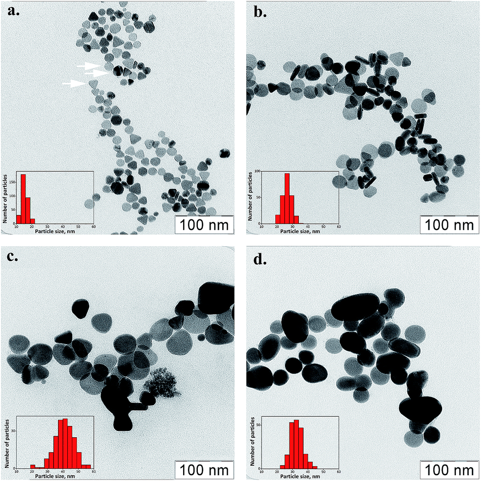 | ||
| Fig. 2 TEM micrographs of silver nanoplatelets obtained using a different amount of the seeding solution, 10240 μl (a), 2560 μl (b) and 640 μl (c), respectively, and the change of the sample (c) morphology with 1% NaCl solution added (d). The arrows show examples of three different morphologies – disks, twinned spheres and triangles. The red insets shows statistics of nanoparticle counts describing particle size distribution (in-plane sizes only). | ||
Anisotropic silver nanoparticles are formed from original silver seeds due to selective grain growth in the presence of PVP and/or citrate ions, as well-known.34–39 The main role of PVP is to stabilize silver seeds with diameters less than a certain critical size required for morphological transformation, while citrate is critical to induce the nanoplate formation, by preferential binding onto (111) planes.34,35 At the same time, they are sensitive to several types of inorganic anions40,47–49 like Cl−, Br−, I−, SCN−. The optical properties changes are then the result of a sculpting effect of halide ions etching the nanoprisms from a triangular shape into a circular shape or even into more isotropic particles. AgNPs formed in the absence of halides and PVP, show truncated triangular and hexagonal morphologies, whereas an addition of bromides leads to rounding of the edges and then to compaction into dominant cubic and bipyramidal geometries. Rounding of AgNPRs occurs due to etching of the most highly energetic protruding edges of AgNPRs.40 Observation of twinned cubic and bipyramidal particles is consistent with the structural transformation since bromide exhibits a strong tendency to stabilize (100) facets in AgNPs and to induce the formation of corresponding 3D shapes. Chlorides bind more weakly to silver ions and to the surface of growing AgNPs as compared to bromide therefore significantly higher chloride concentrations are required to affect AgNP formation.40 However microscopic, optical and particle size distribution measurements reveal48 re-crystallization of the primary silver nanoparticles to one-order larger crystallites already after 15 min after NaCl addition. It is well known that halide anions can serve as a precipitant of Ag+ ions at a low concentration and also as a complexing agent at a high concentration of halides. The halide ions thus can react with the silver ions, coordinate them and then dissociate from the surface of Ag particles transferring silver into a solution.
It is obvious (Fig. 2 and 3) that an addition of pure NaCl in the concentrations comparable to that of the physiological saline to the colored sols containing anisotropic silver nanoparticles causes irreversible, catastrophic changes of their optical properties in a couple of minutes, in particular, leading to a pronounced blue shift by about 100–150 nm and narrowing the peaks (Fig. 3, black and red curves). As seen from Fig. 3 (black curves), mixing of silver sols of different colors (and therefore with different contributions of main fractions, Fig. 2c) results in a superposition of absorption spectra meaning that the nanoparticles do not interact or physically agglomerate at such concentrations of silver and the PVP surfactant. After the addition of chloride ions, the shifted spectra (Fig. 3c) seem to be again a superposition of that we could observe for individual sols treated with NaCl (Fig. 3a and b). The first observation of TEM pictures (see, for example, Fig. 2d) confirms, as described, that triangular silver nanoplates become vanishing while disc-like and other rounded nanoparticles show their amounts increased.
 | ||
| Fig. 3 UV-vis absorption spectra of silver nanoplatelets sols obtained using 480 μl (a) and 7680 μl (b) aliquots of the seeding solution and their equal mixture (c) before and after addition of 1% NaCl (about 2–5 min spent). “mkl” means microliter in the figure. | ||
The estimates of that counted for 450+ particles are shown in Fig. 1d. This statistics tells that even discs and elongated rounded nanoparticles (ellipse-like) transform into the most isotropic, spherical nanoparticles. According to those data, the triangular silver nanoplatelet fraction becomes negligibly small and looses about 10–11% of contribution, the amount of discs drops from about 70% down to ca. 55% while the ellipsoid particle contribution grows up by 10% and the originally small fraction of spheres demonstrates a twofold increase up to about 42%. That explains easily that the 400–410 nm plasmonic band of spherical silver nanoparticles in Fig. 3b looks very sharp, intensive and distinct compared to the original broad plasmonic peak at 500–520 nm with some shoulders. Obviously, the obtained results are in good agreement with literature data discussed above or manifest even stronger changes. This makes us to post an unpleasant statement that anisotropic nanoparticles are not compatible with commonly used saline or similar halide-contained solutions and thus can be hardly used in SERS measurement if a contact with such fluids occurs; oppositely, this seems to be not a problem for isotropic silver nanoparticles.
Silver has only a few soluble salts and the only salt with an anion of a strong acid is known to be silver nitrate, AgNO3. Therefore to prevent the observed recrystallization of silver platelets, which takes place under the action of chloride ions, we should choose another cell incubation medium based on the physiological buffer solution for erythrocytes in which chloride ions are replaced by NO3−. Silver nanoplatelets do not have a tendency to increase their size or change their shape spontaneously if surfactants and nitrate ions are present in the colloidal solution as the remaining counterpart ions after conducting the silver nanoparticle synthesis (Fig. S3,† ESI). This strong proof of nitrate ion indifference with respect to anisotropic silver nanoparticles is highly contrasting with the behavior of the same sols in the presence of chloride ions (Fig. 3).
Nitrate-ions demonstrate no features similar to halide-ions in terms of silver recrystallization and it is not surprising that no paper is reported on such an influence of nitrate-ions on silver. Nitrate-ions are larger than Cl− ions, they are able to act as weak oxidizers and therefore they could be more toxic for living cells than chlorides. In addition, replacing chlorides with nitrates does not limit anymore the concentration of silver ions in solutions like insoluble AgCl did. Hypothetically it allows silver nanoparticles to provide more toxic Ag+ ions in the absence of Cl− and both the factors would cause a synergy effect leading to much faster hemolysis of erythrocytes. To verify these possible influences on the overall integrity of blood cells, we estimated how this new incubation environment with or without nanoparticles affects erythrocyte properties compared to the normal saline by studying the cell shapes and their possible destruction using a light microscopy and cell-by-cell counting in time (Fig. 4).
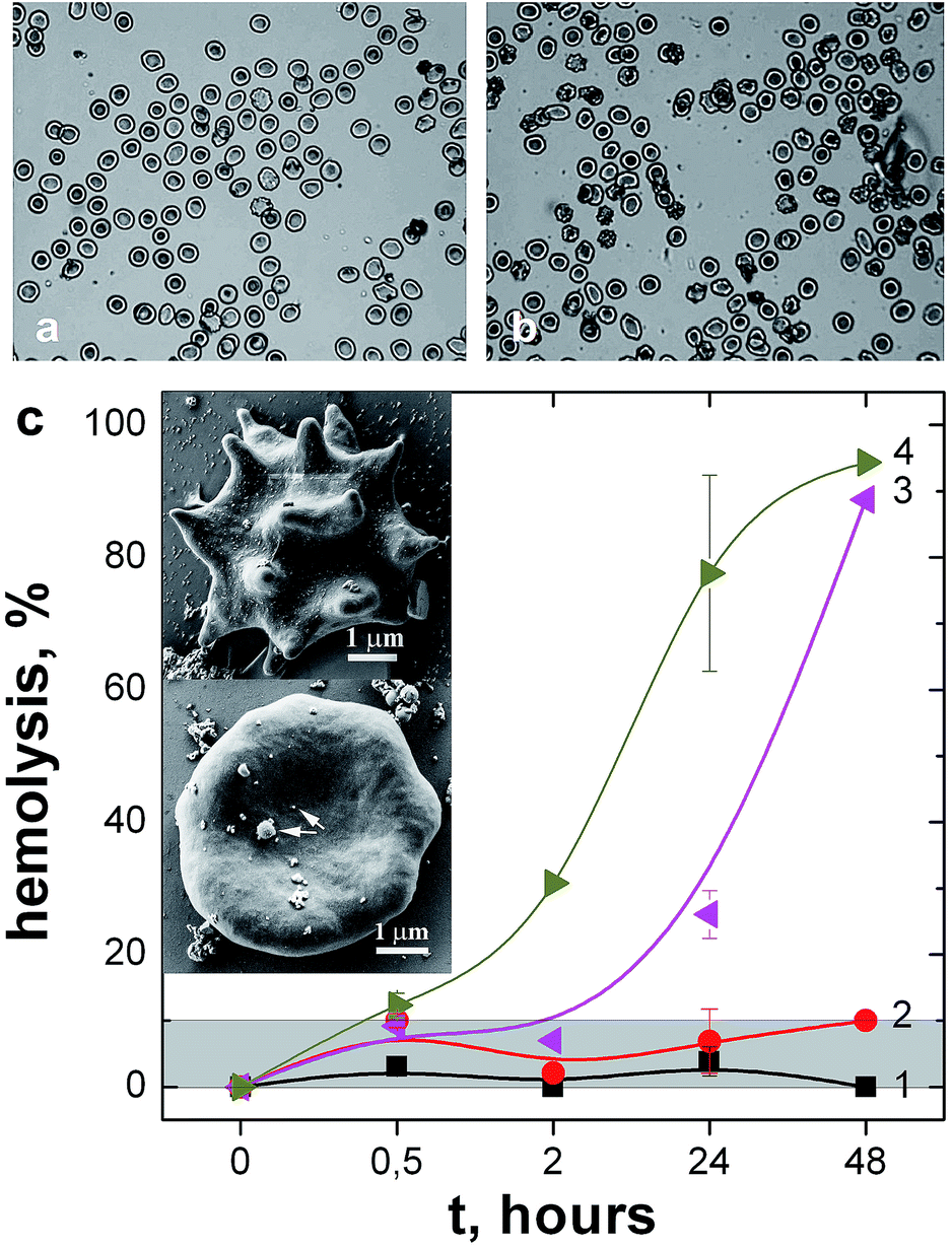 | ||
| Fig. 4 Erythrocyte behavior in different electrolytes (chloride and nitrate-based buffer solutions) (a, b). Erythrocyte morphology, light microscopy study: (a) erythrocytes in a buffer solution with chloride ions (Allen solution without glucose); (b) erythrocytes in the same solution but with chloride ions completely replaced with nitrate ions. (c) Hemolysis rate in the “chloride” (1, 3) and “nitrate” (2, 4) solutions with (3, 4) or without (1, 2) silver nanoparticles. Grey shadowed area corresponds to the intact erythrocytes within the hemolysis measurement error bars. Typical measurement time of SERS spectra of erythrocytes from the preparation of biological samples to the spectra recording does not exceed 5 min. The inset shown a normal erythrocyte form (lower inset) and echinocytes (upper inset). Hemolysis, % = (N0 − N) × 100/N0, where N0 – the cell number at the initial moment, 5 min of incubation, N – the same after a certain period of incubation. Error bars are the standard errors of a mean value (see also ESI, Fig. S8† for details). | ||
First of all, it should be noted that the simultaneous presence of silver nanoparticles and nitrates (Fig. 4c, “3”, “4”) causes gradual degradation of erythrocytes since hemolysis is increased essentially already after 2 h of keeping the cells in the nitrate buffer with silver platelets or after 24 h if silver nanoparticles are mixed with the usual chloride-containing saline. Note that the nitrate saline itself allows to keep the integrity of erythrocytes for almost two days as quite similar to the usual chloride saline, within the measurement error bar (both “1” and “2” sets of data in Fig. 4c stay within the gray shadowed area). The only difference is occasional observation of hedgehog-like echinocyte forms of erythrocytes in the nitrate-based buffer (Fig. 4c the upper inset).
In the chloride-based buffer solution, the largest part of cells have a regular discoidal shape (Fig. 4a) while in the “nitrate” buffer solution, some cells are crenated (have spicules on their surface) (Fig. 4b). Nevertheless, the major fraction of the cells retains its discoid shape, so we can propose that the replacement of chloride ions by nitrates has no dramatic effect on the cell integrity or shapes and therefore this solution is suitable to provide SERS tests with native erythrocytes. At the same time, a commonly discussed theory of cell damage in the presence of silver ions is connected with expected silver ion dissociation from the highly developed surface of metallic silver nanoparticles followed by Ag+ binding to proteins of the cells. This effect seems to be larger in the case of nitrate ions replacing Cl− since the latter do not bind extra Ag+ coming from silver nanoparticles. Certainly, silver nanoparticles themselves seem to be not mechanically damaging erythrocyte membranes30,31 (see ESI, Fig. S8†) therefore the silver ion influence would be predominating. Other reasons of the observed moderate toxicity of the nanoparticles would be also assumed like the influence of residual PVP or other chemical reagents or products naturally remaining together with silver nanoparticles in the produced silver sols.
The data of Fig. 4 are in good agreement with our previous experiments27–30 since we have found there that a period of a safe direct contact of intact erythrocytes with silver nanostructures would exceed 45–60 min while SERS measurement takes only 5 min for erythrocyte landing onto a substrate with silver nanostructures and less than additional 5–10 minutes of repeated SERS measurements with 20–120 seconds taken for each SERS spectrum are needed to achieve a good relation of signal to noise. Thus the suggested application of a special “nitrate” buffer together with anisotropic silver nanoparticles to conduct SERS measurements of living cells seems to be a very reasonable compromise approach on joint surviving of silver nanoplatelets and intact erythrocytes on “rainbow” SERS scaffolds.
A typical example of “rainbow” cellulose scaffolds impregnated with mixed silver platelets is shown in Fig. 5. The picture demonstrates that a mixture of silver nanoplatelets of different sizes and of different optical properties is deposited onto the surface of cellulose bundles of the paper. The paper is porous enough with empty spaces of tens of microns capable to soak intact erythrocytes and land them onto the cellulose bundles with silver nanoparticles during impregnation of the paper with diluted blood. Such a substrate is suitable, from this point of view especially, and also it has a broad light absorption peak (Fig. 6a) since it was created by impregnation with a mixture of various nanoplatelets to provide more spectral sensitivity and universality in its possible applications (see also ESI, Fig. S4†).
 | ||
| Fig. 5 Cellulose scaffold impregnated with mixed silver platelets, (a) a general SEM view of the “rainbow” scaffold (impregnated by sols of many colours), (b) TEM image of the cellulose sheet covered with anisotropic nanoparticles of different sizes (some of them are marked with arrows). | ||
 | ||
| Fig. 6 Absorption and Raman spectra of cellulose scaffolds. (a) Diffuse reflectance UV-vis spectra of the pristine scaffold (1) and the one impregnated with mixed silver platelets (2). (b) A typical SERS spectrum of living erythrocytes on the “rainbow” scaffold with blood 2000 times diluted with a “nitrate” buffer (3) compared to a RS-spectrum of 50 times-diluted blood without nanoparticles (2) and the RS-spectrum of the scaffold impregnated with both mixed silver platelets and the “nitrate” buffer (1) (laser wavelength – 514 nm). The stars mark some peaks from the cellulose substrate. | ||
The prepared cellulose substrate demonstrates a good enhancement of the Raman scattering of intact erythrocytes (Fig. 6b). It is important that the intensity of conventional Raman scattering vanishes as soon as the whole blood is diluted down to what is typically used in SERS experiments (see ESI, Fig. S9†). This guarantees that a spectrum of erythrocytes in such highly diluted samples cannot be taken without a SERS substrate. Moreover, it is usually required to wait for 3–5 minutes before SERS appears since it takes time to land erythrocytes from a droplet of their suspension in saline onto the substrates, after that, the spectra are stable and reproducible. The laser irradiation with an optimal low power kept erythrocytes alive and undamaged.27–29
The SERS spectra of living erythrocytes (Fig. 6) correspond to the enhanced scattering of submembrane hemoglobin27–31 and possess the same set of peaks as conventional Raman spectra of erythrocytes. The Raman spectrum of Hb in erythrocytes has three main peaks that correspond to symmetric vibrations of pyridine half-rings (peak position 1371 cm−1) and vibrations of methine bridges (peak positions 1583 and 1639 cm−1). In the SERS spectra of living erythrocytes, one can observe the strongest peak at 1370–1375 cm−1 and several broad overlapping peaks in the regions of 1550–1600 and 1620–1640 cm−1 due to an enhancement of Raman peaks with maximum positions at 1550, 1564, 1588 cm−1 and at 1622 and 1640 cm−1. The peak assignment is summarized in ESI (Fig. S2 and Table S1†).
The substrate itself is rather suitable for such measurements. It demonstrates a moderate or none luminescence when using a green laser (514 nm wavelength) while no luminescent background is observed at all for the 633 nm laser. The only two cellulose peaks are observed (Fig. 6b, marked with stars) but they do not overlap with fingerprint peaks of hemoglobin. Thus such a substrate could be universally used for SERS studies of at least some important biological objects.
Conclusions
Advanced spectral sensitivity is achieved for biocompatible SERS substrates by means of an application of a mixture of silver nanoplatelet sols impregnating cellulose scaffolds. The ensemble of nanoplatelets with varied shapes and sizes grants multispectral absorption of laser irradiation since a fraction of nanoparticles with a given position of a plasmonic band always exists in such a mixture providing then an effective SERS amplification. We found pronounced negative effects caused by a standard chloride-based saline being crucial to keep the erythrocyte intact but leading to fast recrystallization of the silver nanoplatelets thus neglecting the benefit of anisotropy of the nanoparticles and their unique optical properties. We suggested a replacement of chlorides with nitrates keeping both the intact cells and anisotropic nanoparticles safe and then succeeded in recording SERS data from living erythrocytes using both the suggested scaffold and the new buffer solution. In this terms, the suggested approach gives new insights into the development of SERS devices for biomedical diagnostics.Acknowledgements
This work is supported by the Russian Science Foundation (grant No. 14-13-00871). Authors thank A. S. Baranchikov, V. K. Ivanov, S. V. Savilov for their help with electron microscopy and fruitful discussions.Notes and references
- Y. Sun and Y. Xia, Science, 2002, 298, 2176 CrossRef CAS PubMed.
- J. Kneipp, H. Kneipp, B. Wittig and K. Kneipp, Nano Lett., 2007, 7, 2819 CrossRef CAS PubMed.
- D. E. Charles, D. Aherne, M. Gara, D. M. Ledwith, Y. K. Gun'ko, J. M. Kelly, W. J. Blau and M. E. Brennan-Fournet, ACS Nano, 2010, 4, 55 CrossRef CAS PubMed.
- Y. Lu, G. L. Liu, J. Kim, Y. X. Mejia and L. P. Lee, Nano Lett., 2005, 5, 119 CrossRef CAS PubMed.
- D. D. Evanoff Jr and G. Chumanov, ChemPhysChem, 2005, 6, 1221 CrossRef PubMed.
- Q. Zhang, Y. N. Tan, J. Xie and J. Y. Lee, Plasmonics, 2009, 4, 9 CrossRef CAS.
- R. A. Alvarez-Puebla and L. M. Liz-Marzán, Small, 2010, 6, 604 CrossRef CAS PubMed.
- A. Guerrero-Martínez, S. Barbosa, I. Pastoriza-Santos and L. M. Liz-Marzán, Curr. Opin. Colloid Interface Sci., 2011, 16, 118 CrossRef.
- M. Moskovits, Rev. Mod. Phys., 1985, 57, 783 CrossRef CAS.
- L. Polavarapu, J. Perez-Juste, Q. Xu and L. M. Liz-Marzan, J. Mater. Chem. C, 2014, 2, 7460 RSC.
- Q. Zhang, Y. N. Tan, J. Xie and J. Y. Lee, Plasmonics, 2009, 4, 9 CrossRef CAS.
- R. A. Alvarez-Puebla and L. M. Liz-Marzan, Energy Environ. Sci., 2010, 3, 1011 CAS.
- K. Chaloupka, Y. Malam and A. M. Seifalian, Trends Biotechnol., 2010, 28, 580 CrossRef CAS PubMed.
- B. Lim and Y. Xia, Angew. Chem., Int. Ed., 2011, 50, 76 CrossRef CAS PubMed.
- X. K. Meng, S. C. Tang and S. Vongehr, J. Mater. Sci. Technol., 2010, 26, 487 CAS.
- B. Wiley, Y. Sun, B. Mayers and Y. Xia, Chem.–Eur. J., 2005, 11, 454 CrossRef CAS PubMed.
- J. Fang, S. Liu and Z. Li, Biomaterials, 2011, 32, 4877 CrossRef CAS PubMed.
- B. R. Wood, P. Caspers, G. J. Puppels, S. Pandiancherri and D. Mc-Naughton, Anal. Bioanal. Chem., 2007, 387, 1691 CrossRef CAS PubMed.
- A. März, B. Mönch, P. Rösch, M. Kiehntopf, T. Henkel and J. Popp, Anal. Bioanal. Chem., 2011, 400, 2755 CrossRef PubMed.
- M. S. Kiran, T. Itoh, K. Yoshida, N. Kawashima, V. Biju and M. Ishikawa, Anal. Chem., 2010, 82, 1342 CrossRef PubMed.
- M. Mahato, P. Pal, B. Tah, M. Ghosh and G. B. Talapatra, Colloids Surf., B, 2011, 88, 141 CrossRef CAS PubMed.
- H. Xu, E. J. Bjerneld, M. Käll and L. Börjesson, Phys. Rev. Lett., 1999, 83, 4357 CrossRef CAS.
- N. Shaklai, J. Yguerabide and H. M. Ranney, Biochemistry, 1977, 16, 5585 CrossRef CAS PubMed.
- P. Stein, J. M. Burke and T. G. Spiro, J. Am. Chem. Soc., 1975, 97, 2304 CrossRef CAS PubMed.
- R. T. Tom, A. K. Samal, T. S. Sreeprasad and T. Pradeep, Langmuir, 2007, 23, 1320 CrossRef CAS PubMed.
- A. C. De Luca, G. Rusciano, R. Ciancia, V. Martinelli, G. Pesce, B. Rotoli, L. Selvaggi and A. Sasso, Opt. Express, 2008, 16, 7943 CrossRef CAS PubMed.
- N. A. Brazhe, S. Abdali, A. R. Brazhe, O. G. Luneva, N. Y. Bryzgalova, E. Y. Parshina, O. V. Sosnovtseva and G. V. Maksimov, Biophys. J., 2009, 97, 3206 CrossRef CAS PubMed.
- A. A. Semenova, E. A. Goodilin, N. A. Brazhe, V. K. Ivanov, A. E. Baranchikov, V. A. Lebedev, A. E. Goldt, O. V. Sosnovtseva, S. V. Savilov, A. V. Egorov, A. R. Brazhe, E. Y. Parshina, O. G. Luneva, G. V. Maksimov and Y. D. Tretyakov, J. Mater. Chem., 2012, 22(47), 24530 RSC.
- A. S. Sarycheva, A. A. Semenova, E. Y. Parshina, N. A. Brazhe, A. Y. Polyakov, A. Y. Kozmenkova, A. V. Grigorieva, G. V. Maksimov and E. A. Goodilin, Mater. Lett., 2014, 121, 66 CrossRef CAS.
- A. A. Semenova, N. A. Brazhe, E. Y. Parshina, V. K. Ivanov, G. V. Maksimov and E. A. Goodilin, Plasmonics, 2014, 9(2), 227 CrossRef CAS.
- E. Y. Parshina, A. S. Sarycheva and A. I. Yusipovich, et al., Laser Phys. Lett., 2013, 10(7), 75607 CrossRef.
- A. A. Semenova, V. K. Ivanov and S. V. Savilov, et al., CrystEngComm, 2013, 15(39), 7863 RSC.
- Y. D. Tretyakov and E. A. Goodilin, Russ. Chem. Rev., 2009, 78(9), 801 CrossRef CAS.
- I. Pastoriza-Santos and L. M. Liz-Marzán, J. Mater. Chem., 2008, 18, 1724 RSC.
- T. Huang and X.-H. N. Xu, J. Mater. Chem., 2010, 20, 9867 RSC.
- X.-Y. Zhang, A. Hu, T. Zhang, W. Lei, X.-J. Xue, Y. Zhou and W. W. Duley, ACS Nano, 2011, 5, 9082 CrossRef CAS PubMed.
- B. Pietrobon and V. Kitaev, Chem. Mater., 2008, 20, 5186 CrossRef CAS.
- D. E. Charles, D. Aherne, M. Gara, D. M. Ledwith, Y. K. Gun'ko, J. M. Kelly, W. J. Blau and M. E. Brennan-Fournet, ACS Nano, 2010, 4, 55 CrossRef CAS PubMed.
- J. Zeng, X. Xia, M. Rycenga, P. Henneghan, Q. Li and Y. Xia, Angew. Chem., Int. Ed., 2011, 50, 244 CrossRef CAS PubMed.
- N. Cathcart, A. J. Frank and V. Kitaev, Chem. Commun., 2009, 7170 RSC.
- B. L. Horescer, J. Biol. Chem., 1943, 148, 183 Search PubMed.
- W. G. Zijlstra and A. Buursma, Spectrophotometry of hemoglobin: Absorption spectra of bovine oxyhemoglobin, deoxyhemoglobin, carboxyhemoglobin, and methemoglobin, in. Comparative biochemistry and physiology B – Biochemistry & molecular biology, 1997, vol. 118(4), p. 743 Search PubMed.
- B. R. Wood, B. Tait and D. McNaughton, Biochim. Biophys. Acta, 2001, 1539, 58 CrossRef CAS.
- N. A. Brazhe, E. Y. Parshina, V. V. Khabatova, A. A. Semenova, A. R. Brazhe, A. I. Yusipovich, A. S. Sarycheva, A. A. Churin, E. A. Goodilin, G. V. Maksimov and O. V. Sosnovtseva, J. Raman Spectrosc., 2013, 44(5), 686 CrossRef CAS.
- N. A. Brazhe, A. B. Evlyukhin, E. A. Goodilin, A. A. Semenova, S. M. Novikov, S. I. Bozhevolnyi, B. N. Chichkov, A. S. Sarycheva, A. A. Baizhumanov, E. I. Nikelshparg, L. I. Deev, E. G. Maksimov, G. V. Maksimov and O. Sosnovtseva, Sci. Rep., 2015, 5, 13793, DOI:10.1038/srep13793.
- A. S. Sarycheva, N. A. Brazhe, A. A. Baizhumanov, E. I. Nikelshparg, A. A. Semenova, A. V. Garshev, A. E. Baranchikov, V. K. Ivanov, G. V. Maksimov, O. Sosnovtseva and E. A. Goodilin, J. Mater. Chem. B, 2016, 4, 539 RSC.
- M. Liu, M. Leng, C. Yu, X. Wang and C. Wang, Nano Res., 2010, 3(12), 843 CrossRef CAS.
- R. Prucek, A. Panacek, A. Fargasova, V. Ranc, V. Masek, L. Kvıtek and R. Zboril, CrystEngComm, 2011, 13, 2242 RSC.
- C.-M. Tsai, M.-S. Hsu, J.-C. Chen and C.-L. Huang, J. Phys. Chem. C, 2012, 116, 461 CAS.
- F. Jiang and Y.-L. Hsieh, Biomacromolecules, 2014, 15, 3608 CrossRef CAS PubMed.
Footnote |
| † Electronic supplementary information (ESI) available: Experimental and characterization details. See DOI: 10.1039/c6ra20372h |
| This journal is © The Royal Society of Chemistry 2016 |