T. I. Halenova*a,
I. M. Vareniuka,
N. M. Roslovaa,
M. E. Dzerzhynskya,
O. M. Savchuka,
L. I. Ostapchenkoa,
Yu. I. Prylutskyy*a,
U. Ritterb and
P. Scharffb
aEducational and Scientific Center ‘Institute of Biology’, Taras Shevchenko National University of Kyiv, 64 Volodymyrska Str., 01601 Kyiv, Ukraine. E-mail: galenovatanya@rambler.ru; prylut@ukr.net
bInstitute of Chemistry and Biotechnology, Technical University of Ilmenau, 25 Weimarer Str., 98693 Ilmenau, Germany
First published on 5th October 2016
The development of novel more efficient antioxidants is one of the most perspective approaches for the treatment of the harmful effects of toxins and their metabolites. Due to its remarkable antioxidant properties, pristine C60 fullerene has recently been proposed as a promising candidate for many biomedical applications. The present study aimed to determine whether single oral administration of water-soluble pristine C60 fullerene in small dose (1.5 mg kg−1 bwt) could prevent acute liver injury caused by single intraperitoneal injection of carbon tetrachloride (CCl4; 1.0 mL kg−1 bwt) in rats. We performed structural examination of the liver and monitored the serum markers relevant to hepatocyte integrity as well as liver functionality. The antioxidative potential of pristine C60 fullerene was assessed in vivo by measuring lipid peroxidation. We also measured the activity of catalase and superoxide dismutase in order to evaluate the antioxidative defense system. Biochemical and pathological results obtained in this study indicate that water-soluble pristine C60 fullerene in a single dose of 1.5 mg kg−1 has a hepatoprotective effect against CCl4-induced toxicity via its antioxidant properties. According to histopathological examinations and some biological tests, water-soluble pristine C60 fullerene is more effective against liver injury when administered before a toxicant than after one. Moreover, our results show that the low dose of C60 fullerene used has no acute toxicity in rodents. We concluded that water-soluble pristine C60 fullerene can be considered as a powerful liver-protective agent.
Risk of xenobiotic-induced liver injury has increased in the past decades.7 And nowadays, toxic liver disease is recognized as one of the most common liver disorders in the world while effective pharmacological therapy for this pathological process is absent. The most plausible mechanism of xenobiotic toxicity is an enhancement of free radical production that induces lipid peroxidation and oxidative damage of liver cells.8,9 Thus, the use of antioxidants as protective agents could be a potential therapeutic strategy for xenobiotic-induced toxicity.
Earlier publications examined the biological activity of C60 fullerene, the unique third natural allotropic form of carbon, to show its great potential for biomedical researches.10–15 The results of latter research suggest that the water-soluble pristine C60 fullerenes are nontoxic at low concentrations;16–18 they can penetrate through plasma membrane of cells by passive diffusion or endocytosis19–21 and demonstrate strong antioxidant properties.22,23 It is well documented that C60 fullerene, administered intraperitoneally or intravenous, may act as cardio- and hepato-protector in both doxorubicin- and carbon tetrachloride (CCl4)-induced toxicity.22,24,25 But there are only few available works about the effects of orally applied C60 fullerene.26,27 Also there is little information about the protective effects of extremely low dose of water-soluble pristine C60 fullerene against harmful influence of toxins.28,29
The purpose of this study was to examine the influence of the low dose (1.5 mg kg−1 bwt) water-soluble pristine C60 fullerene in the prevention of acute hepatotoxicity caused by single high dose administration of CCl4 (1.0 mL kg−1 bwt). In order to know whether it can show equal hepatoprotective activity in pre- and post-treatment experiments, we administered C60 fullerenes both 3 h before and 3 h after intraperitoneal injection of toxicant. These results will help clarify the biochemical mechanisms of C60 fullerene action in biological systems.
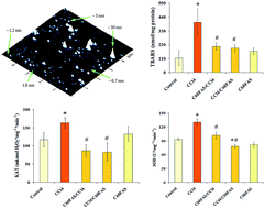
The AFM picture in Fig. 1 clearly demonstrates randomly arranged individual C60 molecules with diameter 0.7 nm and their bulk sphere-like aggregates with a height up to 30 nm. The results obtained are in good agreement with theoretical calculations and experimental measurements32–36 and demonstrate polydispersity of the C60FAS used in our study.
CCl4 is a well-known hepatotoxin.37 The cleavage of CCl4, catalyzed by liver microsomal cytochrome P450, leads to the formation of trichloromethyl radical (CCl3˙), which in the presence of oxygen interacts with it to form the more toxic CCl3O2˙.38 These highly reactive radicals initiate chain lipid peroxidation (LPO) of the hepatocyte membrane causing functional and morphological changes in the liver cell. CCl4-induced accumulation of oxidants results in oxidative liver damage which are characterized by increased necrosis, steatosis, and foamy degeneration followed by the progression of fibrosis.39,40 Liver injury caused by CCl4 is the best characterized model of xenobiotic-induced hepatotoxicity and has been used extensively for decades in preclinical animal studies for the screening of hepatoprotective activities of different drugs.41,42 Thus, to evaluate the protective effect of C60FAS against acute xenobiotic-induced hepatic damage, male Wistar rats injected with CCl4 were used in the present study.
According to previously published papers, several biochemical changes were observed as a result of oxidative stress originating from toxicity induced by CCl4.22,43 Our results well agree with these data. The preliminary assessment of integrity and functionality of liver cells in the CCl4 group was done by determining the serum aspartate aminotransferase (AST), alanine aminotransferase (ALT), alkaline phosphatase (ALP) and γ-glutamyl transpeptidase (GGT) activities as well as total and direct bilirubin levels. Effects of C60FAS on these parameters in rats with CCl4 liver injury model are shown in Fig. 2A–F.
In our experiments, hepatocytes damage, caused by CCl4 application, accompanied by a statistically significant increase in ALT and AST activities compared to the control values. The obtained results showed significant decreased of these enzyme activities in the experimental groups that received a 1.5 mg kg−1 dose of C60FAS either under pretreatment or 3 h after CCl4 treatment condition (Fig. 2A and B). As shown in Fig. 2C and D, activities of ALP and GGT were no significantly changed in CCl4 treated rats compared to control ones. After 24 h of CCl4 treatment, the total bilirubin and direct bilirubin levels significantly raised by 3.5 and 5.6 times respectively, compared to control group (Fig. 2E and F). Pretreatment with 1.5 mg kg−1 of C60FAS decreased significantly the CCl4-induced elevation of serum total bilirubin by 35% as well as direct bilirubin by 45%, whereas treatment with C60FAS after CCl4 administration caused reduced of these parameters by 60% and 70%, respectively (Fig. 2E and F). When animals were given C60FAS alone no changes in the serum enzymatic activities, neither the concentrations of total and direct bilirubin were observed (Fig. 2A–F).
It is well known that CCl4 produces liver damage.37,40 The elevated levels of ALT and AST as well as total and direct bilirubin are indicative for cellular leakage, loss of the hepatocyte membrane integrity and impairment of liver function. In our study, the treatment with C60FAS according to the both scheme of administration suppressed the increment of these parameters induced by CCl4 injection. We supposed that the reversal of increased serum biomarkers in CCl4-induced liver damage by the fullerene may be due to the prevention of the leakage of intracellular content and functional improvement of the hepatic cells by its antioxidant effect.22,23,44 This is confirmed by our findings which showed that C60FAS can overcome oxidative stress caused by CCl4.
LPO in the liver tissue was characterized by measurement of thiobarbituric acid reactive substances (TBARS), which is widely used as a marker of free radical mediated LPO injury. In our experiments, 24 h after a single dose of CCl4, we observed a significantly increased concentration of TBARS in the liver tissue (Fig. 3) that proved activation of oxidative process in the organ. This data is supported by previous studies. C60FAS abolished the toxic effect of CCl4 and maintained LPO at a basal level. Applied alone, the 1.5 mg kg−1 dose of C60FAS did not cause any alteration in the TBARS level. These results support the hypothesis that pristine C60 fullerene is a strong antioxidant and non toxic at low concentration.40,45,46
The increased TBARS level in liver of CCl4-treated rats suggests enhanced LPO leading to tissue damage and failure of antioxidant defense mechanism to prevent formation of excessive free radicals. Superoxide dismutase (SOD, E.C. 1.15.1.1.) has been reported as one of the most important enzymes in the enzymatic antioxidant defense system.47,48 It is found in the cytosol as a zinc/copper-containing enzyme and in mitochondria as a manganese-containing enzyme. SOD dismutates the superoxide radicals by converting them into hydrogen peroxide and oxygen. Catalase (CAT, E.C. 1.11.1.6.) is an enzymatic antioxidant widely distributed in all animal tissues. CAT is known to break hydrogen peroxide down to water and oxygen and can be found in the peroxisome and mitochondria, especially in liver.49
The levels of SOD and CAT in serum as well as in liver tissue were measured for all experimental groups (Fig. 4A–D). High level of CAT in serum was observed 24 h after CCl4 treatment (Fig. 4B), whereas serum SOD activity was statistically similar with this enzyme activity in serum of control group (Fig. 4A). C60FAS is seems to stimulate the serum SOD activity under CCl4-treatment conditions because this enzyme level was increased in both groups with CCl4 injury model treated with C60FAS (Fig. 4A). We have shown that C60FAS did not effect on serum CAT activity of CCl4-treated rats because the enzyme activity in both C60FAS/CCl4 and CCl4/C60FAS groups were approximately equal to this one in serum of group CCl4 (Fig. 4B). In the case of treatment with C60FAS alone, serum SOD and CAT levels close to control group were observed (Fig. 4A and B).
Effects of C60FAS on hepatic SOD and CAT activities are shown in Fig. 4C and D. Application of CCl4 significantly elevated the activity of the examined enzymes compared to the control group. Treatment with C60FAS 3 h before the application of CCl4 as well as 3 h after CCl4 injection succeeded in preventing oxidative stress in liver tissue and maintained the basal activity of SOD and CAT (Fig. 4C and D). The dosage of C60FAS in this study was not toxic to the liver tissue, and the activity of antioxidative enzymes in the C60FAS group did not reveal any significant changes compared to the control group (Fig. 4C and D).
Previous studies have indicated that CCl4 can enhance the activities of antioxidant enzymes.50 Our present work clearly showed that activities of CAT and SOD in the liver as well as in the serum of rats treated with CCl4 alone were significantly higher than those in the control group. The elevated these enzyme activities in rats received CCl4 may indicate a large increase of superoxide anion and hydrogen peroxide after toxicant injection; however, we cannot state in what degree the process of removal of hydrogen peroxide is effective. C60 fullerene was found to be a powerful radical-scavenger for superoxide, hydroxyl and lipid radicals.22,23,44 In this study, C60FAS was potent in decreasing the SOD and CAT activities in rat liver but not serum, which may be resulted from the biodistribution nature of C60 fullerenes. The uptake of C60 fullerene in liver was relatively high, while the clearance from liver was rather slow.51,52 This may eventually lead to the relatively long retention of C60 fullerene in liver and consequently be absorbed into liver cells and acted as an antioxidant there.
The results of our pathohistological analysis of liver tissue conducted in rats after a single dose of CCl4 are similar to the results that were previously reported.53 Liver histopathological changes after 24 h toxicant treatment were noticeable with light microscopy.
The liver in control group had normal histological structure, typical of this organ (Fig. 5A, C and D).
After treatment with C60FAS no histopathological changes were found (Fig. 5K and L), only some hepatocytes had more lighter cytoplasm compared to the control group.
The main pathohistological changes in the liver after treatment with CCl4 were the following: damage of radial structure of the liver cords, hydropic degeneration of hepatocytes, infiltration of inflammatory cells, the appearance of homogeneous eosinophilic content in some sinusoids (seldom), hepatocyte necrosis (seldom) (Fig. 5B, E and F). These changes heterogeneously scattered in the liver (Fig. 5B). Hydropic degeneration of hepatocytes and infiltration of inflammatory cells were significantly greater in the centrilobular zone compared to periportal zone of the liver (Fig. 5B; Table 1).
Treatment with C60FAS 3 h after the application of CCl4 did not alleviate all these lesions (Fig. 5G and H). Percentage of hepatocytes with pathological changes no had significant difference compared with the group that received only CCl4 (Table 1).
After pretreatment with C60FAS 3 h before the application of CCl4 we observed damage of radial structure of the liver cords, but the leukocyte infiltration was lower. In addition, the number of pathologically altered hepatocytes was significantly fewer (Fig. 5I and J; Table 1). Thus, pretreatment with C60FAS 3 h before the application of CCl4 protected the liver against injury induced by CCl4.
These our findings suggest that C60 fullerene more effectively improves liver injury when applied 3 h before toxicant injection. This hypothesis also agree with our results indicating different changes of liver index and liver cytokine profile in rats received C60FAS 3 h before CCl4 injection and 3 h after one.
It was found, that liver index in CCl4 treated rats was significantly higher than in control (55.3 ± 2.4 vs. 45.2 ± 1.9). Animals from the group protected by an oral application of C60FAS 3 h before toxicant had liver index similar to the control (47.2 ± 2.8), while in rats treated with C60FAS 3 h after CCl4 administration liver index was significant higher than in control rats (52.2 ± 3.5) and was comparable to the index of CCl4 group animals, who received toxicant alone. Elevated liver index may indicate inflammation and pathological changes in the organ.
Our findings suggested that intensity of inflammation process was not the same in liver of rats, whom received C60FAS 3 h before CCl4 and 3 h after one. This is confirmed by our results of both macro- and microscopic assessment of the liver as well as the results of cytokine profile measurement.
Cytokines are pleiotropic peptides produced by most of all nucleated cells in the body.54 In variety tissues, including the liver, constitutive production of cytokines is absent or minimal. There is increasing evidence that cytokines mediate not only hepatic inflammation, apoptosis and necrosis of liver cells but also they mediate the regeneration of liver tissue after injury.55
Fig. 6 shows the results of our study of levels of various hepatic inflammatory cytokines under CCl4-induced acute liver injury in rats with or without C60FAS treatment compared to the control rats. In our study we observed significantly increased pro-inflammatory cytokine (IFNγ, IL-1β, IL-12) levels by 25–50% in liver of rats received CCl4 compared with control group (Fig. 6A–C). Such results were expected because it is well known that pro-inflammatory cytokines have emerged as a key factor in various aspects of liver disease, such as fibrosis and/or cholestasis. Different changes of pro- and anti-inflammatory cytokine levels were indicated in the case of treatment of CCl4-administered rats with C60FAS 3 h before toxicant and 3 h after one. When C60FAS was applied after CCl4 injection, the anti-inflammatory cytokine (IL-4, IL-10) levels significantly decreased by 15–20% (Fig. 6D and E) while pro-inflammatory cytokine IL-12 increased by 40% in liver of rats compared with control group (Fig. 6C). When C60FAS was applied before CCl4 injection we did not observe any changes in liver cytokine content. Moreover, animals received C60FAS before toxicant had the same cytokine levels as the control group. These results suggests about more intensive inflammation process in liver of rats received C60FAS treatment after toxicant injection: overwhelming expression of pro-inflammatory cytokines can damage healthy tissues, while decreasing of anti-inflammatory cytokines, which can act as inhibitors of inflammation and are necessary for the regeneration of the tissue after the liver has been injured, might inhibit the process of hepatic recovery.
CCl4 was purchased from Sigma-Aldrich (St. Louis, MO, USA). CCl4 solutions for intraperitoneal (i.p.) injection were prepared ex tempore by dissolving the substances in olive oil. Assays kits for the detection of serum ALT, AST, ALP, GGT, direct and total bilirubin were purchased from ELITechGroup (Paris, France). Anti-rat monoclonal antibodies to INFγ, IL-1β, IL-4, IL-10, IL-12 were purchased from Santa Cruz Biotechnology, Inc. (Dallas, Texas, USA). Horseradish peroxidase-conjugated anti-mouse, anti-goat and anti-rabbit antibodies were purchased from Sigma-Aldrich (St. Louis, MO, USA).
The rats were randomly divided into five experimental groups, namely (Table 2): the control rats (group 1) were treated with olive oil (1.0 mL kg−1) by i.p. as vehicles for CCl4; II–IV groups were treated with 50% CCl4/olive oil (1.0 mL kg−1) by i.p. injection, at once;26,41 C60FAS in a single dose of 1.5 mg kg−1 was given by oral gavage (o.g.), 3 h before (C60FAS/CCl4 group) and 3 h after (CCl4/C60FAS group) administration of CCl4; group V was treated with C60FAS only in accordance to the scheme described above. All animals of I–IV groups were sacrificed 24 h after CCl4/vehicle injection by urethane anesthesia. Animals of V group were anesthetized and sacrificed 24 h after C60FAS treatment.
Group | Description | n | Administration-agenda | 50% CCl4/olive oil (1.0 mL kg−1) | C60FAS (1.5 mg kg−1) |
---|---|---|---|---|---|
I | Control | 6 | i.p. olive oil | − | − |
II | CCl4 | 6 | i.p. in single dose | + | − |
III | C60FAS/CCl4 | 6 | o.g. before 3 h/i.p. | + | + |
IV | CCl4/C60FAS | 6 | i.p./o.g. after 3 h | + | + |
V | C60FAS | 6 | o.g. in single dose | − | + |
It is important to note that a dose of 1.5 mg kg−1 C60FAS applied in our experiments does not present any acute or subacute toxicity in animals: it was significantly lower than the maximum tolerated dose of C60 fullerene, which was found to be 5 g kg−1 both for oral or i.p. administration to rats.22
Finally, it was established that during 48 h the water-soluble C60 fullerene derivatives can be localized in the liver of mice.58
Livers were immediately collected after the animals sacrificed and weighted for calculation of the liver index, which was expressed as mg (wet weight of organ) per g (dead body weight). Liver tissue samples were taken from the left liver lobe and cut into two pieces. One piece was fixed in 10% formalin for histopathological examination. The other piece (1 g) was homogenized in 10 mL ice-cold 50 mM Tris–HCl buffer (pH 7.4) using an automatically homogenizer. The homogenate solution was centrifuged at 12000g for 30 min at 4 °C. The supernatant was collected and stored at −80 °C for further biochemical analysis. The protein concentration was determined by the Bradford method59 using crystalline bovin serum albumin (BSA) as a standard.
CAT activity was measured by the method of Korolyuk et al.:62 0.1 mL serum or liver homogenate (each sample was previously 10-fold diluted with the Tris–HCl buffer, pH 7.4) was incubated in 2 mL freshly prepared 0.03% H2O2 at room temperature for 5 min. Control sample included 0.1 mL buffer instead biological material. The enzymatic reaction was stopped by addition of 1 mL of 4% ammonium molybdate and the yellow complex of molybdate and hydrogen peroxide was measured at 405 nm against blank (mixture of 0.1 mL buffer, 2 mL distillate water and 1 mL ammonium molybdate). The enzyme activity was calculated as the difference of H2O2 content in control and experimental samples by using calibration curve previously prepared with standard H2O2 solutions. Activity of CAT was expressed as μg mg−1 of protein.
This journal is © The Royal Society of Chemistry 2016 |