PDGF-BB, NGF and BDNF enhance pulp-like tissue regeneration via cell homing

Lin Li and Zuolin Wang*
Department of Endodontics, Department of Oral Implantology, School & Hospital of Stomatology, Tongji University, Shanghai Engineering Research Center of Tooth Restoration and Regeneration, Shanghai 200072, China. E-mail: zuolin@tongji.edu.cn; Fax: +86-21-66524025; Tel: +86-21-66311625

Received 11th August 2016 , Accepted 30th October 2016

First published on 2nd November 2016


Abstract

In this study, we investigated the cytobiological effects of platelet-derived growth factor BB (PDGF-BB), nerve growth factor (NGF) and brain-derived neurotrophic factor (BDNF) on the culture of bone mesenchymal stem cells (BMSCs) from rats and explored a viable approach for regenerating ectopic dental pulp-like tissue via cell homing. In vitro, the proliferation, migration, and differentiation of rat BMSCs treated with different dosages of PDGF-BB, BDNF and NGF were evaluated using CCK-8, trans-well and quantitative real-time PCR (qRT-PCR) assays. In vivo, rats were randomly assigned to three groups: the control group, the low concentration group (L group) and the high concentration group (H group). The cytokines were delivered into endodontically treated human teeth, which were then implanted subcutaneously into the rat dorsum for 2 to 4 months. Next, a histologic analysis was employed to identify the regenerated dental pulp-like tissue. The results showed that PDGF-BB/NGF/BDNF facilitated BMSC proliferation in time- and dose-dependent manners. PDGF-BB significantly promoted the migration of BMSCs (P < 0.05). The maximum gene expressions of Runx2, osteopontin (OPN), markers of neural dendrites and somata-microtubule associated protein-2 (MAP2) and β-III-tubulin, were induced following treatment with 100 ng ml−1 PDGF-BB, NGF and BNDF respectively (P < 0.05). In vivo, well-vascularized pulp-like tissue was regenerated in both the H and L groups. This finding was confirmed via CD-34 immunohistological staining, which indicated newly formed vessels. More small vessels were observed in the H group than in the other groups (P < 0.05). In addition, positive signals for S-100 were only detected in the H group, which indicated newly formed nerve fibers. In conclusion, the current study provides evidence supporting the homing of endogenous mesenchymal stem cells via the combined use of a PDGF-BB/NGF/BDNF delivery system to regenerate ectopic pulp-like tissue.

1. Introduction

Dental pulp is the only vascular tissue in the teeth, making it distinctly important for the homeostasis of vital teeth. The loss of pulp due to caries and pulpitis can result in the loss of teeth, consequently affecting chewing function and oral health. The typical treatment for irreversible pulpitis is root canal therapy (RCT), which can eliminate the infection and protect the decontaminated tooth from future microbial invasion.1 Although RCT generally prevents tooth loss, young permanent teeth with pulp necrosis pose a series of challenges for dentists because arrested root development due to RCT usually results in thin and fragile roots. In addition, for patients with short roots, thin dentin walls or coarse root canals, ideal outcomes are difficult to obtain using conventional RCT.

Pulp regeneration is a new method for treating pulp infection or necrosis, and it is also one of the most promising therapeutic strategies for treating irreversible pulp inflammation and periapical disease. This method might extend the longevity of teeth and improve patient quality of life. The major objectives of pulp regeneration are to regenerate the pulp–dentin complex and recover the vitality of normal pulp. Depending on whether exogenous cells are used, tissue regeneration approaches are classified into cell-based and cell-free approaches. Despite its scientific value,2–4 the cell-based approach has also resulted in many problems, including aging-associated phenotypic changes in pulpal mesenchymal stem cells (MSCs), the availability of the tissue sources, safety and regulation involved with the expansion of MSCs, the potential loss of cells during laboratory processing, additional costs, and the acquisition of oncogenes during ex vivo cell processing.5,6 In this regard, the cell-free approach has advantages over the cell-based approach. After mobilization and exogenous infusion, the endogenous MSCs obtained using both methods can capably home various organs, especially injured sites.7,8 Given their homing ability, the cells outside of the dental pulp are attracted to the canal space and can most likely regenerate pulp and dentin tissues.9

Pulp is rich in capillary networks, and the capillaries in pulp provide nutrition and support for dental pulp tissue. Therefore, the blood vessels in the pulp play an important role in maintaining the homeostasis of dental pulp. During tissue regeneration, the survival of inflamed vital pulp and engineered transplanted pulp tissue are closely linked to the process of the angiogenesis at the regeneration sites.10 Angiogenesis establishes the blood supply, thereby enabling the transport of the oxygen, nutrition, and prevascular stem cells that support the regeneration process.11 Moreover, pulp is rich in nerve fibers, which are accompanied by blood vessels. The number of nerve fibers determines the recovery of the sensory function in the regenerated pulp-like tissue. Blood vessels and nerve fibers constitute the basis of the nutrition and sensory function of the pulp tissue, enabling the pulp dentin complex to respond to external stimulation or injury. Thus, revascularization and reinnervation are indispensable in the regeneration of pulp tissue, and these processes are especially essential for maintaining the structure and function of the regenerated pulp.

Cytokines are critical signaling molecules that participate in the process of tissue regeneration. In addition to mobilizing endogenous cells, these factors regulate the proliferation and differentiation of stem/precursor cells. Among the proangiogenic factors, vascular epithelial growth factor (VEGF) and fibroblast growth factors (FGFs) are well established as involved in the tissue regeneration process.12–17 In addition to VEGF and FGFs, platelet-derived growth factor (PDGF) has also been shown to promote the proliferation of fibroblasts in human dental pulp tissue, enhance the expression of VEGF in osteoblasts and promote the formation of blood vessels at the site of pulp injury.18,19 Therefore, PDGF might promote angiogenesis and proliferation and differentiation of MSCs; furthermore, it is an important signaling molecule in the regeneration of dental pulp. Regarding neuronal regeneration, nerve growth factor (NGF) and brain-derived neurotrophic factor (BDNF) have displayed prospective applications. NGF is a target-derived neurotrophic factor that is essential for the development, growth, survival, differentiation and maintenance of sympathetic and sensory neurons. Researchers have demonstrated that pulpal NGF is involved in pulp regeneration by chemo-attracting the invading leukocytes and regulating odontoblastic differentiation. BDNF also plays a role in the survival and differentiation of central and peripheral neurons and non-neural cells. This information suggests that both cell types are closely related to pulp repair/regeneration.

Based on the features of these cytokines, we hypothesized that, in the absence of exogenous cells, NGF and BDNF facilitate pulp regeneration when combined with PDGF-BB and collagen I. We investigated the cytobiological effects of BDNF and NGF as well as the chemotactic effects of PDGF-BB on bone MSCs (BMSCs) in vitro. We also demonstrated that the combined use of PDGF-BB, NGF, and BDNF is a promising in vivo approach for regenerating ectopic dental pulp-like tissue via cell homing. These findings might provide a basis for additional clinical and translational research using this approach.

2. Experimental

2.1. Acquisition of cell lines

Rat BMSCs were isolated from healthy, 4 week-old male SD rats as previously described.20 Briefly, the bilateral iliac bone and femur were obtained, and the rat bone marrow MSCs were isolated and purified via the whole bone marrow adherent method. The animal care and experimental protocols were approved by the Laboratory Animal Center of TongJi University, following International Guiding Principles for Animal Research (1985). Third generation cells were collected for in vitro experiments.

2.2. Cell viability assay

The cell viability of BMSCs was detected using a cell counting kit-8 (CCK-8, Beyotime, China). According to the manufacturer's instructions. Briefly, cells were seeded in 96-well plates (1500 cells per well) and incubated with fresh medium at 37 °C and 5% CO2 atmosphere. After overnight incubation, cells were treated with a fresh medium containing PDGF-BB (0, 10, 50, 100 ng ml−1), or NGF (0, 10, 50, 100 ng ml−1), or BDNF (0, 10, 50, 100 ng ml−1). After 24, 48, 72, and 96 h incubation, the cells were washed three times in phosphate-buffered saline (PBS) and incubated in 100 ml MEM with 10 μl CCK-8 solution for 1 hour. The absorbance was measured at 450 nm using an ELISA reader. A well with fresh medium without cells served as a blank control. After subtracting the OD450 of the blank well, the proliferation rate of the treated cells was calculated based on the percentage of their absorbance compared with that of the control cells.

2.3. Cell migration assay

A trans-well assay was used to assess the migration of the BMSCs in response to PDGF-BB, NGF and BDNF. BMSCs were suspended in 200 μl serum-free MEM medium (2.5 × 10⁴ cells per well) and seeded into the upper chambers, while 600 μl media containing PDGF-BB (0, 10, 20, 50 ng ml−1), NGF (0, 10, 20, 50 ng ml−1) or BDNF (0, 10, 20, 50 ng ml−1) was placed in the lower chambers. After 24 h of incubation, the cells on the upper surface of the chamber that had not migrated were wiped away, and the membranes were washed with PBS. The remaining cells were fixed in ethanol for 15 min and stained with 0.1% crystal violet solution. The average number of cells per five random fields, as observed under an optical microscope, was regarded as the number of cells that had migrated in the trans-well assay.

2.4. Quantitative real-time PCR analysis

BMSCs were seeded in 12-well plates (5 × 10⁴ cells per well) in triplicate. For osteogenic induction, a medium containing α-MEM, 10% FBS, 5 mg ml−1 ascorbic acid, 10 nM dexamethasone, 10 mM β-sodium phosphoglycerol, 2 mM glutamine, 100 U penicillin and 100 mg ml−1 streptomycin was employed. For neurogenic induction, a neural-like induction cocktail (Neurobasal A, B-27, 40 ng ml−1 basic Fibroblast Growth Factor (bFGF), 20 ng ml−1 Epidermal Growth Factor (EGF)) was purchased. Different concentrations of PDGF-BB, NGF, or BDNF (0, 10, 50, 100 ng ml−1) were added to the induction medium, and the cells were incubated for 14 days. The cells were harvested in Trizol solution (Invitrogen, USA), and qRT-PCR (Takara, Japan) was performed to measure the mRNA expression of Runx2, OPN, MAP2 and β-III-tubulin.

2.5. Endodontic treatment of roots from extracted human teeth

The procedures described were performed in accordance with the ethical standards of the Tongji University School of Stomatology. Freshly extracted incisors and premolars were collected and soaked in 0.9% NaCl solution. Residual periodontal and periapical soft tissues were scraped with a scalpel and soaked in 70% ethanol. The teeth were further disinfected with 5.25% NaOCl for at least 1 week. Endodontic treatment was performed as previously described (Kenneth and Stephen 2011). An access opening was made through the lingual crown into the pulp chamber. The pulpal tissue was extirpated and then cleaned and shaped with ProTaper files (Dentsply Maillefer, Ballaigues, Switzerland). All of the teeth were autoclaved to eliminate any biological tissue and dried at 65 °C.

2.6. Reagents and scaffold

The recombinant human growth factors, PDGF-BB, NGF and BDNF, were purchased from R&D systems (USA). Specifically, 50 ng ml−1 PDGF-BB was used to evaluate chemotaxis, angiogenesis, proliferation and differentiation effects on dental pulp cells. In addition, 50 and 100 ng ml−1 NGF and BDNF were used to investigate their effects on cell differentiation and innervation. Neutralized collagen I scaffolds were purchased from BD Biosciences. The cytokines were added to 1.8 mg ml−1 collagen gels, which were injected into the pulp chambers and root canals of endodontically treated human teeth. Approximately 100–150 μl collagen gel solution was used in each tooth. Cytokine-free collagen gels served as a control. The endodontically treated teeth with cytokine-loaded or cytokine-free collagen gel were incubated at 37 °C for 40 min to allow for gel formation.

2.7. Surgery

All animal procedures were conducted in accordance with the International Guiding Principles for Animal Research (1985). The experimental protocol was approved by the Laboratory Animal Center of TongJi University. Thirty male SD rats weighing approximately 250 g each were anesthetized with 10% chloral hydrate (1.2 ml/100 g). A linear incision was made at the dorsum, which was followed by the implantation of the endodontic treatment tooth. The teeth were implanted for 2 and 4 months. The rats were divided into the three following groups based on the cytokines used:

(1) Control group: endodontical treatment tooth + collagen gel.

(2) High concentration group (H group): endodontical treatment tooth + collagen gel + 50 ng ml−1 PDGF-BB + 100 ng ml−1 NGF + 100 ng ml−1 BDNF.

(3) Low concentration group (L group): endodontical treatment tooth + collagen gel + 50 ng ml−1 PDGF-BB + 50 ng ml−1 NGF + 50 ng ml−1 BDNF.

2.8. Sample harvesting and histological analysis

The animals were sacrificed using an overdose of intravenous chloral hydrate (100 mg kg−1) at 2 and 4 months after implantation. The replanted teeth were removed from the dorsum of the rats. The specimens were fixed in 4% paraformaldehyde for 48–72 h, and decalcified in 10% EDTA medium for 6 months. The specimens were then dehydrated in ethanol and embedded in paraffin. Subsequently, serial sections 4 μm thick was created. H&E staining, immunohistochemistry and Masson's trichrome staining were performed on the sections according to the manufacturers' protocols. For immunohistochemistry, polyclonal rabbit antibodies against CD34 (1[thin space (1/6-em)]:[thin space (1/6-em)]200, Abcam) and S-100 (1[thin space (1/6-em)]:[thin space (1/6-em)]200, Santa Cruz) were used as primary antibodies.

2.9. Statistical analysis

SPSS 20.0 was used for data processing. The results are presented as the average ± standard deviation. Analysis of variance (ANOVA) was used to compare the differences among groups. P < 0.05 was considered significant.

3. Results

3.1. The effect of PDGF-BB, NGF or BDNF on cell migration

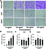
The effects of PDGF-BB, NGF, and BDNF on the migration of BMSCs were tested using a trans-well assay. Cells were cultured with PDGF-BB, NGF or BDNF at concentrations of 0, 10 ng ml−1, 20 ng ml−1, or 50 ng ml−1, for 24 h. The results suggested that PDGF-BB induced cell migration in a dose-dependent manner (Fig. 1A). PDGF-BB led to a significant increase in the number of migrated cells compared with that of the control group (P < 0.05; Fig. 1B). No significant difference was observed between number of migrated cells and the concentration of growth factors in the NGF and BDNF treated groups (P > 0.05).
image file: c6ra20290j-f1.tif
Fig. 1 Migration of bone marrow stromal cells treated with PDGF-BB, NGF and BDNF. (A) The crystal violet staining of BMSCs treated with different concentration of cytokines; (B–D) data analysis of BMSCs migration between different groups. *P < 0.05 vs. control. Data from 3 replicates are presented as the means ± SD, scale bar, 50 μm.

3.2. The effect of PDGF-BB, NGF or BDNF on cell viability

The cell viability of PDGF-BB/NGF/BDNF-treated BMSCs was detected using a CCK-8 assay. Varying concentrations of PDGF-BB, NGF, or BDNF markedly promoted the proliferation of BMSCs, and the proliferation of cells exposed to 10, 50, 100 ng ml−1 of PDGF-BB, NGF or BDNF was significantly promoted compared with that of the cells in the control group (P < 0.05; Fig. 2A–C). The optimum concentration of PDGF-BB for enhancing cell proliferation was 50 ng ml−1 (P < 0.05). When the concentrations of NGF or BDNF were increased, cell proliferation was also enhanced and displayed a significant dose-dependent effect. The optimum concentration of NGF and BDNF for enhancing cell proliferation was 100 ng ml−1 (P < 0.05).
image file: c6ra20290j-f2.tif
Fig. 2 Proliferation and differentiation of BMSCs treated with PDGF-BB, NGF and BDNF. (A–C) The cell proliferation of PDGF-BB, NGF, and BDNF treated BMSCs. CCK-8 assay was taken to analyze the cell proliferation curve of BMSCs treated with different doses of cytokines for 96 h (n = 5); (D–F) qRT-PCR assay of Runx2 and OPN in the PDGF-BB, NGF and BDNF treated groups after osteogenic induction of BMSCs (n = 3); (G–I) qRT-PCR assay of MAP2 and β-III-tubulin in the PDGF-BB, NGF and BDNF treated groups after neuro-like induction of BMSCs (n = 3). *P < 0.05 vs. control.

3.3. The effects of PDGF-BB, NGF or BDNF on cell differentiation

The effects of PDGF-BB, NGF and BDNF on cell differentiation were evaluated using a qRT-PCR assay. Microtubule associated protein-2 (MAP-2) and β-tubulin protein III (β-III-tubulin) were considered markers of neural dendrites and somata, whereas osteopontin (OPN) and Runx2 were considered osteogenic-specific genes. After 14 days of odontogenic induction, the expressions of OPN and Runx2 were increased significantly after PDGF-BB/NGF/BDNF treatment of BMSCs. Moreover, the expressions of MAP-2 and β-III-tubulin were also up-regulated in a dose-dependent manner and were most highly induced when treated with 100 ng ml−1 PDGF-BB/NGF/BDNF during neurogenic induction (P < 0.05; Fig. 2D–I).

3.4. Histological analysis of regenerated pulp-like tissue

All samples were grossly observed. No visible pulp-like tissue was observed in either the opening access or the apical apex in the control group. In groups L and H, however, red congestive pulp-like tissue was exposed at the access opening and the apical foramen (Fig. 3).
image file: c6ra20290j-f3.tif
Fig. 3 Schematic diagrams and gross observation of cell-free based dental pulp regeneration. (A) The cytokines were delivered into endodontically treated human teeth, which were then implanted subcutaneously into the rat dorsum. Groups: the control group, the low concentration group (L group) and the high concentration group (H group); (B) revascularization/revitalization of regenerated pulp-like tissue in endodontically treated real-size teeth received gross observation. No visible pulp-like tissue was observed in either opening access or apical apex in control group. Red congestive pulp-like tissue was exposed to the access opening access of the teeth in PDGF-BB, NGF and BDNF delivered groups (L and H groups).

Under microscopic observation, H&E staining showed that few cells with staggered collagen I scaffold remained in the root canals of the control group. In addition, no organized pulp cells or blood vessels were observed. In contrast, the residual scaffold of the collagen I in the root canals of the L group exhibited a grid-like arrangement, with cells interspersed within. In addition, a small number of blood vessels were observed, and the number of cells in the root canal had increased. Group H showed the presence of new fibroblast-secreted collagen that was distributed along the root canal; some blood vessels were also observed (Fig. 4a–f). The results of Masson's straining also confirmed the histological characteristics of the regenerated dental pulp-like tissue (Fig. 4g–l).


image file: c6ra20290j-f4.tif
Fig. 4 Histological observation of regenerated pulp-like tissue in the root canal. (a–f) The staining observation. (a, d) No organized cells and blood vessels in root canal of control groups; (b, c, e, f) scaffold of collagen exhibited grid-like arrangement with cells interspersed. New blood vessels could be observed in group L and H; (g–l) Masson's staining observation. (g, j) No organized cells and blood vessels in control groups; (h–l) new collagens staining red were secreted by migrated fibroblasts. A lot of blood vessels were found in group L and H. Scale bars, 50 μm. Original magnification level 200×.

Immunohistochemistry showed that pulp-like tissue with good vasculature and innervation was regenerated two months after transplantation. CD-34 positive signals were detected in the newly formed vessels in both the H and L groups compared with the control group. In addition, strand-like collagen fibers interlaced with erythrocyte-filled blood vessels were observed (Fig. 5A). The pulp-like tissue was further regenerated with good innervation and vascularization four months after transplantation. S-100 positive signals were detected in the newly formed nerve fibers in group L (Fig. 6). The nerve fibers had invaded the newly regenerated tissues, indicating the reinnervation of the regenerated tissue. S-100 was immunolocalized to the fiber sprouts, suggesting the myelination of the neural filaments in the regenerated dental pulp following the highly concentrated delivery of PDGF-BB/NGF/BDNF. Microvessel analysis showed that both the microvessel areas and perimeters in the H and L groups were significantly greater than those in the control group (P < 0.05) at the two- and four-month observation time points (Fig. 5B and C). At four months after transplantation, the vascularization in the regenerated tissue of the H group was similar in density and orientation as that of the normal pulp.


image file: c6ra20290j-f5.tif
Fig. 5 CD34 immunohistochemical analysis of regenerated pulp-like tissue. (A)-(a–c) Expression of CD34 two months after transplantation in different groups; (A)-(d–f) Expression of CD34 four months after transplantation in different groups; (A)-(g) human pulp tissue stained with CD34 for positive control; (A)-(h) rat pulp tissue stained with CD34 for positive control; (B) comparison of new blood vessel area in different groups. The new microvessels area value in H group and L group is higher than that in control groups. (C) Comparison of new blood vessel perimeter in different groups. The new microvessels perimeter value in H group and L group is higher than that in control groups. *P < 0.05 vs. control. Scale bars, 50 μm. Original magnification level 200×.

image file: c6ra20290j-f6.tif
Fig. 6 S-100 immunohistochemical staining of regenerated pulp-like tissue. (a) Expression of S-100 two months in group H after transplantation; (b) expression of S-100 four months in group H after transplantation; (c) rat pulp tissue stained with S-100 for positive control; (d) human pulp tissue stained with S-100 for positive control. Scale bars, 50 μm. Original magnification level 200×.

4. Discussion

The following key features of regenerated pulp should be considered: (1) the formation of new odontoblasts lining the existing dentin; (2) newly formed vascularity; and (3) reinnervation.21 In particular, the latter two requirements are essential for maintaining the long-term vitality of the pulp. A key requirement for successful endogenous tissue regeneration is the recruitment of a sufficient number of stem/precursor cells to the injury site.22 Besides BMSCs homing through bloodstream, endogenous cells of periapical residing within neighboring healthy regions in the jaw bone tissue may also be recruited to the site for dental pulp regeneration. Our data showed that, without use exogenous cells, we successfully achieved the revascularization and reinnervation of regenerated pulp-like tissue by the use of PDGF-BB, NGF and BDNF combined with collagen I via cell homing.

PDGF-BB, an isoform of the homodimer PDGF family, attracts pericytes to the sites of blood vessel formation and eventually forms new blood vessels.23 PDGF-B shares a significant homology with VEGF.24 The early expression of PDGF is often found at injury sites, and it promotes the proliferation and migration of fibroblasts and mesenchymal cells as well as the formation of blood vessels.25 PDGF-BB simultaneously stimulates cell proliferation and dentin matrix protein synthesis.26 PDGF-BB enhanced the regeneration of dental pulp-like tissue in an in vivo mouse model.9 The results of the present study showed that PDGF-BB significantly promoted the proliferation and migration of rat BMSCs in a dose-dependent manner compared with the control group. PDGF-BB also increased the gene expression of Runx2, OPN, MAP-2 and β-III-tubulin. In vivo, the PDGF-BB-treated group showed more newly formed blood vessels with higher microvessel perimeters and areas than the control group (P < 0.05). These results indicate that PDGF-BB has the potential to facilitate the formation of blood vessels, promote the differentiation of fibroblasts, and regulate the differentiation of mesenchymal cells, thereby enhancing the regeneration of pulp-like tissue.

NGF is one of the most important bioactive molecules in the nervous system, and the development of dental innervation is highly susceptible to postnatal NGF deprivation.27 NGF promotes the survival and maintenance of sympathetic and sensory neurons, one of the important factors involved in nerve regeneration and repair.28 NGF induces the differentiation of immortalized dental papilla cells into odontoblasts in vitro, suggesting that NGF acts as a stimulant for mineralization.29 As the second member of the neurotrophin family, BDNF plays a role in the survival and differentiation of central and peripheral neurons. BDNF has been implicated in the promotion of angiogenesis and the stability of mouse cardiac cells and rat brain-derived endothelial cells. BDNF might have two mechanisms of action: the direct regulation of the survival and vessel stabilization of endothelial cells and the indirect regulation of the formation of capillary networks through VEGF production.30–32 The present study showed that BDNF and NGF not only significantly promoted the proliferation of BMSCs but also the osteogenic and neural differentiation of BMSCs. Our in vivo study also confirmed the involvement of BDNF and NGF in regulating the differentiation of MSCs, stimulating the formation of peripheral nerve fibers and promoting angiogenesis, thereby enhancing the regeneration of pulp-like tissue.

In addition to successful revascularization and reinnervation of the regenerated pulp-like tissue, this study also employed an interesting experimental animal model.9 That is to say, we use the natural, real-size human exfoliated tooth for dental pulp regeneration. Thus, we could mimic the space and cell homing process of human dental pulp regenerating as much as possible. These results could also provide a new reference for the research of clinical transformation of pulp regeneration. Furthermore, from a clinical point of view, the continued root formation of young permanent teeth could benefit from the revascularization and reinnervation of the regenerated pulp-like tissue. Also, partial recovery the function of the nutrition and sensory of dental pulp tissue would restore the partial vitality of diseased dental pulp, and thereby improve long-term prognosis of diseased teeth. Nevertheless, further study remains to be tested whether cell homing approach in an orthotopic model would still allow dental pulp regeneration. If so, how it might committed the adjacent cells migration into the root canal for dental pulp regeneration.

The present study represents the first attempt to combine the use of chemokines of PDGF-BB and NGFs of NGF and BDNF to achieve pulp-like tissue regeneration via cell homing. Using this approach, we observed the formation of ectopic pulp-like tissue as well as successfully achieved the revascularization and reinnervation of regenerated pulp-like tissue. Collectively, the combined use of chemokines and NGFs via cell homing is a promising approach for enhancing the regeneration of dental pulp-like tissue. Based on these founding of the present study, additional research is needed to further test the orthotopic pulp regeneration with such approach to clarify how nonodontoblastic lineage of MSCs differentiate into odontoblastic lineages. This future work would provide a further understanding of the feasibility of applying this method in clinical practice.

5. Conclusions

The present study depicts a cell-free approach that enables dental pulp regeneration from endogenous stem/progenitor cells via PDGF-BB/NGF/BDNF induced cell homing. These results also provide a novel reference for the research of clinical transformation of pulp regeneration.

Acknowledgements

The Natural Science Foundation of China (No. 81271110), Nature Science and Technology Support Program grants (No. 2014BAI04B07) and the Fundamental Research Funds for the Central Universities of China (No. 20152957) supported this study.

References

  1. K. M. Hargreaves, S. Cohen and L. H. Berman, Cohen's pathways of the pulp, Mosby Elsevier, St. Louis, Mo, 10th edn, 2011 Search PubMed.
  2. G. T. Huang, T. Yamaza, L. D. Shea, F. Djouad, N. Z. Kuhn, R. S. Tuan and S. Shi, Tissue Eng., Part A, 2010, 16, 605–615 CrossRef CAS PubMed.
  3. M. Nakashima and K. Iohara, Adv. Dent. Res., 2011, 23, 313–319 CrossRef CAS PubMed.
  4. M. Nakashima and K. Iohara, J. Endod., 2014, 40, S26–S32 CrossRef PubMed.
  5. Y. Cao, M. Song, E. Kim, W. Shon, N. Chugal, G. Bogen, L. Lin, R. H. Kim, N. H. Park and M. K. Kang, J. Dent. Res., 2015, 94, 1544–1551 CrossRef CAS PubMed.
  6. J. J. Mao, S. G. Kim, J. Zhou, L. Ye, S. Cho, T. Suzuki, S. Y. Fu, R. Yang and X. Zhou, Dent. Clin. North Am., 2012, 56, 639–649 CrossRef PubMed.
  7. K. Fukuda and J. Fujita, Kidney Int., 2005, 68, 1940–1943 CrossRef CAS PubMed.
  8. J. M. Karp and G. S. Leng Teo, Cell Stem Cell, 2009, 4, 206–216 CrossRef CAS PubMed.
  9. J. Y. Kim, X. Xin, E. K. Moioli, J. Chung, C. H. Lee, M. Chen, S. Y. Fu, P. D. Koch and J. J. Mao, Tissue Eng., Part A, 2010, 16, 3023–3031 CrossRef CAS PubMed.
  10. M. A. Saghiri, A. Asatourian, C. M. Sorenson and N. Sheibani, J. Endod., 2015, 41, 797–803 CrossRef PubMed.
  11. M. A. Saghiri, A. Asatourian and N. Sheibani, Oral Surg. Oral Med. Oral Pathol. Oral Radiol. Endod., 2015, 119, 122 CrossRef PubMed.
  12. A. Bagri, H. Kouros-Mehr, K. G. Leong and G. D. Plowman, Trends Mol. Med., 2010, 16, 122–132 CrossRef CAS PubMed.
  13. M. Blaber, J. DiSalvo and K. A. Thomas, Biochemistry, 1996, 35, 2086–2094 CrossRef CAS PubMed.
  14. A. Bronckaers, P. Hilkens, Y. Fanton, T. Struys, P. Gervois, C. Politis, W. Martens and I. Lambrichts, PLoS One, 2013, 8, e71104 CAS.
  15. K. Sagomonyants, I. Kalajzic, P. Maye and M. Mina, J. Dent. Res., 2015, 94, 1582–1590 CrossRef CAS PubMed.
  16. R. Wang, K. Chadalavada, J. Wilshire, U. Kowalik, K. E. Hovinga, A. Geber, B. Fligelman, M. Leversha, C. Brennan and V. Tabar, Nature, 2010, 468, 829–833 CrossRef CAS PubMed.
  17. R. Khurana and M. Simons, Trends Cardiovasc. Med., 2003, 13, 116–122 CrossRef CAS PubMed.
  18. H. Seppa, G. Grotendorst, S. Seppa, E. Schiffmann and G. R. Martin, J. Cell Biol., 1982, 92, 584–588 CrossRef CAS PubMed.
  19. P. J. Bouletreau, S. M. Warren, J. A. Spector, D. S. Steinbrech, B. J. Mehrara and M. T. Longaker, Plast. Reconstr. Surg., 2002, 110, 139–148 CrossRef PubMed.
  20. A. J. Friedenstein, S. Piatetzky II and K. V. Petrakova, J. Embryol. Exp. Morphol., 1966, 16, 381–390 CAS.
  21. G. T. Huang and F. Garcia-Godoy, J. Dent. Res., 2014, 93, 717–724 CrossRef PubMed.
  22. I. K. Ko, S. J. Lee, A. Atala and J. J. Yoo, Exp. Mol. Med., 2013, 45, e57 CrossRef PubMed.
  23. P. Gerwins, E. Skoldenberg and L. Claesson-Welsh, Crit. Rev. Oncol. Hematol., 2000, 34, 185–194 CrossRef CAS PubMed.
  24. P. J. Keck, S. D. Hauser, G. Krivi, K. Sanzo, T. Warren, J. Feder and D. T. Connolly, Science, 1989, 246, 1309–1312 CAS.
  25. S. Barrientos, O. Stojadinovic, M. S. Golinko, H. Brem and M. Tomic-Canic, Wound Repair Regen., 2008, 16, 585–601 CrossRef PubMed.
  26. S. Yokose, H. Kadokura, N. Tajima, A. Hasegawa, H. Sakagami, K. Fujieda and T. Katayama, Cell Tissue Res., 2004, 315, 375–384 CrossRef CAS PubMed.
  27. J. P. Naftel, X. B. Qian and J. M. Bernanke, Brain Res. Dev. Brain Res., 1994, 80, 54–62 CrossRef CAS PubMed.
  28. R. Levi-Montalcini, Science, 1987, 237, 1154–1162 CAS.
  29. N. Mizuno, H. Shiba, W. P. Xu, T. Inui, T. Fujita, M. Kajiya, K. Takeda, N. Hasegawa, H. Kawaguchi and H. Kurihara, Cell Biol. Int., 2007, 31, 1462–1469 CrossRef CAS PubMed.
  30. M. J. Donovan, M. I. Lin, P. Wiegn, T. Ringstedt, R. Kraemer, R. Hahn, S. Wang, C. F. Ibanez, S. Rafii and B. L. Hempstead, Development, 2000, 127, 4531–4540 CAS.
  31. H. Kim, Q. Li, B. L. Hempstead and J. A. Madri, J. Biol. Chem., 2004, 279, 33538–33546 CrossRef CAS PubMed.
  32. T. Ikuta, H. Ariga and K. Matsumoto, Genes Cells, 2000, 5, 913–927 CrossRef CAS PubMed.

This journal is © The Royal Society of Chemistry 2016
Click here to see how this site uses Cookies. View our privacy policy here.