Carbon–carbon vs. carbon–oxygen bond activation in 2- and 3-furonitriles with nickel

Ilnett García-Ventura, Marcos Flores-Alamo and Juventino J. García*
Facultad de Química, Universidad Nacional Autónoma de México, Ciudad Universitaria, Mexico City, 04510, Mexico. E-mail: juvent@unam.mx

Received 10th August 2016 , Accepted 19th October 2016

First published on 20th October 2016


Abstract

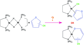
The reaction between [(dippe)NiH]2 (1) (dippe = 1,2-bis(diisopropylphosphino)-ethane) and 2- and 3-furonitrile (2-FN and 3-FN) was carried out at room temperature. Both furonitriles initially react with H2 loss and coordinating a [Ni(dippe)] fragment to the nitrile substituent in a η2-C[triple bond, length as m-dash]N way to yield complexes [(dippe)Ni(η2-C,N-2-FN)] (2) and [(dippe)Ni(η2-C,N-3-FN)] (10), respectively. After monitoring this reaction for 6 days at room temperature in the presence of a slight excess of each nitrile, the C–CN bond activation products [(dippe)Ni(CN)(η1-C-2-FN)] (7) and [(dippe)Ni(CN)(η1-C-3-FN)] (11) were observed as the main products. These complexes were fully characterized by 1H, 31P{1H} and 13C{1H} NMR and by single crystal X-ray determinations. After isolation and characterization of (7) and (11), a thermal stability study was carried out, and it was found that the –C–CN bond activation is reversible at 100 °C for both compounds. Particularly, the thermolysis of complex (7) allowed the further production of a C–O ring opening at the furan to yield complex [(dippe)Ni(κ2-O,C–OCH[double bond, length as m-dash]CH–CH[double bond, length as m-dash]C(CN))] (9), which was also isolated and characterized by multinuclear NMR.

1. Introduction

Activation of strong bonds has been a challenge in organometallic chemistry due to the potential use of a variety of molecules as safe and practical sources of different functional groups.1 One of the most attractive activations that still remain a great challenge is the C–C bond activation.2 Particularly, activation of a C–CN bond in nitriles by transition metals is of high relevance, and in recent years, this has been a useful tool used both, for instance, in the selective and safe transfer of a –CN moiety and in coupling reactions using –CN as a leaving group to produce new C–H, C–C, C–Si or C–B bonds.3

Our group has been interested in these kinds of transformations using nickel-based compounds in the presence of aromatic,4–6 hetero-aromatic6 and alkyl nitriles.7 These studies have contributed to a better understanding of the C–CN oxidative addition process in the presence of a Ni(0) center, allowing an understanding of the observed reactivity by the full characterization of a variety of different intermediates, including the observation of reversibility in some cases of C–CN oxidative addition.4,5 The use of the [Ni(dippe)] fragment has shown that oxidative addition of the C–CN bond is preceded by the η2-coordination of the corresponding nitrile. Additionally, the presence of a bidentate phosphine as an ancillary ligand has been helpful to establish the oxidation state of the metal center by the magnitude of the 2JP–P coupling constant. Thus, Ni(0) complexes with a η2-C[triple bond, length as m-dash]N bond have typical 2JP–P values around 60 Hz, and Ni(II) complexes derived from the C–CN oxidative addition have 2JP–P values lower than 30 Hz.5–7

In this context, the use of the complex [(dippe)NiH]2 as a source of the [Ni(dippe)] fragment by Jones and Vicic8 has been a very useful tool for mechanistic studies related to complex catalytic systems. For example, it was found that the [Ni(dippe)] fragment was able to cleave C–S bond in stable molecules such as thiophene, benzothiophenes and dibenzothiophenes, unveiling mechanistic insights in hydrodesulfurization (HDS) reactions.

Recently, the same nickel complex has been used in a study of reactivity with 2-cyanothiophene;9 for that particular substrate, two competitive reaction pathways can be envisaged: C–CN or C–S bond activation. Both were observed, and several nickel intermediates were characterized at low temperature by considering their 2JP–P values. Additionally, in this study, a full characterization of the Ni(II) complexes derived from C–CN and C–S bond activation was reported, and it was possible to establish that C–S bond activation was reversible at 85 °C to ultimately yield the thermodynamically favored C–CN bond activation complex.

With the continuous interest of our group in the activation of nitriles to be used as building blocks of heterocycles, we decided to explore the reactivity of 2- and 3-furonitriles with the nickel complex [(dippe)NiH]2 to assess the reactivity of these substrates towards a C–CN or C–O bond activation. The reactivity of 3-furonitrile in the presence of a nickel complex containing a N-heterocyclic carbene ligand has been reported by Radius et al.,10 but it was only characterized by 1H-NMR without any further evidence. We report here that both furonitriles reacted with preference for a side on nitrile coordination, followed by C–CN oxidative addition as the kinetic favored reaction at room temperature, which is shown to be reversible at 100 °C for 2-furonitrile to yield the C–O bond ring-opening cleavage as the thermodynamically favored product.

2. Results and discussion

2.1 Reactivity of complex [(dippe)NiH]2 (1) with 2-furonitrile (2-FN)

The reaction between (1) with (2-FN) was carried out according to the conditions specified in Scheme 1.
image file: c6ra20244f-s1.tif
Scheme 1 Reaction between complex [(dippe)NiH]2 and 2-FN in THF-d8.

At room temperature, complex (2) was formed as the major product, and this compound displayed two broad doublets on 31P{1H}-NMR spectrum, at δ 79.7 and 67.9, with 2JP–P = 64 Hz, which is indicative of two asymmetric phosphorous in a Ni(0) complex in agreement with previous reports of closely related compounds.4–7 The 1H-NMR spectrum shows three resonances at δ 7.56, 6.77 and 6.44 (signals for free 2-FN at δ 7.82, 7.31 and 6.63) that shift to high field according with the expected loss of π electron density in the hetero-aromatic ring due to side on coordination of the C[triple bond, length as m-dash]N bond to nickel.

Additionally, the 31P{1H}-NMR spectrum of initial reaction mixture also displays two small broad doublets at δ 66.90 and 58.74 with 2JP–P = 80 Hz, which were assigned to complex (3). To note, this coupling constant is larger compared to the typical values reported for Ni(0) complexes side-on coordinated to nitriles (which are between 60 and 70 Hz). However, larger coupling constants have been observed for aromatic nitriles in which the aromatic or heteroaromatic ring has been coordinated through a double bond in a η2-C[double bond, length as m-dash]C fashion.9,11 Besides, the 1H-NMR spectrum of initial reaction shows another three minor signals at δ 7.22, 6.09 and 5.66. These signals presented a larger change in their chemical shifts compared with the above quoted signals for complex (2), which is consistent with a larger loss of aromaticity in the furan ring due to the direct η2-coordination of nickel to the C[double bond, length as m-dash]C moiety.

A closely related complex to (3) has been reported for 2-cyanothiophene. DFT calculations showed that this kind of complexes are key intermediates in the case of 2-cyanothiophene leading to C–S and later to a C–CN bond activation.9 Considering the above, 2-FN can react either by two ways, i.e., by C–O or by C–CN bond activation, but we expect the C–CN oxidative addition as the favored one in 2-FN, considering the lower Ni(0)–oxygen affinity compared to Ni(0)–sulfur affinity.12

Thus, after 6 days of reaction at room temperature monitored by 31P{1H} NMR, the reaction mixture displayed the above quoted signals for (2) and (3) in a lower amount and the formation of a new signal located at δ 62.5 (ESI, Fig. S1) assigned to complex [(dippe)2Ni2], (4).7 However, the 1H NMR spectrum does not show signals corresponding to free 2-FN (ESI, Fig. S2). This spectrum displays characteristic signals corresponding to compound 2′,2-bifuryl (5) instead, which are assigned by comparison of the reported signals.13 Additionally, complex [(dippe)Ni(CN)2] (6) crystallized out of the solution as yellow crystals, the structure was confirmed by an X-ray structure determination and crystallographic parameters compared with the ones previously reported.7

According to this evidence, it is likely that compounds (2) and (3) yield an oxidative addition product such as [(dippe)Ni(CN)(η1-C-2-FN)] (7) that is not observed under the reaction conditions and that quickly disproportionates to yield compounds (6) and (8) (see Scheme 2). Thus, a further reductive elimination reaction from complex (8) yields 2′,2-bifuryl (5) along with the [(dippe)Ni] fragment; the last of which ultimately forms complex (4) (Scheme 2).


image file: c6ra20244f-s2.tif
Scheme 2 Reactivity for 2-FN with (1) at room temperature in THF-d8.

2.2 Reactivity of complex (1) using an excess of 2-FN

To avoid decomposition due to the formation of [Ni(dippe)] fragment, the reaction between complex (1) and 2-FN was carried out in presence of a slight excess of 2-FN, as depicted in Scheme 3.
image file: c6ra20244f-s3.tif
Scheme 3 Reaction of complex (1) with an excess of 2-FN in THF-d8 at room temperature.

The reaction using an excess of 2-FN with complex (1) monitored by 31P{1H}-NMR initially gave a major set of broad doublets, i.e., the previously quoted signals at δ 79.7 and 67.9 with 2JP–P = 64 Hz, along with the corresponding signals in 1H-NMR spectrum at δ 7.56, 6.75 and 6.44 and the signals for the free 2-FN (ESI, Spectra S1 and S2). These are the same signals assigned before to complex (2). Complex (3) was not observed under the current reaction conditions.

Again, the reaction mixture was monitored for 6 days at room temperature by 1H and 31P{1H}-NMR (ESI, Fig. S3 and S4); after this time, the reaction selectively yielded the formation of complex (7). The 31P{1H}-NMR spectrum for (7) showed two close doublets at δ 83.9 and 83.5 with a 2JP–P = 32 Hz. This P–P coupling constant value is characteristic for a Ni2+ which is formed via oxidative addition of the C–CN bond.4–7 Additionally, the 1H-NMR spectrum shows three signals shifted to high field at δ 7.50, 6.44 and 6.17, as expected for the furan ring. The reaction mixture was placed in a dry box fridge at −30 °C and layered with hexanes to obtain suitable crystals for X-ray diffraction for complex (7), allowing to confirm the characterization of the C–CN oxidative addition product. The corresponding ORTEP representation is shown in Fig. 1.


image file: c6ra20244f-f1.tif
Fig. 1 ORTEP diagram for complex (7), ellipsoids at 50% of probability level. Selected bond lengths (Å): Ni(1)–C(15) = 1.906(3), N(1)–C(19) = 1.145(4), C(15)–C(16) = 1.355(4). Selected angles (deg): C(15)–Ni(1)–C(19) = 88.98(13), P(2)–Ni(1)–P(1) = 88.28(3).

As observed in the ORTEP diagram for complex (7), the nickel center presents a square planar geometry, which is expected for a Ni(II) complex, with a slight elongation of the C[triple bond, length as m-dash]N bond [1.14 Å] due to the back-donation from nickel to –C[triple bond, length as m-dash]N anti-bonding orbitals. The heteroaromatic ring was aligned with the P–Ni–P plane. This is an uncommon orientation of the aromatic fragment compared to Ni2+ complexes that have been reported, which derived from C–CN bond activation in aromatic nitriles.5–7,9 In this case, probably this orientation of the hetero-aromatic ring favored π-back donation from nickel to π* anti-bonding orbitals of C[double bond, length as m-dash]C moiety, which is consistent with a Ni–Cipso bond length in complex (7) that is shorter [1.906(3) Å] compared with related distances observed for benzonitrile [1.935(2) Å] and 2-cyanotiophene [1.918(3) Å] analogues.5,9

The very same crystals were re-dissolved in THF-d8, and in this way, the previous assignations on 1H-NMR and 31P{1H}-NMR spectra were confirmed for complex (7) (vide supra). Additionally, the 13C{1H} NMR spectrum was determined with key resonances assigned to the σ-coordinated C[triple bond, length as m-dash]N ligand at δ 135.2 (dd, 2JP–C = 85, 46 Hz) and also the signal corresponding to ipso heteroaromatic carbon at δ 170.4 (dd, 2JP–C = 98, 39 Hz) (ESI, Spectrum S3).

After leaving a reaction mixture containing complex (7) as a major component in solution (THF-d8) for several days at room temperature, the 31P{1H}-NMR spectrum shows the formation of a new singlet signal at δ 73.5 along with the formation of a yellow crystalline solid precipitating out of the solution (Fig. 2).


image file: c6ra20244f-f2.tif
Fig. 2 31P{1H}-NMR for a sample containing (7) in THF-d8 solution at room temperature.

The yellow solid corresponds again to complex (6), and consequently the new singlet at 73.5 ppm was assigned to complex (8), which is consistent with the increasing intensity of this signal as long as (7) was consumed. As discussed before, complex (8) results in the intermediate formation of 2,2′-bifuryl, which was confirmed by GC-MS determination (ESI, Chromatogram S1 and Spectrum S4).

2.3 Thermolysis for complex (7)

The isolation of pure complex (7) allowed a further study of its thermal stability to assess the potential reversibility of C–CN oxidative addition reaction. Thus, a thermal study was carried out in a sealed NMR tube in THF-d8 by heating the sample at 100 °C and monitoring at 1 hour intervals. After warming for 4 h, the 31P{1H}-NMR spectrum shows two new major broad doublets at 77.3 and 72.5 with a 2JP–P = 40 Hz.

The value of this coupling constant is relatively small to be assigned to a Ni(0) complex and is also quite large for a Ni(II) complex; however, it has a similar value to the reported Ni(II) complex derived from the C–S bond activation of 2-cyanothiophene (2JP–P = 34 Hz).9 Additionally, the 1H-NMR spectrum shows three new up-field signals at δ 7.16 [1H, ddd, J = 11.7, 4.5, 1.8 Hz], δ 6.90 [1H, ddd, J = 16.4, 7.2, 3.6 Hz] and δ 5.10 [1H, dd, J = 7.2, 4.5 Hz] that grow along with the new phosphorous signals as the reaction takes place (ESI, Fig. S5 and S6). The multiplicity of these signals and the coupling constant values are consistent with a 4JP–H coupling.14 Therefore, complex (9) is proposed as the Ni(II) complex derived from the ring opening oxidative addition of C–O bond in 2-FN (Fig. 3).


image file: c6ra20244f-f3.tif
Fig. 3 Proposed structure for complex (9).

Complex (9) was independently synthesized in pure form using a higher quantity of starting complex (7) and toluene as solvent (Experimental section 4.5); thus, 31P{1H} and 1H-NMR assignations were confirmed (ESI, Spectrum S5 and S6).

Additionally, a GC-MS analysis at the end of the thermolysis for complex (7) revealed the presence of compound (5) and free 2-FN (ESI, Chromatogram S2, Spectrum S7 and S8). Since compound (7) was used in pure form, the only reason to obtain free 2-FN is the reversibility of the C–CN oxidative addition at 100 °C, ultimately leading to the formation of complex (9) as the main product. Closely related compounds to this nickelaoxacyclohaxadiene (9) have been reported using iridium and alkylphosphines by Bleeke and coworkers,15 but by using a rather different synthetic procedure.

Considering all these results, a mechanistic proposal is depicted in Scheme 4.


image file: c6ra20244f-s4.tif
Scheme 4 Thermolysis of complex (7) at 100 °C.

2.4 Reactivity of complex (1) with an excess of 3-furonitrile (3-FN)

Similarly to the previous section, the reactivity of an excess of (3-FN) with complex (1) was assessed (Scheme 5); this particular furonitrile owes to the 3-CN position in the heteroaromatic ring and has a smaller contribution to the mesomeric and inductive effects over the –CN group, which can result in a different reactivity.
image file: c6ra20244f-s5.tif
Scheme 5 Reaction of complex (1) with 3-FN in THF-d8 at room temperature.

The reaction initially yields complex (10) as major product. This compound displays two major broad doublets in 31P{1H}-NMR spectrum at δ 79.2 and 66.7 with 2JP–P = 68 Hz, which is characteristic of a Ni(0) complex. The 1H-NMR spectrum shows three up-field signals at δ 7.73, 7.46 and 6.73. The reaction was further monitored by 1H and 31P{1H}-NMR at room temperature for 6 days (ESI, Fig. S7 and S8); after this time, the C–CN oxidative addition complex (11) was obtained as the main product. Complex (11) displays two closely broad doublets at δ 84.2 and 78.4 with 2JP–P = 29 Hz on the 31P{1H}-NMR spectrum, and the coupling constant is characteristic of a Ni(II) complex. The 1H-NMR spectrum exhibits three up-field signals at δ 7.34, 6.63 and 6.26 (ESI, Fig. S9).

The reaction mixture was placed in a vial, layered with hexanes and stored at −30 °C in a dry box fridge to obtain suitable crystals for X-ray diffraction. The corresponding ORTEP diagram for complex (11) is shown in Fig. 4.


image file: c6ra20244f-f4.tif
Fig. 4 ORTEP representation of complex (11) with ellipsoids at 50% of probability level. Selected bond lengths (Å): Ni(1)–C(15) = 1.951(4), N(1)–C(19) = 1.160(6), C(15)–C(18) = 1.335(4). Selected angles (deg): C(15)–Ni(1)–C(19) = 89.32(18), P(2)–Ni(1)–P(1) = 88.79(4).

Complex (11) shows a square planar geometry that is slightly deformed, where the C(15)–Ni(1)–C(19) angle is barely less than 90°; this is in agreement with the values reported for similar compounds, such as the Ni(II) complexes derived from C–CN bond activation in benzonitrile,5 2-cyanoisoquinoline6 and 2-cyanothiopene,9 which displays C–Ni–CN angles values of 88.56(3), 89.15(3) and 88.49(3) degrees, respectively. In complex (11), the furan ring is facing out from the P(1)–Ni(1)–P(2) plane (with an inter-planar angle of 54°). The Ni(1)–C(15) [1.951(4) Å] bond distance is longer in complex (11) compared to the same distance in complex (7) [1.906(3) Å] and C(15)–C(18) distance in complex (11) [1.335(4) Å] is shorter compared to the same distance in complex (7) [1.355(6) Å], probably due to a minor back bonding contribution to π* anti-bonding orbitals on the furan ring in this complex.

As the formation of complex (11) takes place, the formation of complex (6) as a yellow crystalline solid along with complex (12) is observed in 31P{1H}-NMR as small singlets at δ 91.7 and δ 72.6, respectively. Pure crystals for complex (11) were re-dissolved in THF-d8 to confirm the previous assignation on 1H- and 31P{1H}-NMR along with the 13C{1H}-NMR spectrum (ESI, Spectrum S9). These are key signals corresponding to the σ-coordinated C[triple bond, length as m-dash]N fragment assigned at δ 135.7 (dd, 2JP–C = 80 (trans), 30 (cis) Hz), with similar values observed for complex (7) and closely related compounds.5–7

2.5 Thermolysis for complex (11)

Similarly to complex (7), the thermal stability of complex (11) was investigated to assess a possible reversibility in the oxidative addition reaction and a potential C–O bond activation for 3-FN. Thus, a THF-d8 solution of pure complex (11) heated up to 100 °C was monitored by 1H and 31P{1H}-NMR by 1 hour intervals for a total of 4 hours. The 31P{1H}-NMR spectrum shows a gradual decrease of signals corresponding to complex (11) along with the formation of a yellow crystalline solid deposited in the NMR tube characteristic for complex (6), which was confirmed by a small singlet at δ 92.0. In addition, the GC-MS trace showed the formation of 3,3′-bifuryl (13) (ESI, Chromatogram S3 and Spectrum S10). Both are evidence for the disproportionation of complex (11), as represented in Scheme 6.
image file: c6ra20244f-s6.tif
Scheme 6 Thermolysis for complex (11) at 100 °C.

Additionally, the formation of two new, low intensity, broad doublets was observed at δ 80.8 and 68.6 with 2JP–P = 24 Hz. Considering the chemical shifts and the 2JP–P value, these signals can be assigned to the complex derived from the C–O bond activation of 3-FN, (14). However, this complex could not be isolated or crystallized due to its low abundance in the reaction mixture. Also, free 3-FN was detected by GC-MS analysis (ESI, Chromatogram S3 and Spectrum S11) and complex [(dippe)2Ni2] (4) was formed as the main product, showing a singlet at δ 62.5 in 31P{1H}-NMR spectrum.

3. Conclusions

The use of a Ni(0) source allowed the cleavage of the C–CN bond present in 2-FN and 3-FN under mild reaction conditions. The oxidative addition of the C–CN bond proceeds via η2 coordination of the C[triple bond, length as m-dash]N triple bond, and compounds (7) and (11) are evidence of such oxidative addition. Additionally, it was demonstrated that the C–CN cleavage is reversible at 100 °C for both isomers. Particularly for 2-FN, the C–CN bond activation was found to be kinetically favored, whereas the C–O ring opening was the process leading to the thermodynamically stable product. Current work is underway to extend this chemistry to other hetero-aromatic nitriles.

4. Experimental section

4.1 General methods

All reactions were carried out using standard Schlenk and glovebox techniques under an argon atmosphere (Praxair 99.998). THF and hexanes were dried according to standard methods. Deuterated solvents (Cambridge Isotope Laboratories) for NMR experiments were stored over 3 Å molecular sieves in an MBraun glove box (<1 ppm H2O and O2). [Ni(dippe)]2(μ-H)]2 was prepared by the published method.16 2-Furonitrile (99%) was purchased from Aldrich, bubbled with argon (Praxair 99.998) and stored over 3 Å molecular sieves before use. 3-Furonitrile (97%) was also purchased from Aldrich and was used as received. 1H, 13C{1H} and 31P{1H} NMR spectra were recorded at room temperature on a 300 MHz Varian Unity Spectrometer in THF-d8. 1H and 13C{1H} chemical shifts (δ) are reported relative to the residual proton or carbon resonances of the corresponding deuterated solvent. All 31P{1H}-NMR spectra were recorded relative to a 85% H3PO4 standard. All NMR spectra and thermal stability studies were carried out using thin wall (0.38 cm) WILMAD NMR tubes with J. Young valves. Elemental analyses (EAs) were also performed by USAI-UNAM using a Perkin Elmer microanalyzer 2400.

Crystallographic data were collected with an Oxford Diffraction Gemini “A” diffractometer with a CCD area detector with λMoKα = 0.71073 Å and monochromator of graphite at 130 K. Crystals of complex (7) and (11) were covered with Paratone and immediately placed under a cold stream of nitrogen in the diffractometer. CrysAlisPro and CrysAlis RED software packages were used for data collection and integration.17 The double-pass method of scanning was used to exclude any noise. The collected frames were integrated by using an orientation matrix determined from the narrow frame scans. Final cell constants were determined by a global refinement; collected data were corrected for absorbance by using analytical numeric absorption correction using a multifaceted crystal model based on expressions upon the Laue symmetry using equivalent reflections.18

Structure solution and refinement were carried out with the SHELXS-2014 and SHELXL-2014 packages;19 WinGX v2014.1 software was used to prepare material for publication.20 Full-matrix least-squares refinement was carried out by minimising the following: (Fo2Fc2)2. All non-hydrogen atoms were anisotropically refined. H atoms attached to C atoms were placed in geometrically idealized positions and refined as riding on their parent atoms with C–H = 0.95–1.00 Å and with Uiso(H) = 1.2Ueq(C) for aromatic, methylene and methyne groups and Uiso(H) = 1.5Ueq(C) for methyl groups.

4.2 Reaction between 2-FN and complex (1)

Using 2-FN (6.5 μL, 0.0744 mmol) dissolved with 10 drops of THF-d8, the solution was then dropwise added to (1) (0.030 g, 0.0372 mmol) dissolved in a minimum amount of THF-d8 (approx. 0.3 mL), at room temperature in the glovebox. A strong effervescence was immediately observed for approximately 2 minutes, and the solution was stirred for 10 minutes until all H2 gas was vented to the box. Then, the reaction mixture was transferred to a J. Young valve NMR tube, and the necessary quantity of solvent was completed with fresh THF-d8. The sample was immediately analyzed. The reaction mixture was kept in the NMR tube and monitored for 6 days at room temperature.

4.3 Reaction between complex (1) and 2-FN in presence of an excess of nitrile. Preparation of complex (7)

Using 20 mg (0.0200 g, 0.0310 mmol) of complex (1) was dissolved in the minimum amount of THF-d8 (approx. 0.3 mL) and 8.2 μL (0.0087 g, 0.0937 mmol) of 2-FN. Complex (1) and 2-FN was directly added into this solution. A strong effervescence was observed for approximately 2 minutes, then the solution was stirred by 10 minutes, allowing complete H2 venting into the box. Then, the reaction mixture was transferred to a J. Young valve NMR tube, and the necessary quantity of solvent was completed with fresh THF-d8. The sample was immediately analyzed. The reaction mixture was monitored by 31P{1H}-NMR for 6 days at room temperature until quantitative consumption of complex (2). During that time, complex (6) crystallized out from the reaction mixture and separated at the end by decantation. The solution obtained by separation of complex (6) was placed in a vial, added with some drops of hexanes, then placed in a freezer inside the glove box at −28 °C for 5 days. Afterwards, yellow crystals of X-ray quality for complex (7) were obtained. The crystals were washed 3 times with cold hexane/THF 3[thin space (1/6-em)]:[thin space (1/6-em)]1 solution (−28 °C) and dried under vacuum (4 h) (0.0115 g, 0.0278 mmol). Isolated yield: 45%. Anal. calc. for C19H35NNiOP2 (7): % C 55.10; % H 8.52; % N 3.38. Exp: % C 55.30; % H 8.43; % N 3.4. NMR spectra for (7) in THF-d8, 1H: δ (ppm) 0.98–1.29 (m, 24H, CH3), 1.39–1.47 (m, 4H, CH2), 2.44–2.48 (m, 4H, CH), 6.16 (m, 1H, CH), 6.45 (m, 1H, CH), 7.49 (s, 1H, CH). 31P{1H}: δ (ppm) 84.0 (d, J 32 Hz), 83.5 (d, J 32 Hz). 13C{1H}: 18.6–19.3 (m, –CH3), 20.1–20.5 (m, –CH3), 20.9 (dd, JC–P 20, 15 Hz, CH2), 22.5 (dd, JC–P 22, 19 Hz, CH2), 26.5–27.0 (m, CH), 110.6 (m, CH), 122.4 (d, 3JC–P 6.75, CH), 135.2 (dd, 2JC–P 85.1, 45.7 Hz, C(CN)), 143.9–144.0 (m, CH), 170.4 (dd, 2JC–P 98.2, 39.0 Hz C(furan)).

4.4 Thermal stability study of complex (7) in THF-d8

9.1 mg (0.0091 g, 0.0220 mmol) of complex (7) was dissolved in THF-d8 and a J. Young valve NMR tube. The tube was closed and analyzed by 1H, 31P{1H} and 13C{1H}-NMR spectroscopy. The sample was warmed at 100 °C and monitored by 1H and 31P{1H}-NMR spectroscopy. Since complex (6) was formed, it was separated from the final solution by filtration in alumina.

4.5 Thermolysis of complex (7) in toluene. Preparation of complex (9)

57.9 mg (0.0579 g, 0.1398 mmol) of complex (7) were dissolved in 10 mL of fresh dry toluene and heated at 140 °C for 20 minutes with constant stirring in a Schlenk flask. Then, heating was stopped, and the toluene volume reduced down to 2 mL of liquid. This solution was added with dry silica to complete dryness. The solid mixture was then eluted inside the glove box with a mixture 1[thin space (1/6-em)]:[thin space (1/6-em)]1 hexanes/THF (v/v). A red fraction containing pure complex (9) was obtained (0.0095 g, 0.0229 mmol). Isolated yield: 16%. Anal. calc. for C19H35NNiOP2 (9): % C 55.10; % H 8.52; % N 3.38. Exp: % C 55.23; % H 8.5; % N 3.36. NMR spectra for (9) in THF-d8, 1H: δ (ppm) 1.20–1.41 (m, 24H, CH3), 1.50 (d, J 9.0 Hz, 2H, CH2), 1.56 (d, J 6 Hz, 2H, CH2), 2.24–2.37 (m, 2H, CH), 3.07–3.20 (m, 2H, CH), 5.10 (dd, J 7.2, 4.5, 1H, CH), 6.90 (ddd, JH–P 16 Hz, JH–H 7, 3 Hz, 1H, CH), 7.13–7.19 (m, 1H, CH). 31P{1H}: δ (ppm) 72.4 (d, J 40 Hz), 77.8 (d, J 40 Hz).

4.6 Reaction between complex (1) and 3-FN in presence of an excess of nitrile. Preparation of complex (11)

This method is similar to the preparation of complex (7) but uses 60 mg (0.0600 g, 0.0932 mmol) of complex (1) and 26.0 mg (0.0260 g, 0.2796 mmol) of 3-FN. Likewise, this solution was transferred to a J. Young valve NMR tube and monitored by 31P{1H}-NMR until consumption of complex (10). Similarly, complex (6) was decanted from the solution. The solution was layered with hexanes and stored in a freezer inside the glove box at −28 °C for 5 days; after that period of time, brown X-ray quality crystals for complex (11) were obtained. Crystals were washed 3 times with cold hexane/THF 3[thin space (1/6-em)]:[thin space (1/6-em)]1 solution (−28 °C) and dried under vacuum (0.0408 g, 0.0985 mmol). Isolated yield: 53%. Anal. calc. for C19H35NNiOP2 (11): % C 55.10; % H 8.52; % N 3.38. Exp: % C 54.90; % H 8.42; % N 3.31. NMR spectra for (11) in THF-d8, 1H: δ (ppm) 1.03–1.28 (m, 24H, CH3), 1.75–1.89 (m, 4H, CH2), 2.15–2.27 (m, 2H, CH), 2.36–2.49 (m, 2H, CH), 6.25–6.26 (m, 1H, CH), 6.62–6.63 (m, 1H, CH), 7.36–7.37 (m, 1H, CH). 31P{1H}: δ (ppm) 78.4 (d, J 29 Hz), 84.2 (d, J 29 Hz). 13C{1H}: 18.5–19.3 (m, –CH3), 20.0–20.5 (m, –CH3), 20.9 (dd, JC–P 21, 15 Hz, CH2), 22.9 (dd, JC–P 22, 20 Hz, CH2), 25.0–26.7 (m, CH), 119.6–119.8 (m, CH), 135.7 (dd, 3JC–P 80, 30 Hz, C(CN)), 140.9–141.1 (m, CH), 141.2 (dd, 3JC–P 9, 5 Hz, CH).

4.7 Thermal stability study of complex (11) in THF-d8

This study utilized a method similar to complex (7) but used 10.5 mg (0.0101 g, 0.0250 mmol) of complex (11) dissolved in THF-d8 and then transferred into a J. Young valve NMR tube. The solution was then analyzed by multinuclear NMR spectroscopy.

Acknowledgements

We thank CONACYT (0178265), PAPIIT-DGAPA-UNAM (IN-202516) and PAIP-FQ for their financial support for this work. I. G.-V. thanks CONACYT for a graduate studies grant (293314). Finally, we thank Dr Alma Arévalo for her technical assistance.

References

  1. R. A. Gossage and G. Van Koten, in Topics in Activation of unreactive bonds and organic synthesis, ed. S. Murai, Springer-Verlag, Berlin, 1999, ch. 1, pp. 1–8 Search PubMed.
  2. For recent reviews, see: (a) L. Hu and S. Zhang-Jie, Homogeneous catalysis for unreactive bond activation, John Wiley & Sons, USA, 2014, pp. 575–619 Search PubMed; (b) G. Dong, C–C bond activation, Springer-Verlag, New York, 2014, pp. 1–255 Search PubMed.
  3. For recent reviews of this applications, see: (a) Y. Nakao, in C–C bond activation, ed. G. Dong, Springer-Verlag, New York, 2014, ch. 2, pp. 33–58 Search PubMed; (b) Q. Wen, P. Lu and Y. Wang, RSC Adv., 2014, 4, 47806–47826 RSC; (c) R. Wang and J. R. Falck, Catal. Rev.: Sci. Eng., 2014, 56, 288–331 CrossRef CAS PubMed.
  4. T. Li, J. J. García, W. W. Brennessel and W. D. Jones, Organometallics, 2010, 29, 2430–2445 CrossRef CAS.
  5. J. J. Garcia and W. D. Jones, Organometallics, 2000, 19, 5544–5545 CrossRef CAS.
  6. J. J. Garcia, N. M. Brunkan and W. D. Jones, J. Am. Chem. Soc., 2002, 124, 9547–9555 CrossRef CAS PubMed.
  7. J. J. García, A. Arévalo, N. M. Brunkan and W. D. Jones, Organometallics, 2004, 23, 3997–4002 CrossRef.
  8. (a) D. A. Vicic and W. D. Jones, J. Am. Chem. Soc., 1997, 119, 10855–10856 CrossRef CAS; (b) D. A. Vicic and W. D. Jones, Organometallics, 1998, 17, 3411–3413 CrossRef CAS; (c) D. A. Vicic and W. D. Jones, J. Am. Chem. Soc., 1999, 121, 7606–7617 CrossRef CAS.
  9. R. M. Grochowski, T. Li, W. W. Brennessel and W. D. Jones, J. Am. Chem. Soc., 2010, 132, 12412–12421 CrossRef PubMed.
  10. T. Schaub, C. Döring and U. Radius, Dalton Trans., 2007, 1993–2002 RSC.
  11. T. A. Atesin, T. Li, S. Lachaize, J. J. García and W. D. Jones, Organometallics, 2008, 27, 3811–3817 CrossRef CAS.
  12. (a) R. G. Pearson, J. Chem. Educ., 1968, 45, 581–587 CrossRef CAS; (b) R. G. Pearson, J. Chem. Educ., 1968, 45, 643–648 CrossRef CAS.
  13. C. Y. Zhou, P. W. H. Chan and C. M. Che, Org. Lett., 2006, 8, 325–328 CrossRef CAS PubMed.
  14. O. Kühl, Phosphorus-31 NMR Spectroscopy. A Concise Introduction for the Synthetic Organic and Organometallic Chemist, Springer-Verlag, Berlin, 2008, p. 18 Search PubMed.
  15. J. R. Bleeke, T. Haile and M. Y. Chiang, Organometallics, 1991, 10, 19–21 CrossRef CAS; J. R. Bleeke, J. Blanchard and E. Donnay, Organometallics, 2001, 20, 324–336 CrossRef.
  16. D. A. Vicic and W. D. Jones, J. Am. Chem. Soc., 1997, 119, 10855–10856 CrossRef CAS.
  17. Agilent, CrysAlis PRO and CrysAlis RED, Agilent Technologies, Yarnton, England, 2013 Search PubMed.
  18. R. C. Clark and J. S. Reid, Acta Crystallogr., Sect. A: Found. Crystallogr., 1995, 51, 887 CrossRef.
  19. G. M. Sheldrick, Acta Crystallogr., Sect. A: Found. Crystallogr., 2008, 64, 112 CrossRef CAS PubMed.
  20. L. J. Farrugia, Appl. Crystallogr., 1999, 32, 837 CrossRef CAS.

Footnote

Electronic supplementary information (ESI) available: Includes selected NMR spectra, GC-MS determinations of all products and crystallographic tables. The crystallographic material for compounds 7 and 11 has been deposited at the CCDC 1497182 and 1497183 respectively. For ESI and crystallographic data in CIF or other electronic format see DOI: 10.1039/c6ra20244f

This journal is © The Royal Society of Chemistry 2016
Click here to see how this site uses Cookies. View our privacy policy here.