Sustained release of nerve growth factor from highly homogenous cubosomes stabilized by β-casein with enhanced bioactivity and bioavailability

Xin Che, Zhenjie Wang, Yining Liu, Yanhui Sun and Hongzhuo Liu*
School of Pharmacy, Shenyang Pharmaceutical University, No. 103 Wenhua Road, Shenyang 110016, P. R. China. E-mail: liuhongzhuo@syphu.edu.cn

Received 6th August 2016 , Accepted 27th September 2016

First published on 17th October 2016


Abstract

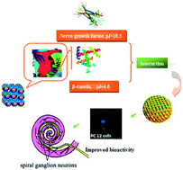
Controlling the specific ionic interactions between drugs and mesophases particles can enable controlled release systems for proteins. Here we present the interaction of basic nerve growth factor (NGF) with highly homogeneous cubosomes based on the glycerol monooleate–water system where β-casein was employed as a stabilizer. Addition of β-casein leads to modification of the internal cubosomal surface to generate highly negative charge. The colloidal structures were characterized with small angle X-ray scattering and visualized using cryogenic transmission electron microscopy. These nanostructured particles are able to sustain the release of hydrophilic NGF since less than 15% of the protein was released over 24 h. Similarly, cationic 1,2-dioleoyl-3-trimethylammonium-propane (DOTAP) immobilized within the mesophase particles provided another specific interactions of the cubosomal surface, from which the release of water soluble bovine serum albumin was retarded. Evaluation of NGF entrapped colloidal dispersions in PC12 cells led to identification of β-casein stabilized cubosomes that induce more than 140% neurite outgrowth enhancing the activity of NGF. Accordingly, the AUC value for RWM administration of NGF-loaded dispersions in guinea pigs was significantly higher than that administrated with NGF, of which the relative bioavailability of the β-casein stabilized dispersions was increased to 337% as compared with free drug.


Introduction

Recent advances in nanotechnology have resulted in multicompartment lipidic nanocarriers and nanoporous templates with hierarchical architecture for encapsulation of biopharmaceutical involving high-resolution structural studies of self-assembled lyotropic liquid crystalline assemblies that may accommodate therapeutic proteins, peptides, or nucleic acids. Over the last decades, significant efforts have been dedicated to apply such liquid crystalline nanoparticles, particularly in vivo applications including drug delivery and biomedical imaging for the administration of these biomacromolecules, which are generally characterized by a low permeability through biological membranes and an insufficient stability in biological environment.1,2

Glycerol monooleate (GMO) is an attractive nonionic lipid since the self-assembled GMO/water liquid crystalline phases represent a biocompatible, nanostructured lipid membrane medium with tunable hydration, which offers new opportunities for design of biomacromolecules carriers with multicompartment architecture and controlled properties. The varied proteins, including apoferritin, transferring, immunoglobulin and fibrinogen have been entrapped in self-assembly of GMO and water with improved protein stabilization.3,4 However, many self-assembled bulk phases, in particular the GMO/water liquid crystalline phases are highly viscous and difficult to handle.5 Toward application of these self-assembled liquid crystalline, the dispersion of the system is required. Nanoparticles based on lyotropic liquid crystalline phases could be simply dispersed by use of a steric stabilizer. Among them, cubosomes (dispersed inverse bicontinuous cubic phase) and hexosomes (dispersed inverse hexagonal phases) are being developed as potential drug delivery systems due to their improved encapsulation efficiency, stabilization and easily accessibility through biological membranes for biomolecules.6 Although these advantages, there are still major limitations, that is, only very few prolonged release of biomacromolecules was found in in vitro investigation of cubosomes and hexosomes. It was proposed that the load distributes between the particles and the continuous water phase according to its partition coefficient and its release is only limited by diffusion. Peptides and proteins showed a burst release from these systems under sink conditions due to the high surface area and the small size of the particles. Otto et al. developed a water-in-oil microemulsion method for loading the proteins into micellar cubic internal structure, which were favourable as sustained delivery carriers for water-soluble proteins such as bovine serum albumin (BSA) and cytochrome c. This preparation procedure, however, sacrificed the size of cubosomes (approximately 620 nm), which are usually too large to be transferred by biological membranes.7

Previous studies demonstrated that β-casein provides steric stabilization to dispersions of lipid nanostructured particles in GMO as well as phytantriol based cubosomes, indicating their potential in drug delivery.8 β-Casein, one of the four main caseins in bovine milk, possesses a pronounced amphiphilic structure that allows it to be employed as an excellent emulsifier. Under appropriate conditions, it forms stable micelle-like structures in aqueous solutions through non-covalent electrostatic interaction or hydrogen bonding.9 Therefore, our goal was to explore the potential of β-casein stabilized liquid crystalline nanoparticles for sustained delivery of biomacromolecules. As we know, it is the first report that β-casein stabilized GMO nanocrystalline worked as a truly drug delivery systems. As a conclusive proof-of-concept, we evaluated the efficacy of these nanocrystallines to transport nerve growth factor (NGF) into the inner ear after round window membrane administration in guinea pigs, which was a promising therapeutic alternative for the treatment of sensorineural hearing loss.10–12

Results

This study focuses on the exploration of the possibility to use lipid crystalline nanoparticles with biomacromolecules as delivery vehicles. It should be noted that any potential delivery candidate for biomacromolecules, such as therapeutic proteins, would be expected to demonstrate an improved performance in their bioactivity and permeability through biological membranes. Correspondingly, biomolecular loading and release profiles of multicompartment lipid nanocarriers governed their further in vivo outcomes. Questions that require further investigations should consider the role of mesostructure formation and additional components for the protein loading and release, the bioactivity of the entrapped proteins and the mechanism of steric stabilization of lipid crystalline nanoparticles by additional components. Toward the aim of preparing proteocubosomes, we investigated self-assembled nanoparticles of GMO and the proteins, with different stabilizer involved F127 and β-casein.

Characterization of cubosomes

The fraction of stabilizer in the lipid mixture need be carefully balanced to be low enough not to affect the liquid crystalline structure of the particles but still be high enough to efficiently disperse the bulk crystal. Our preliminary study demonstrated that the dispersions of 5% of GMO without high energy physical agitation were prepared using 1.25% of stabilizer, while 0.3% of stabilizer was enough to disperse 2% of GMO. Before investigating the release profiles of proteins from the systems, the properties of nanoparticle dispersions were characterized with regards to particle size, charge, morphology, internal structure and entrapment efficiency.

Various compositions of GMO based nanodispersions were prepared, and their size and size distributions were first characterized by dynamic light scattering (DLS). The particle size distributions for the dispersed liquid crystalline systems used in our experiments are shown in Tables 1 and 2. The particle size distributions were all similar with mean diameter of approximately 100–170 nm, and hence particle size was assumed to not be a significant factor in differences in release profiles of proteins from nanoparticles. Particularly, β-casein alone can disperse bulk liquid crystals and stabilize dispersed nanoparticles, as the evidence that there was no significant difference in particles size and distributions between the particles stabilized by β-casein and F127 at equivalent mass ratio.

Table 1 Effect of the ratio of β-casein[thin space (1/6-em)]:[thin space (1/6-em)]Pluronic® F127 (w/w) on particle size, PDI, zeta potential and entrapment efficiency (EE%) of GMO based colloidal dispersion with 2.0% lipid
Formulation parameter The ratio of β-casein[thin space (1/6-em)]:[thin space (1/6-em)]Pluronic® F127 (w/w)
0[thin space (1/6-em)]:[thin space (1/6-em)]1 1[thin space (1/6-em)]:[thin space (1/6-em)]1 1[thin space (1/6-em)]:[thin space (1/6-em)]0
Plain
Size (nm) 124.6 ± 2.1 102.6 ± 0.5 116.2 ± 3.7
PDI 0.227 ± 0.023 0.226 ± 0.007 0.100 ± 0.003
Zeta potential (mV) −23.4 ± 0.6 −29.2 ± 0.2 −32.8 ± 0.2
[thin space (1/6-em)]
Loaded (BSA, 1.0%)
Size (nm) 136.4 ± 0.7 100.1 ± 0.9 119.6 ± 1.9
PDI 0.245 ± 0.025 0.330 ± 0.070 0.189 ± 0.023
Zeta potential (mV) −21.2 ± 1.0 −32.1 ± 1.1 −43.7 ± 3.0
EE% 72.2 ± 1.4 77.7 ± 6.7 77.8 ± 4.2
[thin space (1/6-em)]
Loaded (LZM, 0.02%)
Size (nm) 137.0 ± 3.8 92.4 ± 0.3 100.9 ± 1.9
PDI 0.230 ± 0.017 0.323 ± 0.049 0.168 ± 0.008
Zeta potential (mV) −22.3 ± 0.8 −31.0 ± 4.4 −37.6 ± 2.8
EE% 91.0 ± 4.5 93.1 ± 2.4 96.7 ± 0.2


Table 2 Effect of entrapped proteins on particle size, PDI, zeta potential and entrapment efficiency of GMO based colloidal dispersion with 5% lipid concentration
Protein (%, w/v) Formulation parameter
Size (nm) PDI Z-Potential (mV) EE%
GMO
0 113.2 ± 0.1 0.250 ± 0.001 −21.7 ± 0.8
BSA (1%) 120.3 ± 0.4 0.244 ± 0.021 −21.5 ± 1.4 71.6 ± 0.7
LZM (0.02%) 126.1 ± 1.3 0.243 ± 0.010 −18.6 ± 0.8 90.1 ± 3.1
[thin space (1/6-em)]
DOTAP modified
0 132.6 ± 0.4 0.221 ± 0.018 28.8 ± 0.4
BSA (1%) 170.4 ± 4.1 0.159 ± 0.036 13.6 ± 0.1 78.3 ± 7.2
LZM (0.02%) 132.8 ± 2.7 0.233 ± 0.011 23.5 ± 0.3 92.5 ± 1.9


The zeta potential of nanodispersions was also monitored following the addition of additional components. Results shown in Table 1 indicated that the addition of β-casein maintained a negative zeta potential of GMO cubosomes dispersed with F127, and the value consistently increased, possibly owing to the negative charge rendered by the existence of –COO of casein on the particle surface when finally dispersed in the systems whose pH (∼6.5) is above the isoelectric point of casein (pI = 4.6). On the contrary, it was noticeable that the zeta-potential of the particles is completely different, that is, the zeta potential values shifted to be positive when DOTAP modified GMO (Table 2).

SAXS measurements were performed to determine the internal structures of these particles, and the obtained scattering profiles were shown in Fig. 1.


image file: c6ra19887b-f1.tif
Fig. 1 SAXS profiles for the dispersions of GMO stabilized by Pluronic® F127 and/or β-casein at 25 °C. (A) 5% lipid dispersed in 1.25% Pluronic® F127 with or without 1% BSA. For DOTAP modified dispersions the ratio between GMO and DOTAP was fixed at 98[thin space (1/6-em)]:[thin space (1/6-em)]2. The addition of BSA or DOTAP resulted in no identifiable Bragg peaks observed in SAXS curves (red and blue curves). Interesting, BSA loaded DOTAP modified dispersion was labeled as swollen Im3m cubic phases (dark cyan curve) as compared with 5% GMO dispersion (black curve) with weak Im3m cubic structures; (B) 2% GMO dispersed in 0.3% stabilizer, the ratio between the investigated β-casein and Pluronic® F127 (w/w) was indicated as C[thin space (1/6-em)]:[thin space (1/6-em)]P 0[thin space (1/6-em)]:[thin space (1/6-em)]1, C[thin space (1/6-em)]:[thin space (1/6-em)]P 0[thin space (1/6-em)]:[thin space (1/6-em)]1; 1[thin space (1/6-em)]:[thin space (1/6-em)]1 or 1[thin space (1/6-em)]:[thin space (1/6-em)]0. Increasing the content of β-casein induced the mesophases changes from emulsion (violet curve) to weak Im3m cubic phases (orange curve) and then Pn3m cubic phases (purple curve).

The identifiable Bragg peaks and further calculated theoretical peak position, lattice parameter and the size of water channel were demonstrated in Table 3. Increasing the fraction of GMO in the systems, the phase sequence is microemulsion phase (L2) followed by a reverse cubic phase in coexistence with excess water (the C[thin space (1/6-em)]:[thin space (1/6-em)]P 0[thin space (1/6-em)]:[thin space (1/6-em)]1 dispersion containing 2% GMO to 5% GMO dispersion). The scattering data indicated that the internal nanostructure of cubosomes in the GMO/water cubosomes system (5% GMO) was Im3m with a lattice parameter of 11.8 nm, which is in good agreement with previous findings.13 The addition of BSA and DOTAP resulted in “dissolved” of the cubic phase, which can be explained by a decrease in the cubic phase interfacial curvature by the larger headgroup of the additives relative to GMO. Although the dispersions entrapped with BSA exhibit no remarkable Bragg peaks in the SAXS scatterplot, it has been shown using cryo-TEM that the formation of mesophase seems like the structures formed by membrane fusion of liposomes during their phase transition to the hexagonal phase, coexist with small and medium vesicles (Fig. 2A).14 We observed that BSA had a significant effect on the internal nanostructures of DOTAP modified GMO based dispersion, inducing changes from L2 phase to the swollen Im3m cubic phase structures with increased lattice parameter of 13.9 nm. As ascertained through cryo-TEM (Fig. 2B), despite the vesicular structures, the BSA loaded DOTAP modified dispersion consisted of cubic or micellar cubic mesophase. Interestingly, SAXS studies of nanodispersions with increasing fraction of β-casein showed a vesicle phase (ratio of β-casein[thin space (1/6-em)]:[thin space (1/6-em)]Pluronic® F127 0[thin space (1/6-em)]:[thin space (1/6-em)]1) followed by a weak Im3m cubic phase (ratio of β-casein[thin space (1/6-em)]:[thin space (1/6-em)]Pluronic® F127 1[thin space (1/6-em)]:[thin space (1/6-em)]1) to Pn3m cubic phase structures (ratio of β-casein[thin space (1/6-em)]:[thin space (1/6-em)]Pluronic® F127 1[thin space (1/6-em)]:[thin space (1/6-em)]0) with a smaller lattice parameter (9.3 nm), which is consistence with previous report. Cryo-TEM revealed the β-casein stabilized dispersion that are extremely uniform shape and internal structure displaying typical discrete colloidal nanoparticles, characterized by cubosomes with approximately 150 nm diameter (Fig. 2C).8

Table 3 The phase structure and lattice parameters obtained from SAXS analysis. The ratio between the investigated β-casein and Pluronic® F127 (w/w) was indicated as C[thin space (1/6-em)]:[thin space (1/6-em)]P 0[thin space (1/6-em)]:[thin space (1/6-em)]1; 1[thin space (1/6-em)]:[thin space (1/6-em)]1 or 1[thin space (1/6-em)]:[thin space (1/6-em)]0. For DOTAP modified dispersions the ratio between GMO and DOTAP was fixed at 98[thin space (1/6-em)]:[thin space (1/6-em)]2. a: lattice parameter; Dw: the size of water channel in cubosomes
Formulation Ratio of peaks Phase structure a (nm) Dw (nm)
C[thin space (1/6-em)]:[thin space (1/6-em)]P 0[thin space (1/6-em)]:[thin space (1/6-em)]1 dispersion L2
C[thin space (1/6-em)]:[thin space (1/6-em)]P 1[thin space (1/6-em)]:[thin space (1/6-em)]1 dispersion 21/2[thin space (1/6-em)]:[thin space (1/6-em)]41/2[thin space (1/6-em)]:[thin space (1/6-em)]61/2 Im3m 12.0 5.2
C[thin space (1/6-em)]:[thin space (1/6-em)]P 1[thin space (1/6-em)]:[thin space (1/6-em)]0 dispersion 21/2[thin space (1/6-em)]:[thin space (1/6-em)]31/2[thin space (1/6-em)]:[thin space (1/6-em)]41/2[thin space (1/6-em)]:[thin space (1/6-em)]61/2 Pn3m 9.3 3.3
5% GMO 21/2[thin space (1/6-em)]:[thin space (1/6-em)]41/2[thin space (1/6-em)]:[thin space (1/6-em)]61/2 Im3m 11.8 5.0
BSA loaded 5% GMO L2
DOTAP modified L2
BSA loaded DOTAP 21/2[thin space (1/6-em)]:[thin space (1/6-em)]41/2[thin space (1/6-em)]:[thin space (1/6-em)]61/2 Im3m 13.9 6.5


image file: c6ra19887b-f2.tif
Fig. 2 Cryo-TEM images from (A) BSA loaded 5% GMO stabilized by 1.25% Pluronic® F127. The formation of transitional mesophase nanoparticles (arrow) coexisted with vesicles was observed; (B) BSA loaded DOTAP modified dispersion. The image showed nanoparticles with lattice structures (arrow) consistent with numerous unilamellar vesicles (dish arrow); (C) 2% GMO stabilized by 0.3% β-casein. Highly homogenous cubosomes (arrow) can be seen when β-casein was employed as stabilizer. The scale bar represents 200 nm.

BSA was usually used to protect NGF within their delivery systems, which occupies the interfaces and shields the therapeutic proteins from contact with hydrophobic surfaces.15 For assessing the EE% of proteins in preparing proteocubosomes, BSA (66 kDa, pI 4.6) was used as a representative medium protein, and LZM (14.7 kDa, pI 10.5) was used a representative protein that is in the approximate molecular weight range of many protein factors, including NGF, and widely available, easily assayed and inexpensive.16–18 As shown in Table 2, the EE% of BSA in 5% GMO and DOTAP modified dispersions were 71.6 ± 0.8% and 78.3 ± 7.2% respectively. The EE% of BSA within β-casein stabilized nanoparticles, prepared by varying the ratio of β-casein and F127, was comparable (Table 1). In the case of LZM, the EE% with all prepared formulations (∼90.0%) was significantly greater (p < 0.05) than those dispersions entrapped BSA (∼70.0%).

In vitro release

In vitro release studies were carried out first to establish the influence of β-casein on the entrapped protein release behaviour. Fig. 3 illustrated the protein release profiles from the dispersions, plotted as a percentage of the cumulative released drug against time. As can be seen in Fig. 3A, the control, free BSA, is released relative rapidly with 25.1 ± 7.5% release within the first 2 h, 61.2 ± 7.1% within 8 h and 80.0 ± 4.1% within 24 h. In comparison to the control, the initial burst release of BSA from 5% GMO were lower (17.1 ± 2.3%) in the first 2 h and 49.9 ± 6.5% release over 24 h. For the DOTAP modified dispersions, the burst release is retarded even more significantly with just over 4.7 ± 3.4% of the loaded BSA and 21.8 ± 3.5% for the first 8 h. In contrast, significantly greater release of BSA from GMO based formulation stabilized with β-casein was observed.
image file: c6ra19887b-f3.tif
Fig. 3 Release profiles of BSA and NGF from the dispersions of GMO stabilized by Pluronic® F127 and/or β-casein. Each data point is expressed as mean ± SD (n = 4).

However, the release of NGF from β-casein stabilized GMO based dispersions into the released medium was very slow (Fig. 3B). Within 24 h, release of NGF occurred to a lesser extent from formulations prepared by β-casein stabilized GMO based dispersions when compared to F127 stabilized dispersions although the release profile was comparable for the first 8 h. Afterwards, release was significantly greater from F127 stabilized dispersions (p < 0.05); whereas, release of NGF from β-casein stabilized GMO based dispersions reached a plateau after 8 h with only 8.3 ± 1.0% and 15.8 ± 1.2% of NGF (for the dispersions containing ratio of β-casein[thin space (1/6-em)]:[thin space (1/6-em)]Pluronic® F127 1[thin space (1/6-em)]:[thin space (1/6-em)]1 and 1[thin space (1/6-em)]:[thin space (1/6-em)]0 respectively) released over the release study period.

Bioactivity assay

The neurite outgrowth of neuron-like PC12 cells, which differentiate line neurons when treated with NGF, was used to assess the bioactivity of the protein loaded formulations. Neurite length that showed signs of differentiation for various time points was shown in Fig. 4A. Fig. 4B displayed representative immunocytochemical images of PC12 cells treated for 6 days with four types of nanoparticles. PC12 cells cultured without NGF had round cell bodies and almost no neuritis, suggesting that the delivery vehicles had no bioactivity (data no shown). In contrast, the cells started to differentiate into neuron-like cells and begin to extend neuritis over time upon stimulation by NGF. After 2 days of culture, there was no statistical difference between the protein loaded nanoparticles and the free NGF. However, after 4 days, there was a statistically significant difference between each group (Fig. 4A). As can be seen, the lengths of the neuritis are significantly longer on DOTAP modified GMO based dispersions compared to the other samples. On day 6, the average neurite length of β-casein stabilized GMO based dispersions was 2.4 and 1.5 times longer than that of the free drugs, for 1[thin space (1/6-em)]:[thin space (1/6-em)]0 and 1[thin space (1/6-em)]:[thin space (1/6-em)]1 β-casein[thin space (1/6-em)]:[thin space (1/6-em)]F127 groups respectively, indicating that the delivery vehicles most effectively maintains and potentiates NGF bioactivity. In addition, significant neuronal outgrowth was also observed in the other test GMO based dispersions in comparison to NGF treatment.
image file: c6ra19887b-f4.tif
Fig. 4 PC12 cell differentiation stimulated by 10 ng mL−1 of NGF. (A) Neurite length was evaluated after the treatment of GMO based dispersions over 6 days; (B) immunofluorescence images showed neuronal outgrowth in PC12 cells at day 6. The PC12 cells cultured in the presence of NGF entrapped fully β-casein stabilized GMO dispersion generated an extensive network of neuritis. *P < 0.05, **P < 0.01 when compared with free NGF.

In vivo pharmacokinetics study

Fig. 5 presents the NGF concentrations in cochlear fluid after GMO based dispersions administered onto round window membrane in guinea pigs, as compared with a NGF solution. The major pharmacokinetic parameters obtained were listed in Table 4, including Cmax, Tmax, and AUC0–24 h. The pharmacokinetics curve demonstrated rapid uptake of NGF with a peak concentration of 1811.7 ± 1032.3 pg mL−1 occurring at the 2 h time point for free drug group. After 2 h there was a dramatic decline in NGF concentrations so that there was little drug the cochlear fluid of these animals by 6 h. By contrast, the RWM administration of GMO based dispersions results in maximal NGF level of 9114.4 ± 6869.4, 4979.5 ± 3496.3 and 4422.4 ± 3476.0 pg mL−1 at 2 h post-treatment for the β-casein stabilized, DOTAP modified and F127 stabilized dispersions respectively. Furthermore, the drug distribution into the cochlear fluid, administered using the GMO based dispersions, exhibited a higher level in all time frames compared with that of the free drug, although disappeared rapidly. As a result of it, the AUC value for RWM administration of NGF-loaded dispersions was significantly.
image file: c6ra19887b-f5.tif
Fig. 5 Cochlear concentration–time profiles of NGF after RWM administration. Each data point is expressed as mean ± SD (n = 5).
Table 4 Pharmacokinetic parameters after RWM administration of NGF or NGF-loaded lipid crystalline nanoparticles in guinea pigs. Values are expressed as the mean ± SD (n = 5)
  5% GMO DOTAP modified Casein stabilized Free NGF
Cmax (pg mL−1) 4422.4 ± 3476.0 4979.5 ± 3496.3 9114.4 ± 6869.4 1811.7 ± 1032.3
Tmax (h) 2 h 2 h 2 h 2 h
t1/2 (h) 8.938 ± 3.555 3.431 ± 0.943 16.435 ± 12.021 6.336 ± 3.994
AUC0–24 (pg mL−1 h−1) 13[thin space (1/6-em)]888.8 ± 9231.6 17[thin space (1/6-em)]347 ± 13[thin space (1/6-em)]210.85 31[thin space (1/6-em)]581 ± 25[thin space (1/6-em)]133.5 9360.2 ± 3495.3


Discussion

It has long been known that the bicontinuous cubic protein/lipid assemblies can organize space within the cell, which stimulated the research on meso-porous particles of sophisticated shapes and channel organizations.19,20 While the techniques for establishing entrapped proteins in cubosomes have been well recognized, the cubosomes with a favorable protein release property responsible for resisting dilution upon sink condition are yet in development. In the present work, we offer a possibility for the creation of self-assembled protein loaded lipid nanostructure with proposed release and active functionality. Normally, the release of proteins from cubosomes could be controlled by various factors such as efficient protein entrapment, meso-structure and the interaction of the entrapped molecules with the amphiphilic interface.

EE% of proteins in the lipid nanocarriers

The efficient protein loading in the multicompartment lipid nanocarriers could be proposed toward application in amphiphile nanoarchitectonics. Proteins can have lipophilic domains even if they are water-soluble. Thus, there are three different ways in which hydrophilic proteins can reside in the cubosomes. It can be located in the water channel of organization, partly be arranged in the lipophilic compartment and even be encapsulated in cubosomal pockets of the supramolecular architecture.1 The incorporation of BSA in lipid cubic phases might be hampered as the size of BSA (length ∼ 14.0 nm; diameter ∼ 3.8 nm) that is larger compared with the aqueous nanochannel size of our investigated GMO based vehicles (3.3–6.5 nm). Nevertheless, BSA has binding sites for GMO as the evidence that the interaction between them eventually modified the structure of cubic order, inducing the transformation to other lyotropic phase states. On the contrary, the uploaded LZM could be expected to be more entrapped in nanoconfined state in the lipid assemblies due to its small molecular size (length ∼ 4.5 nm; diameter ∼ 3.0 nm). In general, the release of BSA from GMO based nanocarriers was faster than that of NGF from corresponding test systems except DOTAP modified dispersion (Fig. 3). Particularly, for the 5% GMO dispersions, approximately 40% of the loaded BSA was released in the first 8 h and 50% of the drug was released in one day. Whereas the release of NGF from above system showed much slower rate since only 12% of NGF was released in first 8 h and 36% of NGF was released over one day. This difference can be attributed to the difference in between BSA (∼70%) and NGF (∼assumed 90%) in the test systems.

Meso-structure

It is possible to closely relate the mesophase behavior and the drug release since the release of guest molecules from liquid crystalline phase is found to be a diffusion controlled process regulated by both size of the aqueous channels and the symmetry of the mesophase.21 Normally, enlargement of the aqueous channel size in self-assembled GMO based cubic nanostructures facilitated the release of hydrophilic molecules; the release of encapsulated drugs correlates the diffusion rate of those from mesophase follows: Dcubic > Dhexagonal > Dmicellar cubic.22,23 In our investigation, the release of BSA from each β-casein stabilized system (distinguished as cubosomes) was faster than that from 5% GMO dispersions (vesicles), which was in accordance with previously reports. A possible explanation could be that the difference of internal water compartments between cubosomes and vesicles influenced on the diffusion and release of the proteins. The cubosomes, with either double diamond, primitive or gyroid geometry, consist of a continuous bilayer, mapped over an infinite periodic minimal surface, separating two nonconnected solvent channels, one of which is connected with the outer water compartment.5 While the vesicles consist of internal water compartments surrounded by bilayer of amphiphiles, thus controlling the diffusion of proteins located in water core since hydrophilic molecules need to cross a large number of lipophilic domains before being released to the external aqueous phase. However, as can be observed, the release of BSA from DOTAP modified dispersion (Im3m) was much slower in the initial period than the other systems, which was supposed to demonstrate a fast release profile, indicating the specific interaction between the DOTAP and BSA. In the case of NGF, the drugs showed much slower release from β-casein stabilized cubosomes as compared from 5% GMO and DOTAP modified dispersions, that is opposed to the controlling the release rate of target molecules by size of the aqueous channels and the symmetry of the mesophases, suggesting the specific interaction between proteins and mesophases plays a critical role in the release of encapsulated molecules.

Specific interaction between proteins and mesophases

Specific interaction of proteins with mesophases, such as electrostatic interactions between BSA or NGF and the mesophase, were assumed possible to control the diffusion and thus the release of proteins. Specific ionic interactions between the mesophases and the entrapped drugs provide a straightforward tool to control the transport properties of the matrix. The uncharged GMO were doped by a negatively charged distearoylphosphatidylglycerol (DSPG) was found to provide necessary interaction with a model cationic drug, timolol maleate, thus allowed the suppression the release from the liquid crystalline matrix over a period of several days.24 Along the line, Amar et al. developed a liquid crystalline matrix containing the mixture of GMO and cationic oleyl amine, which demonstrated strong ionic interactions with DNA, quenching their release in excess water.25 In our investigation ionic lipid (DOTAP) or stabilizer (β-casein) immobilized within the mesophase allowed for the specific ionic interactions with proteins. The low solubility of DOTAP ensures themselves insert the lipid domain of GMO, providing the cationic head in the aqueous domains, which allows for ionic interactions with ionic proteins. Negative charged BSA (pI 4.7) was assumed to interact with DOTAP through coulombic forces while positive charges bearing NGF (pI 10.5) was expected to be repelled from the interfaces of mesophases. Fig. 3 clearly demonstrated a slower release of BSA than that of NGF from DOTAP modified dispersions in the first 8 h (21.8% vs 45.5%).

Conversely, because the pI value of β-casein is 4.6, it arranged in the interfacial region and eventually stretched –COO of casein outside the lipid bilayers, providing negative charges to the interfacial region. Importantly, although β-casein is relatively hydrophilic, it can be strongly absorbed on the molecules of GMO as less free β-casein was found in external aqueous phase (<2%) and even less of them were leaked outside the mesophases over 24 h (<5%) based on our previously study. As a result of it, the release profiles of proteins from β-casein stabilized dispersions was the opposite with those from DOTAP modified systems. That is, the charge repelling force between β-casein and BSA led to a faster release from β-casein stabilized dispersions than from the dispersions without β-casein (Fig. 3A). In the case of NGF, the strong multivalent ionic interaction between drugs and β-casein, allowing higher affinity to the interfacial region of mesophases and hence retarded their release (Fig. 3B).

Regarding the bioactivity of NGF encapsulated in the investigated GMO based dispersion, the results demonstrated that the bioactivity of the protein was well maintained or even enhanced compared to the free drugs. There are a few possible non-exclusive mechanisms to explain the enhanced bioactivity of NGF by liquid crystalline nanoparticles: firstly, the protein transfer from aqueous surroundings into the lipid membrane assemblies to produce protein loaded cubic lipid nanoparticles, shielding the proteins from degradation; secondly, the binding of NGF and β-casein further keep the proteins from contact with hydrophobic surfaces directly, then increased the half life of NGF; thirdly, aqueous nano-channels of bicontinuous cubic mesophases for NGF confinement provided a mini-environment to “condense” proteins in optimal spatially manner; last but not least, the accommodation of NGF in highly hydrated cubic mesophases resists upon dilution, allowing above merits to display in vivo.

Experimental

Materials

Glyceryl monooleate (GMO) was a gift from Danisco Ingredients (Denmark) and was used as received. Pluronic F127, with an average molecular weight of 12[thin space (1/6-em)]500, was purchased from BASF (Ludwigshafen, Germany). β-Casein, lysozyme (LZM) and Rhodamine B isothiocyanate (RBITC) were purchased from Sigma-Aldrich (St. Louis, MO, United States). 1,2-Dioleoyl-3-trimethylammonium-propane (DOTAP) was purchased from Invitrogen Life Technologies Corporation (Grand Island, NY, United States). Propylene glycol (PG) and chloroform were purchased from Yuwang Industrial Co. Ltd (Yucheng, China). Recombinant human β-NGF and 2.5S mouse NGF were purchased from Peprotech (Rocky Hill, NJ, USA) and R&D systems (Minneapolis, MN, USA) respectively. Bovine serum albumin (BSA) was purchased from Biosharp (Hefei, China).

Sample preparation

β-Casein was dissolved in water and adjusted to pH 7.0 with 1.0 M NaOH, which was employed as stock solution (8 mg mL−1). The preparation of GMO based nanodispersions were obtained by the method of hydration of a dry lipid film followed by physical agitation.26,27 Briefly, GMO were dissolved in chloroform, and then organic phase was subsequently evaporated under vacuum at 50 °C overnight. When required, Pluronic® F127 were also weighed and mixed with GMO in chloroform. The lipid film was dissolved in propylene glycol (PG), and then emulsified by stabilizer solution containing β-casein by dropwise addition at 55 °C under constant stirring. For preparing drug-loaded nanoparticles, the drugs involved BSA, LZM or NGF (1%, 0.02% or 0.002% with respect to the total dispersions respectively) were incorporated into the nanoparticles by mixing each drug with the dispersed nanoparticles directly. Except bioactivity of NGF assay, recombinant human β-NGF was employed. The final concentration of blank dispersions was 2% of GMO, 0.3% stabilizer, 5.6% PG and 91.1% of water. Varying ratio of β-casein[thin space (1/6-em)]:[thin space (1/6-em)]Pluronic® F127 (1[thin space (1/6-em)]:[thin space (1/6-em)]0, 1[thin space (1/6-em)]:[thin space (1/6-em)]1, 0[thin space (1/6-em)]:[thin space (1/6-em)]1 w/w) were analyzed to study the influence of β-casein on the profiles of drug releases.

For comparison, lyotropic liquid crystalline phase dispersions were prepared at a concentration of 5% of GMO, with 1.25% of steric stabilizer F127 since our previous study demonstrated colloidal dispersion with higher content of GMO facilitates BSA to be carried upon sink condition. To circumvent the possible potent of cationic modified nanoparticles, DOTAP modified dispersions were further compared in vitro and in vivo. Cationic colloidal nanodispersions were also prepared as described above except for cationic lipid mixture (2.0% DOTAP of lipid) were co-solubilizing with F127 in chloroform.28

Characterizations of colloidal dispersions

The mean particles sizes and zeta potentials of GMO based colloidal dispersions were determined using a Malvern Instruments Zetasizer Nano ZS (Malvern, UK) based on photon correlation spectroscopy. Measurements were carried out at 25 °C in triplicate.

Small angle X-ray scattering (SAXS) with synchrotron radiation (Beijing Synchrotron SAXS beamline, China) were employed to determine internal structure in the nanostructured dispersions. Samples were exposed to the 12 keV X-ray beam with a typical flux of approximately 1013 photons per s and diffraction patterns were recorded using a Dectris-Pilatus 1.85 M detector. A silver behenate standard was used to calibrate the reciprocal space vector for analysis. Each sample was exposed for 300 s to obtain the scattering image and the data were further analyzed using fit2D software package. The lattice parameters (a) of the mesophases were determined using image file: c6ra19887b-t1.tif from linear fits of the plots of image file: c6ra19887b-t2.tif versus image file: c6ra19887b-t3.tif. Here, q is the measured peak position, and h, k, and l are the Miller indices. In addition, the size of water channel (Dw) in cubosomes can be estimated the determination of the lattice parameters (a) and the lipid bilayer thickness L (3.30 nm): Dw = 0.707aL.13

The inner phase architecture of colloidal dispersion is also investigated by Cryogenic-Transmission Electron Microscopy (cryo-TEM) analysis. Samples for cryo-TEM studies were examined using Gatan 626 cryoholder (Gatan, Pleasanton, CA, USA) and Tecnai 20 Transmission Electron Microscope (FEI, Eindhoven, The Netherlands) equipped with a Gatan UltraScan 894 CCD digital camera. For the preparation of the sample, a droplet of colloidal dispersion was placed on a perforated carbon film-coated copper grid, blotted with filter paper, and plunged into liquid ethane at its freezing point. Then the specimens were transferred the cooling holder, and observed at 120 kV acceleration voltage in TEM at about 90–100 K.

The entrapment efficiency (EE%) of proteins in colloidal dispersion was carried out with ultrafiltration tubes (Millipore Amicon® ultra, using MWCO 1000 kDa and MWCO 300 kDa membrane for BSA and LZM respectively). Preliminary studies conducted with known concentrations of drug showed that less specific protein bind was observed with the membrane under the optimal experimental condition (lower than 5%). In each experiment, 500 μL of sample was transferred to the upper chamber of a centrifuge tube fitted with an ultrafilter, and then centrifuged at 6000 rpm for 20 min. The non-entrapped drug or free drug form in solution leaked outside the sub-tubes, making it possible to determine each concentration in solution, and thus allowed the estimation of the amount of drug entrapped into colloidal dispersions. For the determination of BSA only coexisting with β-casein, RBITC was covalently coupled to BSA as reported previously.29 Then the BSA content was analyzed quantitatively using a 96-well plate for detection of fluorescence assay by a microplate absorbance reader with excitation and emission fluorescence at 540 nm and 625 nm, respectively. Additionally, the concentration of BSA was determined by BCA mini-assay kit (BCA, Beyotime Institute of Biotechnology, Shanghai, China). To determine the content of lysozyme, Micrococcus luteus (lysodeikticus) (Worthington Biochemical Corporation, Lakewood, NJ, USA), a bacterium sensitive to lysozyme and lyses rapidly when exposed to the enzyme, was employed by determining the solution turbidimetric rate as described by Shugar.30

In vitro release study

The release profile of BSA and NGF was characterized by Franz diffusion cells respectively. Briefly, 500 μL of cubosomes loading RBITC labeled BSA or 150 μL of cubosomes loading NGF were applied onto the donor chamber of the diffusion cells, and the receptor chamber was filled with 7.5 mL PBS. A Biotech membrane (Spectrumlabs, USA, M. W. 1000k) was used to separate the donor and receptor chambers (1.57 cm2 as effective area for diffusion). The donor chamber was then sealed with Parafilm. The solution containing BSA or NGF was employed as controls respectively. The experiments were performed under constant stirring in the receptor chamber at 37 ± 1 °C maintained by a circulating water-bath. At predetermined time intervals, 100 μL of samples were withdrawn from the receptor chamber for assay, and an equal volume of fresh receptor solution was immediately added back to the chamber to maintain a constant volume in the receptor. At the end of experiments, the donor chamber was rinsed with PBS and the rinsing solution was collected to determine the amount of residual drug in the donor chamber. All release experiments were carried out in at least quadruplicate. The content of BSA and NGF in samples were determined by fluorescent/BCA and ELISA assay (Mini Human β-NGF ELISA Development Kits, Peprotech®, Lot# 1211060-M) respectively.

Bioactivity of NGF in vitro

To confirm the bioactivity of NGF after encapsulating into cubosomes, PC12 cells (ATCC Distributor Beijing Zhongyuan Ltd, Beijing, China) were employed as an evaluating system in vitro. PC12 cells differentiate into a neuronal phenotype by extending neurites in response to bioactive NGF.31,32 PC12 cells were cultured in high-glucose Dulbecco's modified eagle's medium (DMEM) containing 10% horse serum and 5% fetal bovine serum. The cell line was kept in a 5% CO2 humidified atmosphere at 37 °C.

For bioactivity analysis, the cells were seeded at a density of 1.0 × 10⁴ cells per well in 2.0 mL culture medium on 12-well culture plate. Loaded colloidal dispersions were added to PC12 cells and then were incubated at 37 °C with free NGF as control. All the treatments were conducted at the same level (10 ng mL−1). Every second day, cell morphology was evaluated under inverted light microscopy. Neurite outgrowth of PC12 cells was measured in a time dependent manner.

After 6 days of treatment by NGF, the PC12 cells were fixed with 4% paraformaldehyde and then washed twice in PBS for 10 min at room temperature. After fixation, the cells were washed three times with PBS and then permeabilized with 0.1% (v/v) Triton X-100 in PBS. Then the samples were incubated in blocking buffer (2% BSA in PBS) for 1 hour. To determine neuronal morphology, a neuronal specific βIII tubulin monoclonal antibody (Hejun Institute of Biotechnology, Beijing, China) was used at 1[thin space (1/6-em)]:[thin space (1/6-em)]100 dilution in blocking buffer. Following overnight incubation with primary antibody at 4 °C, coverslips were washed twice with PBS and secondary antibody FITC labeled Donkey Anti-Mouse IgG (Hejun Institute of Biotechnology, Beijing, China) was used for 2 h at room temperature. Coverslips were washed triple in PBS and incubated with Hoechst-PBS (1 μg mL−1, purchased from Sigma-Aldrich, St. Louis, MO, United States) for 5 min. After washing in PBS, coverslips were mounted on slided with Vectashield and store at 4 °C for image analysis. Imaging was done using an inverted fluorescent microscope (Olympus IX81, Mason Technologies, Dublin, Ireland).33

In vivo pharmacokinetics study

All experimental protocol was carried out in accordance with the Guide to the Care and Use of Experimental Animals (Chinese Council on Animal care) and approved by the Shenyang Pharmaceutical University Committee. One hundred healthy guinea pigs (weighing between 300 g and 400 g, N = 5 per group, per time point) without sex limitation were used. Animals were anaesthetized with an intramuscular injection of ketamine (60 mg per kg body weight) and xylazine (4 mg per kg body weight). After anesthesia, an incision was performed to approach the temporal bone in the left ear. Then a hole was drilled through the bulla to expose the round window niche of the cochlea. 20 μL of loaded NGF colloidal dispersions or free drugs with the same dose were then soaked in small pieces of surgical grade gelatin sponge and carefully inserted the round window niche respectively. Finally, the hole in the bulla was sealed with dental cement. The animals were allowed to recover. After dosing, the cochleae were collected at 1 h, 2 h, 6 h, 12 h and 24 h post-administration. The cochlea fluid was collected by a micro-injection with a sharp tip from the top turn of the cochlea. The cochlea fluid volume was measured (about 3–8 μL) and then diluted for ELISA assay.

Statistical analysis

Data represent the mean and standard deviation in each experiment. Statistical analyses were performed using Student's t-test. Results were considered statistically significant if p < 0.05.

Conclusions

We demonstrated that the formation of cubosomes stabilized by β-casein, which enables uncomplicated preparation of submicron sized particles with reduced energy input. A specific ionic interaction plays a critical role in the release of both proteins from the particles. NGF, a basic protein, was released from the β-casein stabilized cubosomes in a sustained manner and maintains or enhances their bioactivity, indicating good accommodation of proteins in the nanocarriers. Accordingly, nanostructure particles are promising candidates as controlled delivery systems for proteins.

Acknowledgements

This research was undertaken in part on the Small-angle X-ray scattering beamline at the Beijing Synchrotron, Beijing, China. We thank Dr Guang Mo and Dr Zhihong Li of the Beijing Synchrotron for their assistance in the setup of the SAXS beamline and further data analysis. We thank Dr Xiaojun Huang and Dr Gang Ji for technical support in cryo-TEM sample preparation and data collection and gratefully acknowledge the use or TEM facilities at the Center for Biological Imaging (CBI), Institute of Biophysics, Chinese Academy of Science.

Notes and references

  1. A. Angelova, B. Angelov, R. Mutafchieva, S. Lesieur and P. Couvreur, Acc. Chem. Res., 2011, 44, 147–156 CrossRef CAS PubMed.
  2. L. M. Sebastian Puhla and O. Germershaus, Asian J. Pharm. Sci., 2016, 11, 469–477 CrossRef.
  3. J. Clogston and M. Caffrey, J. Controlled Release, 2005, 107, 97–111 CrossRef CAS PubMed.
  4. A. Angelova, M. Ollivon, A. Campitelli and C. Bourgaux, Langmuir, 2003, 19, 6928–6935 CrossRef CAS.
  5. X. Mulet, C. E. Conn, C. Fong, D. F. Kennedy, M. J. Moghaddam and C. J. Drummond, Acc. Chem. Res., 2013, 46, 1497–1505 CrossRef CAS PubMed.
  6. N. M. Lakshmi, P. R. Yalavarthi, H. C. Vadlamudi, J. Thanniru, G. Yaga and H. K., Curr. Drug Discovery Technol., 2014, 11, 181–188 CrossRef CAS.
  7. A. Chemelli, M. Maurer, R. Geier and O. Glatter, Langmuir, 2012, 28, 16788–16797 CrossRef CAS PubMed.
  8. J. Zhai, L. Waddington, T. J. Wooster, M. I. Aguilar and B. J. Boyd, Langmuir, 2011, 27, 14757–14766 CrossRef CAS PubMed.
  9. M. Bachar, A. Mandelbaum, I. Portnaya, H. Perlstein, S. Even-Chen, Y. Barenholz and D. Danino, J. Controlled Release, 2012, 160, 164–171 CrossRef CAS PubMed.
  10. F. Salvinelli, A. Antonelli, B. Stampachiacchiere, F. Bianchi di Castelbianco, M. Casale, L. D'Ascanio, F. Greco and L. Aloe, Ann. Ist. Super. Sanita, 2003, 39, 189–194 CAS.
  11. R. K. Shepherd, A. Coco and S. B. Epp, Hear. Res., 2008, 242, 100–109 CrossRef CAS PubMed.
  12. L. N. Pettingill, R. T. Richardson, A. K. Wise, S. J. O'Leary and R. K. Shepherd, IEEE Trans. Biomed. Eng., 2007, 54, 1138–1148 CrossRef PubMed.
  13. B. Angelov, A. Angelova, M. Ollivon, C. Bourgaux and A. Campitelli, J. Am. Chem. Soc., 2003, 125, 7188–7189 CrossRef CAS PubMed.
  14. Y. Talmon, J. L. Burns, M. H. Chestnut and D. P. Siegel, J. Electron Microsc. Tech., 1990, 14, 6–12 CrossRef CAS PubMed.
  15. H. Liu, Y. Zhou, S. Chen, M. Bu, J. Xin and S. Li, Asian J. Pharm. Sci., 2013, 8, 269–277 CrossRef.
  16. B. Angelov, A. Angelova, S. K. Filippov, M. Drechsler, P. Stepanek and S. Lesieur, ACS Nano, 2014, 8, 5216–5226 CrossRef CAS PubMed.
  17. B. Song, J. Song, S. Zhang, M. A. Anderson, Y. Ao, C. Y. Yang, T. J. Deming and M. V. Sofroniew, Biomaterials, 2012, 33, 9105–9116 CrossRef CAS PubMed.
  18. T. Freier, H. S. Koh, K. Kazazian and M. S. Shoichet, Biomaterials, 2005, 26, 5872–5878 CrossRef CAS PubMed.
  19. A. Zabara, R. Negrini, O. Onaca-Fischer and R. Mezzenga, Small, 2013, 9, 3602–3609 CrossRef CAS PubMed.
  20. B. Angelov, A. Angelova, B. Papahadjopoulos-Sternberg, S. Lesieur, J. F. Sadoc, M. Ollivon and P. Couvreur, J. Am. Chem. Soc., 2006, 128, 5813–5817 CrossRef CAS PubMed.
  21. A. Zabara and R. Mezzenga, J. Controlled Release, 2014, 188, 31–43 CrossRef CAS PubMed.
  22. B. Tenchov, R. Koynova and G. Rapp, Biophys. J., 1998, 75, 853–866 CrossRef CAS PubMed.
  23. K. W. Lee, T. H. Nguyen, T. Hanley and B. J. Boyd, Int. J. Pharm., 2009, 365, 190–199 CrossRef CAS PubMed.
  24. J. E. K. Lindell, M. Jonstroemer, A. Carlsson and S. Engström, Prog. Colloid Polym. Sci., 1998, 108, 111–118 Search PubMed.
  25. J. A. I. Amar-Yuli, S. Blau, A. Aserin, N. Garti and R. Mezzenga, Soft Matter, 2011, 7, 8162–8168 RSC.
  26. H. Liu, Y. Wang, Q. Wang, Z. Li, Y. Zhou, Y. Zhang and S. Li, J. Biomed. Nanotechnol., 2013, 9, 1784–1793 CrossRef CAS PubMed.
  27. S. B. Rizwan, D. Assmus, A. Boehnke, T. Hanley, B. J. Boyd, T. Rades and S. Hook, Eur. J. Pharm. Biopharm., 2011, 79, 15–22 CrossRef CAS PubMed.
  28. H. Liu, S. Chen, Y. Zhou, X. Che, Z. Bao, S. Li and J. Xu, J. Drug Targeting, 2013, 21, 846–854 CrossRef CAS PubMed.
  29. X. Q. Liu, C. L. Zheng and J. B. Zhu, Yaoxue Xuebao, 2011, 46, 115–120 CAS.
  30. D. Shugar, Biochim. Biophys. Acta, 1952, 8, 302–309 CrossRef CAS.
  31. L. Lichvarova, K. Jaskova and L. Lacinova, Gen. Physiol. Biophys., 2012, 31, 473–478 CrossRef CAS PubMed.
  32. S. Lecht, E. Rotfeld, H. Arien-Zakay, R. Tabakman, H. Matzner, R. Yaka, P. I. Lelkes and P. Lazarovici, Int. J. Dev. Neurosci., 2012, 30, 465–469 CrossRef CAS PubMed.
  33. H. Kraskiewicz, B. Breen, T. Sargeant, S. McMahon and A. Pandit, ACS Chem. Neurosci., 2013, 4, 1297–1304 CrossRef CAS PubMed.

This journal is © The Royal Society of Chemistry 2016
Click here to see how this site uses Cookies. View our privacy policy here.