DOI:
10.1039/C6RA19845G
(Paper)
RSC Adv., 2016,
6, 88110-88116
Dispersive solid phase extraction of gold with magnetite-graphene oxide prior to its determination via microwave plasma-atomic emission spectrometry†
Received
5th August 2016
, Accepted 7th September 2016
First published on 8th September 2016
Abstract
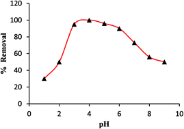
In this paper we demonstrated the quantitative analysis of gold in environmental water samples using microwave plasma atomic emission spectrometry (MP-AES) after dispersive solid phase extraction (DSPE) on magnetite graphene oxide (m-GO). The benefits of the MP-AES are related to cheap analysis costs and improved operational safety with nitrogen used for plasma generation. The detection limit, 3σ m−1, (0.005 μg L−1), inter and intra-day precision (2.6% and 3.2%) and dynamic range (0.02–500 μg L−1) for m-GO coupled MP-AES were compared and matrix effects were evaluated with respect to environmental samples. Recovery of (spiked) analytes ranges from 97.0 to 101%. The developed MP-AES method can offer a comparable or better performance to flame atomic absorption spectrometry and/or inductively coupled plasma optical emission spectrometry with respect to routine analysis for a regulatory program and may be applied as an active method.
Introduction
Gold (Au) is among the noble metals found in nature and at very low content with a wide range of potential applications in biological activity, catalysis, chemical industries and surface technologies due to its specific physico-chemical properties. Due to its low geological abundance and high economic value extraction and preconcentration of this precious metal from complex matrices are necessary in the face of significant demand.1,2 The concentration of gold in environmental samples is rather low, therefore, a separation and preconcentration procedure is required prior to its determination using analytical techniques. Today, the most common sophisticated analytical techniques for elemental analysis in a wide variety of matrices rely on inductively coupled argon plasma, in combination with either optical emission or mass detectors. Therefore, the properties and applicability of argon plasma are well known.3–6 Moreover, the use of micro-plasma which runs on nitrogen gas is typically of interest for many areas of analytical chemistry since the operating cost is significantly lower for instruments that use nitrogen as plasma gas.7–10 The mechanism of microwave plasma atomic emission spectroscopy (MP-AES) is similar to that of the inductively coupled plasma-optical emission spectroscopy (ICP-OES). The major differences between the two types of plasmas are the ways to sustain the plasmas in a gas (argon, nitrogen, helium, etc.). In ICP, plasma is sustained by a radiofrequency generator that generates an oscillating magnetic field around the torch that results in inductive heating of the gas, while microwave plasma is generated by microwave energy. The high temperature in the inductively coupled argon plasma favors the formation of excited ions, which subsequently produces more emission spectral lines that potentially produces more spectral interferences. Using nitrogen plasma, since the temperature is low, most of the elements are in the atomic state resulting in a simpler spectrum than that of argon plasma.
Preconcentration of gold by solid phase extraction (SPE) offers many advantages over classical liquid–liquid extraction such as high enrichment factor, complete recovery, rapid phase separation, less use of organic solvents and possible automation with different detection techniques. Recently, dispersive magnetic solid phase extraction (DSPE) has brought about considerable attention for the preconcentration of trace analytes because of its simplicity, rapidity phase separation, high surface area, possibility for chemical modification, good recovery, low consumption of organic solvents, and the capability of combination with different detection techniques.11–16 Nevertheless, the whole separation process does not require repeated centrifugation or filtration of sample, making sampling comprehensive and faster. It is important that the simplicity of the operation and the flexibility of the working conditions of SPE method are highly dependent on the proper choice of sorbent material. Graphene oxide (GO) with its two dimensional geometrical structure and numerous hydroxyl, epoxy and carboxyl groups was proved an excellent sorbent for SPE.17 The oxygen containing functional groups present at their basal plane and edges can efficiently bind with metal ion through sharing a lone pair of electron. Since, direct application of GO in SPE restricts phase separation due to its small size, high hydrophilicity causes high dispersibility. Thus, the preparation of magnetite GO can overcome this challenge and facilitates its separation from aqueous media after metal ion complexation.
In this article we firstly describe the utility of highly oxygen rich GO18 prepared by a modified method coupled with MP-AES for the extraction and determination of gold from aqueous matrices. Eventually, GO as a sorbent for the preconcentration of gold ions was studied and ligand less dispersive SPE method coupled to MP-AES for the determination of gold was developed. The experimental parameters were optimized in order to reach a high sensitivity and low detection limit of the developed method, and presents the results on the performance of Agilent MP4100.
Experimental
Instrumentation
Nitrogen based Agilent MP4100 microwave plasma atomic emission spectrometer was used to determine the metal ion concentration. The MP 4100 was equipped with a standard glass concentric nebulizer and a double pass cyclonic spray chamber. The pump speed during analysis was kept at 10 rpm and the sample introduction Tygon tube (blue-blue) diameter was 0.88 mm. The analysis steps consisted of 30 seconds rinsing with 1% nitric acid followed by 25 s of sample uptake and then 20 s of torch stabilization time before the reading at pre-selected integration times, whereas for emission measurement of each sample, 5 s read time with 3 replicates were set. Wavelength calibration was made at the beginning of analysis and the integration time was set to 5 s. The analytical signal was evaluated using software MP Expert (Agilent Technologies). The Agilent MP-AES interrogates atomic lines, since it uses relatively cool plasma.
Reagents and solution
All the chemicals used were of analytical reagent grade. Gold standard solution (1000 mg L−1 in 1% HNO3) was purchased from Merck (http://www.merck.de, Darmstadt, Germany). Working solutions were prepared through serial dilutions of stock with triply distilled water (TDW) prior to use. The graphite powder (5–10 μm) was procured from Chengdu organic chemicals (http://www.timesnano.com, China). All glassware was cleaned by soaking in 2% HNO3 and rinsed carefully before use. Calibration solutions for MP-AES were prepared volumetrically by dilution of the Merck calibration solution with 1% nitric acid.
Collection and pretreatment of sample
The sea water samples (A and B) were collected from two different coasts (Pontian district, Malaysia), lake water was collected from university lake (universiti Teknologi Malaysia, Malaysia) and tap water from (Johor bahru, Malaysia). The samples, immediately after collection, were filtered through a cellulose membrane filter (Millipore) of 0.45 μm pore size and acidified to pH 2 with HNO3 and stored in a precleaned polyethylene bottles before analysis.
Synthesis of magnetite graphene oxide (m-GO)
The m-GO was prepared by dual oxidation of graphite powder, following step I oxidation of Hummers method and step II oxidation to increase the population of functional groups on the surface of GO.18 Briefly, a 5.0 g of graphite powder and 2.5 g of NaNO3 were added to 200.0 mL of H2SO4 (95%) under vigorous stirring to avoid agglomeration. Then, 15.0 g of KMnO4 was slowly added over about 2 h under stirring at temperature below 10 °C. The mixture was stirred at 35 °C for 2 h. 350.0 mL of water was slowly added to the paste with continuous stirring and the temperature was kept at 95 °C. The diluted suspension was stirred at 95 °C for 2 h. After the temperature was reduced to 60 °C, 50.0 mL of 30% H2O2 was added into the mixture to eliminate the excess MnO4. After washing with triply distilled water until the pH was 7.0, the mixture was then dried at 80 °C. The obtained grey powdered graphite oxide was exfoliated to GO sheets by bath ultrasonication (500 W, 40 kHz) for 1 h and was subjected to centrifugation at 2415 rcf (g) to remove unexfoliated graphite oxide. The prepared GO sheets were then made magnetic by adding slowly (under ultrasonic agitation) a 300 mL aqueous solution of mixed FeCl3·6H2O (1.86 g) and FeCl2·4H2O (0.96 g) into a GO suspension (0.5 g of GO in 100 mL) at 80 °C, and then 5 mL of 30% ammonia solution was added quickly for the coprecipitation of Fe3O4 nanoparticles on GO sheets. The temperature was raised to 80 °C, and a 5 mL of 25% ammonia solution was added to adjust the pH 10 of the reaction mixture. After being rapidly stirred for 45 min, the resultant black colored Fe3O4 embedded GO suspension was cooled to room temperature and separated from the solution with the aid of external permanent magnet and washed with TDW.
DSPE procedure
A 30.0 mg of m-GO was stirred for 10 min with 25 mL of Au solution (1 μg L−1) maintained at pH 4.0 at 30 ± 0.2 °C. The sorbent was separated from the model solution using an external permanent magnet. The sorbed metal was then eluted with 5 mL of 0.2 M thiourea in 0.2 M HCl shaking for 5 min and subsequently determined by using Agilent MP 4100 MPAES (Melbourne, Australia) with automatic background correction mode at the wavelength of 267.59 nm on nitrogen plasma.
Results and discussion
Choice of material
m-GO was prepared by dual oxidation of graphite powder introducing numerous oxygen containing functional groups18 that chelates to analyte ion. These functional groups results in an increase in the interlayer distance within GO due to presence of intercalated water molecules, thus, leads to high hydrophilicity, high dispersibility and make chelating sites accessible to analyte ion. The step II oxidation increases the population of carboxylic groups, a chelating site, and the relative amount of hydroxyl groups increased with respect to that of epoxy attributed to high dispersibility of solid phase in aqueous media.
Characterization
The surface morphology and incorporation of iron particles were characterized by SEM and TEM studies. The SEM image of m-GO (ESI Fig. S1(A)†) shows a wrinkled surface depicts few layer of GO with crumpled silk wave like structure provides large surface area. The corresponding elemental mapping images (ESI Fig. S1(C, E and F)†) show bright colored spots illustrating homogeneous distribution of matrix carbon, oxygen from functional group, and the chelated Au, respectively. In the TEM image (ESI Fig. S2 (B)†) it was observed that Fe3O4 nano particles are well dispersed onto GO sheets and in the selected area electron diffraction (SAED) pattern (ESI Fig. S2 (D)†), the presence of diffraction dots indicating the crystalline nature of m-GO due to the presence of iron particles. In the FT-IR spectra (ESI Fig. S2†), the characteristics band appeared at 3250 cm−1 corresponds to structural –OH (–COOH and –COH) stretching vibrations of GO. The bands at 1440 cm−1 and 1235 cm−1 were attributes to C
C and C–O stretching vibration. The spectrum also shows the peaks at 2850 and 2900 cm−1 for aliphatic C–H and aromatic C–H stretching vibrations, respectively.
After nitrogen adsorption–desorption isotherm, the multipoint Brunauer–Emmett–Teller (BET) analysis resulted in a good surface area of 703.68 m2 g−1. The pore size distribution analysed using a Barrett–Joyner–Halenda (BJH) isotherm model resulting in a total pore volume of 0.029 cc g−1 and a pore radius of 15.321 Å.
The distribution of acidic sites using pyridine probed FTIR spectra of m-GO after outgassed at 623 K showed that m-GO exhibted absorbance at 1448 and 1544 cm−1 (Fig. ESI 3†). These bands were associated for both Lewis and Bronsted acid sites. An intense peak at 1448 cm−1 indicated the presence of abundance Lewis acid site on m-GO corresponds to GO–OH electron pair acceptor of carboxylic acid which may results to form complex with Au. The adsorption of Au could change the amount of Bronsted acid sites of m-GO due to interaction with Au as a result of dehydrogentaion process leads to increase of Lewis acid sites.
The presence of acidic sites and their strength was also assessed by titration method using Hammett indicator. m-GO showed higher amount of total acid sites. This significant increase in acid sites with acid strength +3.6 ≤ Ho ≤ +6.2 could be attributed to increased number of functional acidic groups.
Optimization of MSPE procedure
The following experimental parameters were optimized: (a) sample pH value (Fig. 1); (b) shaking time (Fig. 2); (c) eluent type and concentration (Fig. 3); (d) effect of interfering ions, respective data are given in the ESI Table S1;† (e) sorption capacity; (f) effect of sample volume and; (g) enrichment factor. The following experimental conditions were found to give best results: (a) a sample pH value of 4.0 for the sorption of Au; (b) a shaking time of 10 min; (c) 5 mL of 0.2 M thiourea in 0.2 M HCl as an eluent; (d) no significant interferences for all the added potentially interfering ions with the recovery >97% in the determination of Au were observed; and (e) a sorption capacity of 192.08 mg g−1 of m-GO. The concentration of Au in all the optimizing procedures was fixed at 1.0 μg L−1 in the initial solution (25 mL) and recovered after DSPE as 5 μg L−1 in the eluent, except batch sorption experiment.
 |
| Fig. 1 Effect of sample pH on the sorption of Au (sorbent amount 30.0 mg; volume 25 mL; Au 1 μg L−1). | |
 |
| Fig. 2 Effect of stirring time on the extraction of Au (sorbent amount 30.0 mg; pH 4.0; volume 25 mL; Au 1 μg L−1). | |
 |
| Fig. 3 Effect of eluent concentration and volume on the recovery of sorbed Au. | |
Sorption capacity. The maximum sorption capacity of m-GO was studied by equilibrating 25.0 mL of Au sample solution (500 mg L−1) with 30.0 mg of m-GO at pH 4.0 in an Erlenmeyer flask stirred for 12 h at 30 ± 0.2 °C. After the sorption process, the m-GO was separated from the model solution using an external permanent magnet. The amount of Au adsorbed (qe) on m-GO was calculated from the difference between the loaded Au concentration Co (mg L−1) and the equilibrium concentration Ce (mg L−1) as follows: qe = (Co − Ce) V/Madsorbent, where V is the volume of the solution, and Madsorbent is the mass of m-GO. The equilibrium sorption capacity of m-Go for Au was found to be 192.08 mg g−1.
Effect of sample volume and enrichment factor. Wrinkeld structure of GO sheets with large surface area and high hydrophilicity favors fast contact with the analyte ions in sample solution to reach equilibrium. In prominence the sorption of analyte ion or sorption equilibrium achieved faster. However, contact time has significant effect on metal ion preconcentration from large sample volume. Using m-GO, quantitative sorption of Au is achieved within 7 min for the sample volume of 25 mL (1.0 μg L−1). For higher sample volume the complete sorption of metal ions (1.0 μg L−1) increases with the increase in contact time and reached upto 10 min for 300 mL. The sorbed analyte ion was subsequently eluted in 5 mL of eluent, thus, gives the enrichment factor of 60. On furthur increasing the sample volume for contact time of 10 min, the recovery of analyte ions gets decreases.
Effect of potentially interfering ions. The potential applicability of the m-GO is prescribed by quantitative sorption and recovery of Au which actually enables its accurate analytical determination. Trace level of Au in real environmental samples inevitably coexists with various matrix ions which may present notable interference in the recovery/determination of Au. To study the tolerance of m-GO towards the extent of recovery of analyte in the presence of various concomitant ions, 25 mL of model solution containing 25 ng of Au and varying amount of interfering ions were stirred under optimum conditions (ESI Table 1†). The sorbed metal was then eluted with 5 mL of 0.2 M thiourea in 0.2 M HCl shaking for 5 min and subsequently determined by using MPAES. The tolerance limit was set as the maximum concentration of the ions causing a deviation of less than ±5% in the recovery of gold. From ESI Table S1,† the result shows that no significant interferences with the recovery/determination of Au for all the added ions upto appreciable molar ratio were observed. The % recovery of Au in presence of these potentially interfering ions was well above 96%. Therefore, under the optimized experimental conditions the procedure showed high selectivity towards the Au in presence of potentially interfering ions.
Reusability test. The re-usability and stability of m-GO for the extraction of 1.0 μg L−1 gold (25 mL) was assessed by performing multiple consecutive separations-desorption steps under the optimized conditions. As shown in Fig. 4, there was no significant change in the performance of the sorbent upto 20 sorption/elution cycles (recoveries higher than 95%), indicating that the fabricated m-GO is a re-usable and stable solid phase sorbent for the extraction/separation of gold.
 |
| Fig. 4 Extraction efficiency of m-GO as number of reuse-cycles increases. | |
Analytical performance of the method
Table 1 illustrates the analytical characteristic data of the developed dispersive solid phase extraction method for the extraction of gold under optimized condition using MP-AES. Under optimum conditions, the analyte calibration curve was obtained after preconcentrating a series of standards (25.0 mL) with increasing analyte ion concentration (0.004–100 μg L−1) were stirred with 30.0 mg of m-GO and the sorbed analyte were recovered with 5 mL of eluting agent (corresponds to 0.02–500 μg L−1) before subjecting it to successive MP-AES determination. The calibration curve was found to have good linearity and correlation coefficient (0.99938). A procedural blank run was performed applying the developed method with 25 mL of aqueous solution prepared by adding suitable buffer (excluding Au) and eluting the same in 5 mL of eluent before serving it to MP-AES. According to the IUPAC definition19 the limit of detection (LOD) and limit of quantification (LOQ) for the MP-AES method evaluated as 3σ m−1 and 10σ m−1 (standard deviation) of the mean blank signal for 20 replicate measurements after preconcentration were found to be 0.005 and 0.015 μg L−1, respectively, where m is the slope of calibration curve. The experimental LOQ obtained after preconcentrating a known amount of analyte ion (N = 3) was found to be 0.02 μg L−1. The method had good precision, relative standard deviation at 1 μgL−1 of gold evaluated by multiple inter-day and intra-day analyses for six successive determinations were found to be 3.8% and 4.5%. As indicated by our analysis, the LOD and LOQ data for MP-AES were comparable to that of ICP-OES.5,20 It is apparent that both the MP-AES and the ICP-OES have lower LOD and LOQ than the flame atomic absorption spectrometry2,13 by more than 10 fold. The small difference in the LOD and LOQ suggested that the performance of MP-AES would be similar to that of ICP-OES. In ICP analysis, water may lead to an increase of the intensity. However, with microwave induce plasma, water may only absorb energy and decrease the emission line intensity. Moreover, both the MP-AES and the ICP-OES utilize the nebulization process to remove the water in the aerosol; the vaporization of water may not be efficient enough so the two instruments share the similar LOD and LOQ.
Table 1 Analytical figures of merits of the proposed DSPE/MP-AES method for Au determination
Analytical parameters |
Au(III) |
Wave length (nm) |
267.59 |
Coefficient of determination (R2) |
0.99937 |
Dynamic range (μg L−1) |
0.02–500 |
Precision (RSD) (n = 5) |
2.6% and 3.2% |
Detection limit (3S/m) (μg L−1) |
0.005 |
Quantification limit (10S/m) (μg L−1) |
0.02 |
Method validation. The accuracy of the developed method was assessed by analysing the gold content in certified reference material MA-1b reference gold ore and by performing recovery experiments after spiking with two levels of known amount of Au into real environmental water samples following optimized DSPE procedure. The Student's t (t-test) value for mean concentration of Au in CRM was found to be less than the critical Student's t-value of 4.3030 at 95% confidence level for N = 3 (Table 2) and the result found was in good agreement with the certified value, indicating absence of bias even in the presence of other elements. Results for recovery experiments after spiking with two levels of known amount of Au are reported in Table 3. The mean percentage recoveries (98.0–102.2%) for the spiked amount with a relative standard deviation (RSD) <5% are in good agreement with the added amount, thus provides the accuracy of the method.
Table 2 Validation of the proposed DSPE/MP-AES method by analysing CRM for Au concentration
CRM |
Matrix composition (μg g−1) |
Certified valuea (μg g−1) |
Foundb (RSD) (μg g−1) |
Calculated Student's t valuec |
95% confidence level. Mean value ± standard deviation; N = 3. At 95% confidence level. |
CRM MA-1b |
Au; 17.0, Si; 24.5, Al; 6.11, Fe; 4.62, Ca; 4.60, K; 4.45, Mg; 2.56, C; 2.44, Na; 1.49, S; 1.17, Ti; 0.38, Ba; 0.18, P; 0.16, Mn; 0.09, Cr; 200.0, Pb; 200.0, Rb; 160.0, Zr; 140.0, Cu; 100.0, Zn; 100.0, Bi; 100.0, Ni; 90.0, Mo; 80.0, Te; 40.0, Co; 30.0, Y; 20.0, W; 15.0, Sc; 13.0, As; 8.0, Ag; 3.9, Sb; 3.0 |
17.0 ± 0.3 |
16.8 ± 0.2 (1.2) |
2.01 |
Table 3 Analytical results for Au preconcentration and recovery in environmental water samples after DSPE/MP-AES determination
Samples |
Amount spiked (μg L−1) |
Amount founda (μg L−1) |
%Recovery (RSD) |
Mean value ± 95% confidence limit; N = 3. Not detected. |
Sea water 1 |
0 |
0.17 ± 0.02 |
−(4.34) |
10 |
10.2 ± 0.35 |
100(1.38) |
20 |
20.1 ± 0.89 |
99.7(1.79) |
Sea water 2 |
0 |
0.29 ± 0.02 |
−(3.29) |
10 |
10.3 ± 0.33 |
100(1.27) |
20 |
20.3 ± 0.76 |
100(1.50) |
Lake water |
0 |
NDb |
— |
10 |
9.73 ± 0.52 |
97.4(2.14) |
20 |
19.9 ± 1.37 |
99.3(2.77) |
Tap water |
0 |
NDb |
— |
10 |
9.80 ± 0.99 |
98.0(4.08) |
20 |
20.1 ± 0.63 |
101(1.25) |
Comparison with other methods
A comparison between the analytical figures of merits of the introduced method and some of the published results for the extraction and trace determination of gold were summarized in Table 4. The developed method has comparable or better results than the other methods. Furthermore, there is no need for centrifugation and filtration step. Also, the developed method is reagent less and does not need organic solvent. The results revealed that the m-GO coupled with MP-AES is a good sample preconcentration technique that can be used for trace analysis of the target analyte in the real samples.
Table 4 Comparision of some previous solid-phase extraction studies for Au
Sorbent |
Mode of operation |
Sorption capacity (mg g−1) |
LOD (μg L−1) |
Technique |
Ref. |
Magnetic GO |
DSPE |
192.08 |
0.02 |
MP-AES |
This work |
Carboxylated pillar[5]arene |
Column |
— |
15.9 |
FAAS |
2 |
CuO microsheets |
Batch |
57.0 |
— |
ICP-OES |
5 |
Polymer monolithic |
Column |
— |
0.006 |
ICP-MS |
6 |
Magnetic functionalized NPs |
Column |
— |
0.16 |
GFAAS |
12 |
Fe3O4–Fir sawdust composite |
DSPE |
188.68 |
0.52 |
FAAS |
13 |
Modified attapulgite |
Column |
66.7 |
0.32 |
ICP-OES |
20 |
Modified carbon nanotubes |
Column |
4.15 |
0.03 |
GFAAS |
21 |
Application of the method
The applicability of the developed method for practical use was checked by analyzing sea water, lake water and tap water samples. The concentrations of Au in all water samples (1 L) were quantified with 95% confidence limit, following optimized DSPE procedure. The results of Au content, along with the recoveries for the spiked samples, are given in Table 3.
Conclusion
The use of nitrogen as plasma gas for MP-AES is an interesting development in analytical sciences, since the running cost can be significantly reduced in comparison to the inductively coupled argon plasma. Here, we successfully used Agilent 4100 MP-AES instrument for the determination of gold in water samples after dispersive solid phase extraction using m-GO as a sorbent. The prepared m-GO not only presents the preconcentration of gold but also simultaneously improves the detection limit of the developed MP-AES method and brings closer to ICP-OES and ICP-MS efficiently.
References
- T. R. Dulski, Trace elemental analysis of metals, Marcel Dekker Inc, 1999, p. 99 Search PubMed.
- S. Zhou, N. Song, S. Liu, D. Chen, Q. Jia and Y. Yang, Separation and preconcentration of gold and palladium ions with a carboxylated pillar[5]arene derived sorbent prior to their determination by flow injection FAAS, Microchim. Acta, 2014, 181, 1551–1556 CrossRef CAS.
- W. Nischkauer, M. Neouze, F. Vanhaecke and A. Limbeck, Extraction and pre-concentration of platinum and palladium from microwave-digested road dust via ion exchanging mesoporous silica microparticles prior to their quantification by quadrupole ICP-MS, Microchim. Acta, 2015, 182, 2369–2376 CrossRef CAS PubMed.
- I. Michalak, K. Chojnacka and K. Marycz, Using ICP-OES and SEM-EDX in biosorption studies, Microchim. Acta, 2011, 172, 65–74 CrossRef CAS PubMed.
- M. M. Rahman, S. B. Khan, H. M. Marwani, A. M. Asiri, K. A. Alamry and A. O. Al-Youbi, Selective determination of gold(III) ion using CuO microsheets as a solid phase adsorbent prior by ICP-OES measurement, Talanta, 2013, 104, 75–82 CrossRef CAS PubMed.
- X. Liu, M. He, B. Chen and B. Hu, Polymer monolithic capillary microextraction on-line coupled with inductively coupled plasma-mass spectrometry for the determination of trace Au and Pd in biological samples, Spectrochim. Acta, Part B, 2014, 101, 254–260 CrossRef CAS.
- Y. Wang, L. A. Baker, E. Helmeczi and I. D. Brindle, Rapid high-performance sample digestion of base metal ores using high-intensity infrared radiation with determination by nitrogen based microwave plasma optical spectrometry, Anal. Chem. Res., 2016, 7, 17–22 CrossRef CAS.
- N. Ozbek and S. Akman, Microwave plasma atomic emission spectrometric determination of Ca, K and Mg in various cheese varieties, Food Chem., 2016, 192, 295–298 CrossRef CAS PubMed.
- N. Ozbek and S. Akman, Method development for the determination of calcium, copper, magnesium, manganese, iron, potassium, phosphorus and zinc in different types of breads by microwave induced plasma-atomic emission spectrometry, Food Chem., 2016, 200, 245–248 CrossRef CAS PubMed.
- Y. Zhao, Z. Li, A. Ross, Z. Huang, W. Chang, K. Ou-yang, Y. Chen and C. Wu, Determination of heavy metals in leather and fur by microwave plasma-atomic emission spectrometry, Spectrochim. Acta, Part B, 2015, 112, 6–9 CrossRef CAS.
- M. Khajeh, S. Laurent and K. Dastafkan, Nanoadsorbents: classification, preparation, and applications (with emphasis on aqueous media), Chem. Rev., 2013, 113, 7728–7768 CrossRef CAS PubMed.
- J. Ye, S. Liu, M. Tian, W. Li, B. Hu, W. Zhou and Q. Jia, Preparation and characterization of magnetic nanoparticles for the on-line determination of gold, palladium, and platinum in mine samples based on flow injection micro-column preconcentration coupled with graphite furnace atomic absorption spectrometry, Talanta, 2014, 118, 231–237 CrossRef CAS PubMed.
- M. Bayat, M. H. Beyki and F. Shemirani, One-step and biogenic synthesis of magnetic Fe3O4–Fir sawdust composite: Application for selective preconcentration and determination of gold ions, J. Ind. Eng. Chem., 2015, 21, 912–919 CrossRef CAS.
- C. Ren, X. Ding, H. Fu, C. Meng, W. Li and H. Yang, Preparation of amino-functionalized CoFe2O4@SiO2 magnetic nanocomposites for potential application in absorbing heavy metal ions, RSC Adv., 2016, 6, 72479–72486 RSC.
- R. Sahraei, K. Hemmati and M. Ghaemy, Adsorptive removal of toxic metals and cationic dyes by magnetic adsorbent based on functionalized graphene oxide from water, RSC Adv., 2016, 6, 72487–72499 RSC.
- A. Islam, H. Ahmad, N. Zaidi and S. Kumar, A graphene oxide decorated with triethylenetetramine-modified magnetite for separation of chromium species prior to their sequential speciation and determination via FAAS, Microchim. Acta, 2016, 183, 289–296 CrossRef CAS.
- R. Sitko, B. Zawisza and E. Malicka, Graphene as a new sorbent in analytical chemistry, Trends Anal. Chem., 2013, 51, 33–41 CrossRef CAS.
- J. H. Kang, T. Kim, J. Choi, J. Park, Y. S. Kim, M. S. Chang, H. Jung, K. T. Park, S. J. Yang and C. R. Park, Hidden Second Oxidation Step of Hummers Method, Chem. Mater., 2015, 28, 756–764 CrossRef.
- G. L. Long and J. D. Winefordner, Limit of detection a closer look at the IUPAC definition, Anal. Chem., 1980, 52, 2242–2249 CrossRef.
- L. Zhanga, Z. Li, Z. Hua and X. Changa, Solid phase extraction of gold(III) on attapulgite modified with triocarbohydrazide prior to its determination in environmental samples by ICP-OES, Spectrochim. Acta, Part A, 2011, 79, 1234–1239 CrossRef PubMed.
- F. H. Moghaddam, M. A. Taher, M. Behzadi and M. Naghizadeh, Modified carbon nanotubes as a sorbent for solid-phase extraction of gold, and its determination by graphite furnace atomic absorption spectrometry, Microchim. Acta, 2015, 182, 2123–2129 CrossRef CAS.
Footnote |
† Electronic supplementary information (ESI) available. See DOI: 10.1039/c6ra19845g |
|
This journal is © The Royal Society of Chemistry 2016 |
Click here to see how this site uses Cookies. View our privacy policy here.