DOI:
10.1039/C6RA19698E
(Paper)
RSC Adv., 2016,
6, 107001-107010
Reinforcement of a new calcium phosphate cement with dopamine-mediated strontium-doped calcium polyphosphate-modified polycaprolactone fibers
Received
4th August 2016
, Accepted 21st October 2016
First published on 26th October 2016
Abstract
To improve some performances of calcium phosphates cement (CPC), such as its mechanical properties, cytocompatibility and bioactivity, in this study, polycaprolactone (PCL) fibers, dopamine (D)/PCL fibers, and strontium-doped calcium polyphosphate (SCPP)/D/PCL fibers were respectively incorporated into Sr-containing calcium phosphate cement (Sr-CPC) for the first time at fiber weight fractions of 0%, 1%, 2% and 5% to develop a novel bone cement. After incubation of the composites in simulated body fluid for 7 days, they were characterized by a three-point bending test for mechanical properties, scanning electron microscopy for morphological observation, a 400 g Gillmore needle with a 1 mm diameter for the setting time, and an MTT test for cytocompatibility. The results indicated that that Sr-CPC reinforced with SCPP/D/PCL fibers had much better mechanical properties and cytocompatibility than pure Sr-CPC. The addition of SCPP/D/PCL fibers into Sr-CPC also dramatically decreased the setting time of cement/SCPP/D/PCL fibers composites. Meanwhile, on the basis of our previous researches, it is expected that this novel bone cement would have a potential efficacy to inhibit aseptic loosening due to the introduction of Sr and SCPP into CPC. The novel SCPP/D/PCL fibers-reinforced Sr-CPC is a promising material for bone substitution.
Introduction
Calcium phosphates cement (CPC) has been developed as a successful material for repairing bone defects due to its easy plasticity, biocompatibility, bone integration and biological activity since first being presented by Brown in 1986.1–3 CPCs have the advantage of self-setting, compared with metal and other bioceramics. They can be arbitrarily shaped and easily sculpted to fill the defect parts according to requirements in the process of an operation.4–6 After CPCs have been implanted in the human body, they self-harden to form calcium-deficient hydroxyapatite similar to the mineral component of bone without any exothermal and toxic reaction, and the surrounding tissues do not produce obvious inflammation.7,8 On this basis, some researchers also developed a novel Sr-containing calcium phosphate cement (Sr-CPC). Compared with CPC, it shows good radiopacity, higher compressive strength, and better degradation performance.9,10 A study reveals that the addition of Sr into CPC improves osseointegration in osteoporotic animals.11 Moreover, Sr-integration into CPC can promote the growth of osteoblasts and significantly attenuate osteoclastic substrate resorption in vitro.12–14
However, some disadvantages, such as long setting time, low strength, and susceptibility to brittle catastrophic fracture, have severely limited the clinic application of CPC.4,15 Moreover, wear particles of CPC might have potential drawbacks, leading to the aseptic loosening of prostheses.16,17 To address these problems and to make CPC the ideal material for bone repair, the researchers have made some corresponding modifications.
The poor mechanical property of CPC is the main reason limiting its application. The addition of hydroxyapatite crystals or ZrO2 nanoparticles can accelerate the nucleation of hydroxyapatite, and then improve its compressive strength and tensile strength.18,19 Nonetheless, the improvement in mechanical properties resulting from the addition of crystals is not obvious. Fibers are widely used to improve the strength and fracture resistance of materials (including CPC). The mechanical properties of CPC have been improved by the addition of fibers, such as polyamide fibers, carbon fibers, polylactic acid fibers and PLGA fibers.20–23 The degradable polymeric fibers are usually incorporated into CPC to reinforce its mechanical properties in order to give the set CPC interconnected macropores or channels suitable for vascular ingrowth when the fibers are dissolved. However, the reinforcement effect of degradable polymeric fibers was limited due to bad interfacial compatibility between polymeric fibers and CPC matrix materials.24,25 Therefore, the surface of the polymeric fibers should be modified to improve the interfacial compatibility between polymeric fibers and CPC matrix materials.
The setting time of CPC is an important parameter that determines the effectiveness of CPC for clinical application. A reduction in setting time can reduce the risk of the displacement and breakup of the bone cement.26 It is well known that 15 min is an appropriate setting time suitable for repairing a bone defect. To overcome the disadvantage of the long setting time of CPC, some authors suggested that the addition of seed crystals, such as hydroxyapatite and tricalcium phosphate crystals, could promote the setting process. The addition of seed crystals can provide the crystal nucleus for heterogeneous nucleation, and thus increase the speed of crystallization.
The side-effect of wear particles of CPC is another important aspect limiting its clinic application. Some researchers suggested that the phagocytosis of CPC-wear particles by monocytes/macrophages might provide a potent stimulus for the release of a variety of cytokines leading to aseptic loosening.27,28 Therefore, methods to prevent osteolysis resulting from CPC-wear particles were explored by many researchers. Afsaneh S. et al. researched the effect of a bisphosphonates-containing bone cement on wear particle induced-bone resorption. The results revealed that this CPC could inhibit particle migration and partially reduce osteolysis.29 However, these methods have some inherent limitations. Compared with the long existence within a body of implants, the release duration of drugs combined with CPC is short, and therefore the local delivery of drugs in joint replacement is not an effective means of preventing and treating aseptic loosening. Moreover, integrating the drugs into CPC can have a pernicious influence on its performance, such as damaging its mechanical properties and influencing its setting time.30 Therefore, we need to develop a new kind of CPC which possesses the pharmaceutical efficacy of protecting patients against aseptic loosening.
To address all of the above-mentioned disadvantages of CPC, there is an urgent need for a modified-CPC with an appropriate setting time that combines good mechanical properties with potential pharmaceutical efficacy to inhibit bone resorption. In this context, in this study, Sr-CPC was prepared as previously described.9 The degradable polycaprolactone (PCL) fibers were selected as test-fibers to reinforce the mechanical properties of Sr-CPC due to their good biocompatibility and nontoxic degradation products; dopamine (DOPA) was introduced to coat the surface of PCL fibers due to its promotion of combination of different materials,24,31 and then the particles of strontium-doped calcium polyphosphate (SCPP) were effectually immobilized onto it to modify the surface of PCL fibers. Finally, the modified-PCL fibers were incorporated into Sr-CPC to prepare a novel modified-CPC with good performance.
SCPP was prepared by introducing strontium (Sr) into calcium polyphosphate (CPP). In our previous studies, 8% SCPP was proved to have good osteoblast-cytocompatibility.12 We also found that 8% SCPP-particles had a potential efficacy to inhibit bone resorption and promote bone formation by increasing the OPG/RANKL ratio.32 Some researchers have suggested that poly (P) plays a role in osteoblastic differentiation, activation, bone mineralization, and the inhibition of osteoclastic bone resorption.33 The Sr supplied by pure Sr-CPC may be not sufficient, and the addition of SCPP could supply poly (P) and more Sr to evoke a better biological effect. Meanwhile, the modification of biomaterials by DOPA has attracted more and more attention in biomaterial applications for its easy operation, solvent-free, non-toxic and excellent adherent properties.34 DOPA could form surface-adherent films onto a great majority of materials, including metals, polymers, semiconductors and ceramics, through self-polymerization. It could firmly combine SCPP-particles and PCL fibers. Therefore, we infer that the incorporation of PCL fibers onto which SCPP-particles are immobilized by DOPA in CPCs can realize the following three purposes: the first one is to improve the reinforcement effect of PCL fibers for Sr-CPC by immobilizing SCPP-particles onto the DOPA-modified surface of PCL fibers to enhance the interfacial compatibility between polymeric fibers and CPC matrix materials; the second one is to endow this novel CPC with the biological efficacy of inhibiting the osteoclast resorption and ability to resist aseptic loosening by incorporating SCPP into it;32 the third one is to reduce the setting time of CPC through the introduction of SCPP-particles which serve as seed crystals to promote the setting process. Based on a survey of the literature, this is the first study to incorporate DOPA-mediated SCPP-modified PCL fibers into CPCs.
Experimental section
Preparation of SCPP powders and Sr-CPC
β-SCPP powders with an average size of 75 μm were prepared as previously described.35,36 Then the powders were put into a refrigeration ball mill (Retsch MM400) and ground for 30 min at 15 Hz. The SCPP powders was placed in a beaker with 100% ethanol. Then the SCPP suspension was stirred with a stirring rod for 2 minutes. After standing for 5 minutes, the supernatant was collected. Finally, the SCPP supernatant was dried to obtain SCPP powders of <1 μm in size.
The specific preparation process of 10% Sr-CPC is shown below. Dicalcium phosphate dihydrate (DCPD), calcium hydroxide and strontium carbonate were mixed in the desired Ca/P ratio (1.67) and the Sr/(Sr + Ca) ratio (10%). Then the mixtures were ground in a planet mill at a speed of 500 rpm to get Sr-ACP. The solid phase was composed of anhydrous calcium hydrogen phosphate (DCPA) and as-prepared Sr-ACP with a weight ratio of 1![[thin space (1/6-em)]](https://www.rsc.org/images/entities/char_2009.gif) :2. 2% chitosan lactate dissolved in distilled water was employed as the liquid phase.23 The liquid phase and the solid phase were blended in a blending instrument with a liquid/solid ratio of 0.4 mL g−1 for 1 minute. Then the paste was molded in a 3 mm × 4 mm × 25 mm mold and Φ 10 × 2 mm cylindrical mold at 24 °C and 40% humidity for 24 h. Finally, the hardened specimens were taken out of the mold and incubated in simulated body fluid at 37 °C and 100% humidity for 7 days.
:2. 2% chitosan lactate dissolved in distilled water was employed as the liquid phase.23 The liquid phase and the solid phase were blended in a blending instrument with a liquid/solid ratio of 0.4 mL g−1 for 1 minute. Then the paste was molded in a 3 mm × 4 mm × 25 mm mold and Φ 10 × 2 mm cylindrical mold at 24 °C and 40% humidity for 24 h. Finally, the hardened specimens were taken out of the mold and incubated in simulated body fluid at 37 °C and 100% humidity for 7 days.
Characterization of SCPP particles
The diameter of the PCL fibers used in this experiment purchased from Beijing Yongkangleye Science and Technology Development Co. Ltd. (China) is 300–800 nm. In order to adhere SCPP particles tightly onto the PCL fibers, the diameter of the particles should be less than that of the fibers. Therefore, the size distribution of SCPP particles was tested. After SCPP particles were dispersed with ultrasonic oscillation in 100% ethanol, the size distribution of the SCPP particles was detected by a laser particle analyser (Mastersizer 2000, Melvin Instrument Co. Ltd., England). A scanning electron microscope (SEM) (JSM-5900LV, JEOL) was used to detect the microstructures of the SCPP particles.
Preparation of DOPA-mediated SCPP-modified PCL fibers (SCPP/D/PCL fibers) and cement/fiber composites
Three kinds of specimens were prepared. SCPP/PCL fibers were prepared by the following steps. First, DOPA (Sigma Company, USA) was dissolved in 10 mM Tris–HCl buffer solution (pH 8.5) to obtain a 2 mg mL−1 DOPA solution. Second, a certain amount of SCPP particles and deionized water was ground in a planet mill for 40 minutes to obtain a 1 mg mL−1 SCPP particles-suspension liquid. Third, PCL fibers were firstly immersed into excess DOPA solution, and then excessive SCPP suspension liquid was also added with the pH value of the reaction system being adjusted to 8.5. The reaction was carried out at 37 °C for 24 h with continuous shaking. Subsequently, specimens were taken out and rinsed with deionized water to remove physically adsorbed SCPP. In order to confirm successful coating of DOPA on the PCL fibers and successful adherence of SCPP particles to the PCL fibers, SEM was employed to observe the surface morphology of the fibers and X-ray photoelectron spectroscopy (XPS) (Shimadzu Co., Japan) was used to detect the elements only present in DOPA and SCPP. SCPP/D/PCL fibers were cut to a size of 3 mm × 3 mm, and mixed with the 10% Sr-CPC paste mixture to obtain cement/fiber composites. The weight fractions of the fibers were 1%, 2%, 5%. The SCPP/D/PCL/Sr-CPC was added to mold. The SCPP/D/PCL/Sr-CPC in the mold was then stored at room temperature for 24 h. After this time, the hardened composite specimen was removed from the mold and incubated in simulated body fluid at 37 °C for 7 days prior to further characterization. D/PCL/Sr-CPC and PCL/Sr-CPC were prepared by the same method as the controls.
Setting time measurement
The final setting times of the cement specimens were assessed according to international standard ISO 9917-1:2007 for water-based cements. A 400 g Gillmore needle with a 1 mm diameter was applied to test the final setting time of the cement. They were tested when the cylindrical cement was fabricated and incubated at 37 °C and in a 100% humidity environment. The final setting time (St) was recorded as the moment when the needle failed to penetrate or produce flaws on the surface of the cement.
Mechanical testing
A standard 3-point flexural test with a span of 20 mm and a crosshead speed of 1 mm min−1 was performed on the specimens (rectangular cross section) with a width of 4 mm, a height of 3 mm, and a length of 20 mm on an Instron material testing machine (Instron Co., USA). Before testing, the 4 mm × 25 mm sides of the specimens were polished using silicon carbide paper with grits of 1200. Flexural strength is given by: S = 3PmaxL/(2bd2), where Pmax is the maximum load on the load-displacement curve, L is the span, b is the specimen width and d is the specimen thickness. The elastic modulus is given by: E = mL3/[4bd3], where m is the slope of the tangent to the initial straight-line portion of the load-displacement curve (N m−1). The work-of-fracture (toughness) is the area under the load-displacement curve divided by the specimen's cross-sectional area. For cases where the cement/fiber composites deformed extensively without catastrophic fracture, the test was stopped at a maximum crosshead displacement of 2 mm for a consistent calculation of the work-of-fracture values.
Morphology observation of the cross-sections of the cement/fiber composites
A scanning electron microscope (JSM-5900LV, JEOL) was employed to observe the morphology of the fibers and the cross-sections of the specimens after the standard three-point flexure test. Gold was sputter-coated on the specimens ahead of observation.
Proliferation of osteoblasts cultured on cement/fiber composites
Osteoblastic cells (MG63) were utilized in this study, which were purchased from West China Hospital, Sichuan University (China). MG63 are human osteoblastic sarcoma cells and display various characteristics of an osteoblast. A colorimetric 3-[4,5-dimethylthiazol-2-yl]-2,5-diphenyl-tetrazolium bromide (MTT) assay was performed to quantify the effect of bone cement-specimens on MG63 proliferation. The size of the bone cement disk is Φ 10 × 2 mm. All bone cement specimens were sterilized by gamma irradiation and pre-incubated in RPMI-1640 medium for 12 h, prior to seeding the cells. The test samples were placed in a 24-well plate. MG63 cells were then micro-seeded at a density of 5 × 10⁴ cells per mL per sample in 600 μL of cell medium, followed by incubation of the samples in cell medium in a humidified environment at 37 °C for periods of 2, 4 and 6 d. One plate was taken out after a specified time interval, and 50 μL per well of MTT was added and the plate was incubated at 37 °C for 4 h to allow the formation of formazan crystals. Then the MTT solution was sucked out and 500 μL of dimethylsulfoxide was added. The 24-well plates were shaken for 15 min, and then the solution was removed to 96-well plates. The optical density (OD) of each well was obtained using a microplate reader (Model550, Bio Rad Corp.) at a test wavelength of 492 nm. Cells without bone cement specimens were provided as the control in this study.
Morphology of MG63 on cement/fiber composites
Scanning electron microscopy (SEM) was used to observe the morphology of MG63 cultured on the specimens. After culture for 2 days, the samples were fixed in 3% glutaraldehyde in 0.1 M phosphate buffer solution (PBS) at 4 °C overnight, washed twice with PBS, then dehydrated by increasing the concentration of alcohol (30, 50, 75, 80, 90, 95 and 100%). The critical point drying of specimens was undertaken with liquid CO2. Finally, the specimens were sputter-coated with gold, and observed by SEM.
Statistical analysis
SPSS (v19.0) was used to perform statistical analysis. Experimental data was presented as means ± standard deviation (SD). Results were analyzed by one-way ANOVA with a Student's t-test. Statistical significance was set at P < 0.05.
Results and discussion
Characterization of SCPP particles
The microstructures of the SCPP particles obtained were observed by SEM, and the size distribution of SCPP particles was detected by a laser diffraction particle-size analyzer. The sub-micron SCPP particles were used to modify the surface of the PCL fibers. Fig. 1 demonstrates that sub-micron SCPP particles were obtained through grinding in the refrigeration ball mill (marked by the black circle). Fig. 2 shows the dispersion of SCPP particles in deionized water and it indicates the good dispersion state of the SCPP particles in water. SEM images (Fig. 2) also confirmed that the size of the SCPP particles was less than 1 μm and that these SCPP particles were sub-micron particles.
 |
| | Fig. 1 Size distribution of SCPP particles. | |
 |
| | Fig. 2 SEM image of SCPP particles. | |
SEM and XPS analysis of PCL fibers and modified PCL fibers
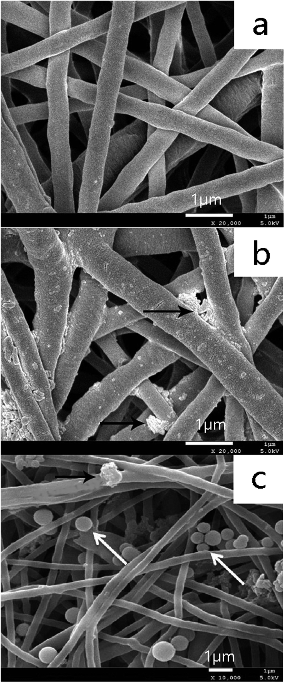
As shown in Fig. 3, the surfaces of the D/PCL fibers were rough (Fig. 3(b)) and the surfaces of PCL fibers were smooth (Fig. 3(b)). The diameters of the fibers modified by DOPA in Fig. 3(b) were bigger than those of the PCL fibers in Fig. 3(a). This also confirmed that dopamine has adhered onto the PCL fibers. As indicated in Fig. 3(c), many SCPP-particles were immobilized on the DOPA-modified PCL fibers.
 |
| | Fig. 3 SEM images of D/PCL fibers and SCPP/D/PCL fibers ((a) PCL fibers ×20000; (b) D/PCL fibers ×20000; (c) SCPP/D/PCL fibers ×10000; white arrows refer to SCPP particles, and black arrows refer to dopamine). | |
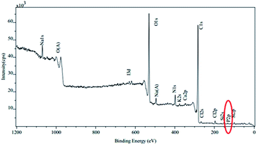
XPS was used to further confirm the successful modification of PCL fibers. As shown in Fig. 4 and 5, after modification by DOPA, a new peak N 1s appeared in the scan spectra of the D/PCL fibers. This suggested that DOPA, the only compound that can introduce nitrogen, reacted with the PCL fibers and formed a stable chemical bond between them. Subsequent to the modification operation of SCPP-particles, the certification of the SCPP-particles being attached onto the D/PCL fibers was supported by the presentation of a new peak P 2p in the scan spectra, which was attributed to the P element.
 |
| | Fig. 4 XPS analysis of D/PCL fibers. | |
 |
| | Fig. 5 XPS analysis of SCPP/D/PCL fibers. | |
Setting time of cement/fiber composites
The final setting times of the mixture of SCPP/D/PCL fibers and Sr-CPC are plotted in Fig. 6. The final setting time was found to decrease with an increase in added SCPP/D/PCL fibers. The reasons for the above results might be related to the following aspects. To begin with, the gap around the fibers could accelerate the infiltration of chitosan solution into the solid phase. Then, the DOPA could enhance the hydrophilicity of the PCL fibers, which could make it easier for the chitosan solution to infiltrate into the solid phase. Finally, the setting process could be promoted by introducing SCPP-particles into Sr-CPC, because SCPP-particles could serve as seed crystals. In a word, the results suggested that the addition of SCPP/D/PCL fibers into Sr-CPC could decrease the setting time of cement/fiber composites dramatically by promoting the setting process, which was helpful to their clinical application.18,37,38 The setting time of bone cement should be about 10 minutes to satisfy the demand of clinical application.37 Other research suggested that the initial setting time of calcium phosphate cement reinforced by fibers was about 2 minutes, which is very short,34 so we thought that the setting time of calcium phosphate cement produced by our lab could be manageable for clinical application.
 |
| | Fig. 6 The setting time of SCPP/D/PCL fibers-reinforced Sr-CPC (* means the difference attained a statistically significant difference compared to Sr-CPC, P < 0.05, n = 5). | |
Mechanical properties of cement/fiber composites
Adding the fiber is an important way to enhance the mechanical properties of bone cement. Fig. 7 shows the effect of different fibers and the fraction of fibers on flexural strength. The flexural strengths of fiber-reinforced Sr-CPC composites were higher than that of the pristine Sr-CPC and the flexural strengths increased with an increase in fiber weight fraction. The flexural strength of SCPP/D/PCL/Sr-CPC sample was the highest, and the flexural strength of SCPP/D/PCL/Sr-CPC with 5% fibers was about 4 times that of the Sr-CPC control without fibers. Fig. 8 shows the effect of fiber type and the fraction of fibers on the elastic modulus. It is obvious that the addition of PCL modified by dopamine or dopamine/SCPP into Sr-CPC could improve the elastic modulus of the cement composites, and SCPP/D/PCL fibers worked better. Fig. 9 shows the effect of fiber type and the fraction of fibers on work of fracture. In general, the work of fracture increased with an increase in fiber fraction and the promotion effect is very obvious. The work of fracture of 5% SCPP/D/PCL/Sr-CPC is the highest. All in all, the mechanical properties of composites could be obviously improved by adding fibers and SCPP/D/PCL fibers achieved the best effect. In addition, adding D/PCL fibers into Sr-CPC exhibited a higher mechanical property compared to those of PCL fibers. The reason for this could be that PCL are hydrophobic. If mixed in aqueous conditions, the hydrophobicity of PCL may hamper its miscibility with the hydrophilic CPCs before self-setting, and dopamine could improve the interfacial compatibility between polymeric fibers and the CPC matrix by means of increasing the hydrophilicity of the PCL fibers.24 Therefore, D/PCL/Sr-CPC obtained better mechanical properties. SCPP-particles introduced onto the surface of PCL fibers can further promote the integration between polymeric fibers and the CPC matrix, which gave SCPP/D/PCL/Sr-CPC the best mechanical properties.
 |
| | Fig. 7 Effect of fiber types on the flexural strength of fiber-reinforced Sr-CPC (values indicated with dissimilar letters are significantly different from each other (P < 0.05), n = 5). | |
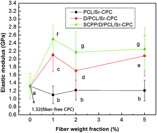 |
| | Fig. 8 Effect of fiber types on the elastic modulus of fiber-reinforced Sr-CPC (values indicated with dissimilar letters are significantly different from each other (P < 0.05), n = 5). | |
 |
| | Fig. 9 Effect of fiber types on the work of fracture of fiber-reinforced Sr-CPC (values indicated with dissimilar letters are significantly different from each other (P < 0.05), n = 5). | |
Morphology observation of cement/fiber composites
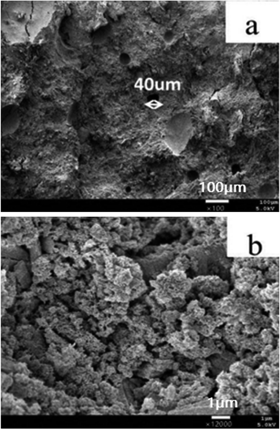
Fig. 10 and 11 show the flexural fracture surface of different cement composites after the mechanical test. A few holes and gaps could be found somewhere in the pure Sr-CPC matrix (see Fig. 10). The hydroxyapatite crystallites in the pure Sr-CPC matrix are arranged in a loose structure (see Fig. 10(b)). This might lead to a significant decrease in mechanical properties and the brittle nature of pure Sr-CPC.
 |
| | Fig. 10 SEM micrographs of the fracture surface of pure Sr-CPC ((a) ×100; (b) ×12000). | |
 |
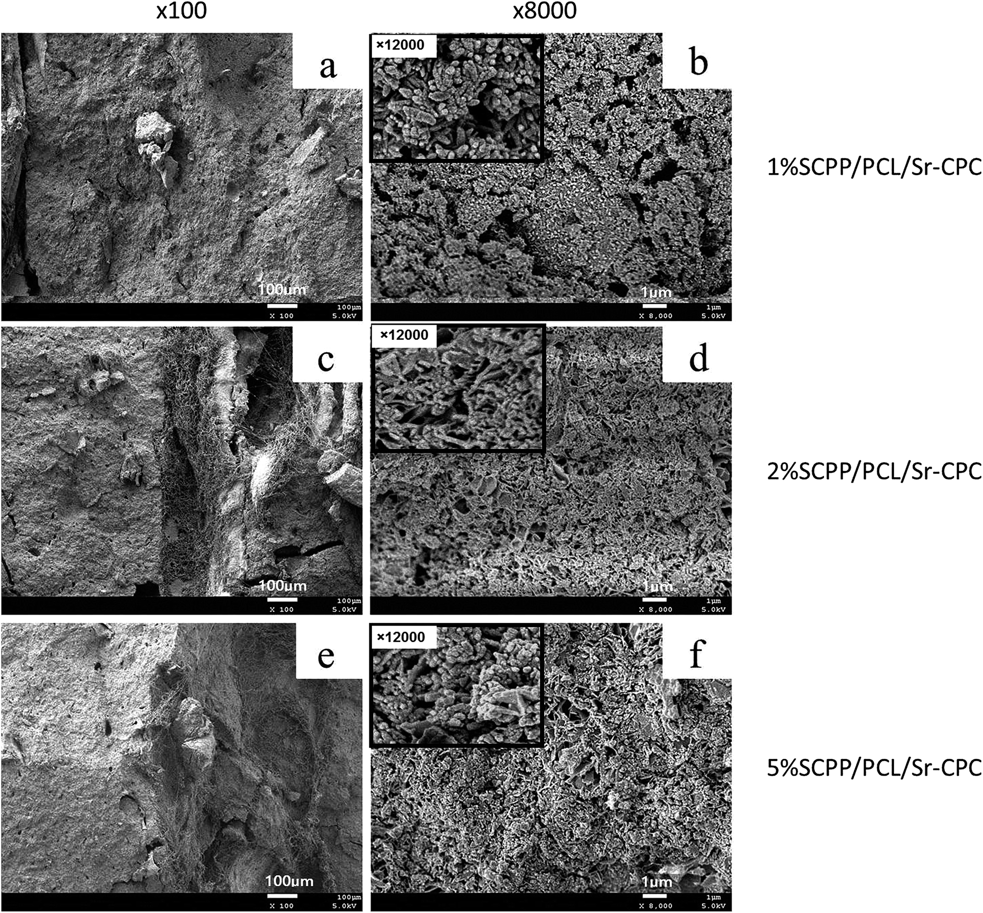
| | Fig. 11 The morphologies of the fractured surface of Sr-CPC reinforced by SCPP/D/PCL fibers (a: 1%SCPP/PCL/Sr-CPC, ×100; b: 1%SCPP/PCL/Sr-CPC, ×8000; c: 2%SCPP/PCL/Sr-CPC, ×100; d: 2%SCPP/PCL/Sr-CPC, ×8000; e: 5%SCPP/PCL/Sr-CPC, ×100; f: 5%SCPP/PCL/Sr-CPC, ×8000). | |
We could deduce that the addition of SCPP/D/PCL fibers could decrease the number of holes and gaps and make the arrangement of the crystallites in the matrix more compact (Fig. 11), comparing Fig. 10 with 11. The addition of SCPP/D/PCL fibers may also increase the amount of hydroxyapatite crystallites formed in the CPC matrix. The increase in the weight fraction of added SCPP/D/PCL fibers could result in a more pronounced effect. This offered significant evidence for the increase in mechanical properties of SCPP/D/PCL/Sr-CPC. This may be attributed to the introduction of SCPP-particles, which served as seed crystals to promote the formation of hydroxyapatite crystallites in the CPC matrix. Moreover, DOPA used in this study can promote the dicalcium phosphate dihydrate (DCPD) conversion into hydroxyapatite after setting for 24 h.31
Fig. 12 shows the fibers on the flexural fracture surface of the Sr-CPC composite cements. As for the SCPP/D/PCL fibers-reinforced Sr-CPC, the surfaces of the pulled-out fibers had numerous remnants of matrix CPC (arrows) attached (Fig. 12(b)), demonstrating that the fibers were relatively well dispersed in and wetted by the CPC. In contrast, as for the pure Sr-CPC, there were almost no CPC pieces adhering to the pulled-out fibers (Fig. 12(c)). Therefore, we could infer that the SCPP/D/PCL fibers-reinforced Sr-CPC has better mechanical properties than the PCL fibers-reinforced Sr-CPC and the D/PCL fibers-reinforced Sr-CPC. This may be due to the following reasons: PCL fibers are hydrophobic materials which cannot integrate well with the CPC cement matrix, and dopamine can improve the interfacial compatibility between polymeric fibers and the CPC matrix by means of increasing the hydrophilicity of the PCL fibers; SCPP-particles introduced onto the surface of PCL fibers can also promote the integration between polymeric fibers and the CPC matrix.
 |
| | Fig. 12 The morphology of PCL fibers on the flexural fracture surface of Sr-CPC composite cements with 5% fiber weight fraction ((a) D/PCL fibers; (b) SCPP/D/PCL fibers; (c) PCL fibers; ×1000) the white arrows indicate the remnants of matrix CPC. | |
Cytocompatibility and bioactivity of cement/fiber composites
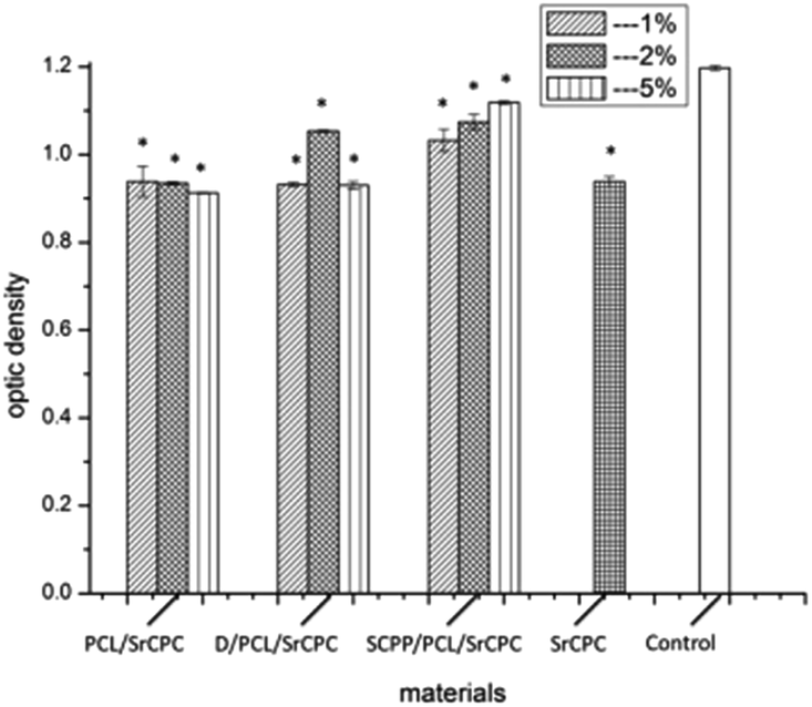
The proliferation of MG63 cultured on different cement/fiber composite-samples on the 3rd and 6th days was determined by MTT assay (Fig. 13 and 14). It was obvious that adding PCL fibers to cement could not promote the growth of MG63. This may be because the addition of PCL fibers could enhance the surface hydrophobicity of cement/fiber composite-samples, which is not good for the adhesion and growth of MG63 on samples. The addition of D/PCL fibers could partially improve the proliferation of MG63 due to the improvement in the surface hydrophilicity of the cement/fiber composites by DOPA. So we concluded that DOPA could promote the proliferation of MG63. Noticeably, the growth of MG63 on SCPP/D/PCL/Sr-CPC samples was always superior to that on other samples in the culture time, which demonstrated that the addition of SCPP/D/PCL fibers could obviously promote the growth of MG63. Moreover, during the proliferation period, in encouraging MG63 proliferation, the role of SCPP/D/PCL/Sr-CPC groups was more and more obvious with an increase in the fiber weight fraction. The reasons for this result may be related to the following aspects. First, as mentioned above, SCPP-particles or DOPA used in this study could promote the formation of hydroxyapatite crystallites in the CPC matrix, and thus the more hydroxyapatite crystallites the CPC matrix contains, the better cytocompatibility the CPC has. Second, the addition of fibers could increase the surface area of cement/fiber composites to enhance cell attachment. Third, the Sr2+ released from CPC or SCPP introduced in this study could promote the growth of osteoblasts.39
 |
| | Fig. 13 The effect of different cement/fiber composite-samples on proliferation of MG63 on 3rd day (* means the difference attained a statistically significant difference compared to control group, p < 0.05, n = 6). | |
 |
| | Fig. 14 The effect of different cement/fiber composite-samples on proliferation of MG63 on the 6th day (* means the difference attained a statistically significant difference compared to control group, p < 0.05, n = 6). | |
The observation results of the morphology of MG63 cultured on cement/fiber composites on the 3rd day were in agreement with the MTT assay. As shown in Fig. 15, few MG63 cells were observed on the surface of pure Sr-CPC. In contrast, a lot of spreading-MG63 cells were observed on the surface of Sr-CPC reinforced by SCPP/D/PCL fibers. In particular, the MG63 on the surface of 5% SCPP/PCL fiber-reinforced Sr-CPC appeared to fully spread and developed a confluent monolayer which nearly reached the continuous cell layer (Fig. 16).
 |
| | Fig. 15 The morphology of MG63 on the surface of pure Sr-CPC on the 3rd day (a: ×200; b: ×500; c: ×100). | |
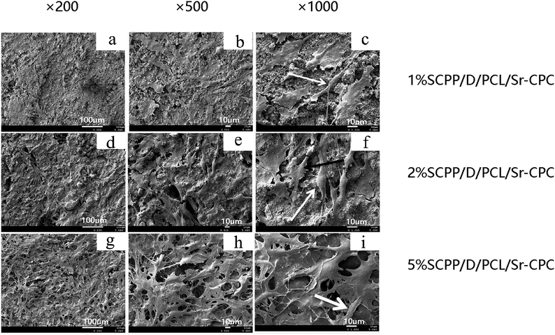 |
| | Fig. 16 The morphology of MG63 on the surface of SCPP/PCL fiber-reinforced Sr-CPC with different fiber weight fractions on the 3rd day (a: 1%SCPP/D/PCL/Sr-CPC, ×200; b: 1%SCPP/D/PCL/Sr-CPC, ×500; c: 1%SCPP/D/PCL/Sr-CPC, ×1000; d: 2%SCPP/D/PCL/Sr-CPC, ×200; e: 2%SCPP/D/PCL/Sr-CPC, ×500; f: 2%SCPP/D/PCL/Sr-CPC, ×1000; g: 5%SCPP/D/PCL/Sr-CPC, ×200; h: 5%SCPP/D/PCL/Sr-CPC, ×500; i: 5%SCPP/D/PCL/Sr-CPC, ×1000;). | |
As is well known, it has been reported that Sr is a bone-seeking element which presents a dual action of improving bone formation and inhibiting bone resorption.40 In our previous study, we also confirmed that SCPP particles can inhibit the expression of TNF-α and RANKL and promote the expression of OPG, and thus has a potential efficacy to inhibit aseptic loosening.32 Based on these researches, we can expect that this novel bone cement developed by our lab also possesses the pharmaceutical efficacy of protecting patients against aseptic loosening. As this Sr/SCPP-containing CPC wears down, SCPP particles can get into the sites around the prosthesis and continue to exist, and therefore play a very important role in the long-term treatment of aseptic loosening due to their potential pharmaceutical efficacy.
Conclusion
To address some disadvantages of CPC, such as poor mechanical properties and long setting time, in this study, PCL fibers, dopamine/PCL fibers and SCPP/D/PCL fibers were respectively incorporated into Sr-CPC at fiber weight fractions of 0%, 1%, 2% and 5% to develop novel bone cements. Our data revealed that Sr-CPC reinforced with SCPP/D/PCL fibers had much better mechanical properties and cytocompatibility compared to pure Sr-CPC. The addition of SCPP/D/PCL fibers into Sr-CPC also dramatically decreased the setting time of cement/SCPP/D/PCL fibers composites. Meanwhile, based on other researchers' and our previous studies, we could infer that this novel bone cement has a potential efficacy to inhibit aseptic loosening. Therefore, we propose that SCPP/PCL fiber-reinforced Sr-CPC represents a potential candidate material for bone substitution, but its efficacy has still to be tested in vivo.
Acknowledgements
The authors also would like to thank the National Natural Science Foundation of China (No. 51503129), the Scientific and Technological Project of Sichuan Province (No. 2012SZ0015), the scientific and technological project of Chengdu (No. 2014-HM01-00207-SF), Sichuan university-Luzhou strategic partnership scientific and technological cooperation projects (No. 2014CDLZ-S27) and China Postdoctoral Science Foundation (No. 2015M570785).
Notes and references
- W. E. Brown, Cem. Res. Prog., 1987, 351–379 Search PubMed.
- D. Knaack, M. E. P. Goad, M. Aiolova, C. Rey, A. Tofighi, P. Chakravarthy and D. D. Lee, J. Biomed. Mater. Res., 1998, 43, 399–409 CrossRef CAS PubMed.
- J. E. Barralet, T. Gaunt, A. J. Wright, I. R. Gibson and J. C. Knowles, J. Biomed. Mater. Res., 2002, 63, 1–9 CrossRef CAS PubMed.
- P. D. Costantino, C. D. Friedman, K. Jones, L. C. Chow and G. A. Sisson, Plast. Reconstr. Surg., 1992, 90, 262–353 Search PubMed.
- L. C. Chow, Calcium Phosphate Cements: Chemistry, Properties, and Applications, MRS Online Proceedings Library Archive, 1999, vol. 599, p. 27, 11 pages, M23-10.1557/PROC-1599-1527 Search PubMed.
- S. Takagi, L. C. Chow, M. Markovic, C. D. Friedman and P. D. Costantino, J. Biomed. Mater. Res., 2001, 58, 36–41 CrossRef CAS PubMed.
- M. L. Shindo, P. D. Costantino, C. D. Friedman and L. C. Chow, Arch. Otolaryngol., Head Neck Surg., 1993, 119, 185–190 CrossRef CAS.
- B. R. Constantz, B. M. Barr, I. C. Ison, M. T. Fulmer, J. Baker, L. McKinney, S. B. Goodman, S. Gunasekaren, D. C. Delaney, J. Ross and R. D. Poser, J. Biomed. Mater. Res., 1998, 43, 451–461 CrossRef CAS PubMed.
- T. Yu, J. Ye and Y. Wang, Acta Biomater., 2009, 5, 2717–2727 CrossRef CAS PubMed.
- D. Guo, K. Xu, X. Zhao and Y. Han, Biomaterials, 2005, 26, 4073–4083 CrossRef CAS PubMed.
- C. C. Wu, C. L. Kuo, F. Y. Fan and K. C. Yang, J. Biomed. Mater. Res., Part A, 2015, 103, 3355–3363 CrossRef CAS PubMed.
- F. Liu, X. Zhang, X. Yu, Y. Xu, T. Feng and D. Ren, J. Mater. Sci.: Mater. Med., 2011, 22, 683–692 CrossRef CAS PubMed.
- S. S. Singh, A. Roy, B. Lee, S. Parekh and P. N. Kumta, Mater. Sci. Eng., C, 2016, 63, 429–438 CrossRef CAS PubMed.
- M. Schumacher, A. S. Wagner, J. Kokesch-Himmelreich, A. Bernhardt, M. Rohnke, S. Wenisch and M. Gelinsky, Acta Biomater., 2016, 37, 184–194 CrossRef CAS PubMed.
- C. D. Friedman, P. D. Costantino, S. Takagi and L. C. Chow, J. Biomed. Mater. Res., 1998, 43, 428–432 CrossRef CAS PubMed.
- A. Grandjean-Laquerriere, O. Tabary, J. Jacquot, D. Richard, P. Frayssinet, M. Guenounou, D. Laurent-Maquin, P. Laquerriere and S. Gangloff, Biomaterials, 2007, 28, 400–404 CrossRef CAS PubMed.
- F. Velard, D. Laurent-Maquin, C. Guillaume, S. Bouthors, E. Jallot, J.-M. Nedelec, A. Belaaouaj and P. Laquerriere, Acta Biomater., 2009, 5, 1708–1715 CrossRef CAS PubMed.
- I. S. Neira, Y. V. Kolen'ko, K. P. Kommareddy, I. Manjubala, M. Yoshimura and F. Guitian, ACS Appl. Mater. Interfaces, 2010, 2, 3276–3284 CAS.
- W. Yu, X. Wang, J. Zhao, Q. Tang, M. Wang and X. Ning, Ceram. Int., 2015, 41, 10600–10606 CrossRef CAS.
- H. H. K. Xu, F. C. Eichmiller and A. A. Giuseppetti, J. Biomed. Mater. Res., 2000, 52, 107–114 CrossRef CAS PubMed.
- X. Wang, J. Ye, Y. Wang and L. Chen, J. Am. Ceram. Soc., 2007, 90, 962–964 CrossRef CAS.
- Y. Zuo, F. Yang, J. G. C. Wolke, Y. Li and J. A. Jansen, Acta Biomater., 2010, 6, 1238–1247 CrossRef CAS PubMed.
- C. Bao, W. Chen, M. D. Weir, W. Thein-Han and H. H. K. Xu, Acta Biomater., 2011, 7, 4037–4044 CrossRef CAS PubMed.
- X. Wang, Z. Gu, B. Jiang, L. Li and X. Yu, Biomater. Sci., 2016, 4, 678–688 RSC.
- Q. Ye, F. Zhou and W. Liu, Chem. Soc. Rev., 2011, 40, 4244–4258 RSC.
- Y. Ueyama, K. Ishikawa, T. Mano, T. Koyama, H. Nagatsuka, T. Matsumura and K. Suzuki, J. Biomed. Mater. Res., 2001, 55, 652–660 CrossRef CAS PubMed.
- E. Buache, F. Velard, E. Bauden, C. Guillaume, E. Jallot, J. M. Nedelec, D. Laurent-Maquin and P. Laquerriere, Acta Biomater., 2012, 8, 3113–3119 CrossRef CAS PubMed.
- E. Gibon, T. Ma, P.-G. Ren, K. Fritton, S. Biswal, Z. Yao, L. Smith and S. B. Goodman, J. Orthop. Res., 2012, 30, 547–553 CrossRef CAS PubMed.
- A. Sabokbar, Y. Fujikawa, D. W. Murray and N. A. Athanasou, Ann. Rheum. Dis., 1998, 57, 614–618 CrossRef CAS PubMed.
- M. P. Ginebra, T. Traykova and J. A. Planell, J. Controlled Release, 2006, 113, 102–110 CrossRef CAS PubMed.
- Z. Liu, S. Qu, X. Zheng, X. Xiong, R. Fu, K. Tang, Z. Zhong and J. Weng, Mater. Sci. Eng., C, 2014, 44, 44–51 CrossRef CAS PubMed.
- H. Chengcheng, L. Li, Y. Xixun, G. Zhipeng and Z. Xu, Biomed. Mater., 2014, 9, 025010 CrossRef PubMed.
- Y. Hacchou, T. Uematsu, O. Ueda, Y. Usui, S. Uematsu, M. Takahashi, T. Uchihashi, Y. Kawazoe, T. Shiba, S. Kurihara, M. Yamaoka and K. Furusawa, J. Dent. Res., 2007, 86, 893–897 CrossRef CAS PubMed.
- J. J. Wilker, Nat. Chem. Biol., 2011, 7, 579–580 CrossRef CAS PubMed.
- Z. Gu, X. Zhang, L. Li, Q. Wang, X. Yu and T. Feng, Mater. Sci. Eng., C, 2013, 33, 274–281 CrossRef CAS PubMed.
- Z. Gu, H. Xie, L. Li, X. Zhang, F. Liu and X. Yu, J. Mater. Sci.: Mater. Med., 2013, 24, 1251–1260 CrossRef CAS PubMed.
- M. Bohner, Eur. Cells Mater., 2010, 20, 3–10 Search PubMed.
- Y. Zhang, D. Wang, F. Wang, S. Jiang and Y. Shu, J. Non-Cryst. Solids, 2015, 426, 164–168 CrossRef CAS.
- Z. Gu, H. Xie, C. Huang, H. Peng, H. Tan, L. Li and X. Yu, RSC Adv., 2014, 4, 2783–2792 RSC.
- W. Zhang, Y. Shen, H. Pan, K. Lin, X. Liu, B. W. Darvell, W. W. Lu, J. Chang, L. Deng, D. Wang and W. Huang, Acta Biomater., 2011, 7, 800–808 CrossRef CAS PubMed.
|
| This journal is © The Royal Society of Chemistry 2016 |
Click here to see how this site uses Cookies. View our privacy policy here.