Nonlinear refraction and absorption activity of dimethylaminostyryl substituted BODIPY dyes

B. Kulyk*a, S. Taboukhatab, H. Akdas-Kiligc, J.-L. Fillautc, Y. Boughalebb and B. Sahraoui*a
aL′UNAM, University of Angers, CNRS UMR 6200, Laboratory MOLTECH-Anjou, 2 bd Lavoisier, 49045 Angers, France. E-mail: bohdan_kulyk@yahoo.com; bouchta.sahraoui@univ-angers.fr
bLaboratory of Bio-Geosciences and Materials Engineering, ENS, University Hassan II of Casablanca, Casablanca, BP 50069, Morocco
cInstitut des Sciences Chimiques de Rennes, UMR CNRS 6226, University of Rennes 1, 263, avenue du General Leclerc, 35042 Rennes, France

Received 27th July 2016 , Accepted 1st September 2016

First published on 1st September 2016


Abstract

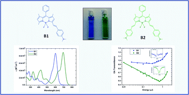
Optical and nonlinear optical properties of difluoroboradiazaindacene (BODIPY) models with attached dimethylaminostyryl substituents were studied. The optical absorption and fluorescence emission of BODIPY compounds were found to be dependent on the number of dimethylaminostyryl substituents. In order to investigate the nonlinear refraction and absorption activity, the Z-scan technique was used employing a laser generating a wavelength at 532 nm with 30 ps pulse duration. The studied dimethylaminostyryl containing BODIPY compounds showed considerable nonlinear refraction and reverse saturable absorption. The BODIPY containing a pair of dimethylaminostyryl substituents demonstrated an increased third order nonlinear optical response mostly due to the extension of its conjugated length.

Introduction

Materials with optimized nonlinear optical (NLO) properties have been the focus of fundamental and applied research in recent years.1 Organic NLO materials have attracted much attention due to their wide scope and ability to maximize a nonlinear response by tailored modification of their molecular structure.2–4 Established NLO organic materials such as the porphyrins, tetrathiafulvalenes and azo-benzenes possess a large number of delocalized π-electrons and hence reasonably high nonlinearities.5–7

During the last decades, the difluoroboradiazaindacene (BODIPY) derivatives have been the subject of intensive studies according to their attractive photophysical properties including relatively sharp absorption and fluorescence emission with high quantum yield.8 The absorption and emission properties of BODIPYs can be tuned conveniently by changing the substitution connected to their core. Thereby, the fluorescence can even be pushed into the near-infrared (NIR) region.9,10 Furthermore, BODIPY dyes are known to be slightly influenced on pH or the polarity of solvent and have excellent thermal and photochemical stability. Due to these remarkable photophysical properties, BODIPYs are widely used fluorescent dyes,11 chemosensors,12 polarity sensitive probe,13 light harvesting systems,14 for laser applications15 and photodynamic therapy16,17 as well as for bio-imaging and optical limiting.18–20 A series of BODIPY-based compounds have been investigated in sense of nonlinear optics, in general, they offer potentially large two-photon absorption cross sections or hyperpolarizabilities.21–31

Herein, we report on two π-conjugated dimethylaminostyryl substituted BODIPY models with favorable tuning of their optical absorption and emission as well as NLO refraction and absorption properties. In fact, it is shown below that BODIPY may possess enhanced nonlinear absorption upon structural modification with dimethylaminostyryl substituent. Up to our knowledge, there is no study on NLO behavior, including nonlinear refraction, multiphoton and excited-state absorption in dimethylaminostyryl substituted BODIPY. Nonlinear optical materials are of interest for several applications including all-optical signal processing, optical limiters, microscopic imaging, etc., therefore, this research may create the perspectives for BODIPYs NLO applications.

Experimental

Materials

BODIPY models have been synthesized in good yield by the established Knoevenagel condensation of 3,5 dimethyl-BODIPY dyes with 4-dimethylaminobenzaldehyde, as previously described by Rurack32 and Akkaya.33 The structures of the resulting difluoroboradiazaindacene chromophores with dimethylaminostyryl substituents (namely B1 and B2) are shown in Fig. 1. The monocondensation was controlled by adjusting the mole ratio and stopping the reaction after few hours, while larger excess of 4-dimethylaminobenzaldehyde and a longer reaction time increased the yield of by-condensed product B2. Experimental details are provided in the ESI.
image file: c6ra19023e-f1.tif
Fig. 1 Structures of the conjugated difluoroboradiazaindacene chromophores with dimethylaminostyryl substituents: (a) B1; (b) B2.

Spectroscopic measurements

The solutions of dimethylaminostyryl BODIPYs in tetrahydrofuran (THF) at 10 μM concentration were prepared. Their absorption spectra were measured by means of Lambda 950 UV/Vis/NIR spectrophotometer (PerkinElmer) in the range 200–1200 nm using the cuvette filled with solvent only on the way of reference beam. Fluorescence emission measurements were carried out by means of spectrofluorometer (Photon Technology International) at room temperature using 514 nm excitation wavelength. For the spectroscopic measurements the 10 mm quartz cuvettes were employed.

NLO measurements

The NLO properties of the B1 and B2 solutions were investigated by means of the Z-scan technique34 using the frequency doubled exit of a 30 ps mode-locked Nd:YVO4 laser with a repetition rate of 10 Hz at 532 nm. In the measurements, the 1 mm quartz cuvettes filled with the solutions at concentration of 0.25 mM were used. The laser beam was focused onto the sample by means of a 20 cm focal length lens, and the spot size of the laser beam at the focus was 17 μm. The Z-scan technique is a simple and effective technique for determining the NLO properties, and it is widely used since it provides simultaneously, by a single measurement, the magnitudes and the signs of the nonlinear absorption and the nonlinear refraction of the investigated sample. In the Z-scan measurements the transmittance of the sample is detected as it moves along the propagation path with the step of 0.25 mm through the focal plane of a focused laser beam. The detection of the transmission has been carried out by appropriate photomultiplier tube, and each experimental point has been the result of a 20 pulse average during the acquisition, performed by the boxcar. In particular two different series of measurements are simultaneously carried out giving access to different information: the “open aperture” (OA) Z-scan, where the totality of the transmitted light is collected, and the “closed aperture” (CA) Z-scan where a small part of the transmitted light is collected after passing through a small circular diaphragm. The former allows the determination of the nonlinear absorption, while the latter includes information related to the nonlinear absorption and refraction of the systems. By dividing the CA with the OA the so-called “divided” Z-scan is obtained, which carries information only related with the nonlinear refraction of the system under investigation. The setup of Z-scan technique used in measurements is shown in Fig. 2.
image file: c6ra19023e-f2.tif
Fig. 2 Experimental setup for the Z-scan measurements: (M) the mirrors, (BS) the beam splitters, (PhD) the photodiode, (P) the Glan polarizer, (λ/2) the half wave plate, (L) the lens, (MS) the moving stage, (S) the sample, (F) the neutral density filters, (KG3) the KG3 filter, (PMT) the photomultiplier tubes.

Results and discussion

Optical absorption and emission spectra

The absorption spectra of B1 and B2 compounds dissolved in THF at the concentration of 10 μM are given in Fig. 3. The spectra of both solutions exhibit wide absorption bands with maximum at 601 and 694 nm for B1 and B2, respectively, which correspond to S0 → S1 (π–π*) transitions. Introduction of second electron-donor dimethylaminostyryl group exerts the 93 nm red-shift of absorption band due to the extension of conjugation length of the system without considerable change of its intensity. The short-wavelength vibronic shoulder of intensive absorption bands can be assigned to a C–H out-of-plane vibration.35 The absorption peaks in the range 300–500 nm could be attributed to a partially forbidden S0 → S2 (π–π*) transitions in BODIPY core36,37 and charge transfer absorption.28 Both compounds present high extinction coefficients to be about 9 × 10⁴ M−1 cm−1. At the wavelengths more than 700 and 800 nm for B1 and B2, respectively, the compounds show high optical transparency.
image file: c6ra19023e-f3.tif
Fig. 3 UV-vis absorption spectra of the B1 and B2 compounds dissolved in THF at the concentration of 10 μM.

The absorption and the emission maxima positions are related to the size of the delocalized π-system.38 The maximal fluorescence emission wavelengths were found to be 643 and 722 nm, with the Stokes shifts of 42 and 28 nm for B1 and B2, respectively (Fig. 4). These emission bands are attributed to the S1 state in B1 and B2. The fluorescence quantum yields in B1 and B2 were determined to be 0.24 and 0.15, respectively, using rhodamine B as a reference (see ESI). The red-shifted peak at 775 nm in emission spectra (more noticeable in B2) is probably caused by the emission from the intramolecular charge transfer (ICT) state. ICT can be also responsible for the considerable decrease of fluorescence quantum yield in B2. The significant bathochromic shift of both absorption and emission bands of dimethylaminostyryl substituent BODIPY takes place as compared to fully unsubstituted one39 due to the extinction of π-conjugated system.


image file: c6ra19023e-f4.tif
Fig. 4 Normalized fluorescence emission spectra of the B1 and B2 compounds dissolved in THF at the concentration of 10 μM.

Z-scan results

In Fig. 5a the characteristic divided Z-scan obtained for the compound B1 is presented, which exhibits a “valley-peak” configuration corresponding to positive nonlinear refraction. The magnitude of nonlinear refraction of B2 could not be estimated from measurements since a considerable distortion of divided Z-scan took place due to huge nonlinear absorption (β/2kn2 > 1), so nonlinear absorption effects dominate over the nonlinear refraction. Only the affirmation that B2 has negative nonlinear refraction can be assumed from its distorted “peak-valley” characteristics. Such opposite positive and negative nonlinear refraction behavior is caused by different polarizability anisotropy in D–π–A (B1) and D–π–A–π–D (B2) structures since molecular orientation contribution of second hyperpolarizability is proportional to the square of difference in molecular polarizabilities along its dielectric axes. The linear behavior between the ΔTp-v values (transmittance difference between the peak and the valley) and the incident laser energy for the B1 was observed till 0.20 μJ, at higher energies the saturation of the NLO response occurred (Fig. 5b). However, the calculation has been made for the region of energies below the saturation.
image file: c6ra19023e-f5.tif
Fig. 5 (a) Characteristic divided Z-scans obtained for B1 compound at the concentration of 0.25 mM and laser pulse energy 0.20 μJ. Solid curves correspond to theoretical fitting. (b) Dependence of ΔTp-v on the incident laser energy for the B1 compound.

The normalized OA Z-scans obtained for B1 and B2 can be seen in Fig. 6. These compounds exhibit a reverse saturable absorption (RSA) attribute corresponding to a decrease of the transmittance around the focal point acting as a RSA based optical limiter of picosecond laser pulses at 532 nm. Observed RSA effect can be due to both two-photon absorption (TPA) to higher singlet states and excited state absorption (ESA) involving the S1 and Sn electronic excited states since the excitation wavelength overlaps a bit the absorption band. It is worth to note that THF showed negligible nonlinear refraction and absence of nonlinear absorption at the applied laser intensities.


image file: c6ra19023e-f6.tif
Fig. 6 Normalized OA Z-scans obtained for B1 and B2 compounds at the concentration of 0.25 mM in THF obtained at 0.400 and 0.025 μJ, respectively. Solid curves correspond to theoretical fitting.

Studied B1 and B2 compounds were found to exhibit strong reverse saturable absorption while with energy increase the saturation appears leading to the accumulation of population in higher states. To explore the NLO absorption behavior, the OA transmittances of B1 and B2 with respect to input laser energy are plotted in Fig. 7a. The linear dependence of transmittance decrease can be seen at low laser pulse energies due to RSA process but at some point transmittance starts rapidly increasing predicting saturation. From the dependence of transmitted laser energy on incident one (Fig. 7b) the saturation energy values were found to be 0.3 and 0.7 μJ, which correspond to laser intensities 2.2 and 5.1 GW cm−2 for B1 and B2, respectively. Relatively faster appearance of saturation in B1 could be due to the fact that its absorption peak resulting from the S0 → S1 (π–π*) transition is closer to the wavelength of the Z-scan measurement (532 nm) comparing to B2,37 therefore the faster electron depletion of ground state occurs.


image file: c6ra19023e-f7.tif
Fig. 7 (a) The dependence of OA transmittance on laser pulse energy in semi-log scale and (b) the dependence of output on input laser energy in B1 and B2. In inset – the OA Z-scans obtained at 1.75 μJ.

The nonlinear refraction index (n2) and the nonlinear absorption coefficients (β) were extracted from the fitting of divided and OA Z-scans using the following equations:40,41

 
image file: c6ra19023e-t1.tif(1)
 
image file: c6ra19023e-t2.tif(2)
where T(z,S) is the normalized transmittance, z0 is Rayleigh range, ΔΦ0 is the on-axis nonlinear phase shift at the focus which related with the nonlinear refraction parameter through the equation:
 
image file: c6ra19023e-t3.tif(3)
where I0 is peak on-axis irradiance at the focus, Leff is the effective thickness of the sample:
 
image file: c6ra19023e-t4.tif(4)
α0 is the linear absorption coefficient at the laser excitation wavelength and L is the thickness of the sample. In order to calculate Re(χ(3)) and Im(χ(3)) in the international system (SI) of units the following equations were used:42,43
 
image file: c6ra19023e-t5.tif(5)
 
image file: c6ra19023e-t6.tif(6)
where n0 is linear refractive index of material. The refractive index of THF at wavelength of laser excitation was used in consideration of low compounds concentration. The derived values of nonlinear refractive index, nonlinear absorption coefficient and cubic susceptibility for the B1 and B2 are given in Table 1. These results show that B2 dye is most efficient at absorbing an additional photon in its excited states with respect to B1 one.

Table 1 Obtained values of nonlinear refractive index, nonlinear absorption coefficient, real, imaginary part and total values of χ(3) for B1 and B2 compounds dissolved in THF
Sample C, mM n2, 10−18 m2 W−1 β, 10−10 m W−1 Re(χ(3)), 10−20 m2 V−2 Im(χ(3)), 10−20 m2 V−2 χ(3), 10−20 m2 V−2
B1 0.25 5.5 0.19 3.8 0.56 3.8
B2 0.25 3.9 11.5 >11.5


In order to characterize the microscopic NLO properties the second order hyperpolarizability γ was deducted from third order susceptibility by following equation:44

 
image file: c6ra19023e-t7.tif(7)
where N is the number of molecules per unit volume and L is the local field correction factor.

As studied compounds have noticeable linear absorption at the wavelength of laser excitation the significant contribution of ESA besides TPA may occur. Since it is difficult to separate these two contributions through the NLO transmittance measurement in the nanosecond–picosecond range, the term “effective NLO absorption cross section” is used to describe the NLO absorption on the microscopic level under these excitation conditions for the studied chromophores. The effective cross section values measured in the nanosecond regime by the nonlinear transmittance method, can be more than 2 orders of magnitude larger than the intrinsic cross section value obtained via a femtosecond measurement.45 The effective NLO absorption cross section σeff which describes the efficiency of a particular molecule in the ground state to reach the excited states via multiphoton absorption and/or ESA processes was calculated as follows:

 
image file: c6ra19023e-t8.tif(8)
where ħ is the reduced Planck constant, NA is Avogadro's number and ρ is the concentration in mole per volume.46,47 Obtained values of effective NLO absorption cross section and second order hyperpolarizability are given in Table 2. The results clearly indicate that the substituent group has a great effect on the NLO absorption cross section of the BODIPY derivatives. B1 forms D–π–A structure since dimethylaminostyryl substituent is electron-donor group and BODIPY core itself has a considerable electron accepting nature, meanwhile B2 forms D–π–A–π–D structure with two dimethylaminostyryl substituents. The extension of π-electron conjugated structure is more appropriative for effective intramolecular charge transfer improves the TPA/ESA properties of B2 compound. It was previously shown, that the increasing of the delocalized π-electron system size drastically changes the ESA cross-section while maintaining a nearly constant ground-state cross-section38 and that moiety with strong electron donor properties and long conjugation lengths increases charge transfer and enhances intersystem crossing and two photon absorption properties.48 As it can be seen, it is possible to optimize nonlinear optical response of BODIPY system with the dimethylaminostyryl substituents and the latter may show great promise toward the design of future NLO organic materials via excited state or multi-photon absorption mechanisms.

Table 2 Obtained values of second order hyperpolarizability and effective NLO absorption cross section
Molecule γ, 10−43 m5 V−2 σeff, 10−52 m4 s
B1 0.82 0.47
B2 >2.49 8.73


Conclusions

In summary, two dimethylaminostyryl substituted BODIPY models have been synthesized and their linear and nonlinear optical properties were explored. The absorption and fluorescence emission spectra are dependent on the number of substituent causing the extinction of conjugation length. The B1 and B2 compounds exhibit opposite (positive and negative) nonlinear refraction behavior likely due to their inherent D–π–A and D–π–A–π–D structures. The considerable RSA is observed in both compounds caused by contribution of ESA to nonlinear absorption. The saturation of RSA takes place with laser energy increasing in both dimethylaminostyryl substituted BODIPY models due to the electron depletion of ground states. These results suggest that modifications on the chemical structure of BODIPYs using various substituents can finely tune their linear and nonlinear optical properties. Obtained results on NLO properties of dimethylaminostyryl substituted BODIPY may have an impact on strategic design of future nonlinear absorbing materials for both biological imaging and optical limiting applications.

Acknowledgements

B. K. acknowledges the Pays de la Loire region for the financial support of research works in the framework of the “Molecular Systems for Nonlinear Optical Application” (MOSNOA) LUMOMAT project. S. T. acknowledges the MESRSFC, IFM, and CNRST of Morocco.

References

  1. D. N. Christodoulides, I. C. Khoo, G. J. Salamo, G. I. Stegeman and E. W. Van Stryland, Adv. Opt. Photonics, 2010, 2, 60–200 CrossRef CAS.
  2. J. Messier, F. Kajzar and P. Prasad, Organic molecules for nonlinear optics and photonics, Kluwer Academic Publishers, Dordrecht, 1991 Search PubMed.
  3. D. S. Chemla, Nonlinear Optical Properties of Organic Molecules and Crystals, Elsevier, 2012 Search PubMed.
  4. B. Kulyk, A. P. Kerasidou, L. Soumahoro, C. Moussallem, F. Gohier, P. Frère and B. Sahraoui, RSC Adv., 2016, 6, 14439–14447 RSC.
  5. H. El Ouazzani, K. Iliopoulos, M. Pranaitis, O. Krupka, V. Smokal, A. Kolendo and B. Sahraoui, J. Phys. Chem. B, 2011, 115, 1944–1949 CrossRef CAS PubMed.
  6. M. Maaza, N. Mongwaketsi, M. Genene, G. Hailu, G. Garab, B. Sahraoui and D. Hamidi, J. Porphyrins Phthalocyanines, 2012, 16, 985–995 CrossRef CAS.
  7. I. Fuks-Janczarek, J. Luc, B. Sahraoui, F. Dumur, P. Hudhomme, J. Berdowski and I. V. Kityk, J. Phys. Chem. B, 2005, 109, 10179–10183 CrossRef CAS PubMed.
  8. A. Loudet and K. Burgess, Chem. Rev., 2007, 107, 4891–4932 CrossRef CAS PubMed.
  9. X. F. Zhang, Y. Xiao, J. Qi, J. L. Qu, B. Kim, X. L. Yue and K. D. Belfield, J. Org. Chem., 2013, 78, 9153–9160 CrossRef CAS PubMed.
  10. K. Umezawa, A. Matsui, Y. Nakamura, D. Citterio and K. Suzuki, Chem.–Eur. J., 2009, 15, 1096–1106 CrossRef CAS PubMed.
  11. K. Umezawa, Y. Nakamura, H. Makino, D. Citterio and K. Suzuki, J. Am. Chem. Soc., 2008, 130, 1550–1551 CrossRef CAS PubMed.
  12. Y. Gabe, Y. Urano, K. Kikuchi, H. Kojima and T. Nagano, J. Am. Chem. Soc., 2004, 126, 3357–3367 CrossRef CAS PubMed.
  13. X. Duan, P. Li, P. Li, T. Xie, F. Yu and B. Tang, Dyes Pigm., 2011, 89, 217–222 CrossRef CAS.
  14. F. Li, S. I. Yang, Y. Z. Ciringh, J. Seth, C. H. Martin and D. L. Singh, et al., J. Am. Chem. Soc., 1998, 120, 10001–10017 CrossRef CAS.
  15. S. Mula, A. K. Ray, M. Banerjee, T. Chaudhuri, K. Dasgupta and S. Chattopadhyay, J. Org. Chem., 2008, 73, 2146–2154 CrossRef CAS PubMed.
  16. A. Gorman, J. Killoran, C. O'Shea, T. Kenna, W. M. Gallagher and D. O'Shea, J. Am. Chem. Soc., 2004, 126, 10619–10631 CrossRef CAS PubMed.
  17. Z. Wang, X. Hong, S. Zong, C. Tang, Y. Cui and Q. Zheng, Sci. Rep., 2015, 5, 12602 CrossRef CAS PubMed.
  18. X. Hong, Z. Wang, J. Yang, Q. Zheng, S. Zong, Y. Sheng, D. Zhu, C. Tang and Y. Cui, Analyst, 2012, 137, 4140–4149 RSC.
  19. Q. Zheng, G. Xu and P. N. Prasad, Chem.–Eur. J., 2008, 14, 5812–5819 CrossRef CAS PubMed.
  20. Q. Zheng, G. S. He and P. N. Prasad, Chem. Phys. Lett., 2009, 475, 250–255 CrossRef CAS.
  21. M. Frenette, M. Hatamimoslehabadi, S. Bellinger-Buckley, S. Laoui, S. Bag, O. Dantiste, J. Rochford and C. Yelleswarapu, Chem. Phys. Lett., 2014, 608, 303–307 CrossRef CAS PubMed.
  22. X. Liu, J. Zhang, K. Li, X. Sun, Z. Wu, A. Ren and J. Feng, Phys. Chem. Chem. Phys., 2013, 15, 4666–4676 RSC.
  23. Y. Wang, D. Zhang, H. Zhou, J. Ding, Q. Chen, Y. Xiao and S. Qian, J. Appl. Phys., 2010, 108, 033520 CrossRef.
  24. M. Zhu, L. Jiang, M. Yuan, X. Liu, C. Ouyang, H. Zheng, X. Yin, Z. Zuo, H. Liu and Y. Li, J. Polym. Sci., Part A: Polym. Chem., 2008, 46, 7401–7410 CrossRef CAS.
  25. P. A. Bouit, K. Kamada, P. Feneyrou, G. Berginc, L. Toupet, O. Maury and C. Andraud, Adv. Mater., 2009, 21, 1151–1154 CrossRef CAS.
  26. D. Zhang, Y. Wang, Y. Xiao, S. Qian and X. Qian, Tetrahedron, 2009, 65, 8099–8103 CrossRef CAS.
  27. D. Potamianos, P. Giannakopoulou, A. Kaloudi-Chantzea, G. Pistolis and S. Couris, Proceedings of the 16th International Conference on Transparent Optical Networks (ICTON), IEEE, Graz, Austria, 6–10 July 2014 Search PubMed.
  28. G. Ulrich, A. Barsella, A. Boeglin, S. Niu and R. Ziessel, Chem. Phys. Chem., 2014, 15, 2693–2700 CrossRef CAS PubMed.
  29. W. J. Shi, P. C. Lo, A. Singh, I. Ledoux-Rak and D. K. P. Ng, Tetrahedron, 2012, 68, 8712–8718 CrossRef CAS.
  30. H. Yılmaz, B. Küçüköz, G. Sevinç, S. Tekin, H. G. Yaglioglu, M. Hayvalı and A. Elmali, Dyes Pigm., 2013, 99, 979–985 CrossRef.
  31. S. Tekin, B. Küçüköz, H. Yılmaz, G. Sevinç, M. Hayvalı, H. G. Yaglioglu and A. Elmali, J. Photochem. Photobiol., A, 2013, 256, 23–28 CrossRef CAS.
  32. K. Rurack, M. Kollmannsberger and J. Daub, Angew. Chem., Int. Ed., 2001, 40, 385–387 CrossRef CAS.
  33. E. Deniz, G. C. Isbasar, Ö. A. Bozdemir, L. T. Yildirim, A. Siemiarczuk and E. U. Akkaya, Org. Lett., 2008, 10, 3401–3403 CrossRef CAS PubMed.
  34. M. Sheik-Bahae, A. A. Said, T.-H. Wei, D. J. Hagan and E. W. VanStryland, IEEE J. Quantum Electron., 1990, 26, 760–769 CrossRef CAS.
  35. A. Schmitt, B. Hinkeldey, M. Wild and G. Jung, J. Fluoresc., 2009, 19, 755–758 CrossRef CAS PubMed.
  36. D. W. Cho, M. Fujitsuka, J. H. Ryu, M. H. Lee, H. K. Kim, T. Majima and C. Ima, Chem. Commun., 2012, 48, 3424–3426 RSC.
  37. B. Liu, L. Li, C. Lin, J. Zhou, Z. Zhu, H. Xu, H. Qiu and S. Yin, Polym. Chem., 2014, 5, 372–381 RSC.
  38. D. K. Kölmel, A. Hörner, J. A. Castañeda, J. A. P. Ferencz, A. Bihlmeier, M. Nieger, S. Bräse and L. A. Padilha, J. Phys. Chem. C, 2016, 120, 4538–4545 Search PubMed.
  39. K. Tram, H. Yan, H. A. Jenkins, S. Vassiliev and D. Bruce, Dyes Pigm., 2009, 82, 392–395 CrossRef CAS.
  40. E. W. Van Stryland and M. Sheik-Bahae, Z-scan Measurements of Optical Nonlinearities in Characterization Techniques and Tabulations for Organic Nonlinear Materials, ed., M. G. Kuzyk and C. W. Dirk, Marcel Dekker, Inc., 1998 Search PubMed.
  41. L. Pálfalvi, B. C. Tóth, G. Almási, J. A. Fülöp and J. Hebling, Appl. Phys. B, 2009, 97, 679–685 CrossRef.
  42. R. Coso and J. Solis, J. Opt. Soc. Am. B, 2004, 21, 640–644 CrossRef.
  43. G. I. Stegeman and R. A. Stegeman, Nonlinear optics: phenomena, materials, and devices, Wiley, Hoboken, 2012 Search PubMed.
  44. R. W. Boyd, Nonlinear Optics, Academic Press, Boston, Amsterdam, 2nd edn, 2003 Search PubMed.
  45. G. S. He, L. S. Tan, Q. Zheng and P. N. Prasad, Chem. Rev., 2008, 108, 1245–1330 CrossRef CAS PubMed.
  46. J. Szeremeta, R. Kolkowski, M. Nyk and M. Samoc, J. Phys. Chem. C, 2013, 117, 26197–26203 CAS.
  47. A. Ajami, W. Husinsky, R. Liska and N. Pucher, J. Opt. Soc. Am. B, 2010, 27, 2290–2297 CrossRef CAS.
  48. B. Kuçukoz, G. Sevinç, E. Yildiz, A. Karatay, F. Zhong, H. Yılmaz, Y. Tutel, M. Hayvalı, J. Zhao and H. G. Yaglioglu, Phys. Chem. Chem. Phys., 2016, 18, 13546–13553 RSC.

Footnote

Electronic supplementary information (ESI) available. See DOI: 10.1039/c6ra19023e

This journal is © The Royal Society of Chemistry 2016
Click here to see how this site uses Cookies. View our privacy policy here.