Multivariate curve resolution-assisted GC-MS analysis of the volatile chemical constituents in Iranian Citrus aurantium L. peel

F. Azimi and M. H. Fatemi*
Department of Analytical Chemistry, Faculty of Science, University of Mazandaran, Babolsar, Iran. E-mail: mhfatemi@umz.ac.ir; Fax: +98-11-35302350; Tel: +98-11-35302395

Received 25th July 2016 , Accepted 7th November 2016

First published on 8th November 2016


Abstract

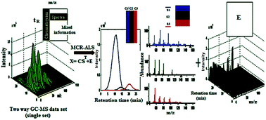
Multivariate curve resolution with alternating least squares optimization (MCR-ALS), as a soft modeling approach based on factor analysis, was proposed to recover the thorough gas chromatography-mass spectrometry (GC-MS) fingerprint analysis of volatile chemical constituents in Iranian Citrus aurantium L. peel. This technique for two-dimensional data was intended to resolve the overlapping and/or embedded GC-MS peaks into the pure chromatogram and mass spectrum of each chemical constituent, overcoming some challenging fundamental chromatographic problems occurring during GC-MS analysis of the Iranian C. aurantium peel chromatographic fingerprint, such as spectral background, baseline offset, and type of noise. In this way, the chromatographic fingerprints of Iranian C. aurantium peel are properly segmented to the appropriate chromatographic regions, and then MCR-ALS is used to achieve pure response profiles of the chemical constituents in each segment, as well as their relative concentrations. Retention indices, together with mass spectral profiles of pure chemical constituents, were considered for qualitative identification by matching against the standard ones through MS library searching; an overall volume integration (OVI) technique was also used for the semi-quantitative analysis (to obtain the relative concentrations of chemical constituents). GC-MS analysis of the C. aurantium L. peel, with the help of the proposed methodology, resulted in extending the number of identified constituents from 45 to 82 with concentrations higher than 0.01%. The lack of fit (LOF), percent of variance explained (R2) under the optimum conditions, and reverse match factor (RMF) were used for the assessment of the MCR-ALS solutions. The LOF values of MCR-ALS models were lower (12.0%) for all segment matrices with RMF values in the range 713–977 and R2 values higher than 97%. It was found that the major constituents of Iranian C. aurantium L. peel are limonene (72.89%), β-myrcene (9.06%), α-pinene (4.74%) and β-pinene (3.44%). It is concluded that the coupling hyphenated GC-MS measurements with the multivariate curve resolution-alternating least squares method is an effective and powerful strategy to solve current problems in GC-MS analysis, to obtain the required analytical selectivity in complex natural products.


1 Introduction

Over recent years, a significant growth in scientific knowledge of the relationship between food and health has been observed. The public's health awareness is an important factor, resulting in the extensive use of natural products, due to them producing fewer side effects in comparison to the synthetic drugs.1–3 One of the most popular natural products that are widely cultivated and have been used worldwide for over 4000 years, are citrus fruits, due to their characteristic aroma, taste and flavour. They are also well-known for multiple health benefits and inhibition of diseases in humans, as they contain important bioactive compounds, such as carotenoids, ascorbic acid (vitamin C), flavonoids, essential oils and phenolic compounds with antioxidant properties similar to those of carotenoids and vitamin-C-type compounds.4–9 The genus Citrus, belonging to Rutaceae or Rue family, comprises various species, varieties and hybrids.8,10,11 Most of these cultivars are found in south, south-eastern and northern parts of Iran. Citrus aurantium L. (the best known species of sour orange) is a hybrid of the lime and citron and is generally used as a rootstock.12 Additional plant descriptions of C. aurantium L. are available in ref. 12 and 13. Among citrus species that have been applied for medicinal purposes in herbal medicine owing to diverse bioactive compounds, this species is a strong appetite suppressant and stimulant. It has also been found to be therapeutically effective against various complaints, such as; indigestion, nausea, cancer, constipation, cardiovascular disease, and anxiety.11,13 The dried outer peel with the removed white pulp layer, leaves, and flowers of the plant are the parts mostly applied for medicinal purposes.11,13 The complex chemical makeup of C. aurantium is best known for the volatile oil in the peel. This is responsible for the strong odor and flavor of C. aurantium L. and many of its medicinal effects.11,14 C. aurantium peel extracts have exhibited several useful biological activities, such as anti-inflammatory, antifungal, and antibacterial.10,11,13–15 So, more research attention towards the identification of volatile chemical constituents in the peel of this plant is important to increase its application for therapeutic purposes to a greater extent.

Chemical fingerprints obtained by hyphenated chromatographic techniques offer a qualitative and integrated profile of all data points related to multiple characteristic constituents for the purpose of identifying complex samples.16,17 Among the different chromatographic fingerprinting techniques, gas chromatography-mass spectrometry (GC-MS), as one of the so-called second order separation methods, is one of the most promising and widespread analytical techniques for the determination of volatile constituents of complex real sample extracts.17–19 However, in GC-MS analysis of highly complex samples, due to the existence of various challenging problems, such as spectral background, baseline drift, different types of noise, and some co-elution (overlapped and/or embedded) peaks, obtaining selective information, even under the best experimental conditions, is not possible.19 These challenging issues may originate from the experimental conditions, the variability of GC-MS systems, such as chromatographic and/or detection systems, and the complexity of natural samples.20 Among these problems, co-elution (peak overlap) is perhaps one of the most important and most observed difficulties in chromatographic analysis, which occurs mainly due to the inadequate selectivity of chromatographic columns, peak capacity, and the need for faster chromatographic analysis. The existence of these problems can reduce the similarity indices (SIs) obtained from a direct search in an MS database and, therefore, correct identification of the constituent cannot be achieved.21,22

The chemical composition of C. aurantium L. peel has been studied by different researchers.11,12,15,23,24 The major constituents found in many studies were limonene, β-myrcene, β-pinene, α-pinene, β-ocimene, linalool, γ-terpinene, sabinene, and octanal. Sarrou et al. investigated the volatile constituents and antioxidant activity of the peel, flowers and leaf oils of Citrus aurantium L. growing in Greece based on GC-MS measurements.11 They reported 26, 20, and 16 constituents, in the flowers, peel, and leaf oils of Citrus aurantium L., respectively. They found that peel oil is composed mainly of monoterpene hydrocarbons (98.30%), mainly limonene, while oxygenated monoterpenes are the dominant compounds of leaf and flower oils. Babazadeh Darjazi reported 29 volatile constituents, including 18 oxygenated terpenes and 11 non-oxygenated terpenes, followed by GC-FID and GC-MS analysis.23 Moreover, Lota et al. reported that 39 components of peel oil and 47 components of leaf oil were identified by mass spectrometry/retention indices (GC-MS analysis), and the major components (≤0.05%) by 13C-NMR spectroscopy.15 Their chemical composition was investigated by capillary GC, GC-MS, and 13C-NMR. The most recent studies analyzed desired peel samples, as a complex matrix, through a comparison of Kovats retention indices (RI), their retention times (RT), and mass spectra of authentic samples or literature data, in order to identify their constituents. Identification of the constituents of C. aurantium L. peel sample by GC-MS was the subject of several investigations, but because of the aforementioned problems with this technique, only a small number of chemical constituents were identified. On the other hand, the minor constituents are responsible for the specific odor and flavor, and they may also have important therapeutic value. Additional information is present in GC-MS chromatograms that cannot be extracted merely by using specialized chromatographic technologies. As a result, the qualitative and quantitative results obtained were not reliable or satisfactory. To overcome these problems, extraction of the required information about the constituents in complex matrices has become possible by means of chemometric resolution methods.

Over the past few decades, different multivariate curve resolution (MCR) techniques have matured to improve the sensitivity and resolve separation issues encountered in GC-MS analysis. These mathematical resolution techniques, by applying a bilinear model, attempt to exploit the chromatographic and spectral differences between all the constituents present in a particularly complex mixture (even if their differences are very small), in order to acquire more informative data from the chemical analysis, both in the spectral identification (qualitative information) and chromatographic separation (quantitative information) of the constituents.21,25,26 Many iterative and non-iterative curve resolution approaches have been offered that can all be classified as multi-component factorization of multivariate data techniques.26 The advantages of applying MCR methods to determine the pure contribution of each constituent involved in the system are obtaining an increased resolution power with reduced chemical use, cost, and time, allowing a better understanding of the chromatographic process.18,26 In this study, the volatile constituents of Iranian C. aurantium L. peel were extracted using a static headspace (SHS) technique and were analyzed by GC-MS under suitable conditions to achieve informative chromatographic fingerprints. Multivariate curve resolution based on alternating least squares optimization (MCR-ALS), along with other chemometric methods, was applied to provide chemically meaningful quantitative and qualitative profiles of the pure constituents present in an Iranian C. aurantium L. peel chromatographic data set. The advantages of MCR-ALS over other MCR methods suggested in the literature are that the application of different constraints during the optimization process and generalization to high-complexity data sets is quite simple.27,28 Parastar et al. have proposed extended MCR-ALS and multivariate clustering methods to improve the analysis of GC-MS fingerprints of secondary metabolites in eighteen citrus samples (including eight lemon (C. limon), five orange (C. sinensis), three mandarin (Citrus reticulate), and two grapefruit (Citrus paradise)) through a multi-set augmented data structure.29 The constituents offered in this work were main and commonly identified metabolites for eighteen citrus samples with noticeable relative concentrations, which were relevant for subsequent principal component analysis (PCA) and k-nearest neighbor (KNN) cluster analysis. Finally, they used a counter-propagation artificial neural network (CPANN) supervised method in order to characterize the chemical markers (chemotypes) responsible for the differentiation of four determined clusters by PCA and KNN. To the best of our knowledge, the constituents of C. aurantium L. peel extract have not been identified using a combination of GC-MS and chemometric resolution methods so far. Therefore, the main aim of the present work is based on the identification and semiquantitative analysis of pure contributions of individual constituents present in an Iranian C. aurantium L. peel sample. Inspection of the results confirmed the potential of MCR-ALS as the most effective strategy, saving work and time in overcoming chromatographic challenges in the GC-MS fingerprints of volatile chemical constituents in a given sample.

2 Experimental

2.1 Sample collection and chemicals

Plant samples, including mature C. aurantium L. were collected from the orchards of Ramsar Citrus Research Institute in the north of Iran (latitude 36° 54′ N, longitude 50° 40′ E; Caspian Sea climate; average rainfall and temperature were 970 mm and 16.25 °C per year, respectively) early in the morning (9 to 10 am), in February 2015.12,23 To properly evaluate and minimize the experimental error associated with the constituent extracts, fruits were picked from different parts of the same tree (top, middle, and base branch, 5 fruits from each part).23,30 Peel samples were dried completely under shadow in the dark under a moderate air flow at room temperature for 4 days. The desired amount of dried peel sample was cut into smaller pieces and then ground into powder and homogenized with a household grinder for later experiments. Powdered peel samples were stored in a glass vessel in a refrigerator until extraction. All chemicals used in this work were analytical grade. Methanol and dichloromethane with purity higher than 99.0% were purchased from Merck Chemicals (Darmstadt, Germany). Pure helium (99.999%) was obtained from Sabalan Co. (Iran). Normal alkane standards (C-8 to C-20) were purchased from Sigma Aldrich.

2.2 Sample preparation: static headspace (SHS)

In order to extract the volatile chemical constituents of a given sample, a static headspace (CombiPAL system), which was provided with a headspace autosampler, agitator, and heater, was used. The exact weight of powdered peel (2.0 g) was placed into a 20 ml headspace vial. This vial was sealed with a rubber septum faced with polytetrafluoroethylene (PTFE) and then placed in the headspace tray. The contents of the vial were continually shaken with an agitator at a controlled temperature of 70 °C for 10 min and with an agitator speed of 500 rpm (high shake mode), before carrying out the static headspace (SHS) extraction; the equilibrium temperature and extraction time were 90 °C and 600 s, respectively. For chromatographic analysis of volatiles in the sample peel, using a gas-tight syringe (with a size of 2.5 ml, at a temperature of 80 °C), 1 ml of headspace vapour phase in the vial was sampled and then injected immediately into the GC column.

2.3 Chromatographic conditions and instrumentation

An Agilent Technologies GC-MS (Santa Clara, CA, USA) consisting of a 7890A gas chromatography system combined with a 5975C network mass spectrometry detector, which was supplied with an electron ionization source and quadrupole, and a DB-5 capillary column (60 m, 0.25 mm i.d.; 0.25 μm d.f., with 0.5 μm film thickness, methyl 5% phenyl polysiloxane) for the separation process, was applied for the analysis of volatile extracts of Iranian C. aurantium L. peel. The oven temperature program started at 40 °C for 7 min, then increased to 240 °C at a rate of 8 °C min−1, and was held constant for 10 min at this temperature for a post-run step. The carrier gas (helium) was adjusted at a constant pressure of 36 psi with a flow rate of 1 ml min−1. A split injection (with split ratio, 1[thin space (1/6-em)]:[thin space (1/6-em)]5) and injector temperature of 250 °C were applied. Other operating conditions were as follows: injection volume, 1 mL; interface temperature, 150 °C; mass spectra were recorded from 42 to 550 amu (ionization energy: 70 eV) in full scan mode. The ionization source and quadrupole temperatures were 230 and 150 °C, respectively.

2.4 Software requirements

A PC interfaced to the GC, using the MSD ChemStation software package (G1701 DA, version D.00.01), was used for the collection and processing of GC-MS data. Data analyses were performed using an Intel (R) Core (TM) i5-based ASUS personal computer. All chemometric resolution techniques were implemented using an MCR-ALS toolbox from the homepage of MCR under the Matlab R2009a environment, and also MCRC software v1.0 for preprocessing, chemical rank determination, local rank analysis, and MCR-ALS analysis.31,32 Identification of individual constituents in the peel extract of Iranian C. aurantium was done based on comparison of retention indices, together with the mass spectral fragmentation pattern of resolved chemical constituents with retention indices and mass spectra of standard components stored in the Wiley 7n.1 MS and National Institute of Standards and Technology (NIST) computer library v2.0.

3 Theory and methodology

3.1 MCR-ALS

Self-modeling multivariate resolution methods (SMCR) are a generic denomination of a family of chemometric methods with the aim of mathematically decomposing a measured complex mixture of signals into pure contributions of constituents solely from the information obtained from an original two-way data matrix (Xm×n).33,34 This method is based on the basic assumption of a chemically meaningful additive bilinear model of the individual contributions of the different constituents in the chemical system, which can be decomposed into the product of two matrices, C and ST, and is defined as follows:35
 
Xm×n = Cm×kSTk×n + Em×n (1)
here, in the GC-MS data, indices m and n refer to the numbers of row and column variables, respectively, for different retention times and spectral channels (e.g., m/z ratios), Cm×k represents the resolved elution (chromatographic) profile matrix of the k component present in the sample, STk×n signifies its corresponding mass spectra matrix, and Em×n denotes the error matrix related to the data variance unexplained by the bilinear model CST. The superscript T is the vector or matrix transposition. It was revealed that this bilinear relation between the pure spectra, the concentrations of the constituents, and the experimental data, is a good approximation in the study of different macromolecular systems using chromatography.36,37 Several different algorithms for solving the above equation exist, but among MCR methods, Alternating Least Squares (ALS, or MCR-ALS) is undoubtedly one of the most often used algorithms due to its special flexibility and simplicity, and proposed by Tauler in 1995.36–39 Several excellent reviews are available.40–43 MCR-ALS solves (eqn (1)) for C and ST, without any previous assumption about the nature and/or composition of the system, applying an iterative algorithm, to optimize the initial estimate of concentration or spectral profiles at each iterative cycle, based on two linear least-squares steps, under the action of suitable constraints until a convergence is achieved (see ESI for more details of the MCR-ALS method).44,45

In order to give physically meaningful and chemically interpretable solutions, and a limited number of possible solutions to maximize the data variance explained by the different constituents, a series of constraints are applied to C and (ST) at each iteration, such as non-negativity46,47 for C and ST matrices and unimodality48 for C, normalization spectra,18,20 and selectivity.49

The convergence criterion can be a difference in fit improvement in two successive iterations, such that if this relative difference in fit is less than a threshold value (predefined cut-off value, usually 0.1%; of course, depending on the step optimization, this may be modified by the user),32 the optimization is finished, or sometimes a preset number of iterations may be applied as the stop criterion.17 In most applications, in MCR-ALS, one opts to normalize the spectral modes in the columns of matrix S to equal length, without changes in data fitting, in order to avoid scale and intensity ambiguities during the ALS optimization algorithm, and so that the retention time profiles can be directly inferred as relative concentrations.40,50 The final MCR-ALS solutions are a set of pure chromatographic and spectral profiles for each instrumental response signal, and the quality of these factors is related to the model fit. The most common methods for this assessment are statistical parameters, such as percent of lack of fit (LOF) and percent of variance explained (R2) under the optimum conditions (see ESI for definitions of these parameters). Also, other parameters related to assessing the resolution results and identifying chemical constituents for two-dimensional GC-MS data are the match factor (MF), or reverse match factor (RMF), and also the comparison of GC retention indices (RIs).

The LOF and R2 quantities allow a simple comparison between different methods and models in a description of the same data set.32,42 In the presence of data with a very low noise level, the LOF offers more distinction between similar models, but in the case of high-noise-level data and a greater number of chemical components, R2 is preferred, as larger model unexplained variances mean that data fitting is not good. Both LOF and R2 values should be judged with expected levels of experimental noise.27,44 The match factor (MF) and reverse match factor (RMF) reflect the likelihood that the resolved mass spectral fragmentation patterns and reference spectra of the standards in the NIST mass spectral search program or the mass spectra from the literature arose from the same compound. However, when studying complex systems containing significant background noise and non-selective separation (co-elution problem) by the GC column, RMF is the preferred technique, because it only matches the abundance values of resolved mass spectra in common with each reference spectrum, rather than a peak-by-peak match as in MF.51 RMF defined the normalized dot product with square root scaling of the MCR-ALS resolved mass spectrum and the library NIST standard mass spectrum, without including the elements of the resolved mass spectra that are not present in the library spectrum.29,51 In addition, to improve determination of the constituents, the temperature-programmed retention index (RI) proposed by Van den Dool and Kratz22,52 in a quasi-linear equation is calculated for all the constituents as follows;

 
RIx = 100n + 100(tRxtRn)/(tRn+1tRn) (2)
where tRn, tR(n+1), and tRx are the retention times in minutes of the reference n-alkane hydrocarbons eluting immediately before and after chemical constituent “x” containing n carbons, n + 1 carbons and the desired constituent, respectively. The identification is done using MF or RMF, and can be verified by comparing GC retention indices (RIs) of resolved constituents with those of authentic constituents and literature data.

3.2 Methodology

In order to give an accurate and reliable description of complex chemical fingerprints, and to find pure signal contributions of a dyad of concentration and spectral profiles of each mixed signal, a set of necessary steps should be considered when performing MCR methods. The flow chart given in the ESI section (Fig. S1) is a representation of the entire strategy for resolving and quantifying the two-way data achieved from a GC-MS fingerprint. The proposed strategy in this study for thorough analysis of GC-MS fingerprints is that, at first, the obtained total ion chromatogram (TIC) is segmented to the desired sub-matrices by applying zero component chromatographic regions based on emerging chromatographic peaks with the elution sequence of extraction. Then, each segment is transformed to a computing environment, such as MATLAB or MCRC for MCR-ALS analysis. The detail of each step is presented as follows;

(1) Data preprocessing: two-way data obtained from GC-MS can suffer from a number of fundamental non-informative defects, such as baseline drift, spectral background, low S/N ratio (heteroscedastic and homoscedastic noise), and many others. The occurrence of these artifacts in both chromatographic and spectrometric dimensions can result in the appearance of more components in each segment matrix than what is actually expected. Therefore, to increase the efficiency of MCR techniques in accurate identification of minor chemical constituents, and to resolve embedded or overlapped peaks, both the artifacts and the number of chemical components should remain at a certain level in a segment matrix.42,44 In order to achieve this goal, some preprocessing methods must be handled for some data sets. Due to the presence of many noise channels in GC-MS data obtained in full scan mode, removing this noise would result in a faster computation. In the present work, a morphological score proposed by Shen et al.53 is applied to distinguish the signal from the noise channels based on their frequency difference. This method was used to decrease the homoscedastic noise in two-way data. The signal channels that had morphological scores below the noise limit were deleted. In addition, a Savitzky–Golay smoothing filter,54 by employing the regression-fitting capacity based on least squares, was used to transform heteroscedastic to homoscedastic noise, then reduce the heteroscedastic noise in each data matrix. In order to quantitatively assess the system under study, the baseline must be stable during analysis. Furthermore, the existence of a spectral background in the MS dimension can affect the identification of the constituents in the system.26 Finally, simultaneous background and baseline correction was performed using a congruence analysis method and least-squares fitting developed by Liang et al. on the chromatographic data.29,53,55 Then, each pre-processed data matrix was scaled for having a maximum signal intensity of 1.0.38

(2) Determination of the number of chemical constituents: most methods applied to estimating chemical rank, and finding the direction of the relevant sources of variation in a bilinear data set, are based on PCA or singular value decomposition (SVD). However, because of the accumulation of noise in complex systems analyzed by GC-MS, achieving a true rank for the full data matrix using PCA is difficult. In addition in the presence of high levels of embedded noise, the vector spaces determined by PCA and SVD are incorrect.31,41,56,57 In order to achieve more reliable results, another category of methods based on finding a group of the purest variables (columns or rows), giving the most dissimilar column and row profiles of the data matrix with the reference profiles, was introduced. The simple-to-use interactive self-modeling mixture analysis (SIMPLISMA), orthogonal projection approach (OPA) and simplified Borgen method (SBM) belong to this category.41,58

In the present work, the number of constituents was preliminarily estimated by means of PCA or by the SVD algorithm.31,58 Also, for complex chromatographic regions, to ensure the correct number of chemical constituents, a morphological score technique was employed, using the OPA and SBM methods for key variable selection.20,41,57 In this technique, after noise elimination in the mass channels in each segment matrix, based on a morphological factor criterion, the number of constituents was estimated using the same technique (morphological score).59 Furthermore, subspace comparison as a purity-based method was also used to confirm the number of constituents present in each submatrix.60

On the other hand, local rank information offered by evolving factor analysis (EFA), and fixed-size moving window-evolving factor analysis (FSMW-EFA) procedures were applied to obtain a more accurate estimation of the number of chemical constituents and peak purity assessment of the bilinear data.61 For a more detailed discussion of factor analysis methods, refer de Juan.62,63 However, finally, the change in lack of fit (LOF) values of the MCR-ALS model by using fewer and more constituents was applied as a criterion to confirm chemical rank.

(3) Initial estimate and chemometric resolution: the preliminary information given by an exploratory analysis during the chemical and local determination process can be used to set up good initial estimates for either concentration or response profiles.31 It is important to note that the use of random and irrational initial guesses during ALS optimization can cause this algorithm to be stuck in local minima instead of global minima, and ultimately result in insufficient curve resolution, since a poor initial estimate does not obey the imposed constraints during the optimization and, as a result, does not provide a profile with clearly definable chemical meanings while saving computational time.43 Hence, methods that can be applied for process-like data based on their evolutionary nature, such as EFA62 and general methods, which can work irrespectively of the presence or absence of a sequential elution structure in the concentration direction, with the aim of direct selection of the uncorrelated variables of the analyzed data matrix, such as OPA and SIMPLISMA,58 were used for initial estimates in this work. Depending on the segment matrix conditions under analysis, one of two methods works better. In the present work, MCR-ALS decomposition was implemented with the introduction of additional knowledge of the natural properties of the constituent profiles through the application of the appropriate constraints of non-negativity in both elution and spectral directions to force the profiles to be equal to or greater than zero, unimodality in the elution direction to allow the presence of only one maximum per peak, and spectral normalization to have a length of one (which means the concentration profiles are not normalized), to improve the recovery of meaningful solutions. Finally, by continuing the iterative method, with successive estimation of C or ST alternately under the mentioned constraints with an increasing number of cycles, convergence was tested. When in two consecutive cycles, the relative differences between the sums of the squares of residuals (SSR) are below a preset selected value, convergence is then fulfilled and the optimization is finished.33

(4) Evaluation of the reliability of resolution results: the statistical terms obtained for each chromatographic segment during the implementation of the MCR-ALS technique, such as lack of fit (LOF in % for ALS optimization, PCA) and percent of explained variance (R2) at the optimum were used for evaluation of the resolution results and choice of the best MCR-ALS model. The best model fitting of the experimental and resolved matrices is achieved when the LOF and R2 are close to zero and one, respectively.27

(5) Qualitative and semi-quantitative analysis: after finding the resolved mass spectral profile for each constituent, the constituents have been identified by similarity match in reverse mode (RMF) using the standard spectra in the NIST MS database. The identification and assessment results were more precisely confirmed with the help of RIs.22,52,64 On the other hand, quantitative analysis of the constituents was performed using the overall volume integration (OVI) algorithm.65,66 This is based on summation of the integrated peak areas at every m/z point for each constituent, and allows the relative amount of the constituents in the whole TIC to be obtained. The OVI algorithm is preferred over the total peak area integration, since all mass spectral points are considered in the calculation. These steps were applied for all of the segment matrices in this work.

4 Results and discussion

4.1 Resolution by MCR-ALS and qualitative analysis

The TIC of the chromatographic fingerprints of Iranian C. aurantium peel extract is displayed in Fig. 1, with the presence of a large number of overlapping and/or embedded peaks with very variable contents, along with other chromatographic artifacts, such as baseline/background contribution and low S/N ratio (i.e. presence of noise), clearly demonstrating the complexity of the studied system of volatiles. Nevertheless, the identification of constituents in complex regions would definitely become very difficult and unreliable using similarity indices (SIs) obtained from simple direct searching with the MS database, especially for the constituents with a low content, because of the unavoidable presence of peaks related to residual gas and column background. In addition, at different scan points of a single chromatographic peak, one can find different constituents through library searching. This means that, if the selective chromatographic signal could not be obtained for each constituent, the traditional search of the MS library would fail.
image file: c6ra18871k-f1.tif
Fig. 1 TIC of volatile chemical constituents from Iranian C. aurantium L. peel extract.

For this purpose, the whole TIC was segmented to 53 chromatographic regions by zero component regions along elution of the volatiles of Iranian C. aurantium peel extract. Some of these segment matrices were single-component. Peaks associated with these constituents could be simply identified and quantified using NIST MS library searching and peak integration in MSD Chemstation software. However, to give more reliable results, single-constituent peak clusters were pretreated using the MCR-ALS method. The results obtained were much better than those achieved with ChemStation with respect to the reverse match factor (RMF) and percentage of each constituent. Accordingly, two-dimensional data obtained from all of the segment matrices was extracted from MSD Chemstation software and was then changed to ASCII format, which is compatible with the MATLAB environment. Each data matrix gives a peak cluster. Therefore, there were 53 peak clusters in the TIC of Iranian C. aurantium L. peel extract, of which the individual peaks were analyzed by MCR-ALS according to the proposed strategy in Section 3. In order to demonstrate the efficiency of the applied chemometric resolution method, three problematic regions were selected and marked as peak clusters A (13.25–14.36 min), B (15.97–16.11 min) and C (21.55–21.73 min), as examples, with their TICs to obtain a better visualization of the enlarged detailed pattern of the GC-MS fingerprint. Here, the figures and results for peak clusters B and C are shown, and also the figures and results for peak cluster A are available in the ESI section.

The exported data matrices of peak clusters B and C are displayed in Fig. 2a and b with sizes of (27 × 151) and (31 × 151), respectively. These specific peak clusters were selected with the aim of showing the performance and then evaluating the application of the MCR-ALS method in multi-component systems by changing the degree of overlapping and the presence of different amounts of other chromatographic challenges.


image file: c6ra18871k-f2.tif
Fig. 2 The exported data matrix of the selected peak clusters B (a) and C (b).

Considering region B in Fig. 1, which is also displayed in Fig. 2a, this appears to be a mixed system of two co-eluted constituents. Furthermore, a direct library search for peak cluster B showed only two constituents, named 1,6-octadien-3-ol,3,7-dimethyl-(β-linalool) (C10H18O) and nonanal (C9H18O). However, after MCR-ALS analysis, different results were achieved. Because first, as mentioned in the proposed strategy in Section 3, baseline/background correction and noise reduction on most of the peak cluster was performed. Congruence analysis and the least squares fitting method introduced by Liang and Kvalheim were applied for baseline/background variation correction. In this method, the essential information for presenting the univariate linear regression with regard to the retention time can be provided by using the local rank analysis of zero component regions. Then, the baseline will be corrected. In addition, homoscedastic noise is reduced by morphological score methods and a Savitzky–Golay filter with a polynomial order of polynomial 2, and a five-point filter was applied for heteroscedastic noise reduction. All of these methods can be easily performed with MCRC software as a chemometric tool for the analysis of two-dimensional chromatographic data,31 and then the morphological score and subspace comparison were used to determine the number of chemical constituents for the pre-processed peak clusters. Fig. 3a shows the results of the morphological score method for cluster peak B. In this figure, to give the purest constituents by the orthogonal projection approach (OPA), the morphological scores of these pure constituents were plotted against the number of constituents. The morphological score for the noise level was estimated by means of an F-test55,59 and is displayed in dotted-line form. Based on the morphological score plot in Fig. 3a, there are three constituents in peak cluster B. This is found by counting the number of significant variables with morphological scores above the noise level. Moreover, subspace comparison methods are used to confirm morphological score method results. Subspace comparison analyzes the key factors similarly to the morphological score method, through the comparison of two subspaces, each of which is determined using a suitable procedure, such as OPA, SIMPLISMA, and PCA, by applying a set of orthonormal vectors in order to select factors.60 These look for the most pure or the most dominant variables. PCA extracts the most dominant linear combination of the actual variable subject to an orthogonality constraint; OPA considers dominant variables among the actual variables in the data matrix, and SIMPLISMA places more importance on purity than on dominance.67


image file: c6ra18871k-f3.tif
Fig. 3 Morphological score plot for chemical rank determination in chromatographic segment B by applying OPA method as factor selection method (a). Noise level is shown as dotted line. Subspace plot for this region (b). Comparison between key selected factors of methods PCA and SIMPLISMA.

The results of PCA-SIMPLISMA subspace plots in Fig. 3b confirm the presence of three constituents. In this method, the key factors or number of constituents could be concluded from the largest value of K (as columns of subspace matrices with n × k dimensions), when D(k) and sin[thin space (1/6-em)]2(θK) are equal and are close to zero. In addition, a local rank map, obtained by EFA and FSMW-EFA methods as a data microscope, was used to obtain a more accurate estimation of the number of chemical constituents, based on the use of local information on the elution patterns of constituents in each region. In contrast with the EFA method, which acts with PCA analyses on an increasing size of window, the FSMW-EFA method performs PCA analyses on a moving window with a fixed size. The FSMW-EFA method particularly gives more reliable results when detecting impurities or minor compounds under a main peak than the EFA method,65,66 due to performing the local analysis on small elution windows. In this method (FSMW-EFA), the noise level is determined by eigenvalue curves with similar numerical values and emerges together at the bottom. Eigenvalue curves exceeding the noise level display the emergence of new constituents. The FSMW-EFA plot with a fixed size 5 window for peak cluster B showed that there are three eigenvalue curves above the noise level within these regions, which indicated that peak cluster B is definitely not a two-constituent cluster. In this plot, the names of the constituents present in cluster B are marked by their elution order. From the FSMWEFA plot in Fig. 4, one can conclude that regions 1 and 3, having one curve above the noise level, are pure regions of the first and third constituents; the regions 1 + 2, 1 + 2 + 3, and 2 + 3, containing two or more curves above the noise level, are overlapping regions of the first and second constituents, by the first, second and third constituents and by the second and third constituents, respectively. According to the results obtained from the FSMWEFA analysis, it can be seen that there are no selective regions for the second constituent. Therefore, the MCR-ALS method is a feasible way of resolving such a peak cluster. Then, peak cluster B was resolved by the MCR-ALS method with the initial estimate of the concentration profile being calculated by the EFA method and under the proper constraints, such as non-negativity, unimodality, and normalization of spectra during ALS optimization. Resolved elution profiles for three constituents in this chromatographic region, obtained by using this technique, are displayed in Fig. 5. Also, their corresponding resolved mass spectra, with the standard spectrum of each constituent from the NIST/Wiley MS database are shown in Fig. 6.


image file: c6ra18871k-f4.tif
Fig. 4 FSMWEFA plot with a size 5 window for analyzing peak cluster B.

image file: c6ra18871k-f5.tif
Fig. 5 Original peak cluster B and pure chromatographic peaks after resolution by chemometric method MCR-ALS: (a) the TIC of peak cluster B from C. aurantium L. peel extract; (b) the corresponding two dimension plot; (c) resolved MCR-ALS chromatographic profiles (1–3).

image file: c6ra18871k-f6.tif
Fig. 6 Resolved mass spectra and their corresponding standard mass spectra for peak cluster B. Resolved (a) and standard (d) mass spectra of β-linalool; resolved (b) and standard (e) mass spectra of dodecane; resolved (c) and standard (f) mass spectra of nonanal.

Theoretical parameters of lack of fit (LOF) as % (for PCA and exp) and explained variance (R2) at the optimum as % were equal to 0.39, 1 and 99.98 respectively. The LOF and R2 values for the optimum MCR-ALS model were satisfactory according to the noise level in this region. Then, each constituent was identified by matching the resolved spectral profile with stored mass spectra in the NIST mass database, and confirmed by comparison of their retention indices. The results showed that β-linalool (C10H18O), dodecane (C12H26), and nonanal (C9H18O) were identified in peak cluster B, with values of the reverse match factor (RMF) equal to 977, 869, and 930, respectively. As can be seen, reliable values of the LOF and R2, together with quite high spectral matches for identified constituents, despite heavy overlap with each other, ascertain the possible identity of the resolved constituents in this chromatographic region. From the resolved chromatographic peaks in Fig. 5, it is clear that the second constituent peak of this peak cluster is embedded in the first constituent peak and also contains a very low quantity over the whole region. Because of these factors, the second constituents were not found in a direct NIST/Wiley MS library search.

Likewise, Fig. 2b shows a TIC curve of peak cluster C, which seems to represent the co-eluted regions of two constituents. Also, the mass spectra of different parts of peak cluster C indicate that there could be more than two constituents or severe noise. However, only three constituents, namely acetic acid, decyl ester (C12H24O2), and dodecanal (C12H24O) can be directly matched in the NIST MS library.

However, after MCR-ALS analysis, different results with more information were achieved for this peak cluster. Firstly, the background and noise were removed using the applied methods for peak cluster B. Then, in a preliminary inspection, morphological score and subspace comparison methods were used for determination of chemical rank. The results of these methods on preprocessed peak cluster C are illustrated in the ESI section (Fig. S6a and b). It can be seen that region C is a four-constituent system.

Also, in order to verify the obtained rank estimation and peak purity control of peak cluster C, as with peak cluster B, EFA and FSMWEFA were applied. The rank map obtained by using the FSMWEFA method with a fixed size 6 window on peak cluster C is presented in the ESI section (Fig. S7). This figure showed that there are four eigenvalue curves above the noise level within this region, which indicated that peak cluster C is definitely not a two-constituent region. According to this figure, regions 1 and 4 are selective regions of the first and fourth pure constituents, and regions 1 + 2, 1 + 2 + 3, 2 + 3 + 4, and 3 + 4 indicate the overlapping regions of constituents 1 and 2, by 1, 2 and 3, by 2, 3 and 4 and also by 3 and 4, respectively. According to the results obtained from FSMWEFA analysis, it can be seen that peak cluster C is much more complex than peak cluster B, since there are no selective regions for some of the constituents (2 and 3). Using this prior knowledge of constituents, MCR-ALS analysis was run using an initial SIMPLISMA estimate of the spectral profile for peak cluster C to start ALS optimization under the applied non-negativity, unimodality, and spectral normalization constraints.

The resolved MCR-ALS chromatographic profiles of peak cluster C and their corresponding resolved mass spectra, together with a standard spectrum of each constituent from the NIST/Wiley MS database, are presented in the ESI (Fig. S8c and S9).

The percent of LOF (for PCA and exp) and R2 values for the optimized MCR-ALS model were 2.35, 4.59, and 99.78, respectively, for peak cluster C. The similarity between the resolved and standard mass spectra and comparison of their retention indices showed the reliability of the resolution method with confirmation of the presence of new detected constituents, cyclohexane, 1-ethenyl-1-methyl-2,4-bis(1-methylethenyl) (C15H24), and benzene, 1,2-dimethoxy-4-(2-propenyl)-(eugenol methyl ether) (C11H14O2) in region C, in addition to constituents that were previously detected by MSD Chemstation software.

The RMF values for the four constituents identified after resolution by the proposed strategy in region C were 936, 969, 862, and 850. Also, according to resolved chromatographic peaks in Fig. S8c, the four constituent peaks of this region have significantly overlapped, and of course due to the presence of background noise in the TIC of this peak cluster, some constituents could not be identified. But after applying the MCR-ALS method, the number of defined constituents in peak cluster C was improved from 2 (direct mass spectra search) to 4 (ESI methods, Fig. S8c) with satisfactory statistical parameters, considering the noise level in this region and high RMF values. All chromatographic segment matrices obtained from the TIC of the studied sample were resolved in a similar way, and the elution and mass spectral profiles for each volatile constituent in a desired sample extract were extracted. In Table 1, qualitative analysis results for the identification of the volatile constituents of C. aurantium L. peel are listed. The chemical name and formula, values of retention time (RT), retention index (RI), RMF, and the percent of the relative concentration of each constituent are also presented. The direct analysis of two-dimensional data obtained from the GC-MS method revealed that only 45 constituents exist in Iranian C. aurantium L. peel extract; however, after resolving all peak clusters by applying chemometric methods, the number of constituents was increased to 82, accounting for 95.93% of the total relative content of constituents from Iranian C. aurantium L. peel, also with fitting values for MCR-ALS models in terms of an LOF (exp) lower than 12% and R2 higher than 97% for all the peak clusters.

Table 1 Identified volatile constituents in Iranian C. aurantium L. peel extract, their retention times (RTs), retention indices (RIs), RMF values, and relative concentration (%) obtained using MCR-ALSa
No. RT (min) RI Chemical name Molecular formula RMF Percentage (%)
a Sum: 95.93%.
1 14.644 1050.6 Limonene C10H16 948 72.895
2 13.616 1006.2 β-Myrcene C10H16 923 9.060
3 12.351 988.8 α-Pinene C10H16 911 4.740
4 13.443 1002.3 β-Pinene C10H16 944 3.440
5 13.310 1028.0 Sabinene C10H16 960 1.230
6 12.380 991.4 α-Thujene C10H16 953 1.058
7 16.036 1076.0 β-Linalool C10H18O 977 1.009
8 13.853 959.4 Octanal C8H16O 975 0.346
9 18.075 1139.8 Decanal C10H20O 939 0.281
10 14.038 1023.1 α-Phellandrene C10H16 915 0.231
11 16.088 1053.3 Nonanal C9H18O 930 0.110
12 17.976 1121.8 α-Terpineol C10H18O 923 0.103
13 19.449 1422.4 Dodecane, 2,6,11-trimethyl C15H32 877 0.093
14 21.267 1479.8 β-Caryophyllene C15H24 953 0.092
15 16.342 1129.8 Cyclohexane, 2-ethenyl-1,1-dimethyl-3-methylene- C11H18 850 0.075
16 16.053 1171.1 Dodecane C12H26 869 0.072
17 18.769 1389.9 β-Cubebene C15H24 853 0.064
18 24.144 1570.9 ±-trans-Nerolidol C15H26O 929 0.053
19 18.300 1118.1 p-Menth-1-en-4-ol C10H18O 886 0.049
20 18.967 1149.5 cis-Carveol C10H16O 882 0.047
21 15.580 1067.6 2-Furanmethanol, 5-ethenyltetrahydro-α,α,5-trimethyl-, trans- C10H18O2 929 0.046
22 15.193 1151.8 Undecane, 2-methyl- C12H26 851 0.044
23 21.465 1380.9 Tetradecane C14H30 970 0.042
24 17.607 1047.3 Benzene, (isocyanomethyl)- C8H7N 856 0.038
25 12.421 996.1 Camphene C10H16 958 0.036
26 18.981 1236.4 Acetic acid linalool ester C12H20O2 889 0.034
27 16.619 1086.4 2,4,6-Octatriene, 2,6-dimethyl-, (E,Z)- C10H16 947 0.034
28 15.378 1010.1 1-Octanol C8H18O 923 0.034
29 24.809 1552.5 Tetradecanal C14H28O 933 0.034
30 15.920 1073.9 Terpinolene C10H16 907 0.033
31 15.337 1063.3 γ-Terpinene C10H16 950 0.030
32 8.880 706.2 Hexanal C6H12O 902 0.029
33 20.367 1599.9 Hexadecane C16H34 876 0.028
34 22.829 1201.7 Perilla alcohol C10H16O 818 0.026
35 18.993 1164.1 (+)-Carvone C10H14O 817 0.026
36 21.650 1347.3 Dodecanal C12H24O 969 0.024
37 18.853 1128.1 Carveol, dihydro C10H18O 818 0.024
38 5.952 589.5 Butanal, 3-methyl- C5H10O 908 0.023
39 10.382 790.6 3-Hexen-1-ol C6H10O 817 0.021
40 10.232 784.4 2-Hexenal, (E)- C6H10O 943 0.021
41 5.250 567.04 3-Buten-2-ol, 2-methyl C5H10O 895 0.020
42 25.438 1554.1 Benzophenone C13H10O 939 0.012
43 6.328 628.9 1-Penten-3-ol C5H10O 970 0.019
44 21.182 1325.1 Geraniol acetate C12H20O2 955 0.018
45 11.329 881.6 1,3,5,7-Cyclooctatetraene C8H8 825 0.018
46 19.880 1091.3 (E)-2-Nonenal C9H16O 728 0.018
47 18.514 1131.9 p-Menth-1-en-9-al C10H16O 763 0.018
48 21.592 1338.8 Acetic acid, decyl ester C12H24O2 936 0.017
49 13.131 959.7 Benzaldehyde C7H6O 889 0.017
50 10.624 778.8 1-Hexanol C6H14O 866 0.015
51 20.408 1315.0 Cyclohexene, 4-isopropenyl-1-methoxymethoxymethyl- C12H20O2 766 0.014
52 12.802 1016.2 Ocimene C10H16 768 0.014
53 7.390 612.1 1-Butanol, 2-methyl- C5H12O 872 0.014
54 21.453 1559.0 7-Hexadecenal, (Z)- C16H30O 847 0.014
55 22.319 1513.0 γ-Elemene C15H24 907 0.014
56 15.880 1083.1 cis-Linalool oxide C10H18O2 920 0.013
57 9.96 808.4 2-Hexen-1-ol, (E)- C6H12O 768 0.013
58 19.928 1254.4 Undecanal C11H22O 913 0.012
59 7.292 713.7 Oxirane, 2-(1,1-dimethylethyl)-3-methyl- C7H14O 897 0.012
60 11.589 894.6 Ethanol, 2-butoxy- C7H14O 933 0.012
61 16.573 1095.9 trans-p-Mentha-2,8-dienol C10H16O 901 0.012
62 21.661 1492.2 Cyclohexane, 1-ethenyl-1-methyl-2,4-bis(1-methylethenyl)- C15H24 862 0.011
63 17.516 1103.4 Terpineol, cis-β- C10H18O 811 0.011
64 16.700 1094.3 trans-3-Caren-2-ol C10H16O 861 0.011
65 16.920 1103.6 Limonene oxide, trans- C10H16O 916 0.011
66 24.785 1654.1 Pentadecanal- C15H30O 850 0.011
67 16.521 1041.9 Octanoic acid, methyl ester C9H18O2 869 0.011
68 17.387 1101.0 β-Citronellal C10H18O 818 0.011
69 20.211 1466.4 Pentadecane C15H32 883 0.010
70 19.518 1154.4 p-Mentha-1,8-dien-3-one, (+)- C10H16O 794 0.010
71 19.760 1164.4 cis-Geraniol C10H18O 822 0.010
72 20.843 1352.7 2-Dodecenal C12H22O 852 0.010
73 19.588 1161.2 Perilla aldehyde C10H14O 854 0.010
74 11.271 885.8 Cardene C8H8 898 0.010
75 16.440 1048.3 Methyl caprylate C9H18O2 819 0.010
76 21.615 1321.3 Eugenol methyl ether C11H14O2 850 0.010
77 22.822 1622.1 α-Humulene C15H24 907 0.010
78 19.270 1157.7 1-Decanol C10H22O 838 0.010
79 19.328 1137.0 α-Citral C10H16O 818 0.010
80 15.296 1154.1 Undecane, 4-methyl- C12H26 847 0.010
81 15.578 1178.4 Bicyclo[6.1.0]nonane, 9-(1-methylethylidene)- C12H20 818 0.010
82 14.512 1047.8 Isoterpinole C10H16 886 0.010


4.2 Semi-quantitative and comparative analysis

Generally, quantitative calculations of the identified constituents from direct GC-MS analysis are done based on relative peak area measurement of each constituents, whereas co-eluted peaks are to some extent treated by peak cut. Nevertheless, the obtained results will certainly be imprecise and even erroneous in some cases. For instance, in the studied unresolved peak cluster C, with vertical cutting at the partition point, a region with only constituents was considered, namely acetic acid, decyl ester (RMF = 934) and dodecanal (RMF = 947). The quantitative results are presented in the ESI section (Table S1).

However, after performing the MCR-ALS method while overcoming some chromatographic artifacts in order to resolve the TIC curve in Fig. 2b into pure chromatographic and mass spectral profiles of four constituents, and the use of overall volume integration, much more accurate and reliable results were found for these overlapped peaks. As can be seen, the peak areas of the original constituents, after preprocessing and resolving, were reduced by MCR-ALS, due to the elimination of baseline and noise, obtaining pure chromatographic profiles.

By using the overall volume integration (OVI) technique to achieve the total relative content of each constituent from its two-way response, as integration based on the TIC, quantitative results were calculated for each of the 82 identified constituents, as shown in Table 1. It should be mentioned that, as a result of applying this technique, the reported results in this work are not real, and absolute concentrations and only some information in relation to the relative composition of every constituent in the whole TIC can be acquired.22,68,69 Therefore the exact quantitative results for each constituent can only be obtained when the real standards for all of the constituents are available. In this regard, after internal normalization of all the resolved peak areas, percentages are calculated. In Table 1, the relative concentrations of constituents are identified using chemometric methods.

In addition, for the sake of comparison, in Table 2, the five most abundant constituents of Iranian C. aurantium L. peel from different countries are presented.11,15,24,70 In Table 2, it can be observed that that limonene is the main constituent of C. aurantium L. peel in all countries. Moreover, inspection of Table 2 indicates that in comparison to other countries, the compositions of the C. aurantium L. peel in Iran and Greece are very similar, having four common constituents between the most abundant constituents. Considering the reported results in Table 2, Iranian C. aurantium L. peel could be a rich source of limonene and β-myrcene. Also, according to the results in these tables, one can observe that of the volatile constituents extracted from C. aurantium L. peel, monoterpene hydrocarbons have the highest content. In addition, sesquiterpenes, oxygenated compounds, alcohols, and esters, in order of content, exist.

Table 2 Comparison of five most abundant constituents of different C. aurantium L. peels
Reference Constituent no.
1 2 3 4 5
Present work Limonene (72.896%) β-Myrcene (9.07%) α-Pinene (4.74%) β-Pinene (3.44%) Sabinene (1.23%)
11 Limonene (94.67%) Myrcene (2.00%) Linalool (0.76%) β-Pinene (0.62%) α-Pinene (0.53%)
15 Limonene (92.70%) Myrcene (1.60%) α-Pinene (1.50%) Linalool (1.10%) β-Bisabolene (0.40%)
24 Limonene (65.80%) Myrcene (2.90%) Linalyl acetate (1.80%) β-Pinene (1.80%) α-Terpinene (0.8%)
70 Limonene (90.90%) β-Myrcene (1.90%) β-Myrcene (1.51%) α-Terpinene (1.22%) Linalool (0.93%)


Parastar et al.29 reported 37 commonly identified metabolites in eighteen citrus peel samples with their relative concentrations, in order to perform clustering and classification analysis, along with MCR-ALS analysis. It was found that the main constituents identified in the C. aurantium L. peel samples studied in this work, are similar to those in other citrus peel samples. The goal in this work was the complete and detailed visualization of the volatile chemical constituents present in C. aurantium L peel, to show the efficiency of the MCR technique in the accurate identification of a large number of minor constituents with contents lower than 0.05%, which are difficult to identify and quantify by using only GC-MS analysis.

5 Conclusions

This work is focused on the use of integrated chemometric methods as complementary tools for GC-MS analysis, in order to achieve comprehensive analysis of the chromatographic fingerprint of Iranian C. aurantium L. peel extract. The application of the MCR-ALS method with the preprocessed data can efficiently assist GC-MS in trace analysis through improvement of the analysis of overlapped or embedded peaks. The proposed methodology not only significantly enhances the separation ability of the hyphenated system to achieve accurate qualitative identification, but also elegantly enhances its ability in quantitative analysis. A total of 82 constituents with concentrations above 0.01% were identified for the C. aurantium L. peel extract, of which only 12 of these constituents had concentrations larger than 0.1%, accounting for 94.5% of the total volatile constituents in a given sample. These results showed that C. aurantium L. peel extract is a very complex system, containing a large number of constituents with contents lower than 0.05%, which is affected by current chromatography problems. This research also demonstrated that C. aurantium L. peel extract is rich in limonene and β-myrcene as monoterpene hydrocarbons. These compounds can play a significant role as flavourings in food and beverage industries, and could also be studied with respect to their pharmacological activities, for use as therapeutic agents.

Acknowledgements

The Citrus Research Institute (Ramsar, Iran) is gratefully acknowledged for helping us in preparing samples, and we would also like to thank Mr Hooman Seifi from the Department of Chemistry, Kashan University, for his guidance.

References

  1. A. Azzurra and P. Paola, in 113th Seminar of European Association of Agricultural Economists, Faunalutics, Crete, Greece, 2009 Search PubMed.
  2. J. F. Guthrie, B. M. Derby and A. S. Levy, America's eating habits: Changes and consequences, United States Department of Agriculture Economic Research Service (USDA), 1999, pp. 243–290 Search PubMed.
  3. D. Jing, W. Deguang, H. Linfang, C. Shilin and Q. Minjian, J. Med. Plants Res., 2011, 5, 4001–4008 Search PubMed.
  4. F. Y. Al-Juhaimi, Pak. J. Bot., 2014, 46, 1459–1462 Search PubMed.
  5. J. Dharmawan, P. J. Barlow and P. Curran, Dev. Food Sci., 2006, 43, 319–322 CrossRef CAS.
  6. Y. Liu, E. Heying and S. A. Tanumihardjo, Compr. Rev. Food Sci. Food Saf., 2012, 11, 530–545 CrossRef CAS.
  7. S. Madhuri, A. U. Hegde, N. Srilakshmi and P. Kekuda, J. Pharm. Sci. Innovation, 2014, 3, 366–368 CrossRef.
  8. E. Meiyanto, A. Hermawan and A. Anindyajati, Asian Pac. J. Cancer Prev., 2012, 13, 427–436 CrossRef PubMed.
  9. R. Rouseff and P. R. Perez-Cacho, in Flavours and Fragrances, Springer, 2007, pp. 117–134 Search PubMed.
  10. S. Najafian and V. Rowshan, Int. J. Med. Arom. Plants, 2012, 2, 488–494 Search PubMed.
  11. E. Sarrou, P. Chatzopoulou, K. Dimassi-Theriou and I. Therios, Molecules, 2013, 18, 10639–10647 CrossRef CAS PubMed.
  12. B. B. Darjazi, J. Curr. Res. Sci., 2014, 2, 468 Search PubMed.
  13. J. A. S. Suryawanshi, Afr. J. Plant Sci., 2011, 5, 390–395 Search PubMed.
  14. F. d. A. Oliveira, L. N. Andrade, É. B. V. de Sousa and D. P. de Sousa, Molecules, 2014, 19, 5717–5747 CrossRef PubMed.
  15. M.-L. Lota, D. de Rocca Serra, C. Jacquemond, F. Tomi and J. Casanova, Flavour Fragrance J., 2001, 16, 89–96 CrossRef CAS.
  16. F. Gong, B. T. Wang, F. T. Chau and Y. Z. Liang, Anal. Lett., 2005, 38, 2475–2492 CrossRef CAS.
  17. Y.-Z. Liang, P. Xie and K. Chan, J. Chromatogr. B: Anal. Technol. Biomed. Life Sci., 2004, 812, 53–70 CrossRef CAS.
  18. E. Konoz, A. Abbasi, H. Parastar, R. S. Moazeni and M. Jalali-Heravi, Int. J. Food Prop., 2015, 18, 316–331 CrossRef CAS.
  19. M. Ahmadvand, H. Sereshti and H. Parastar, RSC Adv., 2015, 5, 11633–11643 RSC.
  20. H. Seifi, S. Masoum and S. Seifi, J. Chromatogr. A, 2014, 1365, 173–182 CrossRef CAS PubMed.
  21. M. Jalali-Heravi, R. S. Moazeni-Pourasil and H. Sereshti, Anal. Methods, 2014, 6, 6753–6759 RSC.
  22. M. Jalali-Heravi, R. S. Moazeni-Pourasil and H. Sereshti, J. Chromatogr. B: Anal. Technol. Biomed. Life Sci., 2015, 983, 83–89 CrossRef PubMed.
  23. B. B. Darjazi, Journal of Pharmaceutical and Health Sciences, 2014, 2, 115–124 Search PubMed.
  24. R. Tundis, M. R. Loizzo, M. Bonesi, F. Menichini, V. Mastellone, C. Colica and F. Menichini, J. Food Sci., 2012, 77, H40–H46 CrossRef CAS PubMed.
  25. H. Parastar, J. R. Radović, M. Jalali-Heravi, S. Diez, J. M. Bayona and R. Tauler, Anal. Chem., 2011, 83, 9289–9297 CrossRef CAS PubMed.
  26. H. Parastar and R. Tauler, Anal. Chem., 2013, 86, 286–297 CrossRef PubMed.
  27. R. Tauler, Anal. Chim. Acta, 2007, 595, 289–298 CrossRef CAS PubMed.
  28. C.-J. Xu, Y.-Z. Liang and F.-T. Chau, Talanta, 2005, 68, 108–115 CrossRef CAS PubMed.
  29. H. Parastar, M. Jalali-Heravi, H. Sereshti and A. Mani-Varnosfaderani, J. Chromatogr. A, 2012, 1251, 176–187 CrossRef CAS PubMed.
  30. M. B. Gholivand, M. Piryaei and M. M. Abolghasemi, J. Sep. Sci., 2013, 36, 872–877 CrossRef CAS PubMed.
  31. M. Jalali-Heravi, H. Parastar, M. Kamalzadeh, R. Tauler and J. Jaumot, Chemom. Intell. Lab. Syst., 2010, 104, 155–171 CrossRef CAS.
  32. J. Jaumot, R. Gargallo, A. de Juan and R. Tauler, Chemom. Intell. Lab. Syst., 2005, 76, 101–110 CrossRef CAS.
  33. A. de Juan, J. Jaumot and R. Tauler, Anal. Methods, 2014, 6, 4964–4976 RSC.
  34. S. Masoum, M. Behpour, F. Azimi and M. H. Motaghedifard, Sens. Actuators, B, 2014, 193, 582–591 CrossRef CAS.
  35. R. C. Henry and B. M. Kim, Chemom. Intell. Lab. Syst., 1990, 8, 205–216 CrossRef.
  36. M. Jalali-Heravi and H. Parastar, Chemom. Intell. Lab. Syst., 2010, 101, 1–13 CrossRef CAS.
  37. R. Tauler, Chemom. Intell. Lab. Syst., 1995, 30, 133–146 CrossRef CAS.
  38. A. de Juan, M. Maeder, T. Hancewicz and R. Tauler, J. Chemom., 2008, 22, 291–298 CrossRef CAS.
  39. R. Wehrens, High-throughput Alternating Least Squares (ALS) with the “alsace” package, 2014, http://https://bioconductor.riken.jp/packages/3.1/bioc/vignettes/alsace/inst/doc/alsace.pdf.
  40. A. De Juan, S. Rutan and R. Tauler, Comprehensive Chemometrics, 2009, 2, 325–344 CAS.
  41. L. W. Hantao, H. G. Aleme, M. P. Pedroso, G. P. Sabin, R. J. Poppi and F. Augusto, Anal. Chim. Acta, 2012, 731, 11–23 CrossRef CAS PubMed.
  42. M. Jalali-Heravi and H. Parastar, Talanta, 2011, 85, 835–849 CrossRef CAS PubMed.
  43. C. Ruckebusch and L. Blanchet, Anal. Chim. Acta, 2013, 765, 28–36 CrossRef CAS PubMed.
  44. M. Antunes, J. Simao, A. Duarte, M. Esteban and R. Tauler, Anal. Chim. Acta, 2002, 459, 291–304 CrossRef CAS.
  45. T. Azzouz and R. Tauler, Talanta, 2008, 74, 1201–1210 CrossRef CAS PubMed.
  46. A. De Juan, Y. Vander Heyden, R. Tauler and D. Massart, Anal. Chim. Acta, 1997, 346, 307–318 CrossRef CAS.
  47. F. C. Sánchez, B. Van den Bogaert, S. Rutan and D. Massart, Chemom. Intell. Lab. Syst., 1996, 34, 139–171 CrossRef.
  48. B. G. Vandeginste, W. Derks and G. Kateman, Anal. Chim. Acta, 1985, 173, 253–264 CrossRef CAS.
  49. R. Tauler, A. Smilde and B. Kowalski, J. Chemom., 1995, 9, 31–58 CrossRef CAS.
  50. H. Malekzadeh and M. H. Fatemi, Bull. Chem. Soc. Jpn., 2015, 88, 706–712 CrossRef CAS.
  51. R. E. Clement and V. Y. Taguchi, Techniques for the Gas Chromatography-mass Spectrometry Identification of Organic Compounds in Effluents, Environment Ontario, Queen's Printer for Ontario, Toronto, 1991 Search PubMed.
  52. Y. He and X. Li, Analysis of Volatile Omponents In Rhizome Zingibers, Zingiber Officinale Roscoe And Ginger Peel By Gas Chromatography-Mass Spectrometry And Chemometric Resolution, CHINESE MEDICINE, 2010, 1(9), WMC00662 Search PubMed.
  53. H. Shen, L. Stordrange, R. Manne, O. M. Kvalheim and Y. Liang, Chemom. Intell. Lab. Syst., 2000, 51, 37–47 CrossRef CAS.
  54. A. Savitzky and M. J. Golay, Anal. Chem., 1964, 36, 1627–1639 CrossRef CAS.
  55. J. Peng, S. Peng, A. Jiang, J. Wei, C. Li and J. Tan, Anal. Chim. Acta, 2010, 683, 63–68 CrossRef CAS PubMed.
  56. Y.-Z. Liang and O. M. Kvalheim, Chemom. Intell. Lab. Syst., 1993, 20, 115–125 CrossRef CAS.
  57. Y.-Z. Liang, O. M. Kvalheim, A. Rahmani and R. G. Brereton, Chemom. Intell. Lab. Syst., 1993, 18, 265–279 CrossRef CAS.
  58. A. G. Frenich, J. Torres-Lapasió, K. De Braekeleer, D. Massart, J. M. Vidal and M. M. Galera, J. Chromatogr. A, 1999, 855, 487–499 CrossRef.
  59. H. Parastar, H. Ebrahimi-Najafabadi and M. Jalali-Heravi, Anal. Chim. Acta, 2013, 772, 16–25 CrossRef CAS PubMed.
  60. H. Shen, Y. Liang, O. M. Kvalheim and R. Manne, Chemom. Intell. Lab. Syst., 2000, 51, 49–59 CrossRef CAS.
  61. N. Hakimzadeh, H. Parastar and M. Fattahi, J. Chromatogr. A, 2014, 1326, 63–72 CrossRef CAS PubMed.
  62. F. Gong, Y.-Z. Liang, H. Cui, F.-T. Chau and B. T.-P. Chan, J. Chromatogr. A, 2001, 909, 237–247 CrossRef CAS PubMed.
  63. R. Tauler, B. Walczak and S. D. Brown, Comprehensive chemometrics: chemical and biochemical data analysis, Elsevier, The Nederland Linacre House, Jordan Hill, Oxford OX2, 8Dp, UK, 2009 Search PubMed.
  64. A. Sonboli, M. R. Kanani, M. Yousefzadi and M. Mojarrad, Nat. Prod. Commun., 2007, 2, 1249–1252 CAS.
  65. R. S. Moazeni-Pourasil, F. Piri, A. Ghassempour and M. Jalali-Heravi, J. Chromatogr. B: Anal. Technol. Biomed. Life Sci., 2014, 949, 1–6 CrossRef PubMed.
  66. H. Seifi, S. Masoum, S. Seifi and E. H. Ebrahimabadi, Phytochem. Anal., 2014, 25, 273–281 CrossRef CAS PubMed.
  67. M. Asadollahi-Baboli and A. Mani-Varnosfaderani, Food Anal. Methods, 2014, 7, 1745–1754 CrossRef.
  68. M. Jalali-Heravi, H. Parastar and H. Sereshti, Anal. Chim. Acta, 2008, 623, 11–21 CrossRef CAS PubMed.
  69. S. Masoum, H. Seifi and E. H. Ebrahimabadi, Anal. Methods, 2013, 5, 4639–4647 RSC.
  70. Y. Qiao, B. J. Xie, Y. Zhang, Y. Zhang, G. Fan, X. L. Yao and S. Y. Pan, Molecules, 2008, 13, 1333–1344 CrossRef PubMed.

Footnote

Electronic supplementary information (ESI) available. See DOI: 10.1039/c6ra18871k

This journal is © The Royal Society of Chemistry 2016
Click here to see how this site uses Cookies. View our privacy policy here.