DOI:
10.1039/C6RA18781A
(Paper)
RSC Adv., 2016,
6, 77358-77365
Facile synthesis of porous CuO polyhedron from Cu-based metal organic framework (MOF-199) for electrocatalytic water oxidation†
Received
24th July 2016
, Accepted 9th August 2016
First published on 10th August 2016
Abstract
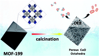
Metal–organic frameworks (MOFs) have been demonstrated as suitable metal sources and sacrificial templates for the preparation of porous transition-metal oxide nanomaterials. Herein, porous CuO micro-polyhedra assembled from numerous nanoparticles are synthesized simply by employing a Cu-based MOF as a precursor via a one-step calcination process under an air atmosphere. Benefitting from their high porosity and large specific surface area, porous CuO materials exhibit excellent electrocatalytic performances toward water oxidation in pH 9.2 KBi solution. A low catalytic onset potential at 1.05 V vs. NHE (NHE = normal hydrogen electrode) is obtained based on electrochemical measurements. A faradaic efficiency of nearly 98% and a stable catalytic current density of 2.2 mA cm−2 are achieved under an applied potential of 1.30 V vs. NHE over an electrolysis period of 10 h. In addition, based on the Tafel plot, the required overpotentials are only ∼410 mV and ∼510 mV for achieving the catalytic current densities of 0.1 mA cm−2 and 1.0 mA cm−2, respectively. The excellent performances make it an appealing candidate as a potential catalyst for green energy applications.
Introduction
Hydrogen energy is a suitable candidate to satisfy the future global need for renewable and carbon-neutral energy resources, due to its high calorific value and the formation of water as the sole combustion product. Electrocatalytic or photo-induced water splitting is considered to be a promising technical scheme for producing hydrogen.1 However, as one of two half reactions for water splitting, water oxidation is deemed as a main bottleneck since it involves complicated endothermic transformation.2 In recent years, some water oxidation catalysts (WOCs) based on transition-metal complexes or oxides have been reported, but most of them are made from noble metals (such as Ru and Ir),3 and the scarcity and high cost would severely hamper their large-scale application. Therefore, it is not preposterous to develop inexpensive and efficient catalysts made from earth-abundant elements for catalysing the oxygen evolution reaction (OER). Among the non-noble transition-metal oxides, copper oxide (CuO) has attracted explosive attention recently due to its low cost, environmentally benign nature and rich redox properties that enable rich redox reactions.4 Since 2014, various CuO-based heterogeneous systems have been reported to activate OER in different pH electrolytes.5 For instance, Du and co-workers reported that the CuO materials with different morphologies were directly synthesized by a hydrothermal procedure from copper salt precursors, the obtained CuO nanowire had a relatively low overpotential of ∼430 mV (0.1 mA cm−2) toward electrocatalytic water oxidation.5a In addition, they utilized an annealing approach to prepare CuO materials on conductive electrodes for electrocatalytic water oxidation in 1.0 M KOH at pH 13.6, the CuO-based catalyst showed excellent electrocatalytic properties and the required overpotentials for current densities of 0.1 mA cm−2 and 1.0 mA cm−2 were 360 and 430 mV, respectively.5b And we also devised a self-supported CuO-based electrocatalyst for water oxidation by electrodepositing a water soluble molecular Cu(II)–TEOA complex (TEOA = triethanolamine).5c Subsequently, the enhanced electrolysis of O2 production by utilizing the H2O2-treated CuO nanostructures were demonstrated by Yeo and a high calculated turnover frequently (TOF) of ∼2.0 × 10⁻³ s−1 was obtained.5d
Although much progress has been achieved for CuO-based WOCs, there is a high demand for further enhancing their electrocatalytic water oxidation activities, and thus improving the energy efficiencies. The nanostructured transition-metal oxides with particular porosity have been confirmed to be good candidates for many electrochemical energy storage and conversion applications, especially in supercapacitors, lithium-ion batteries (LIBs) and water splitting systems,6 since they not only provide high-curvature surfaces to volume ratios and more active sites, but also be in favor of the effective electrolyte penetration and diffusion of substrates, affording high interaction of active species and fast emission of reaction products (e.g., O2). However, the preparation of specific porous nanostructured metal oxides via a facile synthetic approach is still a big challenge to date. In most cases, the synthetic approaches are limited to template-method or water/oil/water emulsion-system with the assistance of organic solvent, and they are usually troublesome and time-consuming.7 Recently, some MOFs have been demonstrated to be ideal sacrificial templates or precursors for preparing porous transition-metal oxide materials through solid-state thermolysis under controllable conditions, due to the periodic permutation of metal nodes and organic motifs in MOF scaffolds and their well-defined porous features.8 For example, Guo et al. obtained porous Co3O4 materials via a two-step thermal decomposition approach from novel Co-based MOF crystals.8a Ai and co-workers also synthesized hierarchically porous Co3O4 architectures with honeycomb-like structures by controlled pyrolysis of Co-based zeolitic imidazolate frameworks, and the obtained Co3O4 materials showed excellent catalytic properties toward water oxidation.8b As for MOF-derived CuO materials, the porous CuO hollow nanostructures were synthesized by a facile carbonization of the Cu-based MOF [Cu3(btc)2, MOF-199] (btc = benzene-1,3,5-tricarboxylate) and these hollow octahedral materials exhibited excellent performance in LIBs with good rate capability.9 Nevertheless, to the best of our knowledge, there is no report available on the utilization of such MOF-derived CuO nanostructures for electrocatalytic water oxidation. Our purpose is to accomplish enhanced OER activities of CuO-based systems by taking advantage of their well-defined porous features. Herein, we employ MOF-199 as metal source and sacrificial template to obtain porous CuO nanomaterials with polyhedral structure through a one-step calcination process under an air atmosphere. Impressively, the as-obtained porous CuO materials exhibited higher intrinsic activity and stability than the commercial CuO catalyst toward electrocatalytic water oxidation in pH 9.2 KBi solution. A small OER overpotential of 360 mV was demonstrated by electrochemical measurements, the large anodic catalytic currents of 2.2 mA cm−2 and long-term durability were achieved under an applied potential of 1.30 V vs. NHE, which are comparable to the most CuO-based WOCs reported so far.5
Experimental section
Materials
All chemicals and metal salts including benzene-1,3,5-tricarboxylate (btc), Cu(NO3)2·2.5H2O, boric acid (HBi), NaOH, KOAc and commercial CuO nanoparticles (diameter: 30 nm) were commercially purchased (Aldrich or Aladdin Co., Ltd.) without further purification unless stated otherwise. The solvents such as absolute ethanol, N,N-dimethyl formamide (DMF) and dichloromethane are analytical grade. All aqueous solutions were prepared with Milli-Q ultrapure water (resistivity: >18 MΩ). The carbon cloth (CC) materials were commercially purchased from Taiwan CeTech Co., Ltd.
Instrumentations and characterizations
Powder X-ray diffraction (PXRD) patterns of all products were collected on a Bruker AXS D8 Advance diffractometer with Cu Kα radiation (λ = 0.1542 nm). Morphologies of MOF templates and MOF-derived CuO materials were observed by field emission scanning electron microscopy (SEM, Hitachi S-4800) and transmission electron microscopy (TEM, JEM-2100). The elemental composition of the samples were characterized by energy-dispersive X-ray spectroscopy (EDX, Oxford instruments X-Max). The thermogravimetric analysis (TGA) was conducted by using a Seiko Exstar 6000 TG/DTA 6300 apparatus with a heating rate of 10 °C min−1 from room temperature to 900 °C in an air stream. X-ray photoelectron spectroscopy (XPS) data were collected by using an ESCALa-b220i-XL electron spectrometer from VG Scientific with 300 W Al Kα radiation, and the binding energies were calibrated with reference to the C 1s peak at 285.0 eV. The specific surface area of the samples was performed by the Brunauer–Emmett–Teller (BET) approach at 77 K using N2 adsorption and desorption isotherms in the Accelerated Surface Area and Porosimetry 2020 (ASAP2020) adsorption apparatus. The pore size and size distribution were measured via the Barrett–Joyner–Halenda (BJH) method.
Preparation of MOF-199
MOF-199 was synthesized according to a modified method from a previous literature.9b 2.5 g of btc (11.9 mmol) was added into 42.5 mL 0.50 M Cu(NO3)2 DMF solution under vigorous stirring. After stirring for 10 min, the above solution was transferred into a jar within 95 mL ethanol/water mixture (v/v, 1
:
1), and the jar was capped tightly and kept at 85 °C for 24 h. After cooled to room temperature, the sky-blue colored products got precipitated and were harvested by filtration. The products were then washed three times each by DMF and ethanol, and finally dried under ambient condition.
Preparation of CuO materials
The porous CuO architectures were obtained by calcination treatment of 4.0 g MOF-199 at 450 °C for 2 h in air with a heating temperature rate of 1 °C min−1.
Preparation of CuO/GC and CuO/CC electrodes
The CuO/GC (GC = glassy carbon) and CuO/CC electrodes were prepared via a drop-casting approach with the assistance of Nafion binders. Initially, 10 mg of CuO materials were dispersed in a mixed solution containing 0.9 mL absolute ethanol and 0.1 mL 5% Nafion solution. The resulting suspensions were then sonicated at room temperature for at least 30 min to make homogeneous CuO inks. Finally, 5 μL of the above inks were dropped onto a polished GC electrode (0.071 cm2) or 30 μL of the inks were dropped onto CC (0.5 cm2) to obtain CuO/GC electrode or CuO/CC electrode, respectively.
Electrochemical measurements
Cyclic voltagrammetry (CV), linear sweep voltagrammetry (LSV), electrochemical impedance spectroscopy (EIS) and controlled potential electrolysis (CPE) measurements were conducted on a Shanghai Chenhua CHI660E electrochemical potentiostat by using a typical three-electrode system. A Pt slice and an Ag/AgCl electrode (saturated KCl solution) were used as counter electrode and reference electrode, respectively. The CuO/GC (0.071 cm2) or CuO/CC (0.5 cm2) electrode was used as working electrode. All potentials measured were converted to NHE scale according to the equation: ENHE = EAg/AgCl + 0.197 V. The CV scans were recorded in a potential range of 0.2–1.6 V vs. NHE with a scan rate of 50 mV s−1. The CPE measurement was performed in a gas-tight single-component electrolytic cell at an applied potential of 1.30 V by using CuO/CC electrode as working electrode, the volume of the KBi solution and the volume of headspace in the electrolytic cell were measured to be 20 mL and 55 mL, respectively. EIS measurement was carried out at the open circuit potential from 10−2 to 105 Hz with an AC amplitude of 5 mV. Prior to each experiment, the electrolyte was degassed by bubbling with high purity nitrogen for 30 min. The amount of generated oxygen during CPE measurement was quantified by gas chromatography (GC) method through manual sampling with a gastight syringe, the calculated amount of O2 was determined by dividing the recorded total passed charge by 4 F.
Faradaic efficiency (FE) = nmeas.O2/ncalcd.O2 × 100%. |
Electrochemical double-layer capacitance (Cdl) and active electrochemical surface area (ECSA) measurement
Cdl for CuO samples was obtained from CV measurements and CV measurements were performed in 0.1 M KBi solution (pH 9.2) under various scan rates from 4 to 36 mV s−1 in the non-faradaic potential range (0.40–0.50 V vs. NHE), the plot of current density (at 0.45 V vs. NHE) against the scan rate has a linear relationship and its slope is equal to Cdl. ECSA was calculated according to the function: ECSA (cm2) = Cdl (without diving the area)/Cs, Cs is the specific capacitance of ideal planar metal oxides having smooth surface, typically taken to be 0.04 mF cm−2.10
Results and discussion
The schematic diagram of the synthetic approach for porous CuO is shown in Fig. 1. The highly uniform MOF-199 with octahedron morphology were obtained via a modified hydrothermal method by using Cu(NO3)2·2.5H2O and btc as metal source and organic ligand, respectively. The PXRD results show that the diffraction peaks of MOF-199 are identical to its simulated patterns (Fig. 1c). The TGA of the MOF template was conducted to identify the calcination temperature. As shown in Fig. 2, the TGA curve for MOF-199 in air presents the first weight loss of 19.1 wt% when the temperature was up to 80 °C, corresponding to the removal of volatile ethanol. The second weight loss of 16 wt% observed in the temperature range from 100 to 300 °C could be attributed to the elimination of terminally coordinated or residual water molecules and DMF. The main weight loss of 40 wt% occurred between 300 and 320 °C and no obvious thermal event happened anymore at higher temperature, indicating the decomposition of MOF-199 and the formation of CuO. Therefore, after thermolysis at 450 °C, the MOF precursors were completely converted to CuO phase.
 |
| Fig. 1 (a) Schematic diagram of the synthetic approach for porous CuO polyhedron; (b) schematic of the crystal structure of MOF-199; (c) PXRD patterns of the as-obtained MOF-199 (black line) and MOF-199 simulated by using the published crystal cif file via Mercury software. | |
 |
| Fig. 2 TGA curves of MOF-199 at a heating rate of 10 °C min−1 from room temperature to 900 °C in an air stream. | |
To further ensure the phase structure of CuO, the sample was investigated by PXRD. As shown in Fig. 3a, all the diffraction peaks are well consistent with the patterns from monoclinic symmetry of CuO (JCPDS card 48-1548), two predominant peaks at 2θ = 35.6 and 38.7 are assigned to the (−111) and (111) phase planes, respectively. In addition, no impurity diffraction peaks was observed, and the EDX spectrum confirms the presence of Cu and O elements in the sample (Fig. S1, ESI†), indicating the high purity of the sample. The morphology and structure of the MOF-199 template and as-prepared CuO product were characterized by SEM and TEM. Fig. 3b and c reveal that MOF-199 templates exhibit perfect octahedron-like morphology, and the octahedral structures consist of an edge length of 40 μm and a diameter of 40–50 μm. Interestingly, after calcination at 450 °C, the morphology of MOF-derived CuO products turns to be polyhedral (Fig. 3d), the magnified SEM images confirm that the obtained CuO are actually porous and composed of numerous CuO nanoparticles with sizes of 100–150 nm, and these nanoparticles would interconnect with each other to form porous agglomerate microstructures (Fig. 3e). The N2 adsorption–desorption isotherms at 77 K and BJH pore size distribution measurements were further performed to evaluate the specific surface area and the porosity of the CuO polyhedron. As shown in Fig. 4, the isotherm presents type IV hysteresis and the loop at relative pressure (P/P0) between 0.7 and 1.0 further suggests its mesoporous structure. The specific surface area of the obtained CuO polyhedron is measured to be 39.13 m2 g−1 and the BJH pore size distribution plot (inset in Fig. 4) shows the pore size mainly distributes centered at 15.0 nm. HR-TEM study was performed to determine the crystal orientation features of CuO products, as shown in Fig. 3f, the observed phase distance between two adjacent lattice fringes is 0.24 nm, corresponding well to (111) plane of monoclinic-phase CuO.9 For comparison, when the calcination temperature was up to 500 °C, the porous CuO polyhedron started to collapse and most of them become irregular agglomerates (Fig. S3, ESI†), indicates that the calcination temperature has a great influence on the morphology of MOF-derived CuO products.
 |
| Fig. 3 (a) XRD patterns of the CuO materials; (b and c) SEM images of MOF-199; (d) SEM image of CuO materials; (e and f) HR-TEM image of CuO materials. | |
 |
| Fig. 4 N2 adsorption–desorption isotherms at 77 K and BJH pore size distribution of the obtained CuO polyhedron. | |
Considering that porous nanostructured CuO might be advantageous for water splitting, their electrocatalytic activities toward water oxidation were investigated by various electrochemical measurements. First, the CuO samples were deposited onto GC electrode via a drop-casting method with the assistance of Nafion binders for CV scans in 0.1 M KBi solution (pH 9.2). In Fig. 5, upon scanning from 0.2 to 1.6 V vs. NHE, one large irreversible oxidation wave beginning at an onset potential of Eonset = 1.05 V appeared with a greatly enhanced current compared with that of using blank GC electrode, accompanied by gas bubbles formation on the surface of working electrode, and the bubbles were further confirmed to be oxygen by gas chromatography analysis, indicating that water oxidation occurred during anodic scan, reflecting a catalytic overpotential of ca. 360 mV. In addition, a broad reduction wave at 0.90 V was observed during the reverse CV scan, corresponding to a reduction of the surface-deposited species.
 |
| Fig. 5 The CVs obtained in a deoxygenated KBi solution (0.1 M, pH 9.2) by using porous CuO polyhedron (black line), commercial-available CuO (red line) and bare GC electrode (blue line) as the catalysts for water oxidation, the scan rate is 50 mV s−1, the contact area of the GC electrode is 0.071 cm2. | |
As comparison, the commercially-available CuO nanoparticles (diameter: 30 nm) showed a higher onset potential at Eonset = 1.25 V and a lower activity. EIS measurement was carried out on as-prepared CuO polyhedron and commercially-available CuO nanoparticles at the open circuit potential from 10−2 to 105 Hz with an AC amplitude of 5 mV. The Nyquist plots (Fig. S4, ESI†) obviously reveal a smaller charge transfer resistance of the as-prepared CuO, indicating the higher conductivity and more efficient charge transfer of porous CuO polyhedron during electrochemical OER process.11 The active surface area were further evaluated by using Cdl and ECSA. The Cdl of porous CuO materials and commercially-available CuO were obtained according to the CV plots recorded at different scan rate in the range of 4–36 mV s−1 in the non-faradaic potential range (0.40–0.50 V vs. NHE). As shown in Fig. S5–S7,† the Cdl of porous CuO materials is 11.2 mF cm−2, whereas the Cdl of commercially-available CuO is only 3.7 mF cm−2. Accordingly, the calculated ECSA for porous CuO materials is 19.9 cm2, which is 3.1 times more than that of commercially-available CuO. Because the values of Cdl and ECSA are proportional to the active surface area of electrocatalysts,12 we can draw the conclusion that the porous CuO materials provide a larger active surface area, which is benefiting from its high porosity and large BET surface area.
The influence of the electrolyte's pH value on the electrocatalytic properties toward water oxidation for porous CuO polyhedron was investigated by LSV measurements in different pH electrolytes. Fig. 6a shows that the CuO samples exhibit highly active toward OER in either neutral or weak basic solution, and the onset catalytic potentials for OER clearly depend on the solution pH value, with a slope of approximately −56 mV per pH in the pH range from 7.20 to 9.20, which can be rationalized by a proton-coupled electron transfer (PCET) process.13 The durability of the porous CuO polyhedron was then assessed by repeated CVs cycled from 0.2 to 1.6 V for one hundred times, after these scans, only a negligible decrease in catalytic current was observed (Fig. S8, ESI†), indicating a strong durability of porous CuO polyhedron during electrolysis.
 |
| Fig. 6 (a) The LSV plots of CuO/GC electrode (0.071 cm2) in different pH electrolytes; (b) the plot of onset potential for electrocatalytic water oxidation vs. pH value. The pH of the electrolyte was adjusted by using controlled microvolumetric additions of 1.0 M KOH or 1.0 M HCl solution and confirmed by pH-meter. | |
To evaluate and quantify the capability of MOF-derived CuO materials, CPE experiment was carried out at the applied potential of 1.30 V without iR compensation in a gas-tight single-compartment electrochemical cell by using CuO/CC (0.5 cm2) as working electrode (because the smooth surface of the GC electrode would lead to the peeling of the coated electrocatalysts during CPE experiments, we used CC to replace GC electrode), and pH 9.2 KBi solution (0.1 M) was utilized as the assistant electrolyte. As shown in Fig. 7, the electrolysis for blank electrode exhibited negligible current at the applied potential. In contrast, the catalytic current was dramatically enhanced for CuO/CC working electrode system, a stable current density of 2.2 mA cm−2 was achieved and retained more than 95% of its maximum value over an electrolysis period of 10 h. During this time, oxygen and hydrogen bubbles were continuously formed at the anode and cathode, respectively, and the amount of produced O2 was quantified by GC analysis, which gave approximately 98 μmol of O2 after 10 h, and the faradaic efficiency was calculated to be nearly 98% for O2 production based on total charge passing through (Fig. S9, ESI†). What's more, the pH decreased by 1.2 units after CPE, consistent with the consumption of OH− by O2 evolution.
 |
| Fig. 7 CPE plots for blank carbon cloth (red line) and CuO/CC cloth at 1.30 V in a closed, deoxygenated single-compartment cell. | |
The phase structure and surface composition for CuO materials after electrolysis were analysed by PXRD and XPS experiments. Firstly, the PXRD patterns of CuO materials recorded after 10 h electrolysis are nearly the same as that from the freshly MOF-derived CuO materials (Fig. 8a). Secondly, after long-term electrolysis, the dried solid was scraped off from CC, and then analysed by XPS. Fig. 8c and d depict the high-resolution XPS spectra of Cu 2p and O 1s, respectively. Two characteristic peaks with binding energies at 933.9 and 953.9 eV are assigned to Cu 2p3/2 and 2p1/2, respectively, consistent with the characteristic peaks of CuO.14 The high-resolution O 1s spectra further confirms the presence of CuO and the binding energy at 529.4 eV is from the oxygen element in CuO. Finally, according to the SEM images, the CuO materials showed almost the same morphologies before and after electrolysis (Fig. S11, ESI†). Based on the above results, no change occurs in the surface properties of porous CuO polyhedron after long-term electrolysis, indicating the good catalyst stability.
 |
| Fig. 8 (a) XRD patterns of porous CuO polyhedron before (red line) and after (black line) 10 h CPE; (b) XPS survey spectra, the binding energies were obtained with reference to the C 1s line at 285.0 eV; (c) high-resolution XPS scan centered on the Cu 2p peak; (d) high-resolution XPS scan centered on the O 1s peak. | |
To gain more insights into the OER kinetics of the porous CuO polyhedron, the Tafel behavior was investigated by using CuO/GC working electrode. The Tafel plot was obtained by measuring the stable current density (j) under various applied potentials as a function of the overpotential (η), where η is defined as the difference between the applied potential minus the cell resistance and the thermodynamic potential toward water oxidation at pH 9.2 (η = Eappl − iR − EpH). The Tafel plot depicted in Fig. 9 shows an appreciable catalytic current density beginning at the overpotential of 360 mV, a slope of 75.5 mV dec−1 for the catalytic current density ranging from 0.10 mA cm−2 to 5.0 mA cm−2 was achieved, indicating that the discharge of OH− is most likely to be the rate-determining step.15 In addition, from the Tafel plot, the required overpotentials are only ∼410 mV and ∼510 mV to achieve a current density of 0.1 mA cm−2 and 1.0 mA cm−2, respectively, demonstrating its favorable kinetics of water oxidation.
 |
| Fig. 9 Tafel plot of CuO/GC electrode measured in pH 9.2 KBi solution. η = Eappl − iR − EpH, Eappl is the applied potential vs. NHE, i is the stable current, R is the uncompensated resistance and EpH is the thermodynamic potential for water oxidation at pH 9.2. | |
Table 1 summarizes the water oxidation performances of in situ-formed CuO from electro-deposition of Cu(II) complexes and as-prepared CuO in different pH electrolytes, the results confirm that the porous CuO polyhedron possess a lower OER onset potential and a higher catalytic activity compared with the in situ-formed CuO.5c,e,f,g,h,i For example, the in situ-formed CuO from Cu–TPA complex requires an overpotential of ∼600 mV to achieve the current density of 1.0 mA cm−2, which is 90 mV more than that of porous CuO polyhedron.5e In addition, the catalytic activities for the porous CuO polyhedron are comparable to those of reported as-prepared CuO-based materials,5a,b,d,j however, for most of them, the catalytic activities are limited to strong alkaline media. Impressively, when performed under weak basic conditions (pH ≤ 9.2), porous CuO polyhedron exhibits greater current and earlier onset of catalytic current, and the mild reaction condition for our system is also appealing and may have great potential in practical applications for clean energy production. These results, when taken together, indicate that the porous CuO polyhedron used as catalyst for water oxidation are remarkable and have great advantages. We believe the following reasons can be attributed to its good catalytic activity. First, the porous CuO polyhedron affords a larger catalytically active surface area in comparison to conventional low-dimensional CuO materials. Second, CuO polyhedron with well-defined porous features provides smooth channels for fast penetration of electrolyte, facilitates the diffusion of reactants as well as the interaction of active species, as demonstrated by a relatively low Tafel slope of 75.5 mV dec−1, indicating its favorable reaction kinetics. Further experimental investigations are under way to use the porous CuO polyhedron as catalyst for photo-catalysing water oxidation under visible light.
Table 1 Comparison of the water oxidation performances for reported CuO-based catalysts
Catalysta |
pH value of the electrolyte |
Onset potential (V vs. NHE)b overpotential (mV) |
η (at 0.1 mA cm−2) |
η (at 1.0 mA cm−2) |
Ref. |
TPA = tris(2-pyridylmethyl)amine; TEOA = triethanolamine; EA = ethylenediamine. Onset potentials of porous CuO polyhedron in this work were defined explicitly by a tangent method.16 |
Cu–TEOA |
pH 12.4 |
— |
550 mV |
780 mV |
5c |
CuO from Cu–TPA |
pH 9.2 |
1.04 V; 350 mV |
470 mV |
600 mV |
5e |
Cu–Bi |
pH 9.0 |
— |
430 mV |
530 mV |
5f |
Cu-bifunctional |
pH 9.2 |
1.04 V; 350 mV |
490 mV |
749 mV |
5g |
CuO–Cu hybrid |
pH 10.8 |
— |
380 mV |
485 mV |
5h |
CuO from Cu–EA |
pH 13.6 |
— |
— |
370 mV |
5i |
CuO nanowires annealed CuO |
pH 9.2 |
1.03 V; 340 mV |
430 mV |
550 mV |
5a |
|
pH 13.6 |
— |
360 mV |
430 mV |
5b |
H2O2-treated CuO |
pH 13 |
0.80 V; 340 mV |
— |
— |
5d |
3D CuO microspheres |
pH 8.5 |
1.22 V; 490 mV |
— |
— |
5j |
Porous CuO polyhedron |
pH 9.2 |
1.05 V; 360 mV |
410 mV |
510 mV |
This work |
Conclusions
In conclusion, our present studies show the facile synthesis of porous CuO polyhedron via solid-state thermolysis by utilizing MOF-199 as metal source and sacrificial template. Thanks to the porous feature and high surface area, porous CuO polyhedron exhibits excellent catalytic activity toward water oxidation in pH 9.2 KBi solution, affords a low overpotential of 360 mV, a high catalytic current density, long-term durability as well as a good faradaic efficiency of nearly 98%. The required overpotentials are only ∼410 mV and ∼510 mV to achieve a current density of 0.1 mA cm−2 and 1.0 mA cm−2, respectively, which are less than those for most reported as-prepared CuO-based OER catalysts. Furthermore, the self-sacrificial template approach for preparation of porous transition-metal materials in our system is steerable and repeatable, and can be easily extended to fabricate the multivariate mixed-metal oxides and carbon material/metal oxide composites with controlled morphologies and structures.
Acknowledgements
This work is financial supported by Ningbo Natural Science Foundation (Grant No. 2016A610069), Ningbo University Research Programs (Grant No. 421401000, 421600580 and XYL16001) and K. C. Wong Magna Fund. T. L. acknowledges the support from Key Laboratory of Photochemical Conversion and Optoelectronic Materials, TIPC, CAS. J. Q. thanks the funding support from the Natural Science Foundation of Zhejiang Province (Grant No. LQ16B010003).
References
-
(a) N. S. Lewis and D. G. Nocera, Proc. Natl. Acad. Sci. U. S. A., 2006, 103, 15729–15735 CrossRef CAS PubMed
;
(b) Solar hydrogen generation-toward a renewable energy future, ed. K. Rajeshwar, R. McConnel and S. Licht, Springer, New York, NY, 2008 Search PubMed
. -
(a) J. Barber, Chem. Soc. Rev., 2009, 38, 185–196 RSC
;
(b) T.-T. Li, Y. Chen, F. Li, W. Zhao, C. Wang, X. Lv, Q. Xu and W. Fu, Chem.–Eur. J., 2014, 20, 8054–8061 CrossRef CAS PubMed
. -
(a) T.-T. Li, W. Zhao, Y. Chen, F. Li, C. Wang, Y. Tian and W. Fu, Chem.–Eur. J., 2014, 20, 13957–13964 CrossRef CAS PubMed
;
(b) H. Over, Chem. Rev., 2012, 112, 3356–3426 CrossRef CAS PubMed
;
(c) Y. Lee, J. Suntivich, K. J. May, E. E. Perry and Y. Shao-Horn, J. Phys. Chem. Lett., 2012, 3, 399–404 CrossRef CAS PubMed
;
(d) Z. Codolà, J. M. S. Cardoso, B. Royo, M. Costas and J. Lloret-Fillol, Chem.–Eur. J., 2013, 19, 7203–7213 CrossRef PubMed
;
(e) C. Rozaina, E. Mayoussea, N. Guilleta and P. Millet, Appl. Catal., B, 2016, 182, 153–160 CrossRef
;
(f) X. Liu, Y. Maegawa, Y. Goto, K. Hara and S. Inagaki, Angew. Chem., Int. Ed., 2016, 128, 8075–8079 CrossRef
. -
(a) E. A. Lewis and W. B. Tolman, Chem. Rev., 2004, 104, 1047–1076 CrossRef CAS PubMed
;
(b) E. Kim, E. E. Chufan, K. Kamaraj and K. D. Karlin, Chem. Rev., 2004, 104, 1077–1133 CrossRef CAS PubMed
;
(c) E. I. Solomon, D. E. Heppner, E. M. Johnston, J. W. Ginsbach, J. Cirera, M. Qayyum, M. T. Kieber-Emmons, C. H. Kjaergaard, R. G. Hadt and L. Tian, Chem. Rev., 2014, 114, 3659–3853 CrossRef CAS PubMed
;
(d) E. I. Solomon, U. M. Sundaram and T. E. Machonkin, Chem. Rev., 1996, 96, 2563–2605 CrossRef CAS PubMed
;
(e) M. B. Gawande, A. Goswami, F.-X. Felpin, T. Asefa, X. Huang, R. Silva, X. Zou, R. Zboril and R. S. Varma, Chem. Rev., 2016, 116, 3722–3811 CrossRef CAS PubMed
. -
(a) X. Liu, S. Cui, Z. Sun and P. Du, Electrochim. Acta, 2015, 160, 202–208 CrossRef CAS
;
(b) X. Liu, S. Cui, Z. Sun, Y. Ren, X. Zhang and P. Du, J. Phys. Chem. C, 2016, 120, 831–840 CrossRef CAS
;
(c) T.-T. Li, S. Cao, C. Yang, Y. Chen, X. Lv and W. Fu, Inorg. Chem., 2015, 54, 3061–3067 CrossRef CAS PubMed
;
(d) A. D. Handoko, A. D. Handoko, S. Deng, Y. Deng, A. W. F. Cheng, K. W. Chan, H. R. Tan, Y. L. Pan, E. S. Tok, C. H. Sow and B. S. Yeo, Catal. Sci. Technol., 2016, 6, 269–274 RSC
;
(e) X. Liu, H. Jia, Z. Sun, H. Chen, P. Xu and P. Du, Electrochem. Commun., 2014, 46, 1–4 CrossRef CAS
;
(f) F. Yu, F. Li, B. Zhang, H. Li and L. Sun, ACS Catal., 2014, 5, 627–630 CrossRef
;
(g) X. Liu, H. Zheng, Z. Sun, A. Han and P. Du, ACS Catal., 2015, 5, 1530–1538 CrossRef CAS
;
(h) J. L. Du, Z. F. Chen, S. R. Ye, B. J. Wiley and T. J. Meyer, Angew. Chem., Int. Ed., 2015, 54, 2073–2078 CrossRef CAS PubMed
;
(i) X. Liu, S. Cui, M. Qian, Z. Sun and P. Du, Chem. Commun., 2016, 52, 5546–5549 RSC
;
(j) X. Q. Du, J. W. Huang, Y. Y. Feng and Y. Ding, Chin. J. Catal., 2016, 37, 123–134 CrossRef CAS
;
(k) N. Cheng, Y. Xue, Q. Liu, J. Tian, L. Zhang, A. M. Asiri and X. Sun, Electrochim. Acta, 2015, 163, 102–106 CrossRef CAS
. -
(a) J. Y. Wang, N. L. Yang, H. J. Tang, Z. H. Dong, Q. Jin, M. Yang, D. Kisailus, H. J. Zhao, Z. Y. Tang and D. Wang, Angew. Chem., Int. Ed., 2013, 52, 6417–6420 CrossRef CAS PubMed
;
(b) Z. Y. Wang, L. Zhou and X. W. Lou, Adv. Mater., 2012, 24, 1903–1911 CrossRef CAS PubMed
;
(c) H. Hu, B. Y. Guan, B. Y. Xia and X. W. Lou, J. Am. Chem. Soc., 2015, 137, 5590–5595 CrossRef CAS PubMed
;
(d) Y. Feng, X. Y. Yu and U. Paik, Chem. Commun., 2016, 52, 6269–6272 RSC
. - M. Fujiwara, K. Shiokawa, Y. Tanaka and Y. Nakahara, Chem. Mater., 2004, 16, 5420–5426 CrossRef CAS
. -
(a) F. Meng, Z. Fang, Z. Li, W. Xu, M. Wang, Y. Liu, J. Zhang, W. Wang, D. Zhao and X. Guo, J. Mater. Chem. A, 2013, 1, 7235–7241 RSC
;
(b) L. Li, T. Tian, J. Jiang and L. Ai, J. Power Sources, 2015, 294, 103–111 CrossRef CAS
;
(c) W. Du, R. Liu, Y. Jiang, Q. Lu, Y. Fan and F. Gao, J. Power Sources, 2013, 227, 101–105 CrossRef CAS
;
(d) S. Chen, M. Xue, Y. Q. Li, Y. Pan, L. Zhu and S. Qiu, J. Mater. Chem. A, 2015, 3, 20145–20152 RSC
;
(e) J. Shao, Z. Wan, H. Liu, H. Zheng, T. Gao, M. Shen, Q. Qu and H. Zheng, J. Mater. Chem. A, 2014, 2, 12194–12200 RSC
;
(f) Y. Lv, W. Zhan, Y. He, Y. Wang, X. Kong, Q. Kuang, Z. Xie and L. Zheng, ACS Appl. Mater. Interfaces, 2014, 6, 4186–4195 CrossRef PubMed
;
(g) T. Ma, S. Dai, M. Jaroniec and S. Qiao, J. Am. Chem. Soc., 2014, 136, 13925–13931 CrossRef CAS PubMed
. -
(a) R. B. Wu, X. K. Qian, F. Yu, H. Liu, K. Zhou, J. Wei and Y. Z. Huang, J. Mater. Chem. A, 2013, 1, 11126–11129 RSC
;
(b) A. Banerjee, U. Singh, V. Aravindan, M. Srinivasan and S. Ogale, Nano Energy, 2013, 2, 1158–1163 CrossRef CAS
. -
(a) F. Song and X. Hu, J. Am. Chem. Soc., 2014, 136, 16481–16484 CrossRef CAS PubMed
;
(b) C. C. L. McCroy, S. Jung, J. C. Peters and T. F. Jaramillo, J. Am. Chem. Soc., 2013, 135, 16977–16987 CrossRef PubMed
. -
(a) X. Chen, N. Q. Zhang and K. N. Sun, J. Mater. Chem., 2012, 22, 15080–15084 RSC
;
(b) Z. L. Zhang, H. Chen, X. L. She, J. Sun, J. Teo and F. B. Su, J. Power Sources, 2012, 217, 336–344 CrossRef CAS
. -
(a) K. Gong, F. Du, Z. Xia, M. Durstock and L. Dai, Science, 2009, 323, 760–764 CrossRef CAS PubMed
;
(b) C. Yuan, L. Yang, L. Hou, L. Shen, X. Zhang and X. Lou, Energy Environ. Sci., 2012, 5, 7883–7887 RSC
;
(c) J. Liao, D. Higgins, G. Lui, V. Chabot, X. Xiao and Z. Chen, Nano Lett., 2013, 13, 5467–5473 CrossRef CAS PubMed
;
(d) K. Wang, H. Wu, Y. Meng and Z. Wei, Small, 2014, 10, 14–31 CrossRef CAS PubMed
. - D. R. Weinberg, C. J. Gagliardi, J. F. Hull, C. F. Murphy, C. A. Kent, B. C. Westlake, A. Paul, D. H. Ess, D. G. McCafferty and T. J. Meyer, Chem. Rev., 2012, 112, 4016–4093 CrossRef CAS PubMed
. -
(a) J. F. Mooulder, W. F. Stickle, P. E. Sobol and K. D. Bomben, Handbook of X-Ray Photoelectron Spectroscopy, ed. J. Chastain, Perkin-Elmer Corp., Eden Prairie, MN, 2nd edn, 1992 Search PubMed
;
(b) M. Durando, R. Morrish and A. J. Muscat, J. Am. Chem. Soc., 2008, 130, 16659–16668 CrossRef CAS PubMed
;
(c) J. F. Moulder, W. F. Stickle, P. E. Sobol and K. D. Bomben, in Handbook of X-ray Photoelectron Spectroscopy: A Reference Book of Standard Spectra for Identification and Interpretation of XPS Data, Perkin-Elmer Corp., Physical Electronics Division, Eden Prairie, MN, 1992, p. 87 Search PubMed
. -
(a) Y. Deng, A. D. Handoko, Y. Du, S. Xi and B. S. Yeo, ACS Catal., 2016, 6, 2473–2481 CrossRef CAS
;
(b) M. Bajdich, M. Garcia-Mota, A. Vojvodic, J. K. Vojvodic, J. K. Norskov and A. T. Bell, J. Am. Chem. Soc., 2013, 135, 13521–13530 CrossRef CAS PubMed
. - L. Zhang, W. He, Y. Li and F. Li, RSC Adv., 2014, 4, 43357–43365 RSC
.
Footnote |
† Electronic supplementary information (ESI) available. See DOI: 10.1039/c6ra18781a |
|
This journal is © The Royal Society of Chemistry 2016 |
Click here to see how this site uses Cookies. View our privacy policy here.