
Influence of thermophilic Bacillus subtilis YB7 on the biodegradation of long chain paraffinic hydrocarbons (C16H34 to C36H74)†
N. Sakthipriyaa,
Mukesh Dobleb and
Jitendra S. Sangwai*a
aFlow Assurance Laboratory, Petroleum Engineering Program, Department of Ocean Engineering, Indian Institute of Technology Madras, Chennai – 600 036, India. E-mail: jitendrasangwai@iitm.ac.in; Fax: +91-44-2257-4802; Tel: +91-44-2257-4825
bBio Engineering Laboratory, Department of Biotechnology, Indian Institute of Technology Madras, Chennai – 600 036, India
First published on 22nd August 2016
Abstract
The long chain paraffinic hydrocarbons (waxes) present in crude oil pose serious issues in the upstream oil and gas industries. The waxy hydrocarbons are reflected in their resistance to biodegradation when released into the environment. Microbial degradation of these paraffins is an alternative way to address the various issues effectively and economically. The present study investigates the utilization of thermophilic Bacillus subtilis isolated from Chennai, India for the degradation of waxes, such as n-hexadecane, n-eicosane, n-tetracosane, n-octacosane, n-dotriacontane and n-hexatriacontane. Several experiments have been conducted to observe the efficiency of Bacillus subtilis at various operating temperatures and concentrations of paraffins. We inspected the primary degradability of paraffins using gas chromatography mass spectrometry and ultimate biodegradability using viscosity analysis. The degradation of paraffins at 50 °C was observed to be 60 to 77% in one day, and 78 to 98% in ten days. The performance of Bacillus subtilis at 50 °C was observed to be higher than the performance observed at 35 and 75 °C. The viscosity of paraffins has reduced with decrease in carbon number and increase in degradation time. The functional groups present after the biodegradation were investigated using Fourier transform infrared spectroscopy. We also observed that the energy required for the activation of paraffin degradation increases with decrease in enzymatic activity and increase in carbon number. The current study with the information on the percentage degradation and viscosity reduction during the degradation of various long chain paraffins will add value in the development of a robust model for microbial degradation of complex mixtures of hydrocarbon systems suitable for upstream oil and gas applications.
1. Introduction
Crude oil is a complex mixture of hydrocarbons comprising of paraffins, aromatics, naphthenes and resins. Crude oil containing long chain paraffins with carbon numbers greater than C15+ (waxes) is referred to as waxy crude oil.1 These paraffins tend to form waxy crystals when the temperature of the system reduces below the wax appearance temperature (WAT) during the flow of crude oil from the reservoir to the surface facilities. The waxy hydrocarbons, thereafter, start depositing on the walls of the production facilities, block the pipeline, challenge the flow assurance and reduce the crude oil recovery.2 It is also known that the total quantity of petroleum hydrocarbons released in the marine environment from various sources is approximately 1.3 MTPA as per the current statistics.3 These long chain paraffins remain persistent on the top of the aqueous layer in the marine environment, due to their low volatility, and cause serious environmental pollution.4 The conventional methods, such as pigging, dispersants, organic solvents, etc., for flow assurance; thermal, chemical, and gas injection for enhanced oil recovery (EOR); and booms, skimmers, sorbents, in situ burning and dispersants for oil spill treatment may not be suitable to address these issues and may also create adverse ecological effects.5–7 It is, therefore, necessary to degrade the long chain paraffins efficiently and economically to overcome the various issues.8The microbial degradation of long chain paraffins is considered to be an economically and environmentally safer method as compared to other conventional techniques.3,9 Bacterial strains able to degrade petroleum hydrocarbons are omnipresent in nature.10 Microbes that can degrade paraffins molecules by producing various by-products may further add value for oilfield operations. Microorganism cracks the long chain paraffin into short chain paraffin with the help of enzymes and by-products, and thus, increases the API (American Petroleum Institute) gravity and reduces the molecular weight of the crude oil. The enzymes present in bacteria act as a catalyst to enhance the rate of paraffin degradation.11 Indeed, the chemical composition of the hydrocarbon and the bacterial metabolism are the deciding factors for the degradation of the long chain paraffins.12
Degradation of alkanes/hydrocarbons using microorganisms has been investigated by many researchers. Al-wasify et al.13 observed 88% of n-alkanes were degraded using microbial consortium at 22 °C. Sakai et al.14 performed the degradation of alkanes using Acinetobacter at 28 °C, and observed that the growth of microorganism and the degradation of hydrocarbon has been enhanced by adding the external agent plysurf. da Cunha et al.15 evidenced the recovery of bacteria from deep subsurface rock samples in oil reservoirs at the depth of about 2000 m. He also utilized the recovered Bacillus species for the degradation of aromatics and observed that the degradation undergoes the ortho pathway. Various researchers have studied the effect of Rhodococcus and Pseudomonas species on the crude oil degradation, particularly focusing on the degradation capabilities on the middle to moderately long chain alkanes using complex nutrient medium.16–19 Tzintzun-Camacho et al.20 studied the effect of Xanthomonas species, Acinetobacter bouvetii, Shewanella species and Defluvibacter lusatiensis for their ability to degrade short chain hydrocarbon, which indeed have shown positive impact. Bacillus, Geobacillus, Thermomicrobium, Thermus are some of the thermophilic bacterial genus that have been researched to degrade hydrocarbons up to octadecane (C18).21 The first study of thermophilic bacteria was proposed by Wang et al.22 He isolated Geobacillus thermodenitrificans and observed that the isolated bacteria can grow on C15 to C36. Table 1 shows a brief review on the few selected microorganisms used for the paraffin degradation along with the concentration of paraffins, degradation conditions and outcome. Few of the studies on Bacillus subtilis strains have been researched at various temperature, but with much lower carbon number.
| S. No. | Microorganisms | Degradation (%) | Concentration of paraffins (g L−1) | Temperature (°C) | Days | Reference |
|---|---|---|---|---|---|---|
| 1 | Achromobacter | 97 | — | 28 | 10 | 23 |
| 2 | Acinetobacter | 82 | 0.5 | 30 | 28 | 3 |
| 3 | Bacillus methylotrophicus | 92 | 2 | 35 | 14 | 4 |
| 4 | Bacillus lincheniformis | 61 | 1 | 35 | 7 | 2 |
| 5 | Corynebacterium | 82 | 1 | 30 | 7 | 24 |
| 6 | Pestalotiopsis | 92 | — | 25 | 30 | 25 |
| 7 | Pimelobacter simplex | 93 | 1 | 15 | 14 | 26 |
| 8 | Pseudomonas aeruginosa | 79 | 1 | 35 | 7 | 2 |
| 9 | Pseudomonas aeruginosa | 30 | — | 30 | 14 | 27 |
| 10 | Pseudomonas aeruginosa | 43 | 0.5 | 30 | 5 | 28 |
| 11 | Pseudomonas | 32 | — | 37 | 12 | 29 |
| 12 | Scenedesmus obliquus | 46 | 0.5 | 25 | 7 | 30 |
It is known that the remedial techniques for oil spill and bioremediation require mesophilic bacteria growing at atmospheric temperature, and EOR techniques require thermophilic bacteria growing at or about 50 °C. However, most of the studies reported in open literature on the microbial degradation of hydrocarbons have been carried out at atmospheric condition. Thus, the growth of microorganism in the presence of hydrocarbons at conditions suitable for hydrocarbon degradation is a particular problem which need to be understood for their successful oilfield applications.
Along with the temperature and carbon chain length, some of the areas that need to be researched are on understanding the viscosity during microbial degradation of hydrocarbon, degradation capabilities for hydrocarbons containing higher carbon numbers at higher temperature and various concentration of hydrocarbons. The solubility of paraffins reduces with increase in carbon chain length (carbon number) and decrease in temperature, and thereby affects the viscosity and bioavailability of the medium.
In our previous work,31 we have performed the studies concerning the production of biosurfactant using Bacillus subtilis in the presence of various paraffinic waxes, such as, such as n-hexadecane (C16H34), n-eicosane (C20H42), n-tetracosane (C24H50), n-octacosane (C28H58), n-dotriacontane (C32H66), and n-hexatriacontane (C36H74). Biosurfactants help the microorganism to breed on paraffinic substrate and in turn increase the bioavailability. Biosurfactant produced by the microorganisms form the emulsion and increase the surface area for the growth of microorganism on hydrocarbon substrate. Due to this, better contact between the hydrocarbon substrate and water interface is developed and the bioavailability is increased. Biosurfactant thus, indirectly helps in improving the degradation of long chain paraffins.31 In addition, biosurfactant may form micelles around the hydrocarbon molecules, thus reducing the viscosity.32,33 Hence, it is motivating to estimate the effectiveness of the bacteria in viscosity reduction of waxes along with the degradation, which is relevant for efficient bioremediation and oil spill tactics for the extensiveness of investigation.
In this study, an attempt has been made to utilize the single strain Bacillus subtilis YB7 for the degradation of various long chain paraffins at different temperature conditions. This work has been focused on the degradation of various long chain paraffins representing waxy nature, such as C16H34, C20H42, C24H50, C28H58, C32H66, and C36H74 using Bacillus subtilis. The degradation has been studied at various temperature conditions with simple nutrient medium suitable for enhanced oil recovery and marine pollution. This study also observed the variation of viscosity throughout the degradation of waxes and viscosity variation with temperature post-degradation of hydrocarbons. Information on the activation energy required for degradation and the effect of enzymes has also been discussed.
The schematic representation of various investigations that have been carried out for this study is shown in Fig. 1. We believe that the outcome of the study on various properties, such as viscosity and the change in composition of post-microbial degradation of pure hydrocarbons will add values for possible robust model development of microbial degradation of complex mixtures of hydrocarbon systems suitable for oilfield application.
 | ||
| Fig. 1 Schematic representation of various investigations carried out for this study. | ||
2. Experimental section
2.1. Cultivation of microorganism on paraffins
Bacillus subtilis YB7 (GenBank accession number: GQ241354), an organism isolated from a polymer dump site near Chennai Coast, India was used in this study.34 A colony from the Petri plate stock was transferred into a 10 mL of sterilized nutrient broth in 50 mL conical flask and incubated overnight in a shaker (Orbitek, Scigenics Biotech, India). After cultivation, 2% of seed culture was added to the sterilized nutrient medium in various flasks containing different hydrocarbon substrates, such as n-hexadecane, n-eicosane, n-tetracosane, n-octacosane, n-dotriacontane and n-hexatriacontane. The nutrient medium contains 0.3 g potassium dihydrogen phosphate (KH2PO4), 0.6 g disodium hydrogen phosphate (Na2HPO4), 0.2 g ammonium chloride (NH4Cl), 0.5 g sodium chloride (NaCl), 0.8 g glucose (C6H12O6), and 0.01 g magnesium sulfate hepta hydrate (MgSO4·7H2O).30 Three replica of each of the six hydrocarbon waxes, and the control were prepared. The two types of controls were used; one was the bacterial isolate in nutrient medium without any hydrocarbon substrate, and another was the hydrocarbon substrates in nutrient medium without bacteria. The shake flasks were incubated at three different temperatures, such as 35, 50 and 75 °C at 180 rpm. The samples were collected from the flasks at different time intervals to determine degradation, viscosity reduction, activity of enzymes, and activation energy. Table S1 in the ESI† provides the list of chemicals, mass fraction purity and the supplier of chemicals used for the present work. The statistical analysis was performed by carrying out all the experiments including the controls in triplicates. The results represented mean ± standard deviation.2.2. Growth of microorganism
The cells were grown on pure paraffins and the growth curve of bacteria in the study was routinely assessed by measuring biomass dry weight and colony forming units (CFU). CFU was used to estimate the viable bacterial count. It was determined by spread plate method reported by Hassanshahian et al.35 The biomass dry weight, which indicates the total weight of the organism was measured according to the reported procedure.7CFU was used under two circumstances. Initially, CFU was determined to find out the growth of bacteria at different temperatures without any hydrocarbon substrate. For this case, Bacillus subtilis was cultivated in flasks for one week without any hydrocarbon substrate at different temperatures, such as 20, 30, 40, 50, 60, 70 and 80 °C. The cultivated bacterial cells were counted using spread plate method to find the CFU. Based on these results the optimum temperature of the growth was observed to carry out the further experiments in the presence of hydrocarbon substrate and again the growth was monitored again using CFU and biomass dry weight.
2.3. Extraction of residual hydrocarbons and its compositional analysis
The residual hydrocarbon and products produced during the degradation of paraffin were extracted from the bacterial cell-free culture supernatant using equal volume of solvent (2 mL). Ethyl acetate was used to extract the hydrocarbon samples degraded from C16H34 and C20H42 while toluene was used to extract the samples degraded from C24H50, C28H58, C32H66, and C36H74. The solvent in the extracted samples were evaporated using rotary vacuum evaporator (Buchi rota vapor, Switzerland), and concentrated again using the same solvent as above and filtered through 0.2 mm filter paper to remove the impurities. The concentrated hydrocarbon extracts were analyzed for degradation and formation of new functional groups using gas chromatography-mass spectroscopy (GC-MS) and Fourier transform infrared spectrometer (FTIR), respectively.The GC-MS (GC-Mate II, JEOL, USA) was operated in the SIM (selective ion monitoring) mode. The injection volume of the degraded paraffin sample was 1 μL, and helium at a flow rate of 1 mL min−1 was used as a carrier gas. The paraffin sample was injected at a flow rate of 4 mL min−1 and the purge flow rate was set at 3 mL min−1. The interface and injector temperatures were maintained at 350 and 220 °C, respectively. Both the control experiment (before degradation) and hydrocarbons after degradation were done in a similar manner. The percentage degradation of long chain paraffins were evaluated by relating the area under the peaks obtained before and after degradation in the chromatograms.
The functional groups and the types of bond present before and after the degradations were analyzed using FTIR (Spectrum 1, Perkin Elmer, USA). For the liquid paraffins, a hydrocarbon drop extracted from the above-mentioned procedure was injected on a highly polished plate. The hydrocarbon extract was spread by fixing another transparent polished plate. These two plates were closed together and positioned in the sample holder on the FTIR apparatus to obtain the spectra. For the pure solid waxes (except hexadecane), potassium bromide pellets were prepared by mixing with respective waxes, and the mixture was placed in the sample holder to get the background spectrum. The spectral readings were performed in a transmittance mode in the range 4000–450 cm−1 with the resolution of 1 cm−1.
2.4. Measurement of viscosity
The discussion about viscosity was concentrated to understand the (1) variation of viscosity during the biodegradation at 35, 50 and 75 °C and (2) viscosity variation with temperature after degradation. The bacterial cells were centrifuged (5804 R, Eppendorf, India) at room temperature at 10![[thin space (1/6-em)]](https://www.rsc.org/images/entities/char_2009.gif) 000 × g for 10 min and the biomass free culture supernatants were measured for viscosity using a stress controlled rheometer (MCR 52, Anton Paar, Austria) fitted with double gap geometry. The samples incubated at 35, 50 and 75 °C in shake flasks were collected on every five days and measured for the viscosity reduction. The degraded samples were also checked for the viscosity reduction at temperatures ranging from 5 to 80 °C to analyze the mobility of the samples at various temperature conditions post-degradation.
000 × g for 10 min and the biomass free culture supernatants were measured for viscosity using a stress controlled rheometer (MCR 52, Anton Paar, Austria) fitted with double gap geometry. The samples incubated at 35, 50 and 75 °C in shake flasks were collected on every five days and measured for the viscosity reduction. The degraded samples were also checked for the viscosity reduction at temperatures ranging from 5 to 80 °C to analyze the mobility of the samples at various temperature conditions post-degradation.
2.5. Activity of enzymes
Alkane monooxygenase, alcohol dehydrogenase and aldehyde dehydrogenase are the enzymes observed to be liable for the long chain paraffin degradation. The activity of enzymes were determined spectrophotometrically at 340 nm by quantifying the formation of NADH according to the reported procedure.36–38 The materials used for this analysis with their purity were given in Table S1.† The time course reading (absorbance) was observed in UV spectrophotometer (V550, Jasco, Germany) at a wavelength of 340 nm for 5 min. The activity of enzyme in units per mL in the presence of paraffins were determined according to the molar extinction coefficient for NADH. One unit of enzyme is the amount of the enzyme, which catalyzes the reaction of 1 μmol of NAD+ per min.37 The activity in the presence of hydrocarbon for each of the enzymes was calculated by eqn (1).
 | (1) |
2.6. Activation energy
Activation energy is the minimum energy essential to initiate the chemical reaction. The activation energy was calculated using Arrhenius equation which contributes the requirement of the rate constant of chemical reactions on temperature and activation energy as shown below.
 | (2) |
Taking logarithm of eqn (2), we get,
 | (3) |
The activation energy was calculated from the slope obtained by plotting lnk versus 1/T. The value of the slope (m) is equal to −Ea/R, whereas A is calculated from the intercept of the graph.
3. Results and discussions
3.1. Effect of temperature on bacterial growth without paraffins
The growth of the bacteria is the prime factor affecting the degradation, since each cell involves in the degradation process. Before investigating the effect of bacteria on hydrocarbon degradation, it is important to know the growth of bacteria in the absence of it, and understanding the optimum temperature at which higher growth is expected. Fig. 2 shows the growth of Bacillus subtilis in minimal nutrient media without any long chain paraffins. The bacterial growth has been observed to be 12, 20, 20, 32, 28, 24 and 20 × 106 CFU mL−1 at 20, 30, 40, 50, 60, 70 and 80 °C, respectively. The bacterial growth has increased until 50 °C, and decreased thereafter. This indicates that the optimum temperature for the growth of the bacteria is 50 °C, and the same has been considered for further comparison. Based on this, we have chosen 35, 50 and 75 °C as operating temperatures to analyses the bacterial degradation of pure hydrocarbons. | ||
| Fig. 2 Growth of Bacillus subtilis after one week as a function of temperature; arrow head indicates optimum growth temperature; results represent mean ± standard deviation of three independent experiments. | ||
3.2. Effect of various concentration of paraffins on bacterial growth
The concentration of the hydrocarbon substrate should be optimized before carrying out the degradation process at temperature where maximum amount of bacteria is produced. Bacillus subtilis was allowed to grow on different concentration of hydrocarbon (0.5, 0.75, 1, 1.25, and 1.5 g L−1) for a week at 50 °C to find the effect of concentration of hydrocarbon substrate on its growth. Fig. 3 shows the growth curve of Bacillus subtilis after one week with change in concentration of various hydrocarbon substrates at 50 °C. Fig. 3a shows the biomass dry weight with various concentration of hydrocarbon substrate, which represents the total weight of the organism. The total weight was maximum at the concentration of 1 g L−1. Fig. 3b shows the CFU with various concentration of hydrocarbon substrate. It is inferred that the bacterial count has found to increase up to the hydrocarbon concentration of 1 g L−1, and reduced further as the hydrocarbon concentration increases. This condition of hydrocarbon concentration (1 g L−1) was taken as benchmark for subsequent investigations. Also when the carbon number increases, the microbial growth is decreased. This is due to the increase in hydrophobicity with increase in carbon number.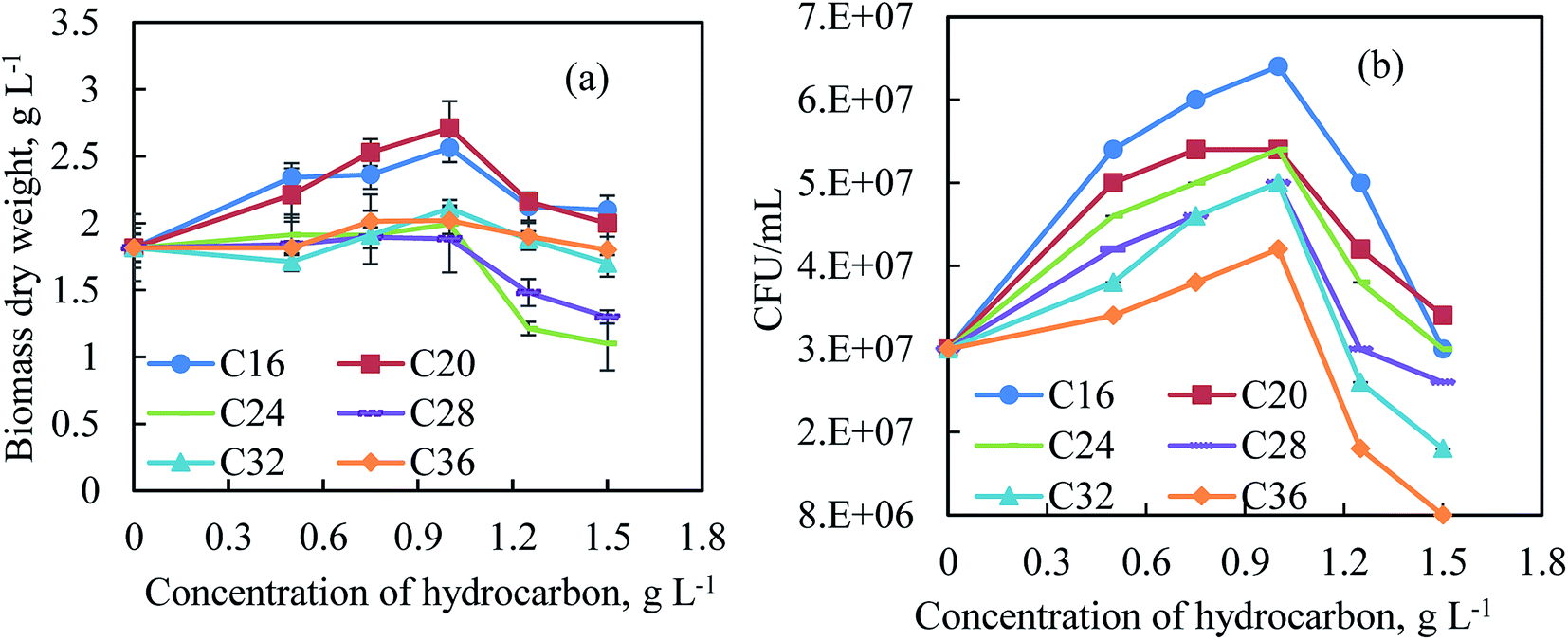 | ||
| Fig. 3 Growth curve of Bacillus subtilis after one week with change in concentration of various hydrocarbon substrates at 50 °C, (a) biomass dry weight; (b) CFU; results represent mean ± standard deviation of three independent experiments. | ||
3.3. Residual hydrocarbon analysis
The mechanism of hydrocarbon degradation involves the direct contact of bacterial cells with the hydrocarbons, and subsequent increase in the cell growth on hydrocarbon substrate results in faster hydrocarbon uptake.39 The paraffin samples incubated with B. subtilis were collected on 0th, 1st, 10th, and 20th day to analyze the extent of degradation. The degree of biodegradation as well as products formed were determined using a GC-MS. Fig. S1a–f† shows the gas chromatograms of pre- and post-degradation of various paraffins using Bacillus subtilis YB7 at 50 °C from 0th day to 15th day post-degradation and the same was used to calculate percentage degradation. Fig. 4 shows the GC-MS of various pure paraffins before (0th day) and after degradation (10th day) using Bacillus subtilis incubated at 50 °C. The hydrocarbon degradation has also been analyzed at the temperatures of 35 and 75 °C. Fig. 5 shows the percentage degradation of paraffins as a function of carbon number and incubation time at different degradation temperatures. The degradation at 50 °C has been observed to be higher than the degradation at 35 and 75 °C. This may be due to the higher growth of bacteria at 50 °C as evidenced from Fig. 2. The degradation of long chain paraffins, C16, C20, C24, C28, C32 and C36, at 50 °C in 10 days were 98, 94, 91, 85, 80 and 78% (w/w), respectively, whereas the respective values in 1 day were 77, 71, 68, 64, 63 and 60% (w/w). There was no significant difference in degradation values between 10th and 20th day post-incubation. Hence, the inoculated bacteria with different hydrocarbon substrates were not sampled after 20 days of incubation for degradation analysis. However, for other studies such as viscosity and enzyme activity, we have extended the investigation above 20 days and presented in subsequent sections. It has been observed that the increase in carbon number in the hydrocarbon system increases the time required for degradation process, and decreases the percentage degradation of paraffins. Though, the extent of degradation reduces with increase in carbon number, about 60 to 70% of C32 and C36 degraded in 1 day. This shows that B. subtilis has good degradation capabilities at 50 °C on pure long chain paraffins. | ||
| Fig. 4 GC-MS images of various pure paraffins before (0th day) and after degradation (10th day) using Bacillus subtilis incubated at 50 °C, (a) n-hexadecane (C16H34); (b) n-eicosane (C20H42); (c) n-tetracosane (C24H50); (d) n-octacosane (C28H58); (e) n-dotriacontane (C32H66); (f) n-hexatriacontane (C36H74). | ||
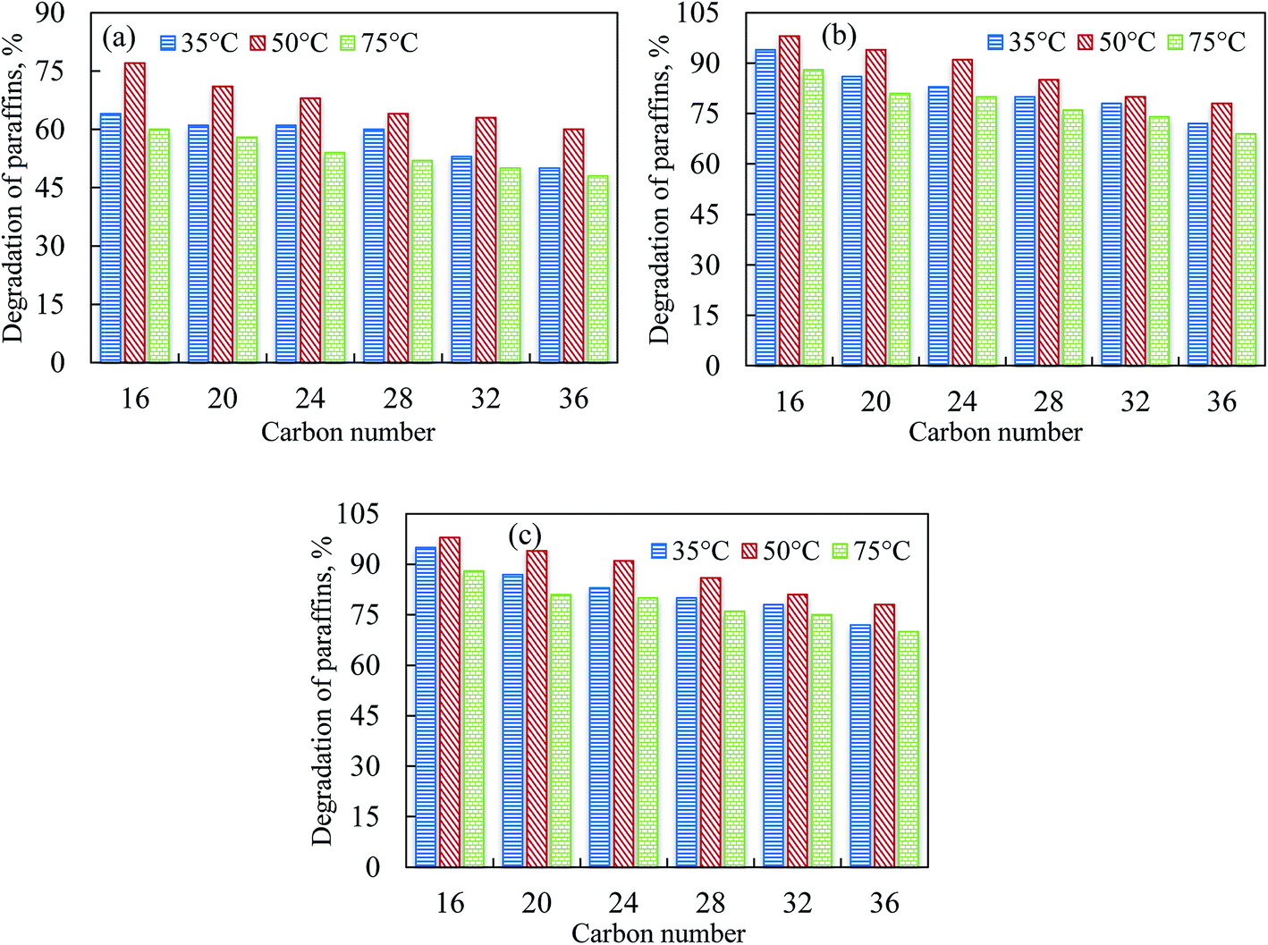 | ||
| Fig. 5 Percentage degradation of hydrocarbons using Bacillus subtilis as a function of carbon number after, (a) 1 day; (b) 10 days; (c) 20 days. | ||
3.4. Analysis of functional groups
The functional groups present in the hydrocarbons pre-, and post-degradation have been confirmed using FTIR spectroscopy. The maximum degradation was found to be around 10th day of post-incubation. Therefore, the functional groups have been analyzed only for 0th day and 10th of incubation for the sake of simplicity. Fig. 6 shows the FTIR spectra of different hydrocarbons pre- and post-microbial degradation at 0th and 10th day, respectively. As seen in a Fig. 6a, the peak has been observed in the range of 2900 cm−1 which is due to –C–H stretching mode of the alkyl chain present in paraffins. The presence of the peak in this range indicates the presence of paraffins (i.e., the hydrocarbon substrate used here) before degradation. It is observed that the initial paraffin band has utmost reduced after the degradation of paraffins, and various other bands, such as carboxylic acid, alcohol, etc., have been formed. The peak in the spectra at 1700 cm−1 stands for the C![[double bond, length as m-dash]](https://www.rsc.org/images/entities/char_e001.gif) O group, and confirms the production of carboxylic acid post-degradation. The weak band around 1400 cm−1 is due to the –C–CH2 and –C–CH3 groups. The stretch at 1200 cm−1 represents the C–O bond, which may be either alcohol or esters that have been produced after degradation of paraffins. The band at around 1100 cm−1 is due to C–O–C stretching, which indicates the presence of ether.40 Similar trends have been obtained for other hydrocarbons as shown in Fig. 6a–f. It has also been observed that the intensity distribution of the spectral bands did not vary significantly among all hydrocarbon substrates, which indicate the consistency of the microorganism to degrade the long chain hydrocarbon. This observation lays in-line with the findings from GC-MS.
O group, and confirms the production of carboxylic acid post-degradation. The weak band around 1400 cm−1 is due to the –C–CH2 and –C–CH3 groups. The stretch at 1200 cm−1 represents the C–O bond, which may be either alcohol or esters that have been produced after degradation of paraffins. The band at around 1100 cm−1 is due to C–O–C stretching, which indicates the presence of ether.40 Similar trends have been obtained for other hydrocarbons as shown in Fig. 6a–f. It has also been observed that the intensity distribution of the spectral bands did not vary significantly among all hydrocarbon substrates, which indicate the consistency of the microorganism to degrade the long chain hydrocarbon. This observation lays in-line with the findings from GC-MS.
 | ||
| Fig. 6 Functional groups present before (0th day) and after degradation (10th day) of various pure paraffins using Bacillus subtilis incubated at 50 °C, (a) n-hexadecane (C16H34); (b) n-eicosane (C20H42); (c) n-tetracosane (C24H50); (d) n-octacosane (C28H58); (e) n-dotriacontane (C32H66); (f) n-hexatriacontane (C36H74). | ||
3.5. Flow analysis during microbial degradation
The culture supernatant of the samples during the course of degradation were taken for the viscosity studies. Experiments were carried out at a constant shear rate of 1000 s−1. The results have been compared against a control (microbial sample without any hydrocarbon) and non-inoculated hydrocarbon sample. Percentage (%) reduction in viscosity with respect to initial viscosity of the paraffinic samples have been analyzed at various conditions. Fig. 7 shows the apparent viscosity profiles with time for various long chain paraffins pre- and post-degradation at 50 °C and at a constant shear rate of 1000 s−1. The viscosity of various paraffins decrease with time and followed an order C16 > C20 > C24 > C28 > C32 > C36 > non-inoculated paraffin samples > control. | ||
| Fig. 7 Viscosity of hydrocarbons as a function of degradation time at 50 °C; (inset-reduction in viscosity (%) of hydrocarbons after 10 days as a function of carbon number at different temperatures); results represent mean ± standard deviation of three independent experiments. | ||
Inset graph in the Fig. 7 shows the % viscosity reduction as a function of carbon number after 10 days of degradation at 35, 50 and 75 °C. It has been observed that the % reduction in viscosity decreases with increase in carbon number. Also, % reduction in viscosity has been found to be higher at 50 °C than 35 °C and 75 °C. A maximum of 46% of viscosity was reduced in 10 days for C16 substrate degraded at 50 °C, while it has been found to be 40% for C36. Thus, the % reduction in viscosity decreases with increase in carbon number which is in-line with the degradation profile. The reduction in viscosity of C16, C20, C24, C28, C32 and C36 have been found to be 66, 62, 60, 59, 58 and 58%, respectively in 40 days, whereas the corresponding values in 1 day were 36, 34, 30, 27, 25 and 19% with respect to the initial viscosity of corresponding paraffins at 50 °C. It has been observed that the drop in viscosity is significant after 5 days, which is due to the increased production of biosurfactant, which is directly related to the biodegradation of long chain hydrocarbons. Table S2 (in ESI†) provides the viscosity of various paraffins at 35 °C and 75 °C as well. The % reduction in viscosity for C16, C20, C24, C28, C32 and C36 was found to be around 30, 31, 29, 25, 22 and 15% at 35 °C, whereas the respective values at 75 °C were 28, 26, 24, 19, 17 and 11% after 1 day of post-incubation. The % reduction in the viscosity after 40th day post-degradation has been observed to be 61, 55, 52, 51, 50 and 46% (35 °C) and 54, 50, 48, 43, 40 and 36% (75 °C).
3.6. Flow analysis after microbial degradation
It is to be noted here that the discussion made in Section 3.5 was more focused towards the viscosity variation during hydrocarbon degradation. It is also essential to know the viscosity variation of hydrocarbon sample post-degradation after discarding the biomass. This study is important to recognize the flow characteristics of crude oil/hydrocarbon after degradation with change in temperature. For example, bacteria may have significant impact on hydrocarbon degradation in the reservoir during enhanced oil recovery in oil and gas industry, but at certain point the biomass has to be removed, and the degraded oil sample has to be flow towards production well or in surface facilities including crude oil pipelines. In such situations, viscosity variation of these hydrocarbon sample post-degradation will be useful for flow analysis.For all the cases as discussed in Section 3.5, it has been observed that the viscosity remains almost stable after 10th day post-incubation. Therefore, the 10th day sample has been taken from the culture broth (incubated at 50 °C) and further investigated for variation in the viscosity at various temperature to find the mobility of the samples after degradation. Fig. 8 shows the variation of viscosity of the culture supernatants containing hydrocarbons degraded using Bacillus subtilis incubated at 50 °C. Viscosity was measured by varying the temperature from 5 to 80 °C along with the non-inoculated hydrocarbon samples (filled symbols) and control. It has been observed that the viscosity of various hydrocarbons, such as C16, C20, C24, C28, C32 and C36, has reduced by the order of 81, 57, 79, 78, 78 and 76% at 80 °C, whereas, the corresponding values at 5 °C are 45, 38, 32, 31, 28 and 18%. At close-to-room temperature conditions (35 °C) the corresponding values have been found to be 62, 52, 53, 69, 66 and 63%. One may argue here that the reduction in viscosity of hydrocarbon sample (as shown in Fig. 8) may be purely due to increase in temperature. However, in contrast to that, that there was no significant reductions in the viscosity of the control sample and non-inoculated samples (filled symbols in Fig. 8) with respect to the temperature. The variation of viscosity for inoculated samples (represented by unfilled symbols in Fig. 8) with temperature has shown rapid decrease as compared to the control and non-inoculated samples. It can be inferred that the viscosity reduction of degraded hydrocarbon (inoculated samples) is primarily due to the microbial degradation, and not much due to the influence of temperature.
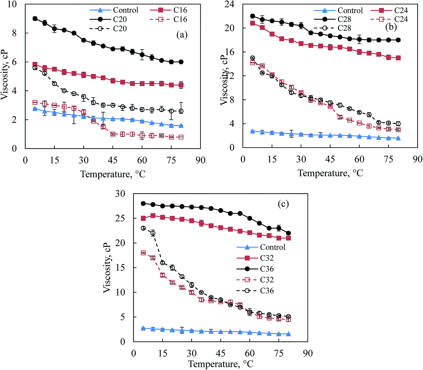 | ||
| Fig. 8 Effect of temperature on the viscosity of various paraffins, (a) C16H34 & C20H42; (b) C24H50 & C28H58; (c) C32H66 & C36H74; filled symbols show non-inoculated samples and unfilled symbol shows inoculated sample after 10th day post-degradation using Bacillus subtilis at 50 °C; results represent mean ± standard deviation of three independent experiments. | ||
3.7. Activity of enzymes
The main step in the aerobic degradation of alkanes by microorganisms is catalyzed by oxygenases.24 This enzyme hosts the oxygen atom derived from the molecular oxygen in alkane substrate, and plays a significant part in crude oil and hydrocarbon degradation. Fig. 9 shows the activity of alkane monooxygenase, alcohol dehydrogenase and aldehyde dehydrogenase enzymes with respect to temperature in the presence of waxy hydrocarbons. The activities of enzymes at 50 °C has been found to be more than the activity at 35 °C and 75 °C. Fig. S2† shows the activity of alkane monooxygenase, alcohol dehydrogenase and aldehyde dehydrogenase at 35, 50 and 75 °C in the presence of waxy hydrocarbons with time. From the results, it has been revealed that the activities of alkane monooxygenase and aldehyde dehydrogenase are higher than the alcohol dehydrogenase. The activity of the enzymes has attained maximum value in 10 days and reduced thereafter as the bacteria reached the dead phase. It has also been inferred that the increase in carbon atoms in hydrocarbon reduces the accessibility of enzymes to the paraffin substrate and in-turn affects the degradation. | ||
| Fig. 9 Enzymatic activity of the hydrocarbon samples on 10th day of incubation, (a) alkane monooxygenase; (b) alcohol dehydrogenase; (c) aldehyde dehydrogenase; one unit of enzyme is the amount of the enzyme, which catalyzes the reaction of 1 μmol of NAD+ per min; results represent mean ± standard deviation of three independent experiments. | ||
3.8. Activation energy
The activation energy and pre-exponential factor required for the degradation reaction at different carbon number hydrocarbon substrates is shown in Fig. 10. The waxes studied here requires higher energy to activate a molecule to start the degradation process, since they are not polar in nature.41 Enzymes are the biological catalysts that lower the activation energy and affect the rate of the reaction. It has been observed that the energy required for degradation increases with the increase in time (Fig. 10). This is due to the fact that the activity of enzymes has found to decrease after 10 days of incubation, and hence, high energy is necessary to overcome the less activity of microorganisms or enzymes. When the activities of enzymes are high, reaction found to proceeds faster, and thus the rate constant (k) increases. The rate constants for each degradation process have been calculated using the Arrhenius expression and are given in Table S3.† The energy required to activate C36 was found to be 7551, 7557 and 8845 J mol−1 on 1st, 10th, and 20th day post-degradation. The energy required for degradation has increased on 10th day, and further increased on 20th day. Also, the activation energy has been found to increase with increases in the carbon number. It requires 2150 J mol−1 to activate C16 molecule, whereas 7551 J mol−1 energy is necessary for C36 hydrocarbon. This implies that the energy required to carry out the degradation of a higher carbon number is lesser than the energy required at the lower carbon number.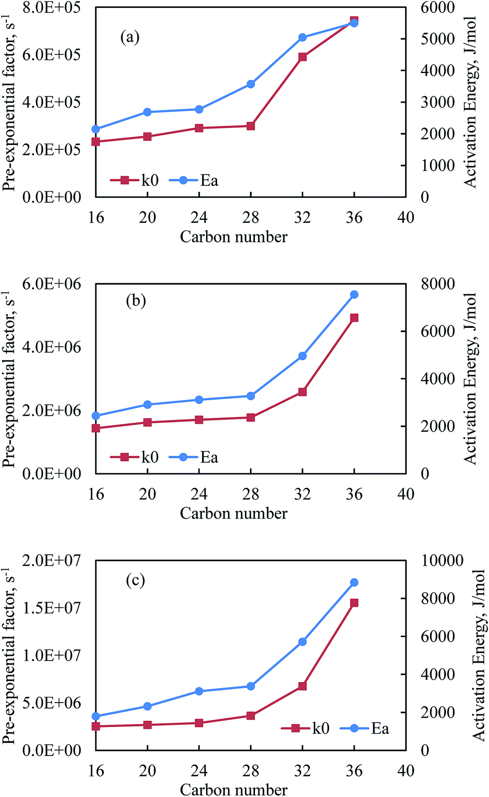 | ||
| Fig. 10 Variation of activation energy and pre-exponential factor as a function of carbon number on (a) 1st day; (b) 10th day; (c) 20th day. | ||
4. Conclusion
The long chain paraffins are the persistent pollutant because of their less volatile and waxy nature. This study presented the biodegradation of six waxy long chain hydrocarbons using Bacillus subtilis isolated from the polymer dump site near Chennai Coast, India, under optimal environmental conditions. Six long chain paraffins representing waxy nature, such as C16H34, C20H42, C24H50, C28H58, C32H66, and C36H74 have been considered for investigations. The information on the optimum concentration of waxes, and temperature required for efficient degradation has been obtained to form a precursor for reverse engineering application of these microorganism. The percentage degradation of hydrocarbons were observed to be in the range of 60 to 77% after 1 day and 78 to 98% after 10 days post-degradation. It was noticed that the hydrocarbons were degraded to carboxylic acid, alcohol, etc. The viscosity reductions of hydrocarbons were found to be in the range of 19 to 36% in 1 day. It is observed that the activity of enzymes produced by Bacillus subtilis is high when grown at 50 °C. It was also observed that the activation energy increased with an increase in carbon number and with an increase in incubation time. All these favorable properties, such as high degradation rate, viscosity reduction, and requirement of less activation energy facilitate the Bacillus subtilis YB7 as an efficient tool in various biotechnological, industrial, and environmental applications, particularly in the oilfield application. The current study on microbial degradation for pure hydrocarbons will add values for possible robust model development of microbial degradation of complex mixtures of hydrocarbon systems suitable for oil spill removal, bioremediation, and enhanced oil recovery applications.Acknowledgements
We thank IIT Madras for providing facilities and infrastructure for carrying out the research. Authors also thank SAIF (IIT Madras) for helping in analyzing the composition of the paraffins before and after degradation.References
- C. Bai and J. Zhang, Energy Fuels, 2013, 27, 752–759 Search PubMed.
- X. Xiao, H. Chen, C. Si and L. Wu, Int. Biodeterior. Biodegrad., 2012, 75, 36–42 Search PubMed.
- M. Bao, Y. Pi, L. Wang, P. Sun, Y. Li and L. Cao, Environ. Sci.: Processes Impacts, 2014, 16, 897–903 Search PubMed.
- R. Chandankere, J. Yao, M. Cai, K. Masakorala, A. K. Jain and M. M. F. Choi, Fuel, 2014, 122, 140–148 Search PubMed.
- A. Aiyejina, D. P. Chakrabarti, A. Pilgrim and M. K. S. Sastry, Int. J. Multiphase Flow, 2011, 37, 671–694 Search PubMed.
- Y. F. Ng, L. Ge, W. K. Chan, S. N. Tan, J. W. H. Yong and T. T. Y. Tan, Fuel, 2015, 139, 523–528 Search PubMed.
- A. Etoumi, J. Pet. Sci. Eng., 2007, 55, 111–121 Search PubMed.
- M. Binazadeh, I. A. Karimi and Z. Li, Enzyme Microb. Technol., 2009, 45, 195–202 Search PubMed.
- G. Moussavi and M. Ghorbanian, Chem. Eng. J., 2015, 280, 121–131 CrossRef CAS.
- K. Masakorala, J. Yao, M. Cai, R. Chandankere, H. Yuan and H. Chen, J. Hazard. Mater., 2013, 263, 493–500 CrossRef CAS PubMed.
- R. Sen, Prog. Energy Combust. Sci., 2008, 34, 714–724 CrossRef CAS.
- A. Elshafie, A. Y. AlKindi, S. Al-Busaidi, C. Bakheit and S. N. Albahry, Mar. Pollut. Bull., 2007, 54, 1692–1696 CrossRef CAS PubMed.
- R. S. Al-Wasify and S. R. Hamed, Int. J. Bacteriol., 2014, 2014, 1–8 CrossRef PubMed.
- Y. Sakai, H. J. Maeng, Y. Tani and N. Kato, Biosci., Biotechnol., Biochem., 1994, 58, 2128–2130 CrossRef CAS.
- C. D. Cunha, A. S. Rosado, G. V. Sebastián, L. Seldin and I. Eid, Appl. Microbiol. Biotechnol., 2006, 73, 949–959 CrossRef CAS PubMed.
- S. H. Ko and J. M. Lebeault, J. Appl. Microbiol., 1999, 87, 72–79 CrossRef CAS PubMed.
- R. S. Norman, R. Frontera-suau, J. Pamela and P. J. Morris, Appl. Environ. Microbiol., 2002, 68, 5096–5103 CrossRef CAS PubMed.
- O. S. Obayori, S. A. Adebusoye, A. O. Adewale, G. O. Oyetibo, O. O. Oluyemi, R. A. Amokun and M. O. Ilori, J. Environ. Sci, 2009, 21, 243–248 CrossRef CAS.
- C. Zheng, L. Yu, L. Huang, J. Xiu and Z. Huang, J. Pet. Sci. Eng., 2012, 81, 49–56 CrossRef CAS.
- O. Tzintzun-Camacho, O. Loera, H. C. Ramírez-Saad and M. Gutiérrez-Rojas, Int. Biodeterior. Biodegrad., 2012, 70, 1–7 CrossRef CAS.
- A. Wentzel, T. E. Ellingsen, H. K. Kotlar, S. B. Zotchev and M. Throne-Holst, Appl. Microbiol. Biotechnol., 2007, 76, 1209–1221 CrossRef CAS PubMed.
- L. Wang, Y. Tang, S. Wang, R. L. Liu, M. Z. Liu, Y. Zhang, F. L. Liang and L. Feng, Extremophiles, 2006, 10, 347–356 CrossRef CAS PubMed.
- M. C. Deng, J. Li, F. R. Liang, M. Yi, X. M. Xu, J. P. Yuan, J. Peng, C. F. Wu and J. H. Wang, Mar. Pollut. Bull., 2014, 83, 79–86 CrossRef CAS PubMed.
- M. Hassanshahian, M. S. Zeynalipour and F. H. Musa, Mar. Pollut. Bull., 2014, 82, 39–44 CrossRef CAS PubMed.
- D. H. Y. Yanto and S. Tachibana, Int. Biodeterior. Biodegrad., 2013, 85, 438–450 CrossRef CAS.
- R. Margesin, C. Moertelmaier and J. Mair, Int. Biodeterior. Biodegrad., 2013, 84, 185–191 CrossRef CAS.
- M. Hasanuzzaman, A. Ueno, H. Ito, Y. Ito, Y. Yamamoto, I. Yumoto and O. Hidetoshi, Int. Biodeterior. Biodegrad., 2007, 59, 40–43 CrossRef CAS.
- R. Pasumarthi, S. Chandrasekaran and S. Mutnuri, Mar. Pollut. Bull., 2013, 76, 276–282 CrossRef CAS PubMed.
- W. Xia, Z. Du, Q. Cui, H. Dong, F. Wang, P. He and Y. Tang, J. Hazard. Mater., 2014, 276, 489–498 CrossRef CAS PubMed.
- M. M. El-Sheekh, R. A. Hamouda and A. A. Nizam, Int. Biodeterior. Biodegrad., 2013, 82, 67–72 CrossRef CAS.
- N. Sakthipriya, M. Doble and J. S. Sangwai, Int. Biodeterior. Biodegrad., 2015, 105, 168–177 CrossRef CAS.
- E. J. Gudiña, J. F. B. Pereira, R. Costa, J. A. P. Coutinho, J. A. Teixeira and L. R. Rodrigues, J. Hazard. Mater., 2013, 261, 106–113 CrossRef PubMed.
- S. A. Rogers, M. A. Calabrese and N. J. Wagner, Curr. Opin. Colloid Interface Sci., 2014, 19, 530–535 CrossRef CAS.
- J. Arutchelvi, S. Bhaduri, P. V. Uppara and M. Doble, J. Appl. Sci., 2009, 9, 3151–3155 CrossRef CAS.
- M. Hassanshahian, H. Tebyanian and S. Cappello, Mar. Pollut. Bull., 2012, 64, 1386–1391 CrossRef CAS PubMed.
- P. Li, L. Wang and L. Feng, J. Bacteriol., 2013, 195, 1892–1901 CrossRef CAS PubMed.
- T. Ishige, A. Tani, Y. Sakai and N. Kato, Appl. Environ. Microbiol., 2000, 66, 3481–3486 CrossRef CAS PubMed.
- A. Tani, Y. Sakai, T. Ishige and N. Kato, Appl. Environ. Microbiol., 2000, 66, 5231–5235 CrossRef CAS PubMed.
- C. W. Zoueki, N. Tufenkji and S. Ghoshal, J. Colloid Interface Sci., 2010, 344, 492–496 CrossRef CAS PubMed.
- W. Ismail, I. S. Al-Rowaihi, A. A. Al-Humam, R. Y. Hamza, A. M. El Nayal and M. Bououdina, Int. Biodeterior. Biodegrad., 2013, 84, 168–178 CrossRef CAS.
- F. Rojo, Environ. Microbiol., 2009, 11, 2477–2490 CrossRef CAS PubMed.
Footnote |
| † Electronic supplementary information (ESI) available. See DOI: 10.1039/c6ra18774a |
| This journal is © The Royal Society of Chemistry 2016 |