DOI:
10.1039/C6RA18432D
(Paper)
RSC Adv., 2016,
6, 84610-84615
Optical thermometry of a Tm3+/Yb3+ Co-doped LiLa(MoO4)2 up-conversion phosphor with a high sensitivity
Received
20th July 2016
, Accepted 23rd August 2016
First published on 31st August 2016
Abstract
Well-crystallized LiLa(MoO4)2 co-doped with 20% Yb3+and 0.5% Tm3+ was successfully synthesized by a sol–gel method and the temperature dependence of its up-conversion (UC) luminescence was investigated systematically. The fluorescence intensity ratio between the UC emission bands centered at around 700 (Tm3+:3F2,3 → 3H6) and 650 nm (Tm3+:1G4 → 3F4) from Tm3+ was measured as a function of temperature in the range of 303–543 K under 980 nm diode laser excitation. The maximum of the relative temperature sensitivity reaches 3.85% K−1 at 300 K, which is superior to other previously reported results based on the FIR technique using the thermally coupled energy levels. This result indicates that the phosphor LiLa(MoO4)2: 0.5% Tm3+, 20% Yb3+ is a promising candidate for accurate optical temperature sensors with a higher relative sensitivity.
1. Introduction
Over the past decade, increasing attention has been paid to non-invasive temperature sensors based on the fluorescence intensity ratio (FIR)1–6 technique using the temperature dependent fluorescence intensities from two thermally coupled energy levels (TCELs) of lanthanide ions (Ln3+). The optical thermometry of the FIR approach has distinctive advantages over conventional temperature sensors for its wide adaptability to harsh environments, the minimal dependence on measurement conditions and the remarkably higher accuracy and reliability.7–9 Among the rare earth ions possessing TCELs in their 4f configuration, Pr3+,10 Nd3+,11 Gd3+,12 Dy3+,13 Ho3+,14 Er3+(ref. 15) and Tm3+,16 have been extensively studied to test their potential for temperature sensing using the FIR technique. As a key parameter, the relative temperature sensitivity (SR) of FIR is used to evaluate the sensing capability of the investigated systems. For TCEL-based temperature sensor, the FIR is governed by the Boltzmann distribution law,17 hence the SR is proportional to the energy gap of the corresponding TCELs.18 However, thermal coupling condition put an upper limit of about 2000 cm−1 on the gap of the TCELs, so make further improvement of the sensing sensitivity difficult to achieve. Therefore, an FIR method different from TCELs strategy is essential to increase relative sensitivity.
On the other hand, since fluorescence intensity of phosphors are largely determined by host materials, it is crucial to choose a suitable host matrix for the phosphors with high quantum efficiency to realize excellent optical temperature sensing. Double molybdates and tungstates ALn(MO4)2 (A = alkali metal ions; Ln = trivalent rare earth ions; M = Mo, W) with scheelite (CaWO4) structure are stable hosts for efficient luminescence19–21 and concentration quenching effect is weak in these compounds.22,23
So, in this work, Tm3+/Yb3+ co-doped LiLa(MoO4)2 phosphor was synthesized to realize higher relative sensitivity exploiting its temperature dependent UC luminescence. Under 980 nm laser excitation, the thermal behavior of the UC emissions originating from 3F2;3 and 1G4 states of Tm3+ ion in Tm3+/Yb3+ co-doped LiLa(MoO4)2 phosphor were investigated systematically by changing the temperature from 303 to 543 K. We have achieved a relative sensitivity value reaching 3.85% K−1 at 300 K remarkably higher than those of previously reported temperature sensors using TCELs-based FIR technique.
2. Experimental procedure
The white powder of phosphor LiLa(MoO4)2: 0.5% Tm3+, 20% Yb3+ was prepared by citric acid chelating sol–gel method.24 In a representative synthesis, rare-earth ions oxides Ln2O3 (Ln = La, Tm and Yb A.R.), alkali metal carbonates Li2CO3 (A.R.) were dissolved in dilute HNO3 (A.R.) in a molar ratio of stoichiometric amount. Transparent solution was obtained by adding suitable volume of deionized water and stirring for a few minutes. Citric acid (A.R.) was added to the solution as chelating agent for the metal ions, which was 3
:
1 in the molar ratio in respect to the total chelated metal cations. Then stoichiometric amount of ammonium molybdate (NH4)6Mo7O24·4H2O (A.R.) was added to the solution. At the end, appropriate amount of dilute NH3·H2O (A.R.) was added to keep the pH value of the final solution to be 7–8, and a highly transparent solution was obtained after stirring for a period of time. The obtained transparent solution was kept in an oven of 70 °C until it has turned into homogeneous pale yellow transparent resin, and the resin was further dried at 120 °C for 24 h to get black dried gel. Finally, we grinded and pre-heated the gel at 500 °C for 5 h in furnace and then resintered for 5 h at 800 °C in furnace at air atmosphere.
X-ray diffractions of the sample was conducted on an X-ray diffractometer (Rigaku-TTR-III) with Cu Kα radiation (λ = 1.5418 Å) in the 2θ range from 10° to 70° to identify the crystal phase. The UC emission spectra of the sample excited by a 980 nm diode laser were dispersed by a Jobin-Yvon HRD-1 double monochromator and detected with a Hamamatsu R928 photo-multiplier. The signals were analyzed by an EG&G 7265 DSP Lock-in Amplifier and stored into computer memories. And the pump power of the laser was adjusted through neutral density filters. The sample was fixed on a copper post and the temperature was controlled by a temperature controller (FOTEK MT48-V-E) with a type-K thermocouple and a heating tube in the range of 303–543 K.
3. Results and discussion
3.1 Characterization of phosphors
The crystal structure of the LiLa(MoO4)2: 0.5% Tm3+, 20% Yb3+ powder sample has been verified by X-ray powder diffraction technique. Fig. 1 shows that all the diffraction peaks of the sample match well with the standard X-ray powder diffraction pattern no. 18-734, indicating that the sample maintained a pure orthorhombic crystal structure. In addition, it is easily observed that the diffraction peaks of the sample exhibit small but unambiguous shift to the large angle compared with that of the standard pattern. This can be attributed to the slight decrease of the crystal lattice size when the substantial portion of larger La3+ ions (ion radius rLa = 0.106 nm) of the lattice are substituted by the smaller Yb3+ (rYb = 0.086 nm) and Tm3+ (rTm = 0.088 nm) ions, hence the interplanar distances d in the Bragg formula 2d
sin
θ = λ will have a shortening on the whole, thus leading to larger θ. This results also evidenced that the dopant Yb3+ and Tm3+ ions have entered the host lattice and occupied the La3+ sites in the host.25
 |
| Fig. 1 X-ray diffraction pattern of the Yb3+/Tm3+ codoped LiLa(MoO4)2. | |
3.2 Up-conversion luminescence
Fig. 2(a) and (b) show the room temperature UC emission spectra in the wavelength range of 400–850 nm and the enlarged part in 610–730 nm, respectively, of LiLa(MoO4)2: 0.5% Tm3+, 20% Yb3+ under excitation of 980 nm. The emission spectrum in Fig. 2(a) possesses two dominant UC emission bands with peaks at 475 nm (blue) and 794 nm (NIR), respectively. The band centered at 475 nm corresponds to the emission of Tm3+ ions from the 1G4 level to the 3H6 ground state, while the notably stronger NIR UC luminescence observed at 794 nm can be ascribed to the transition from 3H4 to the ground state 3H6. As shown in Fig. 2(b), the bands centered at about 650 nm and 700 nm should be assigned to the 1G4 → 3F4 and the 3F2,3 → 3H6 transitions of Tm3+ ions, respectively. Fig. 3 depicts schematically the energy transfer (ET) processes from Yb3+ to Tm3+ in the energy level diagrams. Under 980 nm excitation, Yb3+ ions absorb pumping photons and then transfer energy to Tm3+ ions successively to populate their high energy excited states 3H5, 3F3,2, and 1G4 levels in turn. These energy-transfer processes are as follows:
2F5/2(Yb3+) + 3H6(Tm3+) → 3H5(Tm3+) + 2F7/2(Yb3+) |
2F5/2(Yb3+) + 3F4(Tm3+) → 3F2,3(Tm3+) + 2F7/2(Yb3+) |
2F5/2(Yb3+) + 3H4(Tm3+) → 1G4(Tm3+) + 2F7/2(Yb3+) |
 |
| Fig. 2 (a) The UC emission spectra of LiLa(MoO4)2: Yb3+/Tm3+ phosphor in the wavelength range from 400 nm to 850 nm at room temperature under 980 nm excitation. (b) The zoomed in UC spectra in the wavelength range from 610 nm to 730 nm at room temperature. | |
 |
| Fig. 3 The energy level diagrams and the possible UC mechanisms of the Yb3+/Tm3+ system. | |
To illuminate the UC mechanisms of the observed luminescence bands, the upconverted luminescence intensity I of the relevant transitions was measured as a function of pump power P. In UC process, I is proportional to the nth power of P according to I ∝ Pn, where n is the number of pump photons absorbed per upconverted photon emitted. A plot of lg(I) versus lg(P) yields a straight line with slope n. The power dependence of I of different UC emissions of LiLa(MoO4)2: 0.5% Tm3+, 20% Yb3+ is shown in Fig. 4. Under 980 nm excitation, the obtained n values were 2.87, 2.87, 1.93, 1.81 for the UC emissions of 474, 648, 689, and 795 nm, respectively, which indicated that the 1G4 → 3H6, 1G4 → 3F4, 3F2,3 → 3H6 and 3H4 → 3H6 transitions26,27 of Tm3+ in the Tm3+/Yb3+ co-doped LiLa (MoO4)2 phosphor came from three-photon, three-photon, two-photon, and two-photon UC processes, respectively.
 |
| Fig. 4 The pump power dependence of the emission intensities of the LiLa(MoO4)2: Yb3+/Tm3+ phosphor. | |
3.3 Temperature sensing investigation
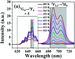
To the best of our knowledge, the heating effect caused by the strong absorption of 980 nm excitation in UC materials doped with Yb3+ should be concerned and avoided as much as possible for the sake of reliability of their temperature sensing applications.15 So, in this work, the temperature dependence of the UC emissions of LiLa(MoO4)2: 0.5% Tm3+, 20% Yb3+ in the interested wavelength range of 610–730 nm were studied under 980 nm laser excitation with low power of 59 mW in order to reduce the heating effect to a negligible extent, and the laser light spot size on the sample was estimated to be 2 mm × 3 mm. The UC spectra recorded at different temperatures from 303 to 543 K are presented in Fig. 5(a), which shows that the emission band located at about 700 nm corresponding to the 3F2,3 → 3H6 transition increases gradually with the increase of temperature. Notably, the intensity enhancement in the same temperature increments becomes more and more remarkable with the increase of temperature. Nevertheless, no significant spectral variation of the two emission bands can be observed at different temperatures, though the FIR of the two emission bands rapidly changes, as indicated in Fig. 5(b) and (c), where the emission spectra at 303 K and 543 K are displayed, respectively.
 |
| Fig. 5 (a) UC spectra in the wavelength range from 610 nm to 730 nm for the Tm3+/Yb3+ co-doped LiLa(MoO4)2 at various temperature from 303 K to 543 K. (b) UC spectra at 303 K from 610 nm to 730 nm. (c) UC spectra at 513 K from 610 nm to 730 nm. (d) Integral emission intensities of 3F2,3 → 3H6 transitions and 1G4 → 3F4 varied with temperature under 980 nm excitation. | |
Fig. 5(d) reveals that distinctly different temperature-dependent behaviours have been observed for the integral emission intensities in the wavelength range of 546–562 nm and 683–715 nm marked with grey areas in Fig. 5(a) of the two emission bands which originate from the 1G4 → 3F4 and 3F2,3 → 3H6 transitions, respectively. It is clearly demonstrated that the intensity of 3F2,3 → 3H6 transition (700 nm) increases monotonously with temperature in the whole range of 303–543 K, while the fluorescence intensity of the 1G4 → 3F4 transition initially increases until 423 K, and then decreases. There are two main reasons accounting for this phenomenon. On the one hand, with the increase of temperature the population of 3F2,3 states from the lower 3H4 state increases due to thermal population, which further promotes the radiative transition from 3F2,3 states18,28,29 and will also reduce the population of 1G4 state due to the fact that the 1G4 state is populated from 3H4 state through the energy transfer ET 3 shown in Fig. 3. On the other hand, there is an obvious mismatch between Yb3+ and Tm3+ in the energy levels involved in the UC processes and the up-converted population of 1G4 can be affected by the participation of phonon, which is temperature dependent. As a result, the UC luminescence intensity of the 1G4 → 3F4 is boosted because of the phonons' assistance as temperature rises in a certain temperature range, but as the temperature rises further, the population of 1G4 may also been brought down owing to the phonon-assisted cross relaxation between Tm3+ and Yb3+, leading to the decrease of the UC luminescence.
Fig. 6(a) shows that the fluorescence intensity ratio R = I1/I2 increased dramatically with the increase of sample temperature, where I1 and I2 stand for the fluorescence intensities of the 3F2,3 → 3H6 and the 1G4 → 3F4 transitions, respectively. The experimental data are well fitted with the following equation:
|
 | (1) |
 |
| Fig. 6 (a) and (b) Temperature dependence of the R between the integral intensities of the 3F2,3 → 3H6 and the 1G4 → 3F4 transitions. | |
We obtained that fitted coefficient A is 2760, B is 9.14 in the temperature range from 303 K to 423 K and A is 4634, B is 13.3 while the temperature is in the range from 453 K to 543 K.
Furthermore, for temperature sensing, the relative sensitivity SR is a key parameter to value the property of the sensors, which is defined as the relative change of the ratio R in response to the variation of temperature.30 The SR could be obtained by the eqn (2).
|
 | (2) |
The relative sensitivity SR of the sample as a function of the temperature from 303 K to 543 K is obtained by applying eqn (2) to the polynomial smooth best fitting of logarithm of the intensity ratio R versus the inverse of temperature 1/T. The result is plotted in Fig. 7. The SR value is 3.85% K−1 at 300 K from eqn (2) which is much better than other previously reported rare earth ions doped temperature sensors based on FIR technique involving TCELs at the same temperature. We summarized several typical temperature sensors based on FIR technique doped with different rare earth ions in Table 1. We conclude that the LiLa(MoO4)2: 0.5% Tm3+, 20% Yb3+ is a potentially excellent candidate for optical temperature sensors with a high relative sensitivity.
 |
| Fig. 7 Plot of the relative sensitivity SR versus temperature for the LiLa(MoO4)2: Tm3+/Yb3+ sample. | |
Table 1 FIR parameters for several typical temperature sensors based on FIR technique doped with different rare earth ions and the corresponding relative sensitivities at 300 K derived from the indicated references
Material |
Transitions |
SR (% K−1) |
Ref. |
AgLa (MoO4)2: 0.02 Er3+/0.4 Yb3+ |
2H11/2 → 4I15/2; 4S3/2 → 4I15/2 |
1.26 |
25 |
Oxyfluoride glass ceramic: Tm3+/Yb3+ |
3F2,3 → 3H6; 3H4 → 3H6 |
0.123 |
18 |
Y2O3: 0.2% Ho3+/3% Yb3+/10% Zn2+ |
3K8 → 5I8; 5F3 → 5I8 |
1.19 |
31 |
β-NaLuF4: 20% Yb3+/20% Gd3+/0.7% Tm3+ |
6P5/2 → 8S7/2; 6P7/2 → 8S7/2 |
0.741 |
12 |
β-NaYF4: 20%Yb3+/0.5%Tm3+/NaYF4: 1%Pr3+ |
3F2,3 → 3H6; 1G4 → 3F4 |
0.418 |
32 |
LiLa(MoO4)2: 20%Yb3+/0.5%Tm3+ |
3F2,3 → 3H6;1G4 → 3F4 |
3.85 |
This work |
4. Conclusions
In conclusion, well-crystallized LiLa(MoO4)2: 20% Yb3+, 0.5% Tm3+ was successfully synthesized by citric acid chelating sol–gel method. The ET from Yb3+ to Tm3+ plays a dominant role in the UC emissions of the sample involving two and three photon processes. In addition, the intensity ratio of the two UC emissions, the 3F2,3 → 3H6 and the 1G4 → 3F4 of Tm3+ in LiLa(MoO4)2: 20% Yb3+, 0.5% Tm3+ under 980 nm laser excitation, exhibits higher relative temperature sensitivity in the range of 303–543 K in comparison with other previously reported rare-earth ions doped temperature sensors based on the FIR approach. The obtained maximum relative sensitivity is 3.85% K−1 at 300 K in the temperature range of 300–550 K for our sample. This result recommends that Tm3+/Yb3+ co-doped double molybdates is an ideal candidate for optical thermometry with high relative sensitivity.
Acknowledgements
This work was financially supported by the National Key Basic Research Program of China (Grant No. 2013CB921800), the National Natural Science Foundation of China (No. 11274299, 11374291, 11574298 and 11404321), and Anhui Provincial Natural Science Foundation (Grant No. 1308085QE75).
References
- S. A. Wade, S. F. Collins and G. W. Baxter, J. Appl. Phys., 2003, 94, 4743–4756 CrossRef CAS.
- F. Vetrone, R. Naccache, A. Zamarron, A. J. de la Fuente, F. Sanz-Rodriguez, L. M. Maestro, E. M. Rodriguez, D. Jaque, J. G. Sole and J. A. Capobianco, ACS Nano, 2010, 4, 3254–3258 CrossRef CAS PubMed.
- Y. Shen, X. Wang, H. He, Y. Lin and C.-W. Nan, J. Alloys Compd., 2012, 536, 161–165 CrossRef CAS.
- B. Chen, B. Dong, J. Wang, S. Zhang, L. Xu, W. Yu and H. Song, Nanoscale, 2013, 5, 8541–8549 RSC.
- D. Li, Y. Wang, X. Zhang, K. Yang, L. Liu and Y. Song, Opt. Commun., 2012, 285, 1925–1928 CrossRef CAS.
- W. Xu, Z. G. Zhang and W. Cao, Opt. Lett., 2012, 37, 4865–4867 CrossRef CAS PubMed.
- C. D. S. Brites, P. P. Lima, N. J. O. Silva, A. Millán, V. S. Amaral, F. Palacio and L. D. Carlos, New J. Chem., 2011, 35, 1177–1183 RSC.
- D. Wawrzynczyk, A. Bednarkiewicz, M. Nyk, W. Strek and M. Samoc, Nanoscale, 2012, 4, 6959–6961 RSC.
- A. Sedlmeier, D. E. Achatz, L. H. Fischer, H. H. Gorris and O. S. Wolfbeis, Nanoscale, 2012, 40, 7090–7096 RSC.
- S. Zhou, G. Jiang, X. Wei, C. Duan, Y. Chen and M. Yin, J. Nanosci. Nanotechnol., 2013, 14, 3739–3742 CrossRef.
- G. Jiang, X. Wei, S. Zhou, Y. Chen, C. Duan and M. Yin, J. Lumin., 2014, 152, 156–159 CrossRef CAS.
- K. Zheng, Z. Liu, C. Lv and W. Qin, J. Mater. Chem. C, 2013, 1, 5502–5507 RSC.
- Z. Boruc, M. Kaczkan, B. Fetlinski, S. Turczynski and M. Malinowski, Opt. Lett., 2012, 37, 5214–5216 CrossRef CAS PubMed.
- W. Xu, X. Y. Gao, L. J. Zheng, Z. G. Zhang and W. Cao, Opt. Express, 2012, 20, 18127–18137 CrossRef CAS PubMed.
- X. Yang, Z. Fu, Y. Yang, C. Zhang, Z. Wu and T. Sheng, J. Am. Ceram. Soc., 2015, 98, 2595–2600 CrossRef CAS.
- H. Lu, H. Hao, G. Shi, Y. Gao, R. Wang, Y. Song, Y. Wang and X. Zhang, RSC Adv., 2016, 6, 55307–55311 RSC.
- M. D. Shinn, W. A. Sibley, M. G. Drexhage and R. N. Brown, Phys. Rev. B: Condens. Matter Mater. Phys., 1983, 27, 6635–6648 CrossRef CAS.
- W. Xu, X. Gao, L. Zheng, Z. Zhang and W. Cao, Sens. Actuators, B, 2012, 173, 250–253 CrossRef CAS.
- F. Cheng, Z. Xia, M. S. Molokeev and X. Jing, Dalton Trans., 2015, 44, 18078–18089 RSC.
- F. Cheng, Z. Xia, X. Jing and Z. Wang, Phys. Chem. Chem. Phys., 2015, 17, 3689–3696 RSC.
- A. Xie, X. Yuan, Y. Shi, F. Wang and J. Wang, J. Am. Ceram. Soc., 2009, 92, 2254–2258 CrossRef CAS.
- Z. Wang, H. Liang, M. Gong and Q. Su, J. Alloys Compd., 2007, 432, 308–312 CrossRef CAS.
- Z. Wang, H. Liang, Q. Wang, L. Luo and M. Gong, Mater. Sci. Eng., B, 2009, 164, 120–123 CrossRef CAS.
- T. Li, C. Guo, Y. Wu, L. Li and J. H. Jeong, J. Alloys Compd., 2012, 540, 107–112 CrossRef CAS.
- T. Li, C. Guo, S. Zhou, C. Duan and M. Yin, J. Am. Ceram. Soc., 2015, 98, 1–5 CrossRef.
- S. Xu, W. Xu, Y. Wang, S. Zhang, Y. Zhu, L. Tao, L. Xia, P. Zhou and H. Song, Nanoscale, 2014, 6, 5859–5870 RSC.
- F. Shi, J. Wang, X. Zhai, D. Zhao and W. Qin, CrystEngComm, 2011, 13, 3782–3787 RSC.
- L. Xing, Y. Xu, R. Wang, W. Xu and Z. Zhang, Opt. Lett., 2014, 39, 454–457 CrossRef CAS PubMed.
- L. Xing, Y. Xu, R. Wang and W. Xu, Opt. Lett., 2013, 38, 2535–2537 CrossRef CAS PubMed.
- S. F. L. Luis, U. R. R. Mendoza, E. Lalla and V. Lavín, Sens. Actuators, B, 2011, 158, 208–213 CrossRef.
- A. Pandey and V. K. Rai, Dalton Trans., 2013, 42, 11005–11011 RSC.
- S. Zhou, G. Jiang, X. Li, S. Jiang, X. Wei, Y. Chen, M. Yin and C. Duan, Opt. Lett., 2014, 39, 6687–6690 CrossRef CAS PubMed.
|
This journal is © The Royal Society of Chemistry 2016 |
Click here to see how this site uses Cookies. View our privacy policy here.