Synthetic methods of CuS nanoparticles and their applications for imaging and cancer therapy

Lu Wang
College of Chemical and Biological Engineering, Zhejiang University, Hangzhou, China. E-mail: ruguoqulvxing@163.com

Received 19th July 2016 , Accepted 22nd August 2016

First published on 26th August 2016


Abstract

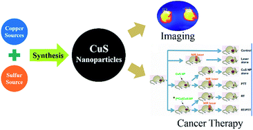
As a single-compartment theranostic nanosystem, copper sulfide nanoparticles have received much attention as they can be used to visualize and treat tumours simultaneously. In this paper, a comprehensive survey of the basic concepts and up-to-date literature results concerning the potential use of CuS nanoparticles for biomedicine applications is presented. The first part summarizes various methods developed to synthesize CuS with varied morphologies and structures, including nanospheres, nanocages, nanoplates, nanotubes, nanorods and nanowires. Then the prepared nanoparticles with designed structures and properties are further explored for their applications in both photoacoustic imaging and cancer therapy. The advancements in photothermal therapy, combinatorial therapy and drug delivery are discussed in detail.


1. Introduction

In the United States, cancer accounts for 1 in 4 deaths. In one's lifetime, the possibilities of developing cancer for men and women are 45% and 38%, respectively.1 Cancer is a complex disease caused by abnormal cell growth with the possibility to spread and invade other organs in the body. The primary challenge in cancer treatment is to efficiently deliver anti-cancer drugs and selectively kill cancer cells without damaging the normal tissue. But conventional anti-cancer drugs suffer from the inadequate accessibility of antineoplastic agents to tumour tissue, high dosage, rapid abolition, poor solubility and inconsistent bioavailability. To overcome these difficulties, the development of a drug delivery system with optimal pharmaceutical action of drugs and minimum toxic side effects tops the scientists' wish list. The increasingly accelerated development of nanotechnology has brought new expectation to be used in as a novel therapeutics for effective cancer treatment.

Nanotechnology is recognized as the fabrication and utilization of materials, devices, and systems through the control of matter on the nanometer-length scale (10−9 m). Since the characteristic structures prepared via nanotechnology are of the nanometer scale, approximating the level of atoms, molecules, and supramolecular structures, they have some particular functions. The special functionalized structures allow researchers to explore their applications in disease diagnostics, drug delivery, and development of nanomedicine.2 Currently, nanotechnology has become an important tool to produce various nanocarriers such as liposomes, dendrimers, micelles, carbon nanotubes (CNTs), polymer–drug conjugates and nanoparticles (NPs), which have the potential to optimize the effect of drugs and reduce toxic side effects. Among these nanocarriers, nanoparticle is an important branch of the rapidly developing nanotechnology.

Nanoparticles, with the nanometer length scale approximating to that of the electronic motion, have some particular properties. For metal-based nanoparticles, the coherent collective oscillation of electrons in the conduction band induces large surface electric fields, and this surface field can interact with resonant electromagnetic radiation, thus enhancing the radiative properties of metallic nanoparticles.3 This makes the absorption cross section of these nanoparticles orders of magnitude stronger than that of the most strongly absorbing molecules and the light scattering cross section orders of magnitude more intense than that of organic dyes. Therefore, these particles are widely used as sensors and novel contrast agents for optical detection because of their enhanced absorption and scattering, respectively. Furthermore, thanks to the electron–phonon and phonon–phonon interaction, the strongly absorbed radiation within particles can efficiently convert into heat on a picosecond time scale, so they show great potential for photothermal therapy application.4 Their applications in drug delivery, cancer cell diagnostics, and therapeutics have been active fields of research. The scattering properties of gold nanospheres have been used for cancer cell imaging using confocal microscopy.5

With different type of inorganic nanomaterials available, CuS nanoparticles become an appealing choice because of their good biocompatibility, low toxicity and reasonable price. Compared with toxic contrast agents containing Cd, copper nanostructures have gained more importance in cancer imaging. In addition, unlike the Au-medicated materials, the p-type semiconductor CuS possesses a d–d transition band showing a near-infrared absorption (700–1100 nm). It does not depend on the dielectric constant of the surrounding medium as Au-related nanoparticles do.

2. Synthetic methods of CuS nanoparticles

Novel categories of CuS nanoparticles have been investigated after the extensive exploration of spherical CuS nanostructures. They contain three-dimensional hollow or solid nanospheres, core–shell nanocages; two dimensional nanorods and nanoplates; and one-dimensional structures of nanowires and nanotubes. The morphological characteristics differ as the synthetic methods change, which further determine the applications of the end products.

Take the structure of CuS nanospheres as an example, they have been widely used in biomedicine field, from optoacoustic imaging6 to photothermal therapy.7 Hollow spherical nanoparticles and nanocages provide suitable carriers for drug delivery,8 while nanowires and nanorods show considerable potential in small molecular sensing.9

Since the applications of CuS nanomaterials depend directly on their shape and dispersion state, it is of great importance to yield monodispersed and uniform nanoparticles which are applicable for biomedical exploitation. Hence, apart from the synthetic procedures, advanced techniques allowing the accurate characterization for chemical and physical properties of the nanostructures prepared are critical. Techniques available abound. Transmission electron microscopy (TEM),10 high resolution TEM (HRTEM),11 scanning electron microscopy (SEM),12 X-ray diffraction (XDR)13 techniques, energy dispersive X-ray spectroscopy (EDS),14 atomic force microscopy (AFM)15 and dynamic light scattering (DLS)16 provide the structural (size and shape) and elemental information, while UV-visible and photoluminescence (PL)17 spectroscopy and Fourier transform infrared spectroscopy (FTIR)18 enable the optical properties characterization of CuS nanomaterials.

2.1. CuS nanospheres and nanocages

In CuS nanoparticle synthesis, hydrothermal/solvothermal method,19–22 sonochemical synthesis23 and microwave irradiation24,25 are generally used to prepare nanostructures. During the preparation, carrying out the reaction of Cu and S element in evacuated tubes is the simplest process. But it has the disadvantages of demanding high temperature for the reaction and large particle diameters of the final product. Thus hydrothermal method, which enables easy fabrication of pure and uniform CuS nanoparticles at a lower temperature without using toxic and complicated reactants, is commonly adopted to avoid these shortcomings. For example, simply add aqueous solutions of sodium citrate and sodium sulfide to CuCl2 liquor, stirred at room temperature for 5 min and then heat the mixture up to 90 °C for 15 min, citrate stabilized CuS sizing about 10 nm can be obtained (Fig. 1).26 By using different precursors, reaction time and temperature to alter the hydrothermal process, nanoparticles with diversified sizes and shapes can be synthesized.
image file: c6ra18355g-f1.tif
Fig. 1 Scheme of TEM characterization of prepared PEG-CuS nanospheres. Inset: the distribution of particle size.27

Moreover, microwave irradiation has become a promising method due to its simplicity and high efficiency.24,25 With the same chemical substances employed above, this technique accelerates the decomposition process tremendously by applying microwave irradiation in aqueous solution. For instance, using copper sulfate and sodium thiosulfate as the starting materials, Y. Ni et al. introduced microwave irradiation to synthesize CuS nanoparticles of different structures (Fig. 2).25 Morphologies of the as-prepared nanostructures varied as the experimental conditions changed. When the initial molar ratio of CuSO4/Na2S2O3 was 1[thin space (1/6-em)]:[thin space (1/6-em)]2, a number of nanotubes and solid nanospheres could be produced. But when the starting molar ratio was either increased or reduced, only solid/hollow nanospheres were obtained. Besides, intensity and duration time of the microwave radiation exerted an influence on the formation of final product as well. Generally, higher power tended to keep the system in a boiling state and hampered the formation of nanotubes. In addition, tubular CuS nanoparticles reduced significantly when the irradiation time was decreased from 20 min to 10 min.


image file: c6ra18355g-f2.tif
Fig. 2 Diagrammatic presentation of CuS nanoparticles' possible forming mechanism possessing varied structures. CuS – CuS stands for the aggregation of CuS NPs.25

The microwave-assisted solvothermal synthesis can increase the reaction kinetics by about one to two orders of magnitude and thus enables a fast heating to the temperature of heat treatment. By this process many novel nanostructures with high crystallinity gradually form in the reaction system. Through this method, T. Thongtem and co-workers prepared copper sulfide nanospheres successfully.24,25 CuCl2·2H2O and CH3CSNH2 were used to construct [Cu(CH3CSNH2)2]Cl2 complexes in ethylene glycol with different pH values. After that the complexes decomposed and produced CuS nanoparticles which were further identified by the FTIR spectrometry and CHNS/O analyse. Researchers indicated that the coordination of Cu2+ and thioacetamide molecules was consistent with the decrease in boding energy of nitrogen–hydrogen bond. The nitrogen atoms in thioacetamide molecules played an important role in the formation of Cu–thioacetamide complexes. SAED, SEM, XRD and TEM analyses demonstrated that in solutions with a very low pH, the obtained products were hexagonal CuS nanospheres. Then when the pH was changed to the value of 13, the final products became hexagonal CuS nanotubes.

Apart from microwave irradiation, many other techniques have been applied to improve the synthetic process. For instance, carboxylic acids were employed as solvents in order to stabilize nanoparticle dispersion and increase nucleation rate. Surfactant-based synthesis was used to prepare CuS nanoparticles (Fig. 3).28 P. Khiew et al. found that the hexagonal system used followed the quasi-Maxwellian fluid behaviour through the sweep measurement. The growth of CuS nanostructures exerted no influence on the rheological response. Besides, the stabilization of the hexagonal microstructures improved after the nanoparticles formed in the reaction system. This phenomenon was consistent with the increase of critical shear stress value obtained from the stress sweep rheogram. Most of the nanoparticles produced were nanospheres with an average diameter between 2–7 nm during the starting phase of the reaction. Then as time went by, these nanoparticles would grow one dimensionally and formed nanorods only due to the restriction caused by surfactant aggregations. In addition, researchers indicated that the Cu2+ ions were abundant in the surface of the as-prepared nanoparticles because of the absorption of excessive metal ions in the precursor liquor.


image file: c6ra18355g-f3.tif
Fig. 3 Polarization pictures of (a) pure hexagonal phase and specimens with varied treatment time: (b) 24 h, (c) 168 h, (d) 336 h (bar: 150 mm).28

Furthermore, enzymatic treatment of CuS nanosuspensions was introduced to produce nanoparticles in a green way (Fig. 4).29 Y. Kim et al. applied dextranase enzyme during the preparation process, which removed the bound dextran bulk and thus produced stable mental sulfide nanostructures. The prepared nanocrystals provided ideal material for medicine and photonics. To be specific, after the Na2S solution was added into the solution containing both dextran and copper nitrate, mild heating was applied to develop the bonding between dextran and copper sulfide nanoparticles. Na2S and CuCl in degassed distilled H2O were utilized as initial reagents for the preparation of copper-rich CuxS nanostructures. Then in order to liberate the surface of the nanoparticles and make them perform effectively in photonic application, enzyme dextranase was introduced to remove the bulk of the polymer shell. By changing the time of enzyme treatment, the relative content of nanoparticles can be altered correspondingly.


image file: c6ra18355g-f4.tif
Fig. 4 Diagrammatic illustrate of stabilized metal sulfide nanomaterial synthesis.29

Compared with solid nanoparticles (Fig. 5a), hollow nanospheres (Fig. 5b) and nanocages offer better capacity for drug delivery and chemical storage, widening the applications of CuS NPs. X. Liu et al. demonstrated the formation of 5–10 nm sized hollow CuS nanospheres and nanotubes at room temperature.34 In this study, researchers indicated that such hollow structures formed after the decomposition of thiourea into hydrogen sulfide, which interacted with copper precursor later and produced carbon dioxide. The CO2 then formed gaseous cavities and provided heterogeneous nucleation centres for the aggregation of copper sulfide nanoflakes and thus contributed to the formation of hollow nanostructures.


image file: c6ra18355g-f5.tif
Fig. 5 SEM images of CuS nanoparticles with respective morphologies. (a) Solid nanospheres,30 (b) hollow nanostructures,31 (c) nanoplates,32 (d) nanorods.33

Besides, hard-template assisted method can be employed to synthesize CuS nanoparticles having large hollow cavities as well (Fig. 6).31 H. Zhu et al. reported a fast and moderate solution method to prepare hollow CuS nanospheres.31 Spherical aggregation of Cu2O nanoparticles were used as the sacrificial templates. Researchers indicated that the formation of incompact aggregation of cuprous oxide nanoparticles was essential for the rapid preparation of hollow nanospheres at a relatively low temperature. Simply by changing the aggregation level of cuprous oxide nanoparticles, shell thickness of the final product could be adjusted accordingly. Except for the synthesis of hollow CuS nanoparticles, this method can be applied to the rapid and mild preparation of other different nanostructures with desirable shell thinness.


image file: c6ra18355g-f6.tif
Fig. 6 Hollow CuS nanoparticles were fast formed after the sacrificing of Cu2O templates with mild conditions.31

Apart from Cu2O nanoparticles,31,35 other sorts of core supports such as surfactant micelle microemulsions30 and poly-(styrene-acrylic) latex particles36 can be adopted to form hollow structures as well. X. Yu and co-workers synthesized nanometer-sized CuS spheres through a surfactant micelles-assisted method under ambient conditions.30 Specifically, Na2S and CS2 were chosen as the S2− source while CuSO4 was chosen as the Cu2+ source. 25 mL cyclohexane and 15 mL Triton X-100 were added into the CuSO4 solution (0.5 M) and stirred to form the micro emulsion system. 15 min later, 0.5 mL CS2 was dropped into the system gradually. After that the flask was heated to the 60 °C and maintained the temperature for 4 hours to form CuS nanoparticles. The products obtained were hollow nanoparticles with an average diameter of around 200 nm. Outside the inner core were thin shells (around 30 nm) formed by lots of tiny nanocrystals. Researchers indicated that these nanoparticles with hollow appearance and small uniform sizes would be ideal candidates for the application of fabricating catalysts and fillers. Besides, due to their optical-limiting effect which is also called as the transmitted energy attenuation with increasing incidence, these as-prepared CuS nanoparticles may play important roles in the application of eyes protectors and intense laser sensors in future.

Y. Huang and co-workers adopted a layer-by-layer self-assembly method to construct core–shell composite and then removed the core template by dispersing them in the solution of toluene. Using this technique, they coated CuS on PSA inner spheres and produced hollow CuS nanoparticles successfully. First, they synthesized CuS/PSA shell/core composites. CuSO4, PSA latex, urea, thiourea and PVP (polyvinylpyrrolidone) were mixed up. Then the mixture was dispersed in deionized H2O with ultrasonic wave. After that the flask was moved into an autoclave at the temperature of 85 °C for up to 8 hours. The suspension obtained was washed by water and ethanol for a couple of times. The as-prepared composite nanoparticles were dispersed in toluene next to remove the PSA cores and became hollow CuS nanoparticles finally. Researchers used TEM, FTIR, XDR and UV-vis absorption spectroscopy to character the products. The final products were shown to have a uniform diameter of around 150 nm with the shell thickness of around 20 nm.

Meanwhile, octahedral nanoparticles along with cubic- and star-shaped nanoparticles were selected as sacrificial templates to enable the formation of hollow particles by solid–liquid reactions.37,38 S. Jiao and co-workers utilized shaped-controlled Cu2O nanocrystals as sacrificial templates and prepared uniform CuS mesocages with monocrystalline shells successfully.37,38 After the formation of Cu2O core, the oxygen atoms in Cu2O aggregation were gradually replaced by divalent sulfur ions in solution and formed the CuxS shell structure. It was quite interesting that if the reaction atmosphere was changed from a nitrogen to an air condition, the compositions of the final product (single-crystal CuS mesocages) would be transferred from Cu2S to Cu1.7S. In another study, H. Xu et al. produced octahedral CuS nanocage structures through the solid–liquid reaction between thiourea solution and cuprous oxide octahedral at 90 °C.37,38 Octahedral Cu2O nanoparticles played the roles of precursor, sacrificial cores and shape-controller during the synthesis. Researchers demonstrated that two parameters, Ostwald ripening and mass diffusion, are key to the transformation procedure.

2.2. CuS nanoplates

Compared to the three-dimensional nanostructures introduced above, two-dimensional CuS nanoparticles (Fig. 5c) primarily in the form of thin films and nanoplates39 are more rarely used. Similarly, they can be prepared via hydrothermal or solvothermal routes,21,32,40–42 producing a range of nanoplates with different sizes (50–200 nm) by varying the conditions of reaction.

Among all the methods reported so far, it becomes a most widely used one to treat the precursor-surfactant aqueous micro emulsions at the temperature between 130 and 180 °C for several hours. Different CuS nanoplates with varied morphologies and dimensions can be obtained by changing the synthetic parameters. P. Zhang et al. reported a route for the synthesis of CuS flakes in CTAB/n-C5H11OH/n-C6H14/H2O micro emulsions via hydrothermal method at the temperature of 130 °C.42 The addition of n-dodecanethiol into micro emulsions led to the formation of smaller hexagonal copper(I) sulfide nanodisks, which further assembled to construct a superlattice consequently. Researchers indicated that dodecanethiol played an important role in the control of CuS stoichiometries and morphologies of the final products.

Y. Zhang21 et al. used Na2S2O3·5H2O as sulphur source to react with different kinds of copper sources (CuO, CuCl2·2H2O and CuSO4·5H2O) respectively. After employed in water for up to 6–24 hours at the temperature between 130–170 °C, plate-like CuS nanocrystallized powders (Fig. 7) possessing spherical and quadrate morphologies were successfully synthesized. Through the adjustments of copper sources, reaction time and temperature, the physical properties (size and morphologies) of final products can be easily and well controlled. Zhang and his co-workers also discussed the formation mechanism during the hydrothermal synthesis. CuSO4·5H2O and CuCl2·2H2O could dissolve in water quickly. When they were applied as the copper sources, Cu2+ released would combine with S2O32− and form stable precipitate such as Cu(S2O3)·(H2O)2 or Cu(S2O3)2. Then this precipitate would decompose and generate H+, SO42− and CuS after the system was heated to the temperature of 150 °C. Things went different when CuO was chosen to be the copper source. As a more stable compound, CuO tended to release free Cu2+ slowly in water. In the meantime, S2O32− was converted to S2− at 150 °C. Then the CuS nanoparticles were formed through the combination of Cu2+ and S2−. Neither organometallic precursors nor complexing agents were required during the preparation of these CuS nanocrystals with high purity and low cost. Therefore, this method provided a promising way to produce semiconductor nanostructure materials.


image file: c6ra18355g-f7.tif
Fig. 7 TEM images of plate-like CuS nanocrystallized powders obtained via different reaction temperatures and time. (a) 150 °C, 12 h; (b) 150 °C, 24 h; (c) 170 °C, 12 h.21

Another publication described a sonochemical approach to synthesize CuS nanoparticles without the usage of surfactant. Based on the in situ copper hydroxide nanoribbon templates,23 single crystalline nanostructures (Fig. 8) can be synthesized under ambient conditions. During the synthesis, Cu(OH)2 was introduced as the precursor and template while ultrasonic irradiation applied contributed to the fabrication of CuS nanoplates. This method provided a quick, low-temperature and mild strategy to prepare two-dimensional CuS nanomaterials.


image file: c6ra18355g-f8.tif
Fig. 8 (a and b) TEM image of the CuS nanoplates obtained. (c) HRTEM image of a single CuS sample. (d) FESEM image of CuS nanostructures.23

Other synthetic methods which can be used to produce CuS nanoplates with cylindrical arrays (Fig. 9) involve chemical vapour method,44 single source reaction45 and high-temperature precursor injection procedure.43 K. Wang and co-workers used a mild chemical vapour reaction to synthesize single-crystalline CuS nanoplates successfully.44 First, they prepared copper films via vacuum evaporation of metal copper on a glass slide. After that, these copper films and sulfur vapour were treated in a vacuum chamber at the temperature of 450 °C for up to 7 hours. Products obtained were found to be single crystals with an average thickness of 20–150 nm and an average length of 200–1800 nm. These CuS nanoplates with unique morphology and properties were ideal semiconductor materials and thus could be used to produce functional components.


image file: c6ra18355g-f9.tif
Fig. 9 (a and b) HRTEM images of product CuS nanostructures under different magnifications; (c) HRTEM image of CuS nanoarrays. (d) Illustrate of a self-assembly nanoplate; (e) HRTEM image of a nanoplate.43

W. Lou et al. put forward a single source precursor route to prepare triangular CuS nanoplates (sizing 9.8 nm) with the existence of oleylamine. Copper dialkyldithiophosphates decomposed at the temperature of 140 °C and then composed uniform faceted CuS nanocrystals, which established the structure of closed-packed hexagonal array (Fig. 10).45 It was obvious that the faceted nanocrystals sizing around 8.9 nm emerged at the temperature of 120 °C (Fig. 7A). Then when the temperature rose to 140 or 160 °C, average diameters of the obtained CuS nanospheres were about 9.8 or 11.4 nm respectively. At 180 °C, the average sizes of the nanocrystals grew to about 13.1 nm and there existed some nanorods with aspect ratio of 4.5 (Fig. 7C). Compared with traditional nanospheres, these triangular CuS nanoparticles were much more difficult to fabricate because they always had relatively high energies. Surfactant molecules dispersed in the solution interacted differently with each crystallographic face of the nanoparticles and led to the phenomenon of shape anisotropy. Researchers expected that these CuS nanostructures were good models for the size-dependent structural phase property study.


image file: c6ra18355g-f10.tif
Fig. 10 Schemes of CuS nanocrystals with different shapes and sizes synthesized under the same reaction conditions.45

H. Wu and W. Chen reported a precursor-injection method to prepare covellite CuS nanoparticles at a high temperature.43 CuS nanoparticles obtained were shown to be one-dimensional arrays which were assembled spontaneously by the CuS nanoplates synthesized. Researchers demonstrated that the temperature of preparation exerted a significant impact on the self-assembly process of the copper sulfide nanoplates and morphology of the final product. The self-assembly process differed as the temperature changed. When the temperature of reaction was between 140 °C and 180 °C, the face-to-face self-assembly behaviour could be observed on the whole substrate. Then when the temperature was increased to about 200 °C, only edge-to-edge arrays were formed. If the temperature went even higher, no self-assembly structures could be found in the final products which were constructed by diverse nanocrystals with different sizes. This study provided a mild route to prepare highly-defined self-assembly nanoparticles which could be applied in the synthesis of other different nanodivices.

2.3. CuS nanotubes, nanorods, and nanowires

One-dimensional CuS nanoparticles (Fig. 5d) possess superior catalytic and electrochemical properties, making them desirable materials for the applications of photocatalysis and sensing.46,47 Abundant methods can be adopted to construct elongated nanostructures. Products gained under different conditions represented multiple morphological and dimensional characters. CuS nanowires5,48 with the diameters ranging from 30–80 nm and CuS nanotubes with the length of 30–120 nm20,49 can be prepared by using different precursors, synthetic methods and growth conditions.

X. Liao and co-workers proposed a synthesizing process induced by microwave heating in mild surroundings.48 5 mmol Cu(NO3)2·3H2O of analytic grade was dissolved in 100 mL SDS (sodium dodecyl sulfate) aqueous solution. Then 10 mmol TAA (thioacetamide) was introduced and produced yellow precipitation which might be the precursor (Cu–SDS–TAA) in the solution. After that the flask was put under microwave for about 20 min and cooled to ambient temperature. Characterized by XRD and TEM, the as-prepared CuS nanorods were proved to be pure, uniform and well crystallized, with diameters varying from 5–10 nm and lengths ranging from 30–50 nm. Researchers indicated that it was a rapid, simple and effective method to synthesize metal sulfide nanocrystallines, not only limited to the preparation of CuS nanostructures. Analogously, nanoparticles mentioned above can be produced via hydrothermal methods, which offer simple and efficient ways to yield uniform and high-quality nanostructures.5,20,33,34,36,49

Based on the hydrogel system, C. Tan et al. prepared CuS nanotubes under an ambient environment. After the reaction of copper acetate with LAA at 70 °C, solution containing copper-LAA compound was cooled down to allow the formation of translucent gel via the hydrogen bonds among adjacent molecules. Subsequently, TAA was add into the system, decomposed and emitted hydrogen sulfide gas, translating the copper-LAA coordination gel into copper sulfide in original positions (Fig. 11).33 The potential formation process of the CuS nanostructures could be represented as follows. Firstly, Cu2+ acetate interacted with LAA and created Cu2+-LAA chemical compound under stirring with the temperature of 70 °C. Then the mixture was cooled to ambient temperature and generated the translucent gel simply through the hydrogen bonds existed between amide among the molecules around. After that TAA was introduced and decomposed in the system which further released hydrogen sulfide gas. Finally, the Cu2+-LAA coordination compound was converted into copper sulfide nanostructures in situ. Through this method different inorganic nanotubes with controllable size could be synthesized simply by selecting other suitable templates. Thus nanotubes which cannot be prepared through conventional route are able to be synthesized via this novel method.


image file: c6ra18355g-f11.tif
Fig. 11 TEM characterization of as-prepared CuS nanostructures: (a and b) images of nanotubes, (c) illustrate of SAED pattern.33

Many methods prepare CuS nanotubes and nanotubes with other different templates. C. Wu et al. synthesized uniform CuS nanotubes of 30–90 nm in inner diameter and 20–50 nm in thickness.49 Large quantities of nanostructures could be obtained via the strategy. To be specific, copper nanowires were chosen as the sacrificial templates and appropriate sulfur sources were added to conduct the sulfuration process in ethylene glycol solutions at the temperature of 80 °C. Researchers indicated that the solvent and sulfur sources used exerted a great influence on the construction of well-organized copper sulfide nanotubes. Ideal sulfur sources like thioacetamide and thiourea would release S2− instead of molecular S when they decomposed at the specific temperature. Compared with S powders, this phenomenon was more helpful to the fabrication of copper sulfide nanotubes. When the experiment was conducted at a relatively high temperature of 140 °C, crystallinity of the final products was improved while the size of the nanotubes became slightly smaller. Besides, similar reaction could not produce CuS nanotubes when the solvent was changed from ethylene glycol to water. This strategy can be used in the preparation of other copper related sulfides and oxide nanostructures. Apart from that, CuS nanotubes obtained may play the role of substituted templates to create other different types of nanotubes which cannot be produced directly.

J. Mao et al. mixed an aqueous solution of CuCl2 with thiourea at room temperature to prepare well-aligned [Cu(tu)]Cl·1/2H2O nanowire precursor (thiourea = tu), which was further used to prepare CuS nanotubes.50 Introduction of tu to the copper chloride solution reduced the Cu2+ to Cu+, enabling the construction of [Cu(tu)]Cl·1/2H2O nanowires with well-organized morphology and desirable chemical purity. Based on these products, size-controllable hollow nanotubes can be fabricated efficiently through self-sacrificial templates. This preparation strategy provided novel insights into the mineralization of inorganic structures at nanometer levels.

Using the intermediate complex Cu3(TAA)3Cl3 as template, the micrometer-scaled hierarchical tubular structures of CuS assembled by nanoflake-built microspheres were first synthesized by Z. Yao et al.51 After the CuCl2·H2O was dissolved in distilled H2O, thioacetamide solution was dropped into the system gradually at ambient conditions without vibration. A few minutes later, a yellow jar was formed and the mixture was heated to 60 °C and kept at this temperature for up to 24 hours. Then the system was cooled to ambient temperature, filtered and washed with water and ethanol to separate the black CuS precipitate. Nanoparticles produced can be used for the storage of hydrogen. However, this template-based synthesis is time consuming and expensive for it requires multiple steps. The difficulty to completely remove the template material would definitely affect the purity of the final product. Therefore, template-free synthesis of CuS tubes was widely studied.

J. Y. Gong et al. reported that CuS microtubes composed of hexagonal nanoflakes can be prepared in an acetic acid-assisted solution.51 X. Zhang et al. prepared CuS nanotubes using oleic acid and poly(vinyl pyrrolidone) (PVP) in a micro emulsion system without using templates.52 PVP (poly(vinylpyrrolidone)), oleic acid and water were mixed to produce the micromulsion solution, in which water was used as the shell while oleic acid was used as the core. 2 hours later, hollow copper sulfide nanospheres were formed in the water phase. Then when the reaction time was expanded to 6 hours, these hollow CuS nanoparticles would assemble and construct nanotubes. After 12 hours, CuS nanotubes/oleic acid performing shell/core structure were prepared. The products obtained were washed with ethanol in order to dissolve the oleic acid template and gain CuS nanotubes eventually. Researchers demonstrated that CuS nanoparticles synthesized performed higher activity to the glucose oxidation and had much lower overvoltage to glucose than the CuS nanotubes prepared via the traditional route did. Therefore, these products might find their potential application of glucose sensors in future.

J. Kundu et al. successfully prepared various CuS nanostructures through a simple, template-free and single-step solution chemistry route.53 Different kinds of solution (100% ethylene glycol or 100% water or their mixture) were applied as the solvents and Cu(NO3)2 and Na2S2O3 were dissolved in this system. After that the mixture was stirred slowly to form a green solution and heated to 70 °C. Four hours later, the system was cooled to the ambient temperature, filtered and washed with water and ethanol. Reaction parameters such as duration time, temperature, counter ions and ratio of precursors were changed to study their influences on the final product. Results of a series of experiments showed that the reaction time exerted a significant influence on the morphology of CuS nanoparticles obtained. When the duration time was 30 min, hexagonal cross-section nanorods were constructed. These nanostructures formed circular cross-section nanoparticles gradually after 4 hours' reaction. Then when the temperature of reaction was changed from 70 to 180 °C, average sizes of the final products grew from 150–200 nm to 400–500 nm. The optimal precursor's ratio was 1[thin space (1/6-em)]:[thin space (1/6-em)]1 (Cu(NO3)2[thin space (1/6-em)]:[thin space (1/6-em)]Na2S2O3) while the best solvent ratio was 1[thin space (1/6-em)]:[thin space (1/6-em)]3 (water[thin space (1/6-em)]:[thin space (1/6-em)]ethylene glycol). Besides, it was quite important that CuS nanotubes could be synthesized only when the counter ions SO42− and NO3 were introduced with the presence of Na+.

Other methods including thermal degradation of copper precursor in a solvent-free condition,54 application of a paired cell under ambient environment55 and amylose-directed synthesis have been established for the synthesis of one-dimensional CuS nanoparticles.56 H. Larsen et al. prepared a solution containing Cu(NO3)2 and chloroform and introduced sodium octanoate to act as a phase transfer catalyst and dissolve the Cu cations into the organic phase.54 The aqueous phase was abandoned after the copper octanoate composite was transferred to the organic solution. Then dodecanethiol was added and contributed to the binding between copper and octanoate. Copper precursors were obtained next by evaporating the organic solvent. This brown material was dissolved by chloroform and washed with ethanol to detach extra surfactant and other unwanted by-products. Monodisperse CuS nanoparticles with uniform diameter could be synthesized through this method. The precursor thiolate might be used in the preparation of other chalcogenides like Nis and CoS. In addition, this solvent less strategy of nanorod construction provided potential method for the synthesis of diverse materials via the selection of suitable molecular precursors.

Some research groups applied microwave irradiation methods to prepare CuS nanotubes and nanorods by utilizing respective experimental routes.24,25,48,59 G. Mao et al. used graphite to assemble arachidic acid monolayers with copper ions in order to fabricate CuS nanorod arrays (Fig. 12).57 This technique offered a controllable way to produce nanowire arrays exhibiting ideal properties for sensing. To be specific, arachidic acid and copper sulfate were mixed to form spin-coated films. Then they were put into the sodium sulfide solution and kept there for 7 days. After that these films were washed with chloroform to get rid of the organic template. This spin-coating strategy provided a controllable and easy route to prepare highly-defined molecular templates which could be applied to develop nanorods of varied materials such as metals, semiconductors and polymers.


image file: c6ra18355g-f12.tif
Fig. 12 Diagrammatic representation of the transformation of copper sulfide nanorods from copper arachidate monolayer templates.57

3. Biomedical application

Originated from the field of radio pharmacology, molecular imaging is an advanced technology which visualizes, characterizes and measures the bioprocesses at molecular/cellular level inside humanity and other living creatures via a non-invasive way.60 The specific techniques involved vary, including but not limited to positron emission tomography (PET),61,62 single-photon emission computed tomography (SPECT),63,64 magnetic resonance imaging (MRI),65 fluorescence or bioluminescence imaging66–68 and targeted ultrasound.69,70

With the advantages of high sensitivity (down to the picomolar level), PET is a good diagnostic tool which can be used in quantitative imaging analyses of in vivo abnormalities. The decay characteristics of Cu have triggered the application of this nuclide as a promising radiotracer for real-time PET monitoring of regional drug concentration, pharmacokinetics and dosimetry during radiotherapy.

In practical use, researchers often combine anatomical and molecular imaging together in order to gain detailed and complemental information.71–73 Recently new-found molecular imaging methods have been emerging.48,74–76 And photoacoustic imaging becomes an appealing one during the development.

Gold nanocages or nanorods, with their particular photothermal properties, are conventionally used as nanomaterial candidates for imaging agents and drugs. However, these gold nanocrystals suffer from low thermal stability when exposed to laser irradiation in a long time stretch, leading to various absorption patterns between in vitro and in vivo correlation studies. Furthermore, the relatively large sizes (typically >50 nm) limit their application in vivo biological settings.77 Fortunately, researcher developed copper-containing semiconductor nanocrystals with advantages of low production cost, high stability, low toxicity, and high photothermal conversion efficiency for biomedical applications. Naturally, copper sulfide (CuS) nanoparticles with strong NIR absorbance have been widely used as a contrast agent in photoacoustic imaging as well as a photothermal agent for ablation of cancer cells.

3.1. Photoacoustic tomography

Photoacoustic tomography (PAT) is a non-invasive biomedical imaging method that synergize optical and ultrasound imaging.78 This tomography is quite useful in biological tissues for it overcomes the resolution disadvantages of pure optical imaging and the contrast and speckle disadvantages of pure ultrasonic imaging.79 This is realized by retaining intrinsic optical contrast characteristics while taking advantage of the diffraction-limited high spatial resolution of ultrasound. This technique is based on the photoacoustic effect, i.e., the absorbed electromagnetic energy of light is transformed into kinetic energy and localized heating, releasing a pressure or radio frequency wave. Once the molecules or contrast agents (such as copper sulfide NPs) absorb the laser pulses, the optical energy will be converted into heat energy and bring ultrasonic signals.76

Both endogenous molecules, such as haemoglobin or melanin, or exogenously delivered contrast agents, such as optical dyes, gold nanoparticles, or single-walled carbon nanotubes cab be used for optical absorption. Haemoglobin strongly absorbs the laser wave less than 530 nm, providing an ideal endogenous contrast agent for vascular imaging. The high concentration of haemoglobin (12 to 15 g dL−1) contribute to a strong light absorption compared with surrounding tissues, making blood vessels visible. But for the nonvascular tissues (e.g., lymph nodes) or intravascular bio signatures (e.g., integrin), exogenous contrast agents have to be introduced for PA imaging. Generally, gold nanoparticles were often used in PA imaging (Fig. 13).80 But this application is discouraged by the high and unpredictable cost of the metal. Moreover, the optical property of gold nanoparticles (i.e., surface plasmon resonance), closely related to particle surface coating and morphology, is modified via expensive and complicated chemistries, which impairs their popularity. Furthermore, this commonly used candidate has another lethal drawback. Their optical characters rely directly on surrounding chemical substances and they may be quickly cleaned by the reticuloendothelial system (RES). These limitations restrict their further application in photoacoustic tomography (PAT).58,81 This problem can be settled by using exogenous contrast agents which absorb near-infrared light.64


image file: c6ra18355g-f13.tif
Fig. 13 Illustrate of Au nanoparticles which can be used as contrast enhancements. (a) G0 dendrimers stabilized Au nanoparticles, (b) Au nanospheres package in G1 dendrimers.58

CuS nanoparticles, a semiconductor material, is able to absorb light in the near-infrared region efficiently, making them suitable contrast agents for molecular PAT of cancer. When selectively delivered to the tumour tissue, CuS nanoparticles can greatly enhance the optical absorption of tumour cells. The absorbed light energy will then be converted into heat, causing transient thermoelastic expansion and wideband ultrasonic emission. The ultrasonic waves generated can be detected by ultrasonic transducers and be analysed to produce tomography.

According to a recent research, copper sulfide nanoparticles were employed as an advanced PAT contrast agent.82 A 1064 nm laser with low absorption in tissue but can be easily absorbed by copper sulfide nanostructures was introduced. Organic dyes and synthesized nanoparticles are contrast agents commonly adopted in PAT. Their optical adsorption is mainly longer than 560 nm and shorter than 840 nm. Therefore, the additive proportion of Cu and S precursors were precisely controlled in order to yield 11 ± 3 nm sized nanoparticles which specifically have the maximum absorption of 1064 nm. In this study, G. Ku and his colleges got the lymph nodes and brain imaging pictures (Fig. 14) based on mouse models successfully. They tested the CuS nanomaterial in chicken breast under the depth of 5 cm. Result showed that these nanoparticles provided high-quality imaging with good sensitivity. Besides, a cell viability assay was conducted to detect their toxicity. After 24 hour-long incubation, minimal cytotoxicity was observed at concentrations of up to 96 μg mL−1. Therefore, CuS nanoparticles have a great potential to be used in clinical testing of lesions in skin, breast, lymph nodes and neck.


image file: c6ra18355g-f14.tif
Fig. 14 Graphical representation of PAT images in vivo of the experimental brain.82

For the first time, D. Pan et al. demonstrated that the potential use of copper as a contrast metal for near-infrared detection of SLN using PAT.83 The prepared structure and its photoacoustic imaging is shown in Fig. 15. They encapsulated multiple copies of Cu as organically soluble small molecule complexes within a phospholipid-entrapped nanoparticle. The sizes of nanoparticles assumed are in the range of 80–90 nm, the optimum sizes for its bio-distribution throughout the lymphatic systems. Compared to natural-light absorbing blood, these particles provided at least six times higher signal sensitivity. They also demonstrated that high SLN detection sensitivity with PAT can be achieved in a rodent model. In addition, dynamic light scattering measurements showed that these CuS nanoparticles exhibited significant shelf stability over a period of more than 60 days from the time of preparation. The maintenance of nanoparticles' integrity kept the copper at low concentration, posing hypo toxicity to tissues.


image file: c6ra18355g-f15.tif
Fig. 15 Cu-neodecanoate nanostructures employed as PAT contrast agents for the imaging of sentinel lymph node.83

3.2. Photothermal therapy

Cancer is one of the most devastating diseases in the world and cost thousands of lives each year. The genetic alterations and cellular abnormalities promotes the aggressive growth of cancer cells leading to significant morbidity and mortality in patients.84 Nowadays, the most commonly used cancer therapies are surgical excision, medical therapies such as chemotherapy and radiotherapy, but they have some inherent drawbacks. Surgical excision cannot remove all cancerous cells completely and thus resulting in serious morbidity. What's more, when tumours are adjacent to critical tissue structures, the difficulty of excision increases tremendously. Furthermore, the severe side effects of chemotherapy and radiotherapy cause great sufferings for patients. For chemotherapy, repeated treatment with chemotherapeutic agents lead to the resistance to the chemotherapies or development of multi-drug resistance.85 Another problem associated with conventional chemotherapy is that the limited accessibility of drug to tumour tissues makes the high dose necessary in cancer treatment, which results in intolerable cytotoxicity and nonspecific targeting.88

Photothermal therapy is a minimally-invasive therapy using light energy and photo absorbers sources and offers an alternative for cancer treatment.89,90 Our tissue consists of water, haemoglobin, oxyhemoglobin and melanin. They can absorb natural light and converse light to heat. It would cause damage to both tumours and healthy tissues because our healthy tissue cells undergo serious damage at temperature of 55–95 °C. But the near-infrared (NIR) light can avoid such damage for it induces minimal photothermal heating with the little light absorption of blood and water in a NIR region (650–900 nm).

In photothermal practice, nanoparticles are often employed as absorbents, whose absorption band must be in the near infrared region to use this near-infrared wave energy.91 To make sure that nanoparticles induce sufficient heating to kill cancerous cells with minimal thermal injury, they are supposed to have a size less than 50 nm, be nontoxic and have functionalized surfaces which can interact with cell recognition moieties. Moreover, the nanoparticles should respond strongly to light excitation with wavelengths in the range of 650 to 950 nm.92 Engineered particles like gold nanoparticles,93–97 carbon nanotubes98 and CuS26,86,99–101 nanostructures have proved to possess photothermal coupling effect and can be used in photothermal treatment. These particles absorb near-infrared light (650–900 nm), experience resonance and then transfer thermal energies to the surrounding tissue to raise the tissue temperature.

Up to date, the optical absorption in gold nanostructures based on the surface plasmon resonance (SPR) is most extensively studied. But since the SPR peak of gold nanoparticles is determined by the dielectric constant of the surrounding medium, the absorption wavelength of gold nanostructures, thereafter, will be greatly influenced by the solvent or the surrounding environment. As an alternative, CuS nanoparticles (CuS NPs) provide researchers another choice for photothermal therapy. Different from gold nanoparticles, the NIR light absorption of CuS NPs derives from the d–d transition of Cu2+ irons,102 which means the absorption wavelength of CuS NPs is not affected by the surrounding environment. Another incentive in CuS NPs study is the less expensive production of CuS nanoparticles.103

CuS nanoparticles can absorb near-infrared light effectively and convert the absorbed optical energy to heat. At high temperatures (>45 °C), thermal energy alone is sufficient enough to damage cancer cells.104 Through reasonable design, CuS nanoparticles are able to selectively target cancer cells and generate a proper amount of heat over a given period of time under NIR laser irradiation, realizing the thermal ablation of cancer without hurting healthy tissue.

Researches on CuS nanoparticles medicated photothermal therapy of cancer have been extensively conducted for the past few years.26,86,87,101,105–115 There are two major approaches aimed at modifying and optimizing the physicochemical properties of CuS nanoparticles in order to improve their photothermal ratio. One strategy is to reveal the complex relationship between nanoparticles' size, chemical composition and shape and their optical properties (the absorption and photothermal conversion efficiency). The other route is to change the NPs' size, optical characters and biocompatibility by introducing different type of surface-coating materials.

In the first approach, for instance, hydrophilic Cu9S5 nanostructures sizing 13 nm × 70 nm exhibited a photothermal conversion ratio of 25.7% when using a 980 nm laser.86 In vivo experiment showed that cancer cells can be effectively destroyed by the combination of Cu9S5 aqueous nanodispersion (40 ppm) and 980 nm laser irradiation with the intensity of 0.51 W cm−2 (Fig. 16). To determine the cytotoxicity of Cu9S5 nanomaterials, a 3-(4,5-dimethylthiazol-2-yl)-2,5-diphenyltetrazolium bromide (MTT) assay was carried out with the human cervical carcinoma cell line. After 24 hour-long incubation at 37 °C, no obvious differences in the cell proliferation were observed in the presence of Cu9S5 nanoparticles with concentrations varying from 1.6 to 100 ppm. Researchers claimed that these as-prepared nanoparticles were promising photothermal nanomaterials for the near-infrared treatment of cancer due to their relatively small size, desirable conversional behaviour, low cytotoxicity and reasonable price. B. Li et al. furthered this investigation by synthesizing Cu7.2S4 nanocrystals, which increased the photothermal conversion efficiency to 56.7% with the same-intensity laser.87 Both in vivo and in vitro cancer therapies approved that these nanoparticles with an average size of 20 nm can efficiently kill the abnormal cells (Fig. 17) by using 40 ppm of Cu7.2S4 nanocrystals in PBS solution under 980 nm irradiation laser (density: 0.72 W cm−2). And no significant cell viability was observed when the concentration of Cu7.2S4 nanoparticles was increased to 40 ppm without laser irradiation.


image file: c6ra18355g-f16.tif
Fig. 16 H & E stained histological pictures of tumor sections after the treatment of Cu9S5 nanoparticles and 980 nm laser irradiation.86

image file: c6ra18355g-f17.tif
Fig. 17 (a) Near-infrared thermal images of mice treatment with Cu7.2S4 NCs (left) and saline (right) after the hypodermic injection. (b) The temperature in different regions changing with time. (c and d) Histological images of tumor sections stained by hematoxylin and eosin, after the treatment of laser irradiation.87

Q. Tian et al. successfully prepared a flower-like hydrophilic copper sulfide superstructure (Fig. 18) via a controlled hydrothermal method.101 They demonstrated that these novel superstructures improved the photothermal conversion efficiency by 50%, compared with the corresponding structure of CuS hexagonal nanoplates at 980 nm laser. Additionally, compared with the control group, the death rate of Hela cells remained almost the same in the presence of these CuS superstructures at the concentration of 0.25 g L−1 without laser treatment. Hollow nanostructures of Cu7S4 exerted strong effect of photothermal therapy for cancer treatment as well.101 And there remain some other cases where CuS nanosuperlattices,108 mesostructures,109 nanoplates108 and nanoclusters110 also showed superior photothermal conversion efficiencies.


image file: c6ra18355g-f18.tif
Fig. 18 Photothermal therapy with CuS superstructures. (a) Scheme of a CuS superstructure, functioning as laser-cavity mirrors for 980 nm laser and photo-thermal conversion. (b) Photothermal characters of CuS superstructures and the building blocks. (c–f) Stained H & E histological illustrates of in vivo tumor sections after the treatment of 980 nm laser (power density: 0.51 W cm−2) for 10 min injected with: (c and d) water; (e and f) CuS superstructure aqueous dispersion.101

As for the second approach, surface-coating materials include citric acid, PEG,26 phospholipid-PEG,113 cysteine112 and bovine serum albumin114 can be employed to improve the nanoparticles' biocompatibility. It has been found that ultra-small CuS nanodots can be prepared via polyvinylpyrrolidone coating.116 Z. Min et al. employed these renal-clearable nanodots to realize image-monitored photothermal treatment. During the initial dose-finding toxicity experiments, no obvious body weight change of the tested mice could be observed when the injection dose of CuS nanodots was increased to 60 mg kg−1. These nanoparticles sizing 5.6 nm were able to incorporate with the 64Cu radioisotope for non-invasive PET stably and be excreted by liver and spleen with little retention within 24 hours. Guided by PET imaging these CuS nanoparticles can accumulate in 4T1 tumour cells and introduce thermal ablation under near-infrared laser irradiation (Fig. 19), providing a promising platform for the imaging and therapy for cancer.


image file: c6ra18355g-f19.tif
Fig. 19 (a and b) Tumor sizes and weights collected with the treatment of CuS nanoparticles and other methods. (c) Photos of tumors from varied groups with varied type of therapies.116

For the development of photothermal nanomaterials, the preparation of small-sized photothermal agents with high thermal stability is a great challenge. More recently, Tian successfully designed and synthesized ultra-small (<10 nm) Fe3O4@Cu2−xS core–shell nanoparticles with excellent photothermal stability and superparamagnetic properties.117 They have proved that these core–shell nanoparticles are effective probes for T2-weighted magnetic resonance imaging and infrared thermal imaging, thanks to their strong absorption at the near infrared region centred around 960 nm. Another great feature of these nanoparticles is their photothermal effect can be precisely controlled by changing the Cu content. Besides, the methyl thiazolyl tetrazolium (MTT) assay was conducted to determine the cytotoxicity of the nanoparticles. Results showed that the cellular viability was higher than 80% after 12 hour-long incubation in the presence of the Fe3O4@Cu2−xS core–shell nanoparticles with concentrations varying from 0 to 100 ppm, demonstrating a low cytotoxicity. These multifunctional nanoparticles show extraordinary performance in vivo and in vitro photothermal ablation of cancer cells. The results promote our understanding of synergistic effect resulting from the integration of magnetism with photothermal phenomenon, providing some clues to design multimode nanoparticle probes for biomedical applications.

3.3. Combinatorial photothermal therapy

It is quite impossible to eradicate residual tumour cells and prevent their recurrence completely by undergoing single medical treatment.118 Therefore, the design of integrating multiple therapeutic methods into one individual nanoparticle system is extraordinary attractive.

In view of the disadvantages inherent in the traditional anti-cancer therapies, the combination therapy has been developed in recent decades. Many clinics have adopted to avoid the drawbacks associated with single chemotherapeutic cancer treatment. Combination therapy generally combines different therapies together, such as chemotherapy, photothermal therapy, immunotherapy and radiotherapy or uses two or more therapeutic agents co-delivered simultaneously.

As a minimally invasive therapeutic approach, photothermal therapy (PTT) is developed based on the fact that carefully-designed nanomaterials can absorb high near-infrared (NIR) optical absorbance to generate heat under laser irradiation to increase tumour local temperature and ablate cancer cells. This emerging technology spurred the researches on photothermal agents such as noble metal nanostructures, nanocarbons, transition metal sulfide or oxides nanomaterials, and many organic nanoparticles. One marked merit of the PTT is that it can be combined with many other different therapeutic strategies such as chemotherapy, radio therapy, photodynamic therapy, and even surgery to enhance the therapeutic effects. For example, X. Liu et al. reported that the imaging-guided PTT to ablate sentinel lymph nodes nearby the tumour, once combined with surgical resection of the primary tumour, has an improved effect on preventing lymphatic metastasis of tumour cells. PEGylated single-walled carbon nanotubes (SWCNTs) were used as the theranostic agent, and their translocation from the injected primary tumour to the nearby sentinel lymph nodes was monitored via NIR-II fluorescent imaging. First injected in the primary tumour and then retained in the sentinel lymph nodes, the SWCNTs make it possible to ablate the primary tumour and the metastatic cancer cells under NIR laser irradiation, which greatly improved animal survival and remarkably inhibited metastasis.119 Additionally, Chen and his colleagues discovered that the combination of PTT with immunotherapy is an effective way to kill cancer cells and inhibit tumour metastasis by triggering potent immunological responses.120

3.3.1. Combinatorial photothermal and radio therapy. Radiotherapy (RT) is a technique to use ionizing radiation, such as X-rays, gamma rays, electron beams or protons, to kill or damage cancerous tumours and stop them from growing and multiplying. This technique is developed based on the fact that ionizing radiation would damage the DNA of malignant cells and thus lead to cellular death. It is a localized treatment and generally affects the specific part of the body where the radiation is directed. It has two categories: the internal RT, which means the radiation from radionuclide administrated into tumours and the external RT, which uses with externally applied radiation. Both techniques are widely adopted as the strategy for cancer treatment. But the RT will be ineffective in the later stage of cancer dominated by tumour metastasis. In this stage, metastasis would generally target the sentinel lymph nodes (SLNs) in the vicinity of the primary tumour via the lymphatic pathway and then expand to large areas.121 It has been reported that more than 90% of cancer deaths are attributed to the metastatic spread of tumour cells.122 Therefore, the identification of the locations of SLNs, and treatment of those nodes potentially invaded by metastasis tumour cells become highly important in order to prevent further spread of cancer cells to other organs.

In view of minimally invasive therapeutic effects of PTT, the combination of PTT with RT becomes a new trend to cure cancers. In Yi's study, CuS nanoparticles are used for imaging agent and photo absorber. He prepared iodine-doped CuS (CuS/I) nanoparticles with radionuclide 131I as the label. Here 131I is selected for it is a commonly used radio isotope in RT, and thus this nanoparticle can be used for multimodal imaging-guided combined internal RT and PTT in cancer treatment. As is shown in Fig. 20, their work demonstrated that PEG-CuS/[131I]I NPs, integrating the photothermal and radio therapy together, showed noticeable effect for subcutaneous 4T1 breast tumours treatment. In addition, researchers found that PEG-CuS/[131I]I NPs were able to target and then retain in the sentinel lymph nodes after intratumoral drug injection, thereby restraining lung metastasis and prolonging the survival time of experimental animals.123 In addition, 4T1 cancer cells were incubated with CuS nanoparticles with or without 131I labelling in order to detect the in vitro cytotoxicity. No obvious cytotoxicity was observed for CuS/I-PEG NPs without 131I doping. Finally, they concluded that the combinational treatment has great efficacy not only to destruct subcutaneous tumours but also to prevent tumour metastasis.


image file: c6ra18355g-f20.tif
Fig. 20 Combination therapy with CuS/[131I]I-PEG to assist surgery in the treatment of tumour metastases. (a) A scheme showing the design of the animal experiment. 4T1 cancer cells were injected into the right hind food sole of each mouse. After 12 d to allow the lymphatic metastasis, mice were randomly divided into different groups and injected with free 131I, CuS/I-PEG, or CuS/[131I]I-PEG into their primary tumours. PTT was conducted 2 h after injection of nanoparticles on SLNs. After 24 h, amputation was conducted to remove primary tumours on mice in all groups. (b) Morbidity free survival of mice after different treatments (seven mice per group). (c) Photographs of India-ink stained whole lungs and micrographs of H & E stained lung slices collected from different groups of mice. Tumour metastasis sites are highlighted by dashed circles.

Z. Min et al. designed a single agent nanoparticle – polyethylene glycol-coated [64Cu]CuS nanoparticles (PEG-[64Cu]CuS NPs) to enhance the therapeutic efficacy of radiotherapy (RT) combined with photothermal therapy (PTT).124 This agent combines the radio therapeutic property of 64Cu with the plasmonic properties of CuS NPs and can be sued to treat thyroid cancer recently.124 64Cu was introduced for radio therapy due to its desirable half-life (T1/2 = 12.7 h), decays via β emission and electron capture.126,127 As is shown in Fig. 21 about 50% of the injected dose of PEG-[64Cu]CuS NPs was retained in tumour 48 h after intratumoral injection. Their antitumor experiments proved that tumour growth was delayed by PEG-[64Cu]CuS NP-mediated RT, PTT, and combined RT/PTT, with combined RT/PTT being most effective. Moreover, total body weight of the tested mice was recorded to evaluate the toxicity of these nanoparticles. Neither significant decrease of body weight nor mortality was observed in the treatment or control groups. Another great advantage of combined RT/PTT over laser treatment alone or NP treatment alone, is that the survival of Hth83 tumour-bearing mice can be significantly prolonged without producing acute toxic effects.


image file: c6ra18355g-f21.tif
Fig. 21 Combinatorial photothermal and radio therapy. (a) Synthesis of PEG-CuS nanoparticles. (b) TEM image of obtained nanomaterials. (c) Antitumor effect research by nude mice bearing subcutaneous ATC tumors. (d) Tumor growth curves after different methods of treatment.124
3.3.2. Combinatorial photothermal and photodynamic therapy. Photodynamic therapy (PDT) is another promising treatment modality in cancer. It uses a photosensitiser or photosensitizing agent as drug, and exposes the agent to a specific wavelength of light to produce a form of oxygen that initiate an irreversible photochemical destruction to cells.128 This wavelength usually determines how far the light can travel into the body, and thus doctors use specific photosensitisers and wavelengths of light to treat different areas of the body with PDT. Even if the PDT has been successfully used in the clinical trials since 1995, it is with its own limitations. In addition to the side effects of photo toxicity, the dependence of the activation of the photosensitisers on the light of wavelengths limits its penetration into tissues, restricting the treatment area only to superficial regions.129

The photothermal therapy (PTT) uses the deep penetrating near infrared (NIR) lasers to interact with photosensitiser. Taking the advantage of the fact the activating NIR light propagates through the tissue with minimum absorption and maximum transmission to reach the underlying tumour, the photosensitisers convert photoenergy to medium heat to kill abnormal tissues while causing least damage to the surrounding healthy tissues.

In view of the drawbacks of PDT and the merits of PTT, researchers designed a new treatment modality to combine these two method, hoping to make use of their corresponding advantages while avoiding the disadvantages. J. Kah et al. explored this combinatorial treatment by using gold nanoshells as the drug.129 In their study, gold nanoshells were chosen for their tunable surface plasmon resonance coupled and large absorption cross enable them to strongly absorb light in the NIR wavelength with maximum tissue penetration. Another consideration is that the structures are photo stable, biocompatible and easy conjugation with targeting moieties such as antibodies and oncoproteins, which improves the specificity for targeted tissues.130 Their results show that the cell viabilities achieved by using PDT or PTT alone are only 30.9% and 44.0% respectively, while a combined treatment regime can reduce the cancer cell viability to 17.5%. This finding demonstrates that a combined PDT and PTT treatment modality is more effective compared with conventional PDT or PTT treatment method.

Using lower cost copper sulfide nanoparticles to replace the expensive gold nanostructures, S. Wang et al. studied the application of copper sulfide nanoparticles in both PDT and PTT. This is the first reported research which detected the dual PTT and PDT cytotoxic effects of NIR plasmonic copper sulfide (Cu2−xS) when activated by NIR laser light. As illustrated in Fig. 22, the in vitro and in vivo study indicated that the combined treatment using Cu2−xS nanoparticles with near-infrared light can inhibit the growth of B16 melanoma tumor by 90%, far better than the therapy using Cu2−xS nanoparticles or NIR laser alone.125 ROS generation upon NIR excitation of Cu2−xS was proven both by ESR spectroscopy and DCF fluorescence assays. In addition, a 2-(4-iodophenyl)-3-(4-nitrophenyl)-5-(2,4-disulfophenyl)-2H-tetrazolium (WST-1) assay was conducted to determine the viability of the cancer cells, which further helped researchers evaluate the in vitro cytotoxicity of Cu2−xS nanoparticles with or without NIR laser irradiation. No significant adverse impact on the cell viability was observed during the 4 hour-long incubation in treatment or control groups. Another conclusion from their study is that the PEG-coated Cu2−xS nanostructures have a reasonable biocompatibility, good photothermal conversion efficiency, and unique photodynamic capability under NIR laser light illumination.


image file: c6ra18355g-f22.tif
Fig. 22 Comparative efficacy research of single intratumoral injection in nude mice bearing B16 xenograft. (a) Mean volumes of tumor in diversified experimental groups. (b) Schemes of mice bearing B16 (taken before and after the therapy of NCs-NIR).125
3.3.3. Combinatorial photothermal and immunotherapy. Photothermal therapy using near-infrared light-responsive inorganic nanoparticles to convert optical energy into thermal energy and cure cancerous cells has found some clinical applications. However, the CuS NPs-medicated photothermal therapy is mainly used as a local treatment method in the primary position, making it less efficient for cancer metastatic control.

For an ideal therapy, eradicating the treated primary tumours is not enough, the systemic antitumor immunity capable of controlling metastatic tumours and long-term tumour resistance should be induced at the same time.132 For normal tumour cells, their inefficient expression of molecules was important for antigen processing and presentation prevent the introduction of potent antitumor immune responses. Nevertheless, laser-induced tumour cell death can release tumour antigens into the surrounding milieu. Furthermore, the immunoadjuvants for cancer immunotherapy can improve antigen uptake and presentation by professional antigen-presenting cells, and thus induce specific antitumor immunity.133 Therefore, the combination of photothermal therapy and immunotherapy can address this problem (Fig. 23)134,135 by inducing a systemic antitumor immunity,131 which enables the control of metastasis and tumour resistance.134


image file: c6ra18355g-f23.tif
Fig. 23 Scheme of photothermal and immune therapy medicated by HCuSNPs-CpG nanoparticles for both local and distant tumours.131

Immunotherapy is an anti-cancer treatment based on certain parts of a person's immune system such as a collection of organs, special cells and substances. By slowing the growth and spread of cancer cells, and eventually helping the immune system destroy existing cancer cells, immunotherapies show their great strength in cancer fighting.136 Three major types of immunotherapy are adopted in practices – monoclonal antibodies, nonspecific immunotherapies, and cancer vaccines.

When our body is attacked by harmful viruses, bacteria and other substances, antibodies are naturally produced to target parts of cancer cells to inhibit their growth. Monoclonal antibodies may be designed to change cancer cells in different ways. For nonspecific immunotherapies, the cytokines, proteins produced by white blood cells to control immune responses, are produced to help the body's immune system destroy cancer cells. Cancer vaccines are synthetic medicines that trigger the body's immune system to fight cancer cells. They can be divided into two types. Preventive vaccines may prevent cancer cells from developing, and are only effective for cancers derived from infections. Therapeutic vaccines, on the other hand, prompt the immune system to fight existing cancer cells.

Among the many kinds of immunoadjuvants, oligodeoxy nucleotides containing cytosine–phosphate–guanosine (CpG) motifs are proved to be potent modulators of cancer immunotherapy.137 Their anti-cancer treatment relies on the activation of Toll-like receptor 9 signalling in plasmacytoid dendritic cells, which regulate the activation of innate and adaptive immune responses. CpG oligodeoxynucleotides can be an effective drug used either as monotherapy or as vaccine adjuvants. Despite their advantages, CpG oligodeoxynucleotides suffer from in vivo instability, unfavourable pharmacokinetic and poor bio distribution characteristics, the assistance of intracellular uptake for Toll-like receptor 9 is located in the endosomal compartment.138

Previous study show that nanoparticles can promote CpG uptake into Toll-like receptor 9-rich endosomes of plasmacytoid dendritic cells, thus improving efficacy.139 Based on this result, L. Guo and his co-workers proposed a near-infrared laser-induced transformative nano-CpG platform, hollow CuS nanoparticles-chitosan-CpG (HCuSNPs-CpG), to combine immuno and photothermal therapy in tumour-bearing mice.131 Hollow CuS nanoparticles disintegrated under the stimulation of laser irradiation, allowing the conjugation to recombine and generate chitosan-CpG nanoparticles (Fig. 24). Chitosan-CpG NPs played an important role in enhancing their tumour retention ratio/time and thus promoted the uptake of CpG by plasmacytoid dendritic cells. Compared to using photothermal or immuno therapy alone, this combinatorial treatment has the obvious advantage of bring more quick and strong systemic immune response and eliciting both primary and distant anticancer activities. In a nutshell, photothermal therapy may have the functions of making the residual and metastatic tumours more susceptible to immune-induced killing and therefore enhance the original immune responses.


image file: c6ra18355g-f24.tif
Fig. 24 (A) Hollow CuS nanoparticles disintegrated under the stimulation of laser irradiation, allowing the conjugation to recombine and generate chitosan-CpG nanoparticles. (B) TEM images of HCuSNPs-CpG before and after the treatment of near-infrared laser. (C) Agarose gel electrophoresis of free CpG, HCuSNPs-CpG before and after near-infrared laser treatment for different times.131

3.4. Drug delivery

For up to 90% of patients with cancer, the main cause leading to death is metastases.141 In current available treatments for most terminal cancers, chemotherapy is a widely used one. But this method is hardly curative and can bring unavoidable severe toxic side effect due to the non-specific bio distribution of chemotherapeutic drugs. This phenomenon largely restricts the maximum dosage and undermines the effectiveness of therapy.142 Worse still, after injection antineoplastic will be rapidly eliminated and spread over healthy organs and tissues. Therefore, a large dose of antineoplastic is required to perform therapeutic effect, proposing a vicious cycle of massive doses and subsequent toxicity. For instance, when monoclonal antibodies are administered intravenously, only 1 to 10 parts per 100[thin space (1/6-em)]000 molecules reach their targets in vivo.19 The “undelivered” drug molecules are not only wasteful, but some drugs can also cause side effects if they reach healthy cells.143

Since the efficacy of a therapeutic is adversely subject to its difficulty in delivery, developing an efficient delivery system becomes an important work. To reach this goal, two problems should be addressed in the future research. To begin with, the delivery system should protect the active drug compound and help overcome the biological barriers in the human body. Secondly, the targeting selectivity needs to be improved to decrease the drug dosages and side effects.144 Therefore, the designed delivery system ought to be able to across biophysical barriers as well as identify the biomarker capable of distinguishing cancer cells from healthy cells.

Nanoparticle-based delivery system of anticancer drugs is a promising alternative for it has great advantages over conventional chemotherapeutics in the following aspects: (1) better delivery efficiency of drugs with poor solubility in water and larger load of a therapeutic agent targeting cancerous cells; (2) greater protection of a drug from harsh environments, such as the acidic environment in the stomach and the high levels of proteases or other enzymes in the blood stream, enables drug to have an extended plasma half-life in the systemic circulation; (3) specifically-targeted delivery of drugs on the cell or tissue level to improve the treatment efficacy while reducing the side effects; (4) controlled-release of drugs at desired time with precise doses; (5) co-delivery of different types of drugs and/or diagnostic agents in combination therapy.140

Therefore, nanotechnology-assisted drug delivery system with desirable biocompatibility and targeting ability provides a promising solution to overcome the difficulties thwarting the chemotherapy. The massive surface area, high drug-loading capacity and diversified chemical composition make nanoparticles perfect carriers for anticancer medicine and targeted tumour treatment. The application of nanoparticles for drug delivery has been illustrated in Fig. 25.


image file: c6ra18355g-f25.tif
Fig. 25 A summary of nanoparticles that have been explored as carriers for drug delivery in cancer therapy, together with illustrations of bio physicochemical properties.140

S. Ramadan et al. synthesized hollow copper sulfide nanoparticles sized smaller than 55 nm to implement ablation-induced transdermal drug delivery (Fig. 26).48 After mediation of as-prepared CuS nanomaterials on skin, short pulsed NIR laser with the intensity of 1.3–2.6 W cm−2 was applied, resulting in the specific thermal ablation of selected stratum corneum. This short pulse laser played an important role in fast heating of the applied nanomaterials to a relatively high temperature, helping them transmit rapidly into the contiguous tissues and then cooled down. Researchers demonstrated that the skin temperature never exceeded 50 °C in the primary positions coating by drug-loading CuS nanomaterials gel. Without causing any considerable damage, this temperature was sufficient enough to cause the disruption and decomposition of keratin networks and make the stratum out of order. As a result, the uptake ratio of hollow copper sulfide nanospheres loading the hydrophilic “medicine”-FITC-labelled dextran was facilitated. The procedures of near-infrared laser medicated heating of local epidermis and following strengthened penetration of FITC-dextran were characterized by the microscopy of thermography/fluorescence. When using a macromolecular drug (human growth hormone) as an alternative, outcome results were consistent with the conclusion obtained former. In a nutshell, this method offered a potential solution for the effective delivery via stratum corneum barrier of proteins, vaccines and other hydrophilic medicines which are not suitable for oral/intravenous administration.


image file: c6ra18355g-f26.tif
Fig. 26 Graphic representation of NIR laser-induced photothermal ablation of skin mediated by HCuSNPs.103

There exist large amount of reports intending to favour the development of diversified type of nano platforms for drug delivery, such as organic or inorganic materials and nanopolymers with various structures. Each come up with their own pros and cons.58,145–150 Considering of the varied chemical composition and microstructures, hybrid organic–inorganic nanomaterials (e.g. coordination polymer complexes) become attractive nanosystems for substantial studies.151,152

Along with the emerging formations, innovative methods including specific targeting delivery, excellent drug bearing and controllable/stimuli-responsive drug release with enhanced drug-loading capacity develop rapidly as well. With their photothermal characters, CuS nanopolymers can be used as advanced drug delivery agents by reasonable design.

4. Conclusion

Cancer research has been attached to tremendous importance for it closely related to millions of lives. In the modern era, researchers attempt to develop cancer treatment able to kill as many cancer cells as possible while leave the healthy cells unaffected simultaneously. To improve accuracy and efficiency of both clinic diagnosis and therapy, selectively kill cancer cells without damaging the normal tissue becomes a great challenge. Nanotechnology provides us a powerful tool for both ex vivo sensing applications and in vivo imaging/therapy applications, and becomes an emerging alternative for cancer therapy.

CuS nanoparticles were extensively researched for they are highly versatile and readily tunable and thus have many desirable features for biomedical applications. In the first part a detailed and overall investigation of the various synthetic routes to obtain different morphologies of CuS nanoparticles has been presented. Since the synthetic methods have a direct influence on the properties and potential applications of CuS nanoparticles, various nanoparticles with particular functions can be obtained by employing different methods. To be specific, the hydrothermal or solvothermal method,18–22 sonochemical synthesis and microwave irradiation hard-template assisted method and chemical vapour method were developed and modified at different situations to obtain a range of different nanoparticles. The prepared materials involve three-dimensional hollow or solid nanospheres, core–shell nanocages; two dimensional nanorods and nanoplates; and one-dimensional structures of nanowires and nanotubes.

The medical uses of CuS nanoparticles have also been studied in preclinical studies, including molecular imaging with various techniques, cancer therapy based on the photothermal properties of CuS, as well as drug delivery. Based on their excellent sensitivity and specificity under the optimal wavelength of laser, CuS nanomaterials enable researchers to specifically target the tumour, and show the promise to be applied in clinical test. As to photothermal therapy, a lot of researches dedicating to the modifying and optimizing the physicochemical properties of CuS nanoparticles in order to improve their photothermal ratio. One strategy is to reveal the complex relationship between nanoparticles' size, chemical composition, shape and their optical properties (the absorption and photothermal conversion efficiency). The other route is to change the NPs' size, optical characters and biocompatibility by introducing different type of surface-coating materials. Furthermore, to achieve a more accurate and efficient therapy, the integration of multiple therapeutic methods into one individual nanoparticle system is widely investigated, such as the combinatorial photothermal and radio therapy, the combinatorial photothermal and photodynamic therapy, and the combinatorial photothermal and immuno therapy. Developing a drug delivery system with desirable biocompatibility and targeting ability has been a decades-long difficulty for research scientist and worth substantial investigations. CuS nanoparticles, by virtue of their typically have massive surface area, high drug-loading capacity and diversified chemical composition, become a promising carriers for anticancer medicine and serve the propose of targeting tumour quite well.

Nevertheless, the researches on the biomedical applications of CuS nanoparticles is still in its infancy and still have a long way to go. A series of problems have to be explored and addressed. To begin with, the prohibitively high power of laser that is needed for CuS nanoparticle activation prevent their wide application in cancer therapy and drug delivery. Moreover, the difficulty lies in the surface modifications and precise control in shape/size distribution need to be overcome for successful future applications. For commercial production, a simple, easy, reproducible, and scalable techniques need to be developed so that more uniform, appropriately sized, and bio inert CuS nanoparticles would be possible. Finally, the fundamental understanding between CuS nanoparticles with the body needs to be clarified, and thus facilitating the successful clinical and commercial translation of these systems. More research effort on elucidating the pharmacokinetics and potential toxicity of CuS nanoparticles in mammalian systems are required before any clinical applications of these nanosystems can be envisaged. Despite of its promising usage in bio imaging, cancer therapy and drug delivery, much more work remaining to be done before the clinical operation. That also means this area contains so many things to be tapped and thus offers ample opportunities.

Since the final goal of the CuS-based nanoparticle study is to develop effective imaging and therapeutic agents for clinical applications, some questions related to these medical applications should be answered. Specifically speaking, the emerging generation of more complicated nanostructures and the corresponding multicomponent properties may bring in complex commercial and regulatory issues. But when considering to their infinite feasibility, it is still worthwhile to concentrate on the innovative design of nanoparticles for cancer imaging and therapy. Facing the challenges of cancer heterogeneity and adaptation, it seems essential to realize the combination of diagnostic imaging ability with therapeutic function. Novel techniques are expected to be derived from the unique nature of CuS nanoparticles through rational modification or powerful combination with other nano-platforms. These technologies would certainly bring fresh insights into molecular diagnostics and therapy.

References

  1. R. Siegel, D. Naishadham and A. Jemal, Ca-Cancer J. Clin., 2013, 63, 11–30 Search PubMed .
  2. K. K. Jain, Technol. Cancer Res. Treat., 2005, 4, 407–416 Search PubMed .
  3. M. A. El-Sayed, Acc. Chem. Res., 2001, 34, 257–264 Search PubMed .
  4. X. Huang, I. H. El-Sayed, W. Qian and M. A. El-Sayed, J. Am. Chem. Soc., 2006, 128, 2115–2120 Search PubMed .
  5. P. Roy and S. K. Srivastava, Cryst. Growth Des., 2006, 6, 1921–1926 Search PubMed .
  6. F. Hou, L. Huang, P. Xi, J. Cheng, X. Zhao, G. Xie, Y. Shi, F. Cheng, X. Yao, D. Bai and Z. Zeng, Inorg. Chem., 2012, 51, 2454–2460 Search PubMed .
  7. G. Yan, G. T. Nie, X. Q. Kong, M. Kuang and J. H. Rong, Acta Polym. Sin., 2016, 256–263 Search PubMed .
  8. L. Zhang, Y. Li, Z. Jin, K. M. Chan and J. C. Yu, RSC Adv., 2015, 5, 93226–93233 Search PubMed .
  9. H. Q. Wang, S. S. Song, J. C. Hao and A. X. Song, Chem.–Eur. J., 2015, 21, 12194–12201 Search PubMed .
  10. Y. Zhao, H. Pan, Y. Lou, X. Qiu, J. Zhu and C. Burda, J. Am. Chem. Soc., 2009, 131, 4253–4261 Search PubMed .
  11. W. M. Du, X. F. Qian, X. D. Ma, Q. Gong, H. L. Cao and H. Yin, Chem.–Eur. J., 2007, 13, 3241–3247 Search PubMed .
  12. J. Xu, X. Yang, Q.-D. Yang, T.-L. Wong and C.-S. Lee, J. Phys. Chem. C, 2012, 116, 19718–19723 Search PubMed .
  13. J. S. Chung and H. J. Sohn, J. Power Sources, 2002, 108, 226–231 CrossRef CAS .
  14. P. Roy and S. K. Srivastava, Cryst. Growth Des., 2006, 6, 1921–1926 CAS .
  15. A. A. Sagade and R. Sharma, Sens. Actuators, B, 2008, 133, 135–143 CrossRef CAS .
  16. S. Biswas, S. K. Hait, S. C. Bhattacharya and S. P. Moulik, J. Dispersion Sci. Technol., 2004, 25, 801–816 CrossRef CAS .
  17. Y. Y. Lu, Y. Y. Zhang, J. Zhang, Y. Shi, Z. Li, Z. C. Feng and C. Li, Appl. Surf. Sci., 2016, 370, 312–319 CrossRef CAS .
  18. V. K. Gupta, D. Pathaniac, S. Agarwal and P. Singh, J. Hazard. Mater., 2012, 243, 179–186 CrossRef CAS PubMed .
  19. A. Ghezelbash and B. A. Korgel, Langmuir, 2005, 21, 9451–9456 CrossRef CAS PubMed .
  20. Q. Lu, F. Gao and D. Zhao, Nano Lett., 2002, 2, 725–728 CrossRef CAS .
  21. Y. C. Zhang, T. Qiao and X. Y. Hu, J. Cryst. Growth, 2004, 268, 64–70 CrossRef CAS .
  22. S. Gorai, D. Ganguli and S. Chaudhuri, Cryst. Growth Des., 2005, 5, 875–877 CAS .
  23. H. Xu, W. Wang and W. Zhu, Mater. Lett., 2006, 60, 2203–2206 CrossRef CAS .
  24. T. Thongtem, A. Phuruangrat and S. Thongtem, Mater. Lett., 2010, 64, 136–139 CrossRef CAS .
  25. Y. Ni, H. Liu, F. Wang, G. Yin, J. Hong, X. Ma and Z. Xu, Appl. Phys. A: Mater. Sci. Process., 2004, 79, 2007–2011 CrossRef CAS .
  26. M. Zhou, R. Zhang, M. Huang, W. Lu, S. Song, M. P. Melancon, M. Tian, D. Liang and C. Li, J. Am. Chem. Soc., 2010, 132, 15351–15358 CrossRef CAS PubMed .
  27. M. Zhou, R. Zhang, M. Huang, W. Lu, S. Song, M. P. Melancon, M. Tian, D. Liang and C. Li, J. Am. Chem. Soc., 2010, 132, 15351–15358 CrossRef CAS PubMed .
  28. P. S. Khiew, S. Radiman, N. M. Huang and M. S. Ahamd, J. Cryst. Growth, 2004, 268, 227–237 CrossRef CAS .
  29. Y.-Y. Kim and D. Walsh, Nanoscale, 2010, 2, 240–247 RSC .
  30. X. L. Yu, C. B. Cao, H. S. Zhu, Q. S. Li, C. L. Liu and Q. H. Gong, Adv. Funct. Mater., 2007, 17, 1397–1401 CrossRef CAS .
  31. H. Zhu, J. Wang and D. Wu, Inorg. Chem., 2009, 48, 7099–7104 CrossRef CAS PubMed .
  32. L. Chu, B. Zhou, H. Mu, Y. Sun and P. Xu, J. Cryst. Growth, 2008, 310, 5437–5440 CrossRef CAS .
  33. C. Tan, Y. Zhu, R. Lu, P. Xue, C. Bao, X. Liu, Z. Fei and Y. Zhao, Mater. Chem. Phys., 2005, 91, 44–47 CrossRef CAS .
  34. L. Xiaoyan, X. Guangcheng, L. Yankuan, X. Shenglin, C. Lanlan and Q. Yitai, J. Nanosci. Nanotechnol., 2007, 7, 4501–4507 CrossRef .
  35. Z.-H. Yang, D.-P. Zhang, W.-X. Zhang and M. Chen, J. Phys. Chem. Solids, 2009, 70, 840–846 CrossRef CAS .
  36. Y. Huang, H. Xiao, S. Chen and C. Wang, Ceram. Int., 2009, 35, 905–907 CrossRef CAS .
  37. S. Jiao, L. Xu, K. Jiang and D. Xu, Adv. Mater., 2006, 18, 1174–1177 CrossRef CAS .
  38. X. Haolan, W. Wenzhong, Z. Wei and Z. Lin, Nanotechnology, 2006, 17, 3649 CrossRef .
  39. C. N. R. Rao and K. P. Kalyanikutty, Acc. Chem. Res., 2008, 41, 489–499 CrossRef CAS PubMed .
  40. F. Li, T. Kong, W. Bi, D. Li, Z. Li and X. Huang, Appl. Surf. Sci., 2009, 255, 6285–6289 CrossRef CAS .
  41. J. Zhang and Z. Zhang, Mater. Lett., 2008, 62, 2279–2281 CrossRef CAS .
  42. P. Zhang and L. Gao, J. Mater. Chem., 2003, 13, 2007–2010 RSC .
  43. H. Wu and W. Chen, Nanoscale, 2011, 3, 5096–5102 RSC .
  44. K.-J. Wang, G.-D. Li, J.-X. Li, Q. Wang and J.-S. Chen, Cryst. Growth Des., 2007, 7, 2265–2267 CAS .
  45. W. Lou, M. Chen, X. Wang and W. Liu, J. Phys. Chem. C, 2007, 111, 9658–9663 CAS .
  46. Y. Zhang, J. Tian, H. Li, L. Wang, X. Qin, A. M. Asiri, A. O. Al-Youbi and X. Sun, Langmuir, 2012, 28, 12893–12900 CrossRef CAS PubMed .
  47. C. Shifu, J. Mingsong and Y. Yunguang, J. Nanosci. Nanotechnol., 2012, 12, 76–79 CrossRef .
  48. S. Ramadan, L. Guo, Y. Li, B. Yan and W. Lu, Small, 2012, 8, 3143–3150 CrossRef CAS PubMed .
  49. C. Wu, S.-H. Yu, S. Chen, G. Liu and B. Liu, J. Mater. Chem., 2006, 16, 3326–3331 RSC .
  50. J. Mao, Q. Shu, Y. Wen, H. Yuan, D. Xiao and M. M. F. Choi, Cryst. Growth Des., 2009, 9, 2546–2548 CAS .
  51. J. Y. Gong, S. H. Yu, H. S. Qian, L. B. Luo and X.-M. Liu, Chem. Mater., 2006, 18, 2012–2015 CrossRef CAS .
  52. X. Zhang, G. Wang, A. Gu, Y. Wei and B. Fang, Chem. Commun., 2008, 5945–5947,  10.1039/b814725f .
  53. J. Kundu and D. Pradhan, ACS Appl. Mater. Interfaces, 2014, 6, 1823–1834 CAS .
  54. T. H. Larsen, M. Sigman, A. Ghezelbash, R. C. Doty and B. A. Korgel, J. Am. Chem. Soc., 2003, 125, 5638–5639 CrossRef CAS PubMed .
  55. X. Yang, W. Lu, J. Hou, X. Li and S. Han, J. Nanosci. Nanotechnol., 2011, 11, 9818–9822 CrossRef CAS PubMed .
  56. Y. Li, J. Hu, G. Liu, G. Zhang, H. Zou and J. Shi, Carbohydr. Polym., 2013, 92, 555–563 CrossRef CAS PubMed .
  57. G. Mao, W. Dong, D. G. Kurth and H. Möhwald, Nano Lett., 2004, 4, 249–252 CrossRef CAS .
  58. E. Boisselier and D. Astruc, Chem. Soc. Rev., 2009, 38, 1759–1782 RSC .
  59. C. Ding, H. Zhong and S. Zhang, Biosens. Bioelectron., 2008, 23, 1314–1318 CrossRef CAS PubMed .
  60. D. A. Mankoff, Journal of Nuclear Medicine Official Publication Society of Nuclear Medicine, 2007, 48, 18–21 Search PubMed .
  61. S. S. Gambhir, Nat. Rev. Cancer, 2002, 2, 683–693 CrossRef CAS PubMed .
  62. M. M. Alauddin, Am. J. Nucl. Med. Mol. Imaging, 2012, 2, 55–76 CAS .
  63. P. Bhargava, G. He, A. Samarghandi and E. S. Delpassand, Am. J. Nucl. Med. Mol. Imaging, 2012, 2, 221–231 Search PubMed .
  64. M. L. James and S. S. Gambhir, Physiol. Rev., 2012, 92, 897–965 CrossRef CAS PubMed .
  65. D. E. Sosnovik and R. Weissleder, Curr. Opin. Biotechnol., 2007, 18, 4–10 CrossRef CAS PubMed .
  66. X. Huang, S. Lee and X. Chen, Am. J. Nucl. Med. Mol. Imaging, 2011, 1, 3–17 CAS .
  67. Y. Wu, W. Zhang, J. Li and Y. Zhang, Am. J. Nucl. Med. Mol. Imaging, 2013, 3, 1–15 CAS .
  68. C. H. Contag and M. H. Bachmann, Annu. Rev. Biomed. Eng., 2002, 4, 235–260 CrossRef CAS PubMed .
  69. P. A. Dayton and J. J. Rychak, Front. Biosci., 2007, 12, 5124–5142 CrossRef CAS .
  70. H.-D. Liang and M. J. K. Blomley, Br. J. Radiol., 2003, 76, S140–S150 CrossRef CAS PubMed .
  71. D. D. Nolting, M. L. Nickels, N. Guo and W. Pham, Am. J. Nucl. Med. Mol. Imaging, 2012, 2, 273–306 Search PubMed .
  72. S. Balyasnikova, J. Löfgren, R. de Nijs, Y. Zamogilnaya, L. Højgaard and B. M. Fischer, Am. J. Nucl. Med. Mol. Imaging, 2012, 2, 458–474 Search PubMed .
  73. G. E. Fakhri, Am. J. Nucl. Med. Mol. Imaging, 2012, 2, 415–417 Search PubMed .
  74. M. V. Yigit and Z. Medarova, Am. J. Nucl. Med. Mol. Imaging, 2012, 2, 232–241 CAS .
  75. D. L. J. Thorek, R. Robertson, W. A. Bacchus, J. Hahn, J. Rothberg, B. J. Beattie and J. Grimm, Am. J. Nucl. Med. Mol. Imaging, 2012, 2, 163–173 Search PubMed .
  76. L. V. Wang and S. Hu, Science, 2012, 335, 1458–1462 CrossRef CAS PubMed .
  77. Y. Xia, W. Li, C. M. Cobley, J. Chen, X. Xia, Q. Zhang, M. Yang, E. C. Cho and P. K. Brown, Acc. Chem. Res., 2011, 44, 914–924 CrossRef CAS PubMed .
  78. X. Wang, Y. Pang, G. Ku, X. Xie, G. Stoica and L. V. Wang, Nat. Biotechnol., 2003, 21, 803–806 CrossRef CAS PubMed .
  79. H. Tokuno, N. Hatanaka, M. Takada and A. Nambu, Neurosci. Res., 2000, 36, 335–338 CrossRef CAS PubMed .
  80. X. Cai, W. Li, C.-H. Kim, Y. Yuan, L. V. Wang and Y. Xia, ACS Nano, 2011, 5, 9658–9667 CrossRef CAS PubMed .
  81. A. De La Zerda, C. Zavaleta, S. Keren, S. Vaithilingam, S. Bodapati, Z. Liu, J. Levi, B. R. Smith, T.-J. Ma, O. Oralkan, Z. Cheng, X. Chen, H. Dai, B. T. Khuri-Yakub and S. S. Gambhir, Nat. Nanotechnol., 2008, 3, 557–562 CrossRef CAS PubMed .
  82. G. Ku, M. Zhou, S. Song, Q. Huang, J. Hazle and C. Li, ACS Nano, 2012, 6, 7489–7496 CrossRef CAS PubMed .
  83. D. Pan, X. Cai, C. Yalaz, A. Senpan, K. Omanakuttan, S. A. Wickline, L. V. Wang and G. M. Lanza, ACS Nano, 2012, 6, 1260–1267 CrossRef CAS PubMed .
  84. C. Mohanty, M. Das, J. R. Kanwar and S. K. Sahoo, Curr. Drug Delivery, 2011, 8, 45–58 CrossRef CAS .
  85. P. Parhi, C. Mohanty and S. K. Sahoo, Drug Discovery Today, 2012, 17, 1044–1052 CrossRef CAS PubMed .
  86. Q. Tian, F. Jiang, R. Zou, Q. Liu, Z. Chen, M. Zhu, S. Yang, J. Wang, J. Wang and J. Hu, ACS Nano, 2011, 5, 9761–9771 CrossRef CAS PubMed .
  87. B. Li, Q. Wang, R. Zou, X. Liu, K. Xu, W. Li and J. Hu, Nanoscale, 2014, 6, 3274–3282 RSC .
  88. S. K. Sahoo, S. Parveen and J. J. Panda, Nanomedicine: Nanotechnology, Biology and Medicine, 2007, 3, 20–31 CrossRef CAS PubMed .
  89. M. B. Paiva, K. E. Blackwell, R. E. Saxton, M. Bublik, C. D. Liu, A. A. P. P. Paolini, T. C. Calcaterra and D. J. Castro, Lasers Surg. Med., 2002, 31, 64–69 CrossRef PubMed .
  90. M. B. Paiva, K. E. Blackwell, R. E. Saxton, T. C. Calcaterra, P. H. Ward, J. Soudant and D. J. Castro, Laryngoscope, 1998, 108, 1277–1283 CrossRef CAS PubMed .
  91. R. Weissleder, Nat. Biotechnol., 2001, 19, 316–317 CrossRef CAS PubMed .
  92. C. M. Hessel, V. P. Pattani, M. Rasch, M. G. Panthani, B. Koo, J. W. Tunnell and B. A. Korgel, Nano Lett., 2011, 11, 2560–2566 CrossRef CAS PubMed .
  93. E. B. Dickerson, E. C. Dreaden, X. Huang, I. H. El-Sayed, H. Chu, S. Pushpanketh, J. F. McDonald and M. A. El-Sayed, Cancer Lett., 2008, 269, 57–66 CrossRef CAS PubMed .
  94. L. R. Hirsch, R. J. Stafford, J. A. Bankson, S. R. Sershen, B. Rivera, R. E. Price, J. D. Hazle, N. J. Halas and J. L. West, Proc. Natl. Acad. Sci. U. S. A., 2003, 100, 13549–13554 CrossRef CAS PubMed .
  95. J. Chen, C. Glaus, R. Laforest, Q. Zhang, M. Yang, M. Gidding, M. J. Welch and Y. Xia, Small, 2010, 6, 811–817 CrossRef CAS PubMed .
  96. S. E. Skrabalak, J. Chen, L. Au, X. Lu, X. Li and Y. Xia, Adv. Mater., 2007, 19, 3177–3184 CrossRef CAS PubMed .
  97. W. Lu, C. Xiong, G. Zhang, Q. Huang, R. Zhang, J. Z. Zhang and C. Li, Clin. Cancer Res., 2009, 15, 876–886 CrossRef CAS PubMed .
  98. M. O'Connell, J. A. Wisdom, H. Dai and H. B. Gray, Proc. Natl. Acad. Sci. U. S. A., 2005, 102, 11600–11605 CrossRef PubMed .
  99. Y. B. Li, W. Lu, Q. A. Huang, M. A. Huang, C. Li and W. Chen, Nanomedicine, 2010, 5, 1161–1171 CrossRef CAS PubMed .
  100. S. Song, C. Xiong, M. Zhou, W. Lu, Q. Huang, G. Ku, J. Zhao, L. G. Flores, Y. Ni and C. Li, J. Nucl. Med., 2011, 52, 792–799 CrossRef CAS PubMed .
  101. Q. Tian, M. Tang, Y. Sun, R. Zou, Z. Chen, M. Zhu, S. Yang, J. Wang, J. Wang and J. Hu, Adv. Mater., 2011, 23, 3542–3547 CrossRef CAS PubMed .
  102. Y. Li, W. Lu, Q. Huang, M. Huang, C. Li and W. Chen, Nanomedicine, 2010, 5, 1161–1171 CrossRef CAS PubMed .
  103. S. Ramadan, L. Guo, Y. Li, B. Yan and W. Lu, Small, 2012, 8, 3143–3150 CrossRef CAS PubMed .
  104. J. van der Zee, Ann. Oncol., 2002, 13, 1173–1184 CrossRef CAS PubMed .
  105. Y. Li, W. Lu, Q. Huang, M. Huang, C. Li and W. Chen, Nanomedicine, 2010, 5, 1161–1171 CrossRef CAS PubMed .
  106. X. Bu, D. Zhou, J. Li, X. Zhang, K. Zhang, H. Zhang and B. Yang, Langmuir, 2014, 30, 1416–1423 CrossRef CAS PubMed .
  107. G. Song, L. Han, W. Zou, Z. Xiao, X. J. Huang, Z. Qin, R. Zou and J. Hu, Nano-Micro Lett., 2014, 6, 169–177 CrossRef CAS .
  108. J. Cui, R. Jiang, S. Xu, G. Hu and L. Wang, Small, 2015, 11, 4183–4190 CrossRef CAS PubMed .
  109. L. Tan, Z. Wu, X. Wang and J. Sun, RSC Adv., 2015, 5, 35317–35324 RSC .
  110. D. Yoon, H. Jin, S. Ryu, S. Park, H. Baik, S. J. Oh, S. Haam, C. Joo and K. Lee, CrystEngComm, 2015, 17, 4627–4631 RSC .
  111. H.-Y. Wang, X.-W. Hua, F.-G. Wu, B. Li, P. Liu, N. Gu, Z. Wang and Z. Chen, ACS Appl. Mater. Interfaces, 2015, 7, 7082–7092 CAS .
  112. X. Liu, B. Li, F. Fu, K. Xu, R. Zou, Q. Wang, B. Zhang, Z. Chen and J. Hu, Dalton Trans., 2014, 43, 11709–11715 RSC .
  113. Y. Huang, Y. Lai, S. Shi, S. Hao, J. Wei and X. Chen, Chem.–Asian J., 2015, 10, 370–376 CrossRef CAS PubMed .
  114. C. Zhang, Y.-Y. Fu, X. Zhang, C. Yu, Y. Zhao and S.-K. Sun, Dalton Trans., 2015, 44, 13112–13118 RSC .
  115. F. Wang, Q. Li, L. Lin, H. Peng, Z. Liu and D. Xu, J. Am. Chem. Soc., 2015, 137, 12006–12012 CrossRef CAS PubMed .
  116. M. Zhou, J. Li, S. Liang, A. K. Sood, D. Liang and C. Li, ACS Nano, 2015, 9, 7085–7096 CrossRef CAS PubMed .
  117. Q. Tian, J. Hu, Y. Zhu, R. Zou, Z. Chen, S. Yang, R. Li, Q. Su, Y. Han and X. Liu, J. Am. Chem. Soc., 2013, 135, 8571–8577 CrossRef CAS PubMed .
  118. M. Zhou, M. Tian and C. Li, Bioconjugate Chem., 2016, 27, 1188–1199 CrossRef CAS PubMed .
  119. C. Liang, S. Diao, C. Wang, H. Gong, T. Liu, G. Hong, X. Shi, H. Dai and Z. Liu, Adv. Mater., 2014, 26, 5646–5652 CrossRef CAS PubMed .
  120. Q. Chen, C. Liang, X. Wang, J. He, Y. Li and Z. Liu, Biomaterials, 2014, 35, 9355–9362 CrossRef CAS PubMed .
  121. M.-E. Bonnet, J.-B. Gossart, E. Benoit, M. Messmer, O. Zounib, V. Moreau, J.-P. Behr, N. Lenne-Samuel, V. Kedinger, A. Meulle, P. Erbacher and A.-L. Bolcato-Bellemin, J. Controlled Release, 2013, 170, 183–190 CrossRef CAS PubMed .
  122. M. B. Sporn, Ann. N. Y. Acad. Sci., 1997, 833, 137–146 CrossRef CAS PubMed .
  123. X. Yi, K. Yang, C. Liang, X. Zhong, P. Ning, G. Song, D. Wang, C. Ge, C. Chen, Z. Chai and Z. Liu, Adv. Funct. Mater., 2015, 25, 4689–4699 CrossRef CAS .
  124. M. Zhou, Y. Chen, M. Adachi, X. Wen, B. Erwin, O. Mawlawi, S. Y. Lai and C. Li, Biomaterials, 2015, 57, 41–49 CrossRef CAS PubMed .
  125. S. Wang, A. Riedinger, H. Li, C. Fu, H. Liu, L. Li, T. Liu, L. Tan, M. J. Barthel, G. Pugliese, F. De Donato, M. Scotto D'Abbusco, X. Meng, L. Manna, H. Meng and T. Pellegrino, ACS Nano, 2015, 9, 1788–1800 CrossRef CAS PubMed .
  126. C. J. Anderson and R. Ferdani, Cancer Biother. Radiopharm., 2009, 24, 379–393 CrossRef CAS PubMed .
  127. J. S. Lewis, R. Laforest, T. L. Buettner, S. K. Song, Y. Fujibayashi, J. M. Connett and M. J. Welch, Proc. Natl. Acad. Sci. U. S. A., 2001, 98, 1206–1211 CrossRef CAS PubMed .
  128. R. Xing, K. Liu, T. Jiao, N. Zhang, K. Ma, R. Zhang, Q. Zou, G. Ma and X. Yan, Adv. Mater., 2016, 28, 3669–3676 CrossRef CAS PubMed .
  129. J. C. Y. Kah, R. C. Y. Wan, K. Y. Wong, S. Mhaisalkar, C. J. R. Sheppard and M. Olivo, Lasers Surg. Med., 2008, 40, 584–589 CrossRef PubMed .
  130. A. M. Gobin, M. H. Lee, N. J. Halas, W. D. James, R. A. Drezek and J. L. West, Nano Lett., 2007, 7, 1929–1934 CrossRef CAS PubMed .
  131. L. Guo, D. D. Yan, D. Yang, Y. Li, X. Wang, O. Zalewski, B. Yan and W. Lu, ACS Nano, 2014, 8, 5670–5681 CrossRef CAS PubMed .
  132. T.-W. Wong, S. Tsao and J. Y.-Y. Lee, Dermatologica Sinica, 2014, 32, 90–92 CrossRef .
  133. W. R. Chen, A. K. Singhal, H. Liu and R. E. Nordquist, Cancer Res., 2001, 61, 459–461 CAS .
  134. W. R. Chen, A. K. Singhal, H. Liu and R. E. Nordquist, Cancer Res., 2001, 61, 459–461 CAS .
  135. M. F. Naylor, W. R. Chen, T. K. Teague, L. A. Perry and R. E. Nordquist, Br. J. Dermatol., 2006, 155, 1287–1292 CrossRef CAS PubMed .
  136. R. Toy and K. Roy, Bioengineering & Translational Medicine, 2016, 1, 47–62 Search PubMed .
  137. A. M. Krieg, Oncogene, 2008, 27, 161–167 CrossRef CAS PubMed .
  138. K. D. Wilson, S. D. de Jong and Y. K. Tam, Adv. Drug Delivery Rev., 2009, 61, 233–242 CrossRef CAS PubMed .
  139. M. Wei, N. Chen, J. Li, M. Yin, L. Liang, Y. He, H. Song, C. Fan and Q. Huang, Angew. Chem., Int. Ed., 2012, 51, 1202–1206 CrossRef CAS PubMed .
  140. T. M. Sun, Y. S. Zhang, B. Pang, D. C. Hyun, M. X. Yang and Y. N. Xia, Angew. Chem., Int. Ed., 2014, 53, 12320–12364 CAS .
  141. P. Mehlen and A. Puisieux, Nat. Rev. Cancer, 2006, 6, 449–458 CrossRef CAS PubMed .
  142. B. A. Chabner and T. G. Roberts, Nat. Rev. Cancer, 2005, 5, 65–72 CrossRef CAS PubMed .
  143. M. Ferrari, Nat. Rev. Cancer, 2005, 5, 161–171 CrossRef CAS PubMed .
  144. R. A. Petros and J. M. DeSimone, Nat. Rev. Drug Discovery, 2010, 9, 615–627 CrossRef CAS PubMed .
  145. M. Liong, J. Lu, M. Kovochich, T. Xia, S. G. Ruehm, A. E. Nel, F. Tamanoi and J. I. Zink, ACS Nano, 2008, 2, 889–896 CrossRef CAS PubMed .
  146. Z. P. Xu, Q. H. Zeng, G. Q. Lu and A. B. Yu, Chem. Eng. Sci., 2006, 61, 1027–1040 CrossRef CAS .
  147. K. S. Soppimath, T. M. Aminabhavi, A. R. Kulkarni and W. E. Rudzinski, J. Controlled Release, 2001, 70, 1–20 CrossRef CAS PubMed .
  148. T. Murakami and K. Tsuchida, Mini-Rev. Med. Chem., 2008, 8, 175–183 CrossRef CAS PubMed .
  149. J. M. J. Fréchet, J. Polym. Sci., Part A: Polym. Chem., 2003, 41, 3713–3725 CrossRef .
  150. S. Svenson and D. A. Tomalia, Adv. Drug Delivery Rev., 2005, 57, 2106–2129 CrossRef CAS PubMed .
  151. I. Imaz, J. Hernando, D. Ruiz-Molina and D. Maspoch, Angew. Chem., Int. Ed., 2009, 48, 2325–2329 CrossRef CAS PubMed .
  152. I. Imaz, M. Rubio-Martinez, L. Garcia-Fernandez, F. Garcia, D. Ruiz-Molina, J. Hernando, V. Puntes and D. Maspoch, Chem. Commun., 2010, 46, 4737–4739 RSC .

This journal is © The Royal Society of Chemistry 2016
Click here to see how this site uses Cookies. View our privacy policy here.