Solvent-controlled platinum nanocrystals with a high growth rate along 〈100〉 to 〈111〉 and enhanced electro-activity in the methanol oxidation reaction

Zhanzhong Wang, Quanyu Suo, Caixia Zhang, Zhanli Chai* and Xiaojing Wang*
Chemistry and Chemical Engineering Department, Inner Mongolia University, Inner Mongolia 010021, People's Republic of China. E-mail: chai_zl@hotmail.com; wang_xiao_jing@hotmail.com

Received 17th July 2016 , Accepted 12th September 2016

First published on 12th September 2016


Abstract

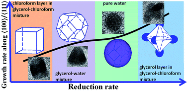
Platinum nanocrystals with different growth rates along 〈100〉 to 〈111〉 are selectively shaped using the reaction solvent, which is due to the different reduction rates in diverse reaction solutions. Electrocatalytic results show that the branched and cubic Pt/C catalysts exhibit much higher ECSA and enhanced activity of MOR, in addition, the branched Pt/C catalyst shows improved durability.


Platinum based catalysts are used extensively, particularly for the reforming of hydrocarbon and the reduction of oxygen in fuel cells.1–3 Recent works have shown that the catalytic activity and selectivity of such catalysts can be altered significantly by the nature of the Pt facets exposed.4–6 As we know, shape control of a colloidal nanocrystal is believed to be a kinetic process, with high-energy facets growing faster then vanishing, leading to nanocrystals enclosed by low-energy facets.7,8 Identifying a surfactant that can specifically bind to a particular crystal facet is critical.9–11 Thereby almost all possible shapes of Pt crystal, including cube,12 octahedron,13 tetrahedron,14 decahedron,15 multipod,16 polyhedron,17 etc., have been revealed under the well-control of surfactants in literatures. However, it is impossible to remove all the excessive surfactants from as-obtained Pt nanocrystals, which is responsible for the dramatic reduction of catalytic properties.18 More recently, specific molecules, such as carbon monoxide,19 amine,20 peptide,21,22 can be specifically selected to recognize a chosen surface through a molecular evolution process, and thereby control the shapes of Pt nanocrystals. But to date, surfactant-free preparations of shaped Pt nanocrystals have often been obtained by means of a trial-and-error process for identifying the appropriate capping agents and synthetic environments.

For Pt single crystal (fcc phase), the surface energies associated with the low-index crystallographic planes are in the order of γ(111) < γ(100) < γ(110).23–25 From the viewpoint of Wulff construction, the thermodynamically favored shape of Pt single crystal would be a truncated octahedron with the exposed facets of (111) and (100).26 And the growth rate along the 〈100〉 is much faster than that of the 〈111〉 under high reduction rate.2 Thus, the formation of Pt nanocrystal with different morphologies is achieved by controlling the growth rate of different crystal facets. Kinetics and mechanisms of such crystal growth are intensively explored.27 According to the report, precious metal nanocrystals with fcc phase can be selectively shaped using reaction kinetics by varying reaction conditions, such as precursor's concentration,28 temperature,29 etc. Although the solvent has the capability to control the reduction rate of Pt monomer in solution synthesis, there are limited reports about elaborate control of solvent on the morphology of Pt nanocrystals.

Herein, the predictable synthesis of platinum nanocrystals with exposed crystal surfaces and particular shapes was demonstrated by controlling the type of reaction solution in solvothermal approach. In particular, platinum acetylacetonate (Pt(acac)2) was firstly added into glycerol–chloroform mixture (or water, glycerol–water mixture) dissolved with benzoic acid and poly(vinyl-pyrrolidone) (PVP). The mixture was fully stirred and subsequently heated to 180 °C for 12 h in autoclave. The experimental details are revealed in ESI. High resolution transmission electron microscope (HRTEM) images were shown that the formation of Pt nanocrystals with different morphologies was dependent on the reaction solution. And the relationship between the reduction rate and the growth rate along 〈100〉 to 〈111〉 of Pt crystal was investigated. In addition, the catalytic actives for methanol electro-oxide were discussed among the Pt/C catalysts based on as-obtained different shaped Pt nanocrystals and commercial Pt nanoparticles.

As a result, branched and cubic Pt nanocrystals are obtained from glycerol–chloroform mixture in typical synthesis and collected in glycerol layer and chloroform phase, respectively. As shown in Fig. 1a, small branched nanoparticles of around 10 nm in size with well monodispersion are the main products, which are collected in glycerol layer. And the arms of nanoparticle vary from two to dozens in number as seen in the inset of Fig. 1a. From the HRTEM image in Fig. 1b and corresponding fast Fourier transformation (FFT) pattern in Fig. 1c, the measured distances of the neighboring lattice fringes for two arms of one nanoparticle are 0.201 and 0.228 nm, which are exactly the main exposed crystal facets of (100) and (111) for Pt crystal, respectively. According to the HRTEM image, structure carton is drawn in Fig. 1c, which shows octahedron with extended growth along the axis of 〈100〉. In the literature by Dr Wang,30 the function of the ratio (R: the growth rate along the 〈100〉 to that of the 〈111〉) was calculated to describe different shape of fcc crystal. The value of R for the branched Pt crystal should be higher than the function of ratio for the octahedron shape of fcc crystal according to the report (R > 1.73).30 However, the products collected in chloroform layer show obvious cubic nanostructures with dimensions of 9 nm and accompanied by 10% of nanorods (Fig. 1e). The HRTEM image in Fig. 1f and the FFT pattern in Fig. 1h exhibit cubic crystal structure and present the measured interplanar spacing of 0.201 nm, which corresponds to the exposed facet of (100) of Pt (fcc) crystal. Fig. 1g reveals the shape model of cubic Pt crystal with a very low function of the ratio according to the literature (R = 0.58).30


image file: c6ra18171f-f1.tif
Fig. 1 TEM, HRTEM images, stimulated structure and FFT patterns of Pt nanoparticles obtained in glycerol–chloroform mixture and collected in glycerol layer (a–d) and chloroform layer (e–h) in typical synthesis.

In order to prove the formation of the different morphologies of Pt nanocrystals is related to the use of the reaction solvent, glycerol–chloroform mixture are replaced with pure water and glycerol–water mixture, respectively. When the mixture of glycerol–water with volume ratio of 1/1 was used as reaction solution in typical synthesis, different Pt nanostructures were obtained as seen in Fig. 2a–d. They are mostly (>95%) polyhedral nanocrystals with average particle size of 10 nm and a few nanoparticles with diameter less than 10 nm. As shown in Fig. 2a, most polyhedral nanoparticles reveal cubooctahedral structures and have well monodispersion. HRTEM image for one cubooctahedron in Fig. 2b shows a typical hexagonal-shaped nanocrystal that can be identified as truncated octahedrons bound by (100) and (111) facets, which could be indicated by the FFT pattern (Fig. 2d) and stimulated by the structure carton in Fig. 2c. According to the literature,30 the value of R for the cubooctahedron is equal to 1.0. As seen in Fig. 2e–h, spherical nanoparticles of 5–7 nm in size are the products obtained in typical synthesis with pure water as reaction solvent. HRTEM image in Fig. 2f and corresponding FFT pattern in Fig. 2h confirm that the nanocrystal shows isotropic shape and has the fringes with lattice spacing of 0.228 and 0.201 nm, which can be indexed as (111) and (100) facets of Pt crystal, respectively. The stimulated shape in Fig. 2g shows the structure is close to another type of cubooctahedron with R value of 1.15 in the literature.30


image file: c6ra18171f-f2.tif
Fig. 2 TEM, HRTEM images, stimulated shape and FFT patterns of Pt nanoparticles obtained in typical synthesis with glycerol–water mixture (a–d) or pure water (e–h) as reaction solution.

As we know, the growth rate of different facet of nanocrystal could influence the final morphology and be adjusted by the reduction rate, when the nanoparticle's growth follows kinetic process instead of thermodynamic pathway. Fig. S1 shows the UV-vis spectra of the time evolution of substrate concentration for as-obtained polyhedral nanocrystals in typical synthesis with glycerol–water as solution. The characteristic absorption peaks of C[double bond, length as m-dash]O at around 350 nm in Fig. S1 gradually increase with time, which indicates the amount of reduced Pt is increased due to the fracture of Pt–O bond. Accordingly, Pt nanocrystals experience a kinetic growth process in the typical synthesis. And many factors could influence the reduction velocity, such as the reduction ability of reducing agent, solvent viscosity, the concentration ratio of precursor to reducing agent, reduction temperature, and so on. In glycerol–chloroform mixture in typical synthesis, there are two incompatible phases, one is chloroform layer in which Pt(acac)2 has much better solubility, the other one is glycerol phase in which more of PVP and benzoic acid are dissolved. Accordingly, the concentration ratio of Pt precursor to reducing agents (Pt/RA) is relatively high in chloroform layer, but the value of Pt/RA is fairly lower in glycerol phase. So a very high reduction rate is achieved in glycerol layer, which is beneficial to the growth along 〈100〉 and contributes to the formation of branched Pt nanocrystals with much more exposed facet of (111). Conversely, cubic Pt nanocrystals with much more exposed facets of (100) are obtained in chloroform phase, which would be attributed to the decrease of growth rate along 〈100〉 under lower reduction rate. Due to the poor solubility of Pt(acac)2 in water, the ratio of Pt/RA is lower than that in glycerol–water mixture, which results in much higher reduction rate and thus the formation of sphere-like nanocrystal with slightly higher value of R in pure water solution (Fig. 2e–h). Comparing the four different morphologies in Fig. 1 and 2, we can find that the reduction rates in four different reaction solutions have the following relationship: υ (branched crystal, R > 1.73) > υ (spherical crystal, R ≈ 1.15) > υ (polyhedral crystal, R = 1.0) > υ (cubic crystal, R = 0.58), which indicates the high reduction rate is beneficial to the growth along the axis of 〈100〉. The above relationship could be simulated by the curve in Fig. 3, in which reduction rate is directly proportional to the growth rate along 〈100〉 to 〈111〉.


image file: c6ra18171f-f3.tif
Fig. 3 Schematic for the relationship between the reduction rate and the growth rate along 〈100〉 to 〈111〉 for Pt nanocrystal.

To prove the relationships seen in Fig. 3, the reducing agent, solvent, and temperature were explored. When the glycerol in glycerol–water mixture was replaced by ethylene glycol (EG), multipolyhedral nanostructures with average diameter of 30 nm were obtained, as shown in Fig. S2. Although the single crystal is still polyhedral shape, a few polyhedral particles are connected together by growing along axis of 〈100〉 and expose more (111) planes. Due to the lower viscosity of EG than glycerol, the reduction rate would be enhanced and much higher growth rate along 〈100〉 to 〈111〉 could be obtained based on the curve in Fig. 3, thereby multipolyhedral nanocrystals with more exposed facet of (111) are produced in EG–water mixture. After the substitution of oxalic acid for benzoic acid, the reduction ability is elevated and the reduction rate is increased. As shown in Fig. S3, the flower-like nanostructures with much higher growth rate along 〈100〉 to 〈111〉 are assembled by polyhedral particles, which is similar with the result in Fig. S2. In addition, when the reaction temperature was increased to 200 °C, branched nanostructures with much longer arms (much higher growth rate along 〈100〉 to 〈111〉) than that in Fig. 1a–d were synthesized as shown in Fig. S4, since high temperature was beneficial to the increase of reduction rate. The above results all show that a number of reaction conditions could achieve the increase of reduction rate, thereby promoting the formation of Pt nanocrystals with much higher growth rate along 〈100〉 to that of 〈111〉, which is consistent with the result in Fig. 3.

In order to further characterize and compare the electro-catalytic properties of Pt nanoparticles with different morphologies, Vulcan XR-72 commercial carbon supporters were loaded with above four as-synthesized Pt nanocrystals and commercial Pt nanoparticles. As shown in Fig. 4, the primary morphologies of Pt nanoparticles are well preserved, and the nanoparticles have good monodispersion on the surface of carbon carriers. After calculation, the loading capacities of Pt in above five catalysts were all around 10%. For comparison, the electrochemical measurements are performed in the same conditions (2.0 mg cm−2).


image file: c6ra18171f-f4.tif
Fig. 4 TEM images of Vulcan XC-72 carbon loaded with as-obtained Pt nanoparticles with four different morphologies: (a) spherical Pt nanoparticles shown in Fig. 2e–h, (b) polyhedral Pt nanoparticles shown in Fig. 2a–d, (c) branched Pt nanoparticles shown in Fig. 1a–d, (d) cubic Pt nanoparticles shown in Fig. 1e–h.

The as-obtained four Pt/C catalysts and commercial Pt/C catalyst for the oxidation of methanol was studied using cyclic voltammetry (CV). The CV curves in Fig. S5 were measured in an aqueous solution of H2SO4 (0.5 M) between −0.2 and 1.0 V with a sweep rate of 50 mV s−1. The electrochemically active surface area (ECSA) is calculated from the CV curve in Fig. S5 and described in Table S1 in the ESI. The ECSA's relationship of as-obtained four catalysts and commercial Pt/C is as follows: branched Pt/C (62.2 m2 g−1) > cubic Pt/C (47.8 m2 g−1) > polyhedral Pt/C (26.1 m2 g−1) > spherical Pt/C (15.2 m2 g−1) > commercial Pt/C (14.2 m2 g−1). The morphology effects of Pt crystal on the activity in MOR are studied in H2SO4 (0.5 M) and methanol (0.5 M) solutions between 0.0 and 1.0 V, as seen in Fig. 5a. Among the as-obtained four Pt/C catalysts, Vulcan XR-72 catalyst loaded with branched Pt nanocrystals exhibits the highest electrocatalytic activity at 151.1 mA cm−1 mg−1 Pt, which is 1.2-, 3.2-, 3.9- and 3.5-fold higher than those catalysts based on cubic Pt nanocrystal (130.4 mA cm−1 mg−1 Pt), Pt nanopolyhedrons (47.5 mA cm−1 mg−1 Pt) and spherical Pt nanoparticles (39.0 mA cm−1 mg−1 Pt), commercial Pt nanoparticles (43.1 mA cm−1 mg−1 Pt), respectively. The catalytic activities of MOR and ECSA results all show that branched Pt/C has the highest catalytic efficiency, which is most likely due to the high-index facet in the edge of the branched Pt/C nanoparticles. According to literature,31 high-index surfaces of a face-centered cubic metal (e.g., Pd, Pt) have a high density of low-coordinated surface atoms and therefore possess enhanced catalysis activity in comparison with low-index faces. However, the cubic Pt/C exposed with facet of (100) has relatively high catalytic ability because the electrocatalytic ability of facet (100) is better than that of facet (111).3 The peak current ratio of the forward scan (If) to backward scan (Ib), If/Ib, could represent the ability of resistance to the poisoning of carbonaceous species.32 The values of If/Ib for as-obtained five Pt/C catalysts are all around 1.00, which indicate they have comparable CO poison tolerance. As shown in Fig. 5b, chronoamperometric curves were recorded at 0.65 V vs. Hg/Hg2Cl2 for 3500 s to evaluate the stability of the electroctatalysts. The chronoamperometry generates high charging currents owing to the initial high concentration of methanol on the surface of the electrocatalysts, but at the initial stage the current intensity decays rapidly because of the decrease of the concentration gradient and poisoning by the intermediate species. Clearly, the current density induced by the branched Pt/C catalyst decays more slowly than others before the initial 2000 s. However, the current decay of cubic Pt/C catalyst is most conspicuous in the initial 500 s among all the catalysts. The big differences in chronoamperometric curves could mainly be ascribed to the much more stability of facet (111) than (100) due to the low surface energy.


image file: c6ra18171f-f5.tif
Fig. 5 (a) Cyclic voltammetric curves and (b) chronoamperograms for the MOR at 0.65 V vs. Hg/Hg2Cl2 of Vulcan XR-72 loaded with as-obtained four different shaped Pt nanocrystals and commercial Pt nanoparticles in 0.5 M H2SO4 + 0.5 M methanol.

In summary, we have synthesized Pt nanocrystals with four different shapes in diverse solvent mixtures by solvothermal approach. HRTEM measurements clearly show that the difference in the morphologies of Pt crystals is attributed to the different growth rate along 〈100〉 to that of 〈111〉. In addition, higher reduction rate is proposed to be beneficial to the growth along the axis of 〈100〉, which could be induced by comparing the results under different reaction conditions. Among the electrocatalytic performances of four as-obtained Pt/C catalysts and commercial Pt/C catalyst, the branched Pt/C exhibits the highest ECSA, enhanced activity of MOR, and improved durability, which is probably due to the high-index facet in the edge and stability of facet (111) of the branched nanocrystal. Meanwhile, cubic Pt/C has relatively high catalytic ability but poor durability, because the facet (100) has better electrocatalytic efficiency and inferior stability than that of facet (111).

Acknowledgements

This work was supported by National Natural Science Foundation of China (Grant No. 21306075 and 21567017), and Natural Science Foundation of Inner Mongolia (Grant No. 2015MS0213).

Notes and references

  1. M. K. Debe, Nature, 2012, 486, 43 CrossRef CAS PubMed.
  2. J. Wu and H. Yang, Acc. Chem. Res., 2013, 46, 1848 CrossRef CAS PubMed.
  3. Y. Xu and B. Zhang, Chem. Soc. Rev., 2014, 43, 2439 RSC.
  4. S. Cheong, J. D. Watt and R. D. Tilley, Nanoscale, 2010, 2, 2045 RSC.
  5. N. S. Porter, H. Wu, Z. Quan and J. Fang, Acc. Chem. Res., 2013, 46, 1867 CrossRef CAS PubMed.
  6. J. Ding, X. Zhu, L. Bu, J. Yao, J. Guo, S. Guo and X. Huang, Chem. Commun., 2015, 51, 9722 RSC.
  7. T. S. Ahmadi, Z. L. Wang, T. C. Green, A. Henglein and M. A. El-Sayed, Science, 1996, 272, 1924 CrossRef CAS PubMed.
  8. Y. N. Xia, Y. J. Xiong, B. Lim and S. E. Skrabalak, Angew. Chem., Int. Ed., 2009, 48, 60 CrossRef CAS PubMed.
  9. Y. D. Yin and A. P. Alivisatos, Nature, 2005, 437, 664 CrossRef CAS PubMed.
  10. J. Xiao and L. Qi, Nanoscale, 2011, 3, 1383 RSC.
  11. S. Sreedhala and C. P. Vinod, Chem. Commun., 2015, 51, 10178 RSC.
  12. C. Kim, S. S. Kim, S. Yang, J. W. Han and H. Lee, Chem. Commun., 2012, 48, 6396 RSC.
  13. H. Song, F. Kim, S. Connor, G. A. Somorjai and P. D. Yang, J. Phys. Chem. B, 2005, 109, 188 CrossRef CAS PubMed.
  14. C. Burda, X. B. Chen, R. Narayanan and M. A. El-Sayed, Chem. Rev., 2005, 105, 1025 CrossRef CAS PubMed.
  15. M. E. Scofield, H. Liu and S. S. Wong, Chem. Soc. Rev., 2015, 44, 5836 RSC.
  16. S. Sun, G. Zhang, D. Geng, Y. Chen, R. Li, M. Cai and X. Sun, Angew. Chem., Int. Ed., 2011, 50, 422 CrossRef CAS PubMed.
  17. X. Huang, Z. Zhao, J. Fan, Y. Tan and N. Zheng, J. Am. Chem. Soc., 2011, 133, 4718 CrossRef CAS PubMed.
  18. N. R. Wu, Q. Y. Suo, H. Wang, Z. Z. Wang, Z. L. Chai and X. J. Wang, CrystEngComm, 2015, 17, 6716 RSC.
  19. Z. Peng, C. Kisielowski and A. T. Bell, Chem. Commun., 2012, 48, 1854 RSC.
  20. X. Huang, Z. Zhao, J. Fan, Y. Tan and N. Zheng, J. Am. Chem. Soc., 2011, 133, 4718 CrossRef CAS PubMed.
  21. Y. Li and Y. Huang, Adv. Mater., 2010, 22, 1921 CrossRef CAS PubMed.
  22. C. Y. Chiu, Y. Li, L. Ruan, X. Ye, C. B. Murray and Y. Huang, Nat. Chem., 2011, 3, 393 CrossRef CAS PubMed.
  23. J. Chen, B. Lim, E. P. Lee and Y. Xia, Nano Today, 2009, 4, 81 CrossRef CAS.
  24. J. Wu and H. Yang, Acc. Chem. Res., 2013, 46, 1848 CrossRef CAS PubMed.
  25. Z. Peng and H. Yang, Nano Today, 2009, 4, 143 CrossRef CAS.
  26. C. Li, M. Imura and Y. Yamauchi, Phys. Chem. Chem. Phys., 2014, 16, 8787 RSC.
  27. F. D. Wang, V. N. Richards, S. P. Shields and W. E. Buhro, Chem. Mater., 2014, 26, 5 CrossRef CAS.
  28. X. F. Ma, R. Lin, R. Y. Ofoli, Z. Mei and J. E. Jacksonm, Mater. Chem. Phys., 2016, 173, 1 CrossRef CAS.
  29. B. Lim, H. Kobayashi, P. H. C. Camargo, L. F. Allard, J. Y. Liu and Y. N. Xia, Nano Res., 2010, 3, 180 CrossRef CAS.
  30. Z. L. Wang, J. Phys. Chem. B, 2000, 104, 1153 CrossRef CAS.
  31. X. Q. Huang, Z. P. Zhao, J. M. Fan, Y. M. Tan and N. F. Zheng, J. Am. Chem. Soc., 2011, 133, 4718 CrossRef CAS PubMed.
  32. A. M. Hofstead-Duffy, D. J. Chen, S. G. Sunb and Y. J. Tong, J. Mater. Chem., 2012, 22, 5205 RSC.

Footnote

Electronic supplementary information (ESI) available: Experimental details and characterization details. See DOI: 10.1039/c6ra18171f

This journal is © The Royal Society of Chemistry 2016
Click here to see how this site uses Cookies. View our privacy policy here.