DOI:
10.1039/C6RA18091D
(Paper)
RSC Adv., 2016,
6, 81719-81725
Adsorption of radioactive iodine on surfactant-modified sodium niobate†
Received
15th July 2016
, Accepted 20th August 2016
First published on 22nd August 2016
Abstract
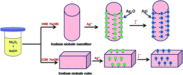
Iodine radioisotopes have been released into the environment both by the nuclear industry and as a result of medical treatment using radioactive materials, potentially posing a serious threat to human health. Thus, when developing applications of radioactive iodine, emergency procedures for dealing with it, and preventing its leakage, are constant concerns. In this study, nanofibers and cubes of Ag2O anchored sodium niobate composites were synthesized by a wet chemical process, and characterized by means of XRD, SEM, TEM and BET techniques. It was found that the well-dispersed Ag2O nanocrystals (3–5 nm) were firmly anchored on the external surface of sodium niobate along the planes with crystallographic similarity to those of Ag2O. These composites can efficiently capture I− anions by precipitating AgI, firmly attaching it to the substrate and allowing it to be recovered easily for safe disposal. Variations in the iodine removal abilities with differing pH value, adsorption time, adsorption temperature, initial I− concentration, and in the presence of competing ions, were studied. The maximum monolayer adsorption capacities of I− anions for Ag2O anchored sodium niobate nanofibers and cubes were found to be 296 and 193 m2 g−1 respectively. TEM, XRD, XPS and Raman results indicated that I− anion adsorption was mainly due to the Ag2O nanocrystals on the sodium niobate surfaces. We conclude that Ag2O deposited on sodium niobate hass a great potential for removal of radioactive iodine from waste water.
1 Introduction
Radioactive iodide ions are the products of uranium fission. They are highly radioactive and acutely toxic, and can easily be dissolved in water during an accident at a nuclear reactor, potentially entering the food chain leading to significant health risks;1,2 examples include incidents such as those that occurred at Chernobyl in 1986, at Three Mile Island in Pennsylvania in 1979, and in 2011 at Fukushima, Japan. In particular after the nuclear accident at Fukushima, global concern about nuclear waste leakage rose significantly.3 Radioactive iodine is also used in the treatment of thyroid cancer, and as a result, radioactive wastewater is generated by a large number of medical treatment facilities. The wide used of radioisotopes requires effective technology to remove radioactive ions from the environment, to allow their subsequent safe disposal.4 The key issue in the development of such a technology is to devise materials which are able to absorb radioactive iodine irreversibly, selectively, and efficiently.
Many materials, such as saponite,5 activated carbon,6 zeolite,7 heavy metal (Hg2+) compounds,8 and silver oxide composites,9 have been investigated as candidates for removing radioactive iodide ions from wastewater by formation of precipitates or sparingly soluble phases on the their surfaces, which can then be separated from solution and disposed of safely. However, these compounds displayed low adsorption capacities and slow uptake dynamics due to their small specific surface areas. To solve this problem, Ag+ and Hg2+ ions have been loaded onto the surface of natural zeolites to remove iodide anions from gaseous and aqueous wastes.10,11 However, such zeolite adsorbents suffer from slow adsorption kinetics, and thus, poor efficiency. In addition, concern has also been raised about the possible release of radioactive species from the used sorbents if the precipitate particles did not bind tightly to the zeolite substrate.
Recently, such Ag2O grafted nanomaterials as Ag2O grafted titanate,12,13 and Ag2O grafted layered sodium vanadate,14 have shown better I− removal ability. The Ag2O nanocrystals were be firmly bonded to the surface of those substrates by means of a coherent interface. In addition, the morphology of the substrate provides a large surface area, providing more space for attachment of Ag2O nanocrystals, and increasing the probability of contact with I− anions. These results gave a new direction for capturing of radioactive iodine using anchored Ag2O nanocrystals. However, the application of such materials is limited to a narrow pH range due to instability of the nanostructure of supports under changing conditions. Finding other absorbents suitable for wider pH ranges is necessary.
The properties of the support materials have an important effect on the adsorption capacities of the absorbent. For example, a larger specific surface provides more space for Ag2O nanocrystals. Ideally, the inorganic materials used as supports should possess the following features: (1) large specific surface area, (2) the ability to firmly attach Ag2O nanocrystals to its surface, (3) good stability in the presence of radiation, and chemicals, (4) good temperature stability. All of these should be taken into account when designing efficient absorbents.
In this work, we successfully synthesized nanostructured Ag2O-anchored sodium niobate with unusual morphologies, namely Ag2O-anchored sodium niobate nanofibers (Ag2O–SNF) and Ag2O-anchored sodium niobate cubes (Ag2O–SNC). These Ag2O nanocrystals are not aggregated randomly on the surface of the niobate substrate, but are firmly anchored via stable coherent interfaces between Ag2O and niobate phases. We found the nanofibers of Ag2O–SNF possess a large surface area, which can provide more space for attachment of Ag2O nanocrystals, and displayed high adsorption capacity, selectivity, and kinetics in the removal of I− anions from basic media.
2 Experiments
2.1 Synthesis
All chemical used were standard reagents unless otherwise specified. The sodium niobates were prepared by employing hydrothermal methods. In a typical procedure, 1 g Nb2O5 and 60 mL of 10–12 mol L−1 NaOH solution were mixed by stirring at room temperature. The mixture was transferred into a 100 mL Teflon-lined autoclave and allowed to react at 180 °C 232 for 2 h; afterwards, the autoclave cooled down naturally. The finally products were collected by glassware, then washed with deionized water three times, and dried in air at 80 °C.
The Ag2O nanocrystals anchored sodium niobate nanofibers (Ag2O–SNF) and cubes (Ag2O–SNC) were prepared by chemical deposit. Initially, 0.2 g sodium niobate was dispersed in 200 mL pure water. The pH value of the suspension was adjusted to 11 by dropwise addition of dilute NaOH solution. The nanoparticles were collected by centrifuging and then dispersed in 200 mL silver nitrate aqueous solution. After stirring vigorously for 24 h, the precipitate was collected and washed with deionized water three times, and then dried at 80 °C for 24 h. The processes of fabricating Ag2O–SNF and Ag2O–SNC composites and subsequent I− adsorption are illustrated in Scheme 1.
 |
| Scheme 1 Processes for fabricating Ag2O–SNF and Ag2O–SNC composites, and subsequent capture of I−. | |
2.2 Characterization
The crystalline structure of the prepared samples was characterized using X-ray diffraction (PXRD, X'Pert PRO, PANalytical, Almelo, Netherlands) with Cu-Kα radiation (λ = 0.15406 nm at 40 kV and 45 mA). The sizes and shapes of the nanostructures were observed on a field emission scanning electron microscope (FE-SEM Philips XL30 FEG, Eindhoven, Netherlands) and transmission electron microscopy (TEM, JEM200CX, 120 kV). The composition and chemical state were determined by X-ray photoelectron spectroscopy (XPS) performed on an RBD upgraded PHI-5000C ESCA system (Perkin–Elmer) using Mg-monochromatic X-rays at a power of 25 W with an X-ray-beam diameter of 10 mm, and a pass energy of 29.35 eV. The binding energy was calibrated by the C 1s hydrocarbon peak at 284.8 eV. Nitrogen adsorption and desorption isotherms were measured at 77 K with a Beckman Coulter SA 3100 surface area analyzer. To determine the surface area, the Brunauer–Emmett–Teller (BET) method was used. Raman spectra were recorded on a microscopic confocal Raman spectrometer (Renishaw 1000NR) with an excitation of 514.5 nm laser light.
2.3 Adsorption experiment
In this experiment, anion adsorption experiments were carried out with nonradioactive iodine in aqueous solution to avoid high radiation doses and the toxicity of 131I or 129I. The equilibrium uptake capacities of the Ag2O-anchored sodium niobate absorbents for I− anions were conducted through batch experiments which were implemented using a series of concentrations of I− solutions ranging from 50 mg L−1 to 1000 mg L−1 (the amount of adsorbent = 0.1g L−1, 30 °C, pH = 9.0). For comparison, the same amount of Nb2Na2O6·H2O was used as a control. The effects of pH, equilibrium time, ionic strength and competition ions were examined. All samples with iodide solutions were equilibrated for over 48 h at room temperature with magnetic stirring. Afterward the solids and solutions were separated by centrifugation and the supernatants were analyzed by ultraviolet spectrophotometer (UV-vis) to determine the amount of remaining iodide anions in the solutions.
3 Results and discussion
3.1 Characterization of Ag2O–SNF and Ag2O–SNC adsorbents
XRD measurements were carried out to determine the microstructure of sodium niobate coated with Ag2O nanocrystals, as depicted Fig. 1. They clearly showed that the concentration of NaOH solution used to prepare the sodium niobate had a key effect on the structure of the final products. The intensities and positions of the diffraction peaks for the product prepared using 12 mol L−1 NaOH solution are consistent with the values in the literature11 for orthorhombic sodium niobate (Ag2O–SNC). However, the XRD pattern for the product prepared with 10 mol L−1 NaOH solution (Ag2O–SNF) are attributed to Sandia octahedral molecular sieves (SOMS, Na2Nb2−xMxO6−x) with x = 0, Na2Nb2O6·H2O; the peak intensities and positions are consistent with the values presented by Nyman.15
 |
| Fig. 1 XRD patterns of Ag2O–SNF and Ag2O–SNC samples. | |
The morphologies and microstructures of the synthesized samples were further determined by SEM and TEM. Again it was found that they depend on the concentration of NaOH used to prepare the niobate. As shown in Fig. 2a, the morphologies and microstructures of Ag2O–SNF obtained by reacting Na2O5 with 10 mol L−1 NaOH solution. Many nanofibers with a uniform diameter of around 200 nm and lengths of several to hundreds of microns were obtained. TEM (Fig. 2b) and HRTEM (Fig. 2c) clearly show Ag2O nanoparticles with a size of 2–5 nm; these were anchored on the external surface of sodium niobate nanofibers by dispersing them into an aqueous silver nitrate solution. In neutral or basic conditions (pH ≥ 7) most silver is in the form of Ag2O nanoparticles and the remainder is in the form of Ag+ ions in the interlayer region because of exchange with Na+.16 These Ag2O nanoparticles efficiently capture I− anions as the nanoparticles are exposed on the surface of the fibers and so are readily accessible to anions. The selected area electron diffraction (SAED) patterns (inset in Fig. 2b) reveal the signal crystalline nature of each nanofiber. However, the morphology of the product changed markedly when increasing the concentration of the NaOH solution. Fig. 3a presents an SEM image of Ag2O–SNC obtained by reacting Na2O5 with 12 mol L−1 NaOH solution. The main products were large, sharp-edged cubes and thick plates with depths and widths of 1–3 microns (Fig. 3b). The inset image in Fig. 3b shows the SAED pattern of the [101] zone axis, revealing the single crystalline nature of each cube. It was also found that the Ag2O nanoparticles with a particle size of about 5 nm are located on the surface of Ag2O–SNC by dispersing them into an aqueous silver nitrate solution in neutral or slightly basic condition (Fig. 3c).
 |
| Fig. 2 SEM and TEM images of sodium niobate nanofibers with anchored Ag2O nanocrystals (Ag2O–SNF). (a) SEM image of sodium niobate nanofibers. (b) TEM image showing abundant Ag2O nanocrystals dispersed on the sodium niobate nanofibers. Inset: SAED image of a single nanofiber. (c) HRTEM image of Ag2O nanocrystals. (d) IFFT image of the selected area A in (c). | |
 |
| Fig. 3 SEM and TEM images of sodium niobate microcubes with anchored Ag2O nanocrystals (Ag2O–SNF). (a) SEM image of sodium niobate microcubes. (b) TEM image showing abundant Ag2O nanocrystals dispersed on the sodium niobate microcubes. Inset: SAED image of a signal microcube. (c) HRTEM image of Ag2O nanocrystals. (d) IFFT image of the selected area B in (c). | |
To capture and immobilize I− anions from wastewater, the Ag2O nanoparticles must be firmly attached to the sodium niobate nanostructures. If the nanocrystals readily detach from the fibers or cubes, it will be extremely difficult and costly to recover the fine nanoparticles from a solution. Furthermore, the small Ag2O nanocrystals may aggregate to form a solid with low surface area and thus poor ability to precipitate I− anions. Fortunately, the Ag2O nanoparticles are attached to the external surface of the sodium niobate nanostructure by means of their similar surface crystal structure, as shown in the inverse fast fourier transform (IFFT) images (Fig. 2d and 3d) of the selected areas in Fig. 2c and 3c respectively. Fig. 2d shows that the (111) planes of the Ag2O nanocrystals are parallel to the (110) plane of the sodium niobate. The interplane distance of the (111) planes of Ag2O nanocrystals is 0.2748 nm, which is close to that of the (110) planes of the sodium niobate nanofibers (0.2750). If (111) planes of Ag2O match (110) planes of sodium niobate nanofibers where the Ag2O nanocrystals meet the sodium niobate substrate, the number of oxygen atoms shared by the two phases is maximized and full coordination can be achieved, to form a well-matched (coherent) interface. Similarly, Ag2O nanocrystals are anchored on the surface of sodium niobate cubes by such a coherent interface (Fig. 3d).
The good I− anion removal ability of Ag2O–SNF and Ag2O–SNC are due to the large amount of Ag2O uniformly loaded on the large specific surface area of the sodium niobate fibers or cubes acting as substrate. The N2 adsorption–desorption isotherms of Ag2O–NNF and Ag2O–SNC samples were measured, and the Brunauer–Emmet–Teller (BET) method was applied to the N2 sorption isotherms to determine the specific surface area of the samples. As observed in Fig. 4, the samples exhibited typical type II isotherms with a hysteresis loop that is indicative of the presence of small slit-shaped pores in the samples. The specific surface area is 148.31 m2 g−1 for Ag2O–SNF and was much higher than that of Ag2O–SNC (92.24 m2 g−1), resulting in the higher iodine uptake capacity of Ag2O–SNF.
 |
| Fig. 4 N2 adsorption–desorption isotherms for Ag2O–SNF and Ag2O–SNC. | |
3.2 Adsorption experiment
3.2.1 Effect of initial pH. It is known that solution pH significantly affects the adsorption process. In order to investigate pH effects on adsorption capacity for I− anions, the adsorption process was studied in a batch experiment with initial ion concentration of 100 mg L−1 at 30 °C, as shown in Fig. 5. As can be seen, the uptake of I− anions by Ag2O–SNF is much greater than for Ag2O–SNC at any pH value. The adsorption of I− anions by both Ag2O–SNF and Ag2O–SNC experienced similar rapid rises as pH increased from 3.0–6.0, and reached optimum adsorption capacities over the pH range 6.0–9.0. However, the uptake efficiencies of the Ag2O–SNF and Ag2O–SNC adsorbents decreased slowly when the solution pH was up to 9.0. This is because the increased OH− concentration at high pH results in an increase in negative charges on the external surface of the adsorbent. Overall, the pH range of 6.0–9.0 is best suited for removal of I− anions from aqueous solution.
 |
| Fig. 5 Effect of solution pH on the adsorption capacity of Ag2O–SNF and Ag2O–SNC. | |
3.2.2 Adsorption isotherm. Adsorption isotherm measurements for Ag2O–SNF and Ag2O–SNC samples were carried out by static adsorption experiments. The results are exhibited in Fig. 6, where it can clearly be seen that the adsorption capacity of Ag2O–SNF is significantly higher than for Ag2O–SNC at the same initial concentrations and temperatures. This may be explained by the different numbers of available active adsorption sites on the surfaces of Ag2O–SNF and Ag2O–SNC. To further determine the parameters associated with I− anion adsorption, the experimental data were analyzed using Langmuir and Freundlich adsorption isotherm models. The Langmuir model assumes that monolayer surface adsorption occurs on specific homogeneous sites and no interaction occurs between the adsorbed pollutants. The equation for this model can be expressed as follows:
 |
| Fig. 6 Effect of initial I− anion concentration on the adsorption of I− by Ag2O–SNF and Ag2O–SNC. | |
while the Freundlich adsorption isotherm is used to describe adsorption on a heterogeneous surface and can be expressed as follows:
here,
Ce (mg L
−1) is the equilibrium concentration of the bulk solution,
qe (mg L
−1) is the equilibrium concentration of the solid phase,
b (L mg
−1) is the Langmuir constant related to the free energy or net enthalpy of adsorption, and
qm (mg g
−1) is the adsorption capacity at isotherm temperature.
kf and
n are equilibrium Freundlich constants denoting adsorption capacity and adsorption intensity, respectively.
Table 1 summarizes the corresponding maximum adsorption capacity
qmax. It is clear that the Langmuir model is more appropriate than the Freundlich model for representing the adsorption of I
− anions as it has high correlation coefficients (
R2 = 0.9913 and 0.9316 for Ag
2O–SNC and Ag
2O–SNF, respectively), indicating that adsorption is localized in a monolayer. The monolayer maximum adsorption capacities calculated from the Langmuir isotherm are 295.91 and 193.04 mg g
−1 for the prepared Ag
2O–SNF and Ag
2O–SNC, respectively. The specific surface area measured by BET as above for the prepared Ag
2O–SNF was considerably higher (142.93 m
2 g
−1) than for Ag
2O–SNC (92.24 m
2 g
−1), making it suitable for high-performance wastewater treatment. Compared with other adsorbents in previous studies,
10,11,17–20 the Ag
2O–SNF absorbent described here exhibited much higher adsorption capacities for I
− anions, and has great promise for the effective removal of I
− anions in environmental remediation.
Table 1 Adsorption isotherm constant of I− anions adsorption process on the Ag2O–SNF and Ag2O–SNC
Model |
Parameters |
Ag2O–SNC |
Ag2O–SNF |
Langmuir isotherm |
qm (mg g−1) |
193.04 |
295.91 |
b (L mg−1) |
0.088 |
0.0356 |
R2 |
0.9316 |
0.9913 |
Freundlich isotherm |
kf [mg g−1(L g−1)1/n] |
42.43 |
55.16 |
n |
2.87 |
3.18 |
R2 |
0.8812 |
0.9658 |
3.2.3 Adsorption kinetics. The effect of contact time on the adsorption of I− anions by Ag2O–SNC and Ag2O–SNF was investigated to determine the time taken for the adsorption equilibrium. As Fig. 7 shows, the adsorption rate is fast before 110 min and slowly reaches equilibrium at approximately 110 min. This can be explained by positing that there are sufficient available active sites (Ag2O nanocrystals) on the surface of the samples in the initial stages, and these are gradually occupied by iodine (AgI nanocrystals) as time passes. In order to examine the controlling mechanism of adsorption, two kinetic models were used to describe the adsorption process: a pseudo-first-order model and a pseudo-second-order model. The pseudo-first-order kinetic model can be expressed in the following linearized form:
where t (min) is the contact time, k1 (min−1) is the pseudo-first-order rate constant, qe (mg g−1) and qt (mg g−1) represent the uptake of ions by the adsorbent at equilibrium and time t, respectively. k1 and qe can be calculated from the slope and the intercept of the plot of log(qe − qt) versus t.
 |
| Fig. 7 Effect of contact time on the adsorption of I− anions by Ag2O–SNF and Ag2O–SNC. | |
The pseudo-second-order kinetic model is described by the following equation:
where
k2 (g mg
−1 min
−1) is the pseudo-second-order rate constant. The values for
k2 and
qe can be calculated from the slope and the intercept of the plot of
t/
qt.
The parameters of these two kinetic models were calculated and are given in Table 2, which shows that the pseudo-second-order model provides better correlation coefficients (R2 = 0.9908 and 0.9936 for Ag2O–SNC and Ag2O–SNF, respectively). Therefore, the pseudo-second-order model appropriately describes the kinetics of the adsorption process. This implies that chemical sorption may be the main rate limiting factor during the adsorption process.21,22
Table 2 Kinetic parameters for I− anions adsorption onto Ag2O–SNF and Ag2O–SNC (T = 308 °C)
Model |
Parameters |
Ag2O–SNC |
Ag2O–SNF |
Experiment |
qe (mg g−1) |
191.89 |
293.46 |
Pseudo first-order kinetic model |
k1(min−1) |
0.028 |
0.035 |
qcale (mg g−1) |
202.54 |
349.17 |
R2 |
0.8756 |
0.9502 |
Pseudo second-order kinetic model |
k2 (g mg−1 min−1) |
0.0028 |
0.0056 |
qcale (mg g−1) |
229.32 |
337.32 |
R2 |
0.9908 |
0.9936 |
3.2.4 Effect of temperature. The effect of temperature on the adsorption capacity of Ag2O–SNF for I− anions is presented in Fig. 8. The increase in I− anion adsorption capacity of Ag2O–SNF with increasing temperature indicates the endothermic nature of the adsorption process. To investigate the mechanism involved in the adsorption, the thermodynamic behavior of I− anion adsorption onto Ag2O–SNF was evaluated using the following equations:
 |
| Fig. 8 (a) Effect of temperature on I− adsorption onto Ag2O–SNF; (b) relationship between ln kd and 1000/T. | |
where Kd (mL g−1) is the distribution coefficient, R (8.314 J mol−1 K−1) is the gas constant, and T (K) is the absolute temperature of the aqueous solution. ΔH° and ΔS° were respectively calculated from the slope and intercept of the linear variation between ln
Kd and (1/T) (see Fig. 8). Thermodynamic parameters were shown in Table 3.
Table 3 Thermodynamic parameters for I− adsorption
T (K) |
ΔG° (kJ mol−1) |
ΔS° (J K−1 mol−1) |
ΔH° (kJ mol−1) |
288 |
−52.94 |
15.4 |
−48.5 |
298 |
−53.08 |
|
|
308 |
−53.24 |
|
|
The negative ΔG° indicates that the adsorption process is spontaneous. This value decreases with increasing temperature, indicating that better adsorption performance is obtained at higher temperature. The positive value of ΔH° confirms the endothermic nature of the overall adsorption process, and the positive value of ΔS° suggests increasing randomness at the solid/solution interface during the adsorption process.
3.2.5 Effects of co-existing anions. Contaminated water contains a variety of other anions such as Cl− and CO32− which may compete with I− anions for the adsorption sites on the surface of the adsorbents. It is thus necessary to study the influence of co-existing anions on iodine adsorption. As shown in Fig. 9, more than 98% of I− anions were taken up by Ag2O–SNF in 0.001∼0.1 mol L−1 NaCl. Clearly, the Ag2O–SNF is highly selective for I− anions and competitive Cl− anions had little effect on I− anion uptake capacity. However, the uptake capacity for I− anions decreased in the presence of 0.001 mol L−1 CO32− anions. These differing results may be ascribed to the large differences in Gibbs energy of the reaction between Ag2O and I−, Cl−, and CO32− anions respectively.9,23 The energy value for the reaction between Ag2O and I− (−32 kJ mol−1) is lower than that for the reaction between Ag2O and Cl− (+41 kJ mol−1), but is higher than that for the reaction between Ag2O and CO32− (−21 kJ mol−1). Thus, Ag2O–SNF absorbent displays a higher selectivity for Cl− than CO32−.
 |
| Fig. 9 Effect of competitive anions on the removal of I− anions by Ag2O–SNF. | |
3.2.6 Adsorption mechanism. The difference in I− anion adsorption between Ag2O–SNF and Ag2O–SNC can be attributed to the different numbers of available active adsorption sites (the number of Ag2O nanocrystals) on their surfaces. The Ag2O–SNF prepared here had a large amount of space available, allowing the attachment of many Ag2O nanocrystals, providing more contact opportunities for I− anions. In addition, the larger surface area and nanofiber structure allowed fast adsorption of I− anions from solution without diffusion problems.After contact with I− anions, the yellow Ag2O–SNF powder turned grey due to the formation of AgI–SNF (Fig. S1†). To investigate the interaction mechanism between I− anions and the Ag2O–SNF, the adsorbent after uptake of I− anions was washed and characterized by TEM and HRTEM. As shown in Fig. 10a and b, the adsorbent appears to maintain the fiber-like morphology after the uptake of iodide. The AgI nanocrystals formed were slightly larger (5–8 nm) than the initial Ag2O nanoparticles anchored on the surface of the Ag2O–SNF. The presence of iodine in the used absorbents was also confirmed by EDX (Fig. S2†), XRD (Fig. S3†), and IR (Fig. S4†). The IFFT of the used adsorbent (Fig. 10c, showing a selected area of Fig. 10b) showed the distance between the (11
0) planes in the AgI nanocrystal was 0.2290 nm, close to that of the (111) planes of the niobate (0.2250 nm). Thus, the two phases were able to join to form a well-matched interface that bonded the newly deposited AgI nanocrystals firmly to the sodium niobate nanofibers. This also indicated that the used absorbents could easily be recovered for safe disposal.
 |
| Fig. 10 TEM images of sodium niobate nanofibers anchored with AgI nanocrystals. (a) TEM image of AgI nanocrystal formed on a single sodium niobate nanofiber. Inset: SAED image of a single nanofiber. (b) HRTEM image of AgI nanocrystals on sodium niobate nanofibers (c) IFFT image of the selected area A in (b). | |
The composition of the adsorbents before and after adsorption of I− anions was further analyzed by XPS spectrum. Fig. 11 is a typical XPS survey spectrum giving high-resolution XPS spectra of different atoms. The whole spectrum of the absorbent before and after adsorption of I− anions is shown in Fig. 11a. It indicates that the I− anions were successfully adsorbed onto the surface of the Ag2O–SNF. The O 1s core level spectrum (Fig. 10b) can be fitted well with two peaks. The first strong peak at 529.8 eV corresponds to crystal oxygen whereas the second strong peak at 531.4 eV corresponds to surface adsorption oxygen.23 After adsorption of I− anions by forming AgI on the surface, the O 1s spectrum of the adsorbent is quite different. As shown in Fig. 11c, the main peak at 529.5 eV remains after uptake of I− anions, but the peak at 531.4 eV (OH) becomes substantially weaker. Furthermore, the I 3d core level appears at the binding energies of around 630.2 eV (I 3d3/2) and 618.7 eV (I 3d5/2), respectively (Fig. 11d), which is ascribed to I− in AgI,23 is in good agreement with those reported in other Ag2O grafted absorbents.9,17
 |
| Fig. 11 XPS spectra of Ag2O–SNF before and after adsorption of I−anions: (a) survey scan, (b) O 1s of Ag2O–SNF, (c) O 1s of AgI–SNF and (d) I 3d of AgI–SNF. | |
From the above discussion, we may conclude that the mechanism of I− adsorption is based on chemical reaction between I− and Ag2O rather than physical adsorption, following the reaction below:
Ag2O(s) + 2I−(aq) + H2O → 2AgI(s) + 2OH−(aq) |
4 Conclusions
In this paper, novel composites of Ag2O–SNF and Ag2O–SNC, with specific surface areas of 148.31 m2 g−1 and 92.24 m2 g−1 respectively, were prepared by a simple method. SEM, TEM and XRD results reveal that Ag2O nanocrystals were firmly anchored on the surface of sodium niobate nanofibers and cubes. The Ag2O–SNF and Ag2O–SNC composites were then used as absorbents for iodine anion removal from water. Maximal I− anion adsorption was achieved in neutral media, and the maximum monolayer adsorption capacity of I− anions by the Ag2O–SNF was determined to be 293.46 mg g−1. The specific surface areas of substrate were responsible for I− anion adsorption. In summary, the composite Ag2O–SNF exhibits great potential for the removal of iodine from contaminated water in engineering practice.
Acknowledgements
We wish to acknowledge the financial support from the Foundation of China Academy of Engineering Physics and the National Natural Science Foundation of China (No. 21501159)
Notes and references
- H. A. Robertson and I. R. Falconer, Nature, 1959, 184, 1699–1702 CrossRef CAS PubMed.
- T. H. Woo, Ann. Nucl. Energy, 2013, 53, 197–201 CrossRef CAS.
- M. Asami and M. Akiba, Journal of National Institute of Public Health, 2011, 60, 306–313 Search PubMed.
- J. Sun, D. J. Yang, C. H. Sun, L. Liu, S. G. Yang, Y. Jia, R. S. Cai and X. D. Yao, Sci. Rep., 2014, 4, 7313–7317 CrossRef CAS PubMed.
- K. Morimoto, K. Tamura, Y. Umemura, H. Sato and A. Yamagishi, Chem. Lett., 2011, 40, 867–869 CrossRef CAS.
- K. Kosaka, M. Asami, N. Kobashigawa, K. Ohkubo, H. Teracia, N. Kishicla and M. Akiba, Water Res., 2012, 46, 4397–4404 CrossRef CAS PubMed.
- I. Sato, H. Kudo and S. Tsuda, J. Toxicol. Sci., 2011, 36, 829–834 CrossRef CAS PubMed.
- S. D. Balsley, P. V. Brady, J. L. Krumhansl and H. L. Zhang, Environ. Sci. Technol., 1996, 30, 3025–3027 CrossRef CAS.
- D. Yang, H. Liu, L. Liu, S. Sarina, Z. Zheng and H. Zhu, Nanoscale, 2013, 5, 11011–11018 RSC.
- G. Lefevre, A. Walcarius, J. J. Ehrhardt and J. Bessire, Langmuir, 2000, 16, 4519–4523 CrossRef CAS.
- G. D. Gradev and J. Radioanal, Nucl. Chem., 1987, 116, 341–349 CrossRef CAS.
- S. Sarin, B. Arixin, L. Dejun, H. W. Liu, D. J. Yang, C. F. Zhou, N. Maes, S. Komarneni and H. Y. Zhu, Chem. Mater., 2014, 26, 4788–4795 CrossRef.
- D. J. Yang, H. W. Liu, L. Liu, Z. Z. Zheng, S. Sarina and H. Y. Zhu, Nanoscale, 2013, 5, 2232–2242 RSC.
- D. J. Yang, S. Sarina, H. Y. Zhu, H. W. Liu, Z. Z. Zheng, M. X. Xie, S. Smith and S. Komarneni, Angew. Chem., Int. Ed., 2011, 50, 10594–10598 CrossRef CAS PubMed.
- M. Nyman, A. Tripathi, J. B. Parise, R. S. Maxwell and T. M. Nenoff, J. Am. Chem. Soc., 2002, 124, 1704–1710 CrossRef CAS PubMed.
- T. Y. Ke, H. A. Chen, H. S. Sheu, J. W. Yeh, H. N. Lin, C. Y. Lee and H. T. Chiu, J. Phys. Chem. C, 2008, 112, 8827–8831 CAS.
- S. S. Liu, N. Wang, Y. C. Zhang, Y. R. Li, Z. Han and P. Na, J. Hazard. Mater., 2015, 284, 171–181 CrossRef CAS PubMed.
- J. B. Zhou, S. Hao, L. P. Gao and Y. C. Zhang, Ann. Nucl. Energy, 2014, 72, 237–241 CrossRef CAS.
- I. Marya, M. Yoshihiko, S. Yute, M. Taku and S. Nobutaka, Water Res., 2015, 68, 227–237 CrossRef PubMed.
- A. Bo, S. Sarina, Z. F. Zheng, D. J. Yang, H. W. Liu and H. Y. Zhu, J. Hazard. Mater., 2013, 246–247, 199–205 CrossRef CAS PubMed.
- Y. C. Chen, S. L. Lo and J. Kuo, Colloids
Surf., A, 2010, 361, 126–131 CrossRef CAS.
- L. L. Wang, F. Luo, L. L. Dong, J. Q. Li, X. L. Wu, S. J. Liu and M. B. Luo, J. Mater. Chem. A, 2015, 3, 13724–13730 CAS.
- L. Liu, W. Liu, X. L. Zhao, D. Chen, R. C. Cai, W. Y. Yang, S. Komarneni and D. J. Yang, ACS Appl. Mater. Interfaces, 2014, 6, 16082–16090 CAS.
Footnote |
† Electronic supplementary information (ESI) available. See DOI: 10.1039/c6ra18091d |
|
This journal is © The Royal Society of Chemistry 2016 |
Click here to see how this site uses Cookies. View our privacy policy here.