CuO nanomaterials for p-type dye-sensitized solar cells

Tengfei Jiang*ab, Martine Bujoli-Doeuffa, Yoann Farréc, Yann Pellegrinc, Eric Gautrona, Mohammed Boujtitac, Laurent Carioa, Stéphane Jobic*a and Fabrice Odobel*c
aInstitut des Matériaux Jean Rouxel (IMN), Université de Nantes, CNRS, 2 rue de la Houssinière, BP 32229, 44322 Nantes, Cedex 03, France. E-mail: tengfei.jiang@cnrs-imn.fr; Stephane.Jobic@cnrs-imn.fr
bSchool of Chemistry and Chemical Engineering, Yangzhou University, 180 Siwangting Road, Yangzhou, 225002, P. R. China
cUniversité LUNAM, Université de Nantes, CNRS, Chimie et Interdisciplinarité: Synthèse, Analyse, Modélisation (CEISAM), UMR 6230, 2 rue de la Houssinière, 44322 Nantes cedex 03, France. E-mail: Fabrice.Odobel@univ-nantes.fr

Received 13th July 2016 , Accepted 15th November 2016

First published on 22nd November 2016


Abstract

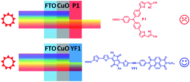
In p-type dye-sensitized solar cells (p-DSSCs), NiO is the most commonly used p-type semiconductor. Nevertheless, because of the drawbacks of NiO, much effort has been made to search for suitable substitutes. Herein, three different morphologies of CuO nanomaterials were used to prepare photocathodes for p-DSSCs, which have a deeper valence band and a higher dielectric constant compared to that of NiO. We observe that CuO is unstable in the presence of iodide/triiodide electrolyte, while cobalt complexes with bipyridine ligands are more suitable redox shuttles. We also note that the average transport time in CuO is shorter than that in NiO. Finally, the deep absorbance of CuO in the visible range indicates that suitable sensitizers for the CuO p-DSSC must exhibit high extinction coefficient and absorption bands located in the lower energy part of the solar spectrum (>600 nm) to be exploitable. In this case such CuO based photocathodes represent valuable systems to exploit the near-infrared (NIR) region.


Introduction

p-Type semiconductor (p-SC) based dye-sensitized solar cells (p-DSSCs) have become a research focus because they open the way for the construction of tandem DSSCs1 and dye sensitized photoelectrosynthetic devices.2,3 In p-DSSCs, the operation mechanism is reverse to that of conventional Grätzel cells (n-DSSCs), since the holes are photoinjected into the valence band of the p-SC from a photoexcited dye.4,5 NiO is the most commonly used p-type semiconductor to prepare cathode for p-DSSCs.6,7 Recently, NiO based p-DSSCs reach a power conversion efficiency (PCE) as high as 2.5%, which represents a real breakthrough in the field of p-DSSCs.8 However, most p-DSSCs exhibit PCEs of only few tenths of percent in spite of the fact that they might achieve similar PCEs as those reported with n-DSSCs.9 The low performances of the current p-DSSCs stem mainly from the intrinsic properties of NiO. First, its valence band position is as low as +0.3 V vs. SCE (at pH 7),10 which limits the maximum achievable open circuit voltage (Voc). Second, the photocurrent of NiO based p-DSSCs can be restricted by the low hole mobility in the p-SC.11 Finally, NiO displays a quite low dielectric constant of ε = 9.7 (ref. 4) compared to that of TiO2 (ε = 80). As a consequence, the injected hole might be strongly bound with the electron residing on the dye, limiting thus their dissociation and consequently the charge collection efficiency as it was proposed for ZnO based n-DSSC, which also shows a low dielectric constant (ε = 8).12 Accordingly, the development of new p-type semiconductors with better suited properties has become a priority if one wishes to advance with this new DSSC technology. Copper oxides, and particularly cuprite (CuO) or cuprous (Cu2O) oxides have been much investigated for photovoltaic and photoelectrochemical proton reduction applications, because copper has a high natural abundance on the earth crust and a low toxicity.13–15 On the other hand, cupric oxide (tenorite as mineral name) is another attractive p-SC, which can be valuable to fabricate p-DSSCs, because it has a higher dielectric constant (ε = 18.1)16 than NiO. As far as we are aware, there are only two reports dealing with the preparation of p-DSSCs with nanocrystalline films of CuO. First, Suzuki and co-workers investigated four different dyes with iodide/triiodide redox couple on CuO based p-DSSCs, which led to modest efficiencies (PCE around 0.01%).17 Very recently, Guldi and co-workers demonstrated interesting performances (around 0.2%) with zinc phthalocyanine dyes.18 These favourable features prompted us to investigate further the potential of CuO for developing p-DSSCs. Towards this goal, we have fabricated and analysed the performances of p-DSSCs prepared with CuO nanomaterials of different morphologies (nanowires and nanorods) sensitized with three different organic dyes (P1 and two diketopyrrolopyrrole derivatives) and using two different redox mediators (iodide/triiodide and cobalt bipyridine complexes).

Experimental section

Synthesis

The CuO nanowires were synthesized by a facile wet chemical method.19 Cu(CH3COO)2·H2O (1 g) and NaOH (2 g) were dissolved in 50 mL H2O, respectively. Then the two solutions were mixed and large amount of blue precipitate was formed immediately. After filtering and washing the precipitate, the Cu(OH)2 precursor was further sintered at 300 °C for 1 h under air and black CuO nanowires were obtained. The two other CuO nanorods (nanorods 1 and nanorods 2) were purchased from US Research Nanomaterials, Inc.

Characterization

The X-ray diffraction (XRD) patterns were collected via a Bruker, D8 X-ray diffractometer with Cu-Kα radiation (λ = 0.15418 nm). The UV-vis reflectance of CuO was recorded on a UV-vis spectrophotometer (Perkin Elmer, Lambda 1050). The morphology of CuO was observed with a scanning electron microscope (SEM, JEOL, 7600F) while transmission electron microscopy (TEM) was performed on a Hitachi H-9000 NAR at 300 kV (point to point resolution of 0.18 nm). The X-ray photoelectron spectra (XPS) were measured on a Kratos Nova X-ray photoelectron spectrometer. More details are given in the ESI.

Results and discussion

CuO nanomaterials were first characterized by X-ray diffraction (XRD). As displayed in Fig. 1, all the diffraction peaks can be indexed to monoclinic CuO with C2/c space group (JCPDS card, no. 45-0937).20 No additional diffraction peaks possibly assigned to impurities could be detected suggesting that the samples do not contain any by-product.
image file: c6ra17879k-f1.tif
Fig. 1 XRD patterns of CuO nanowires, nanorods 1, and nanorods 2.

Transformed Kubelka–Munk reflectance spectra of CuO are given in Fig. 2. The threshold of the reflectance is 880 nm corresponding to a band gap of 1.41 eV, which is consistent with the literature.21 Clearly, CuO has a strong absorbance in the visible region until 880 nm, which can prevent the dye from absorbing incident solar light. The near infrared absorption in the region from 1000 to 2000 nm would be related to interband transitions within the Cu-d block.22,23


image file: c6ra17879k-f2.tif
Fig. 2 Transformed Kubelka–Munk reflectance spectra of CuO samples. Inset: Zoom of figure in visible region.

The SEM and TEM images show that CuO nanowires exhibit a diameter of 20 nm and lengths that range from 1 to 10 μm (Fig. 3a and b, length/diameter > 10). Indeed, each nanowire has to be regarded as an assembly of well crystallized nanoparticles (Ø ∼ 20 nm). CuO nanorods 1 are also characterized by a diameter of about 20 nm but lengths range from 100 nm to 200 nm (Fig. 3c and d, length/diameter ≤ 10). At the end, CuO nanorods 2 consist of rod-like crystals with diameter of about 30 nm and length of 50 to 100 nm approximately (Fig. 3e and f, length/diameter < 5). High resolution transmission electron microscopy (HRTEM) images, as well as selected area electron diffraction (SAED) patterns, are displayed in Fig. S1 while extra low magnification SEM images of CuO samples are given in Fig. S2.


image file: c6ra17879k-f3.tif
Fig. 3 SEM and TEM images of CuO nanowires, (a) and (b), CuO nanorods 1, (c) and (d), and CuO nanorods 2, (e) and (f).

The specific surface area (SSA) of these CuO nanomaterials was determined by Brunauer–Emmett–Teller (BET) method (Table 1). CuO nanorod 1 exhibits a SSA (49 m2 g−1) which is twice as large as that of CuO nanorod 2 (25 m2 g−1), while that of CuO nanowire is intermediate (31 m2 g−1).

Table 1 SSA and dye loading amount of CuO of different dye
Sample SSA (m2 g−1) Dye loading (nmol cm−2)
P1 DPP-NDI
CuO nanowires 31 7.2 9.2
CuO nanorods 1 49 7.2 8.5
CuO nanorods 2 25 6.7 9.1


The top of the valence band position of CuO in these nanomaterials was determined by electrochemical impedance spectroscopy (EIS) measurements. The flat band potential (VFB) was determined from the intercept of the reciprocal square capacitance axis with the potential axis, based on the Mott–Schottky equation:24

1/C2 = (2/0εNa)[(VVFB) − kT/e]
where C represents the capacitance of the space charge region, ε0 is the vacuum permittivity, ε is the dielectric constant of CuO, e is the electron charge, V is the electrode applied potential, k is the Boltzmann constant, T is the absolute temperature, and Na is the acceptor concentration. The negative slops of Mott–Schottky plots (Fig. 4) confirm the p-type conductivity of all CuO samples. The measured VFB at pH 9.4 are 0.27 V, 0.41 V, and 0.31 V (vs. SCE) for CuO nanowires, nanorods 1, and nanorods 2, respectively (0.15 V vs. SCE at pH 9.2 from K. Nakaoka25). The difference in VFB along the series may be associated to slight changes in the stoichiometries and/or surface states. However, a little positive shift of VFB compared to that of NiO measured in the same conditions (VFB = 0.1 V vs. SCE), is clearly evidenced. This confirms that the valence band of CuO is deeper than that of NiO. With a ε value of 18.1,16 the acceptor concentration of CuO sample were determined from the slope of Mott–Schottky plots (electrode surface of 0.50 cm2). They turn to be 1.50 × 1016 cm−3, 2.28 × 1013 cm−3, and 1.23 × 1014 cm−3 for CuO nanowires, nanorods 1, and nanorods 2, respectively. For comparison, the usually reported charge carrier concentration for NiO nanoparticles is much higher since it is estimated around 1020 cm−3.7


image file: c6ra17879k-f4.tif
Fig. 4 Mott–Schottky plots of CuO nanowires (a), CuO nanorods 1 (b), and CuO nanorods 2 (c) in 1 M LiClO4 at pH 9.4.

Next, mesoporous CuO films were prepared by screen printing on FTO glass of an ink made of the CuO nanomaterials and additives of terpineol and ethyl cellulose. Finally, the films were sintered at 350 °C under the air and solar cells mounted with Pt as counter-electrode (see ESI for details). First, the classical benchmark and highly efficient push–pull dye P1 (Fig. 5),26,27 which has maximum absorption peaks at 468 nm in solution, was investigated with I3/I as redox mediator. The photovoltaic performances of the solar cells made with the three morphologies of CuO nanomaterials are gathered in Table 2. The Voc of CuO based p-DSSCs are similar with that of NiO. However, the short circuit photocurrents (Jsc) are much lower than in NiO p-DSSC, which results with a lower overall PCE. From the photoaction spectra shown in Fig. 6, the maximum value is located at 370 nm, which is most certainly mainly due to the contribution of triiodide photoredox activity as previously reported.28,29 However, the IPCE band around 540 nm can be attributed to the photoactivity of P1 dye as it matches very well that of this dye measured on NiO p-DSSCs.26,30


image file: c6ra17879k-f5.tif
Fig. 5 Structure of the dyes used in this study.
Table 2 Photovoltaic performances of p-DSSCs fabricated with CuO photocathodes under simulated AM1.5 (100 mW cm−2). The thickness of the CuO photocathodes were ∼1 μm. NW = nanowire; NR1 = nanorod 1 and NR2 = nanorod 2
CuO Dye Electrolyte Voc (mV) Jsc (mA cm−2) FF (%) η (%)
NW P1 I/I3 91 0.69 42 0.026
NR1 P1 I/I3 88 0.77 45 0.030
NR2 P1 I/I3 89 0.67 42 0.025
NR2 No dye I/I3 20 0.21 20 0.0008
NW DPP-NDI CoIII/II 298 0.16 38 0.019
NR1 DPP-NDI CoIII/II 332 0.20 43 0.029
NR2 DPP-NDI CoIII/II 271 0.23 42 0.026
NR2 No dye CoIII/II 112 0.053 26 0.0016
NW YF1 CoIII/II 302 0.55 35 0.059
NR1 YF1 CoIII/II 283 0.51 40 0.059
NR2 YF1 CoIII/II 316 0.72 43 0.097


image file: c6ra17879k-f6.tif
Fig. 6 IPCE spectra of p-DSSCs fabricated with CuO nanorod 2 photocathodes using P1 dye (red) with I3/I redox mediator and DPP-NDI (black) or YF1 (blue) with cobalt electrolyte.

Interestingly, the intrinsic photovoltaic activity of CuO is weak in this configuration as indicated by the cell without dye (Table 2). This may indicate that the electron/hole pair generated by direct absorption of CuO probably recombines before reaching the electrolyte/CuO interface and is therefore not efficiently collected. Secondly, although the performances of these cells are higher than those previously reported by Suzuki et al.17 with nanoparticles of CuO, the photocurrent density is much lower than that recorded with P1 in NiO based p-DSSCs.26,30 One main limitation of these devices comes from the high absorbance of CuO, which outcompetes with P1 for light collection; because the spectrum of P1 strongly overlaps with that of CuO. Thirdly, the morphology of CuO has low impact on the PCE, because it is not the critical factor governing the PCE. Finally, the stability of these DSSCs is very poor, since after few hours the photovoltaic performances completely collapsed owing to the corrosion of CuO by the iodide electrolyte, most probably induced by the transformation of CuO into CuI.31 This prompted us to investigate tris(4,4′-di-tert-butyl-2,2′-bipyridine)cobalt(III/II)32 as alternative redox mediator (Fig. S3), because it must be compatible with the stability of CuO and can also lead to higher Voc than iodide/triiodide.27 However, P1 performs very badly with the latter redox mediator owing to the very fast geminate charge recombination of this dye.32,33 Accordingly, we have selected the DPP-NDI dye (Fig. 5), which works well on NiO based DSSCs with this cobalt electrolyte (the NiO electrode is stable in both I3/I and cobalt electrolyte).34 DPP-NDI was selected because it allows the formation of a long lived charge separated states following hole injection into NiO, which requires a slower electron acceptor (e.g. CoIII/II). The characteristics of the DSSCs mounted with the CuO photocathodes sensitized with DPP-NDI are gathered in Table 2. As expected, the Voc is much enhanced relative to that measured with iodide electrolyte, but the Jsc values are still very low, most certainly because of the absorption of CuO nanomaterials that shades the DPP-NDI dye from the incoming sunlight. Dye loading measurements were undertaken with P1 and DPP-NDI dyes using desorption experiments since Jsc can be related to this parameter. However, there is only very few difference between the dye loading within the three CuO nanomaterials, which means that the dye loading amount is not closely related to the SSA of the materials. However, it is worthwhile noting that the dye loading on CuO is 10 times lower than that of NiO.7,30,35 Deeper understanding on the reasons of lower dye loading on CuO is in progress (binding affinity of COOH, surface accessibility, etc.).

Finally, YF1 (Fig. 5), a blue dye with higher absorption coefficient than DPP-NDI and having a maximum absorption band at 540 nm (Fig. S4), was chosen to increase the ratio of the absorption of the sensitizer relative to that of CuO. The photovoltaic performances of p-DSSCs sensitized with YF1 dye are gathered in Table 2 and the photoaction spectra are shown in Fig. 6. Satisfyingly, the photocurrent densities are higher than those recorded with DPP-NDI and higher than P1 with I/I3 especially if we subtract the photocurrent produced by triiodide. This indicates that sensitizers with intense absorption bands which span above the absorbance of CuO are better suited than red dyes, whose maximum absorption band occurs below 500 nm. The positioning of energy levels relative to CuO, the dye, and the redox mediator is sketched in Fig. S5.

On the other hand, the fill factor (FF) of CuO nanomaterials based p-DSSC is higher (above 40%) than that of NiO (around 30–35%) (Table 2). To further understand the mechanism, CuO nanowires with the largest length/diameter ratio (>10) were selected to be investigated by intensity modulated photocurrent spectroscopy (IMPS). Then the transport time of carriers in CuO nanowires based p-DSSC were calculated and compared with NiO as shown in Fig. 7. Clearly, CuO has a shorter transport time than NiO, which could explain the higher FF. Taking into account that Jsc is proportional to the injection rate and that NiO based devices always deliver higher photocurrent densities with the same dye and the same electrolyte,2,25 therefore the shorter charge transport time in CuO cannot be linked with higher charge carrier concentrations. Indeed, in NiO based DSSC the hole concentration is higher than that in CuO.


image file: c6ra17879k-f7.tif
Fig. 7 Transport time of p-DSSCs fabricated with CuO nanowires with DPP-NDI dye and cobalt redox mediator.

Conclusions

In summary, we have fabricated p-DSSCs with three morphologies of CuO nanomaterials, which were investigated with three different dyes and two different electrolytes. This study leads to the following three main conclusions. First, CuO is instable in presence of iodide/triiodide electrolyte, which precludes the use of this later as redox mediator. Cobalt complexes with bipyridine ligands are certainly suitable redox shuttles with CuO based p-DSSCs. Secondly, we note that the average transport time in CuO is shorter than in NiO, probably due to the higher conductivity of CuO coming from the wire like morphology. Thirdly, the deep absorbance of CuO itself cannot significantly contribute to the production of photocurrent, most certainly because of the short diffusion length of the electron/hole pair generated in CuO. Conversely, the intrinsic absorbance of CuO shades the absorbance of the sensitizer or can quench the dye excited state by energy transfer which limits the photocurrent density of the cell. This effect is all the more pronounced than the dye absorbs below 500 nm, such as P1 and DPP-NDI. However, if the sensitizer exhibits high extinction coefficient and absorption bands above 600 nm, the contribution of the dye can be significant. This is clearly demonstrated by the comparison of the IPCE of P1, DPP-NDI and YF1 (Fig. 6). It can be therefore concluded that CuO could represent a valuable cathode material for solar cells provided that it is sensitized with a dye harvesting low energy photons such as those above 700 nm. In that case, it can certainly be successfully used for the fabrication of tandem DSSCs with a photoanode exploiting most of the visible sunlight photons and leaving the lower energy part of the solar spectrum for the photocathode.

Acknowledgements

For financial support, we are grateful to the ANR program, POSITIF (No. ANR-12-PRGE-0016-01) and for Région Pays de la Loire for LUMOMAT.

Notes and references

  1. A. Nattestad, A. J. Mozer, M. K. R. Fischer, Y.-B. Cheng, A. Mishra, P. Bauerle and U. Bach, Nat. Mater., 2010, 9, 31–35 CrossRef CAS PubMed.
  2. F. Li, K. Fan, B. Xu, E. Gabrielsson, Q. Daniel, L. Li and L. Sun, J. Am. Chem. Soc., 2015, 137, 9153–9159 CrossRef CAS PubMed.
  3. L. Tong, A. Iwase, A. Nattestad, U. Bach, M. Weidelener, G. Gotz, A. Mishra, P. Bauerle, R. Amal, G. G. Wallace and A. J. Mozer, Energy Environ. Sci., 2012, 5, 9472–9475 CAS.
  4. F. Odobel and Y. Pellegrin, J. Phys. Chem. Lett., 2013, 4, 2551–2564 CrossRef CAS.
  5. F. Odobel, L. Le Pleux, Y. Pellegrin and E. Blart, Acc. Chem. Res., 2010, 43, 1063–1071 CrossRef CAS PubMed.
  6. F. Odobel, Y. Pellegrin, E. A. Gibson, A. Hagfeldt, A. L. Smeigh and L. Hammarström, Coord. Chem. Rev., 2012, 256, 2414–2423 CrossRef CAS.
  7. D. Dini, Y. Halpin, J. G. Vos and E. A. Gibson, Coord. Chem. Rev., 2015, 304–305, 179–201 CrossRef CAS.
  8. I. R. Perera, T. Daeneke, S. Makuta, Z. Yu, Y. Tachibana, A. Mishra, P. Bäuerle, C. A. Ohlin, U. Bach and L. Spiccia, Angew. Chem., Int. Ed., 2015, 54, 3758–3762 CrossRef CAS PubMed.
  9. M. Awais, D. D. Dowling, F. Decker and D. Dini, SpringerPlus, 2015, 4, 564 CrossRef PubMed.
  10. J. He, H. Lindström, A. Hagfeldt and S.-E. Lindquist, J. Phys. Chem. B, 1999, 103, 8940–8943 CrossRef CAS.
  11. L. D'Amario, L. J. Antila, B. Pettersson Rimgard, G. Boschloo and L. Hammarström, J. Phys. Chem. Lett., 2015, 6, 779–783 CrossRef PubMed.
  12. J. A. Anta, E. Guillén and R. Tena-Zaera, J. Phys. Chem. C, 2012, 116, 11413–11425 CAS.
  13. C. G. Morales-Guio, S. D. Tilley, H. Vrubel, M. Gratzel and X. Hu, Nat Commun, 2014, 5, 3059 Search PubMed.
  14. S. Sun, Nanoscale, 2015, 7, 10850–10882 RSC.
  15. K. M. Haynes, C. M. Perry, M. Rivas, T. D. Golden, A. Bazan, M. Quintana, V. N. Nesterov, S. A. Berhe, J. Rodríguez, W. Estrada and W. J. Youngblood, ACS Appl. Mater. Interfaces, 2015, 7, 830–837 CAS.
  16. S. Das, S. Saha, D. Sen, U. K. Ghorai, D. Banerjee and K. K. Chattopadhyay, J. Mater. Chem. C, 2014, 2, 1321–1330 RSC.
  17. S. Sumikura, S. Mori, S. Shimizu, H. Usami and E. Suzuki, J. Photochem. Photobiol., A, 2008, 194, 143–147 CrossRef CAS.
  18. O. Langmar, C. R. Ganivet, A. Lennert, R. D. Costa, G. de la Torre, T. Torres and D. M. Guldi, Angew. Chem., Int. Ed., 2015, 54, 7688–7692 CrossRef CAS PubMed.
  19. A. S. Ethiraj and D. J. Kang, Nanoscale Res. Lett., 2012, 7, 1–5 CrossRef PubMed.
  20. Y. Duan, X. Liu, L. Han, S. Asahina, D. Xu, Y. Cao, Y. Yao and S. Che, J. Am. Chem. Soc., 2014, 136, 7193–7196 CrossRef CAS PubMed.
  21. J. Yu, S. Zhuang, X. Xu, W. Zhu, B. Feng and J. Hu, J. Mater. Chem. A, 2015, 3, 1199–1207 CAS.
  22. H. Peelaers, E. Kioupakis and C. G. Van de Walle, Appl. Phys. Lett., 2012, 100, 11914 CrossRef.
  23. D. Dorfs, T. Härtling, K. Miszta, N. C. Bigall, M. R. Kim, A. Genovese, A. Falqui, M. Povia and L. Manna, J. Am. Chem. Soc., 2011, 133, 11175–11180 CrossRef CAS PubMed.
  24. L. Wang and M. Tao, Electrochem. Solid-State Lett., 2007, 10, H248–H250 CrossRef CAS.
  25. K. Nakaoka, J. Ueyama and K. Ogura, J. Electrochem. Soc., 2004, 151, C661–C665 CrossRef CAS.
  26. L. Li, E. A. Gibson, P. Qin, G. Boschloo, M. Gorlov, A. Hagfeldt and L. Sun, Adv. Mater., 2010, 22, 1759–1762 CrossRef CAS PubMed.
  27. E. A. Gibson, A. L. Smeigh, L. Le Pleux, J. Fortage, G. Boschloo, E. Blart, Y. Pellegrin, F. Odobel, A. Hagfeldt and L. Hammarström, Angew. Chem., Int. Ed., 2009, 48, 4402–4405 CrossRef CAS PubMed.
  28. H. Zhu, A. Hagfeldt and G. Boschloo, J. Phys. Chem. C, 2007, 111, 17455–17458 CAS.
  29. E. A. Gibson, L. Le Pleux, J. Fortage, Y. Pellegrin, E. Blart, F. Odobel, A. Hagfeldt and G. Boschloo, Langmuir, 2012, 28, 6485–6493 CrossRef CAS PubMed.
  30. G. H. Summers, J.-F. Lefebvre, F. A. Black, E. Stephen Davies, E. A. Gibson, T. Pullerits, C. J. Wood and K. Zidek, Phys. Chem. Chem. Phys., 2016, 18, 1059–1070 RSC.
  31. M. Yang, J.-Z. Xu, S. Xu, J.-J. Zhu and H.-Y. Chen, Inorg. Chem. Commun., 2004, 7, 628–630 CrossRef CAS.
  32. M. Raissi, Y. Pellegrin, S. Jobic, M. Boujtita and F. Odobel, Sci. Rep., 2016, 6, 24908 CrossRef PubMed.
  33. P. Qin, J. Wiberg, E. A. Gibson, M. Linder, L. Li, T. Brinck, A. Hagfeldt, B. Albinsson and L. Sun, J. Phys. Chem. C, 2010, 114, 4738–4748 CAS.
  34. L. Favereau, J. Warnan, Y. Pellegrin, E. Blart, M. Boujtita, D. Jacquemin and F. Odobel, Chem. Commun., 2013, 49, 8018–8020 RSC.
  35. D. Ameline, S. Diring, Y. Farre, Y. Pellegrin, G. Naponiello, E. Blart, B. Charrier, D. Dini, D. Jacquemin and F. Odobel, RSC Adv., 2015, 5, 85530–85539 RSC.

Footnote

Electronic supplementary information (ESI) available. See DOI: 10.1039/c6ra17879k

This journal is © The Royal Society of Chemistry 2016
Click here to see how this site uses Cookies. View our privacy policy here.