DOI:
10.1039/C6RA17525B
(Paper)
RSC Adv., 2016,
6, 96223-96228
Facile one-pot synthesis of magnetic Prussian blue core/shell nanoparticles for radioactive cesium removal
Received
8th July 2016
, Accepted 30th September 2016
First published on 30th September 2016
Abstract
Magnetic Prussian blue/Fe3O4 core/shell nanoparticles were successfully fabricated via a facile one-pot method. The results of transmission electron microscope (TEM), X-ray diffraction patterns (XRD) and Fourier transform infrared spectra (FT-IR) reveal that the as-prepared products are 20–40 nm in diameter and have obvious core/shell structures, which consist of face-centered-cubic lattice Prussian blue and Fe3O4. They can be well dispersed in water and show obvious magnetism, which means they can be rapidly separated by a magnet. The results of adsorption experiments indicate that their maximal adsorption capacities for Cs+ are 145.8 and 132.6 mg g−1, respectively. After magnetic separation, the Cs+ removal efficiency remains at more than 90% when the Cs+ concentration in solution is 50 μg mL−1. The magnetization and Cs+ adsorption properties can be easily adjusted by changing the content of Fe3O4 in nanocomposites. The synthesis strategy and mechanism are also generally discussed. The resulting magnetic Prussian blue nanoparticles can be widely applied to treat radioactive waste water containing cesium ions.
1. Introduction
Radioactive cesium ions (Cs+) are one of the main fission products from nuclear power stations, and are very harmful to the environment and humans due to their long half-life.1,2 Many kinds of methods and materials have been developed to separate Cs+ from radioactive waste solutions.3–6 Inorganic absorbents, such as zeolite, activated carbon, and clay, have been used to remove metal ions due to their stable skeletal structures and high surface areas.7–9 Polymer absorbents, such as chitosan, alginate, and cellulose, have many functional groups which are beneficial to the adsorption of metal ions.10–12 Recently, more and more attention has been paid to the selective removal of radioactive ions.
Prussian blue, a kind of zeolite-like inorganic material with a face-centered-cubic lattice, can exchange its potassium ions with cesium ions.13 Recently, Ishizaki et al. also found that as-prepared Prussian blue nanoparticles (Fe4[Fe(CN)6]3·xH2O) with many hydrophilic defect sites had a supreme Cs+ adsorption capability and proposed that the hydrated Cs+ was preferably adsorbed via hydrophilic defect sites accompanied by proton-elimination from the coordinated water.14 Prussian blue and its analogues have been widely applied in the selective removal of cesium ions from radioactive waste water.15–17 Prussian blue functionalized magnetic nanoparticles have been widely studied because they can realize highly efficient removal of radioactive cesium and rapid magnetic separation of adsorbents from the waste water. Prussian blue-coated Fe3O4 core/shell magnetic nanocomposites have been fabricated via many kinds of methods for effective cesium removal from solution.18–22 Fe3O4@SiO2@KTiFC core–shell-structured magnetic microspheres were developed for radioactive cesium removal from seawater and their maximum adsorption capacity was up to 43.09 mg g−1.23 Prussian blue-coated PDDA@iron oxide magnetic nanoparticles were fabricated for the removal of radioactive cesium.24 Magnetic Prussian blue/graphene oxide (PB/Fe3O4/GO) nanocomposites were fabricated and their maximum adsorption capacity for Cs+ was 55.56 mg g−1.25,26
In this paper, magnetic Prussian blue/Fe3O4 core/shell nanoparticles were successfully fabricated via a facile one-pot method and studied in detail. A transmission electron microscope (TEM), X-ray diffraction patterns (XRD) and Fourier transform infrared spectra (FT-IR) were employed to characterize their morphology and structures. Their magnetization and Cs+ adsorption properties were investigated. Their synthesis strategy and mechanism are also generally discussed. Compared with Prussian blue-coated Fe3O4 magnetic nanoparticles, Prussian blue is difficult to release from the as-prepared magnetic Prussian blue/Fe3O4 core/shell after adsorbing Cs+, which can elevate the Cs+ removal efficiency.
2. Experimental
2.1. Materials
Potassium hexacyanoferrate(III) (K3[Fe(CN)6]·3H2O, AR, Sinopharm Chemical Regent Company, China), iron(III) chloride (FeCl3·6H2O, AR, Nanjing Chemical Reagent Company, China), sodium borohydride (NaBH4, AR, Aladdin Company, China) and deionized water.
2.2. Synthesis of magnetic Prussian blue core/shell nanoparticles
Pure Prussian blue nanoparticles (PB) and two kinds of magnetic Prussian blue core/shell nanoparticles (MPB-1 and MPB-2) were separately fabricated using the same facile method by changing the ratio of reactants (Table 1). A typical route to synthesize magnetic Prussian blue core/shell nanoparticles is as follows: certain amounts of K3[Fe(CN)6] and FeCl3·6H2O were dissolved in 100 mL of deionized water to form solution A. A certain amount of NaBH4 was dissolved in 50 mL of deionized water to form solution B. The NaBH4 solution was gradually added into the K3[Fe(CN)6]/FeCl3 solution under stirring (VA![[thin space (1/6-em)]](https://www.rsc.org/images/entities/char_2009.gif) :VB = 2:1). Then, the mixed solution stood for 20 min until all depositions were completed. After that, the depositions were separated from the solution via centrifugation (3000 rpm, 5 min) and freeze-dried in vacuum freeze-drying equipment (−55 °C, 48 h). The freeze-dried depositions were placed in air for 30 min and directly oxidized to form magnetic Prussian blue core/shell nanoparticles. The obtained MPB samples were dispersed in the deionized water again and purified via magnetic separation using two pieces of neodymium magnet (N45, 50 mm × 10 mm × 5 mm). The PB samples were synthesized using similar processes except for the magnetic purification. The ratio of reactants, addition method of chemicals and vacuum freeze-drying conditions were crucial to the formation of magnetic Prussian blue core/shell nanoparticles.
:VB = 2:1). Then, the mixed solution stood for 20 min until all depositions were completed. After that, the depositions were separated from the solution via centrifugation (3000 rpm, 5 min) and freeze-dried in vacuum freeze-drying equipment (−55 °C, 48 h). The freeze-dried depositions were placed in air for 30 min and directly oxidized to form magnetic Prussian blue core/shell nanoparticles. The obtained MPB samples were dispersed in the deionized water again and purified via magnetic separation using two pieces of neodymium magnet (N45, 50 mm × 10 mm × 5 mm). The PB samples were synthesized using similar processes except for the magnetic purification. The ratio of reactants, addition method of chemicals and vacuum freeze-drying conditions were crucial to the formation of magnetic Prussian blue core/shell nanoparticles.
Table 1 The ratio of reactants in the experiments
| Samples |
K3[Fe(CN)6] (g) |
FeCl3 (g) |
NaBH4 (g) |
| PB |
0.33 |
0.17 |
0.19 |
| MPB-1 |
0.33 |
0.40 |
0.19 |
| MPB-2 |
0.33 |
0.45 |
0.19 |
2.3. Characterizations
The morphology and structures of the samples were characterized in detail. Macroscopic pictures of PB and MPB solutions were taken at 0, 5, 10, 20 min after they were put in the magnetic field. The above-mentioned solutions were prepared by adding 0.1 g samples into 18 mL of deionized water and putting them in transparent glass bottles. The magnetic field was produced by two pieces of neodymium magnet (N45, 50 mm × 10 mm × 5 mm). Transmission electron microscope (TEM) images were taken on a Philips CM100 transmission electron microscope. X-ray diffraction (XRD) patterns were recorded on a BRUKER D8-ADVANCE X-ray diffractometer with Cu-Kα radiation (λ = 0.154178 nm). Fourier transform infrared (FT-IR) spectra were obtained on a Bruker OPUS 80V FT-IR spectrometer. The magnetic properties were tested on a MPMS XL-7 superconducting quantum interference device (SQUID) at 300 K.
2.4. Cesium adsorption experiments
Cesium chloride was dissolved in deionized water to prepare solutions containing Cs+ to simulate the radioactive waste water. The initial Cs+ concentrations ([Cs+]initial, Ci) were 50, 200, 500, 1000, 1500, 2000 and 2500 μg mL−1. Certain amounts of PB or MPB adsorbents were put into Cs+ solutions. The ratios between adsorbents and Cs+ solutions (madsorbent/Vsolution) were 1, 2, 4, 6, 8 and 10 mg mL−1. The Cs+ solutions with adsorbents were mixed well and kept for a period of time (contact time 6 h) at 25 °C. After adsorption, the adsorbents were separated from the solution via different methods. In order to examine the adsorption capacity under different initial Cs+ concentrations, the adsorbents were separated from the solution via centrifugation (4000 rpm, 5 min). In order to examine the Cs+ removal efficiency under different concentrations of adsorbents in Cs+ solution, the adsorbents were separated from the solution via magnetic separation using two pieces of N45 neodymium magnet. After the separation, the equilibrium Cs+ concentration (Ce) was tested on VARIO AAS-990 atom adsorption spectrum equipment.
The Cs+ removal efficiency (%) and adsorption capacity (Qe, mg g−1) were calculated from the following equations:
| | |
Removal efficiency = (Ci − Ce)/Ci × 100%
| (1) |
where
Ci (μg mL
−1) and
Ce (μg mL
−1) were the initial and equilibrium Cs
+ concentration, respectively,
V (mL) was the volume of solution, and
W (mg) was the weight of dried sorbent.
3. Results and discussion
3.1. Synthesis strategy and mechanism of magnetic Prussian blue core/shell nanoparticles
Pure Prussian blue nanoparticles and magnetic Prussian blue core/shell nanoparticles can be fabricated using the same facile one-pot method by changing the ratio of reactants. Their synthesis routes are shown in Fig. 1. FeCl3 and K3[Fe(CN)6] are mixed together in the initial solution. NaBH4 is a kind of strong reductive agent, which has been widely used. When NaBH4 is gradually added into above solution, oxidation–reduction reaction will be induced. The standard reduction potentials of Fe3+/Fe2+ and [Fe(CN)6]3+/[Fe(CN)6]4+ in aqueous solution are 0.771 V and 0.355 V.27 The standard reduction potential of Fe3+/Fe2+ is higher than that of [Fe(CN)6]3+/[Fe(CN)6]4+. So, Fe3+ is firstly reduced by the NaBH4 to form Fe2+. Then, the Fe2+ can rapidly react with K3[Fe(CN)6] to form Prussian blue. If the initial K3[Fe(CN)6] is excessive, all the Fe2+ can react with K3[Fe(CN)6] to obtain pure Prussian blue. If the initial Fe3+ is excessive, only part of the Fe2+ can react with K3[Fe(CN)6] to obtain a Prussian blue core. The excessive Fe2+ can be sequentially reduced to Fe0 by the NaBH4 and forms a shell on the surface of the Prussian blue core. H2, which is produced in the above-mentioned oxidation–reduction reactions, also plays an important role in preventing the aggregation of nanoparticles. After vacuum freeze-drying, the very thin Fe0 layer on the surface of the Prussian blue core has very high activity and can be directly oxidized to Fe3O4 when it comes into contact with oxygen in the air, which will lead to the formation of magnetic Prussian blue core/shell nanoparticles.28 The excessive Fe3+, sufficient NaBH4, order of addition of reactants and vacuum freeze-drying are crucial in the above fabrication processes. The particle size and shell thickness of the MPB core/shell nanoparticles can be adjusted by changing the concentration and ratio of reactants.
 |
| | Fig. 1 Synthesis schematic of magnetic Prussian blue core/shell nanoparticles. | |
3.2. Morphology of magnetic Prussian blue nanoparticles
Fig. 2 shows the photos of PB and magnetic PB solutions under a magnetic field. PB and magnetic PB nanoparticles can all be well dispersed in water and exist stably for a long period of time. The solution containing pure PB shows no obvious changes under a magnetic field. The color of the magnetic PB solutions gradually becomes lighter under a magnetic field as the processing time goes on, which indicates that the magnetic PB can be successfully magnetically separated. MPB-2 is more rapidly separated than MPB-1. Fig. 3 shows the TEM images of as-prepared samples. The PB nanoparticles are 15–30 nm in diameter. The magnetic PB nanoparticles are 20–40 nm in diameter and exhibit obvious core/shell structures. The shells of the MPB-2 samples are thicker than those of MPB-1, which indicates that more Fe3O4 was formed in MPB-2. The core/shell structures of magnetic PB nanoparticles can be easily adjusted by changing the concentration and ratio of reactants.
 |
| | Fig. 2 Macroscopic pictures of PB and magnetic PB nanoparticles under magnetic field. 0.1 g samples in 18 mL water; N45 neodymium magnets; time: 0, 5, 10, 20 min. | |
 |
| | Fig. 3 TEM images of PB and magnetic PB nanoparticles. | |
3.3. Structure analysis of magnetic Prussian blue nanoparticles
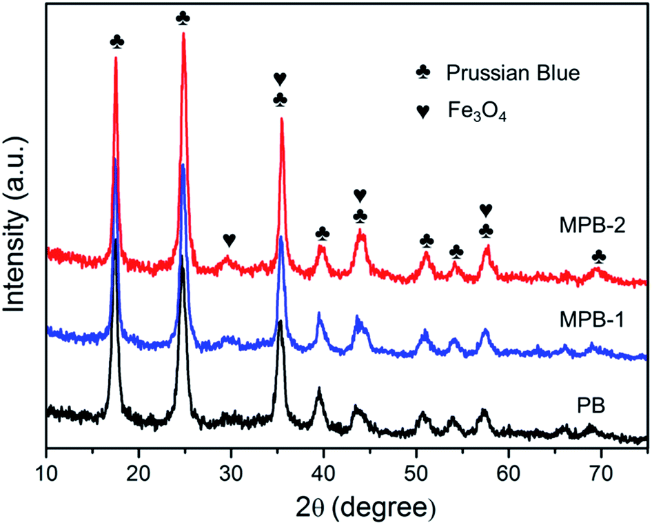
The XRD patterns of the samples are shown in Fig. 4. The obtained PB have obvious peaks at around 17.5°, 24.8°, 35.4°, 39.7°, 43.8°, 51.0°, 54.2°, 57.4° and 69.4°, which can be assigned to the (200), (220), (400), (420), (422), (440), (600), (620) and (642) planes of face-centered-cubic lattice Prussian blue (JCPDS 73-0687).29 Compared with the patterns of pure PB, a new diffraction peak at around 30.1° is observed in MPB-1 and MPB-2, which can be assigned to the (220) plane of face-centered-cubic Fe3O4 (JCPDS 19-0629).23 Diffraction peaks of (311), (400) and (511) planes in Fe3O4 are at around 35.5°, 43.1° and 57.1°, which are similar to the characteristic peaks of PB. The content of Fe3O4 in the magnetic PB nanocomposite is very low. So, there are no very obvious differences between PB and MPB in XRD patterns.
 |
| | Fig. 4 XRD patterns of PB and magnetic PB nanoparticles. | |
The FT-IR spectra of PB and magnetic PB nanoparticles ranging from 400 cm−1 and 4000 cm−1 are given in Fig. 5. The peaks at 2088 cm−1 and 499 cm−1 correspond to the stretching vibrations of C![[double bond, length as m-dash]](https://www.rsc.org/images/entities/char_e001.gif) N and Fe–CN–Fe, which are the typical signals of PB.19 The peak at 3446 cm−1 is ascribed to the O–H group, indicating the presence of interstitial water in the PB.25 The peak at 1610 cm−1 in PB shifts to 1621 cm−1 and 1630 cm−1 in MPB-1 and MPB-2, respectively. The peak at around 597 cm−1 corresponds to the Fe–O bond, which is the characteristic peak of Fe3O4. This peak becomes stronger in MPB and indicates the existence of Fe3O4 in MPB. A new peak at around 670 cm−1 is observed in MPB, which also indicates the formation of Fe3O4.
N and Fe–CN–Fe, which are the typical signals of PB.19 The peak at 3446 cm−1 is ascribed to the O–H group, indicating the presence of interstitial water in the PB.25 The peak at 1610 cm−1 in PB shifts to 1621 cm−1 and 1630 cm−1 in MPB-1 and MPB-2, respectively. The peak at around 597 cm−1 corresponds to the Fe–O bond, which is the characteristic peak of Fe3O4. This peak becomes stronger in MPB and indicates the existence of Fe3O4 in MPB. A new peak at around 670 cm−1 is observed in MPB, which also indicates the formation of Fe3O4.
 |
| | Fig. 5 Macroscopic pictures of PB and magnetic PB nanoparticles under a magnetic field. | |
3.4. Magnetic properties of as-prepared Prussian blue nanoparticles
Fig. 6 shows the field-dependent magnetic hysteresis curves of samples at 300 K. It reveals that the pure PB shows no obvious magnetization. MPB-1 and MPB-2 exhibit obvious magnetic behaviors. MPB-1 has a saturation magnetization of 0.53 emu g−1 and a coercive force of 258 Oe. MPB-2 has a saturation magnetization of 1.7 emu g−1 and a coercive force of 150 Oe. The saturation magnetization of MPB-2 is larger than that of MPB-1, which is consistent with their macroscopic performance (Fig. 2). The magnetizations of MPB-1 and MPB-2 are not very strong, because of the low content of Fe3O4 in the nanocomposites. In order to retain the good adsorption ability of MPB, the content of Fe3O4 is controlled with the promise that they can be successfully magnetically separated. Their magnetization can be elevated by adjusting the content of Fe3O4 according to practical demands.
 |
| | Fig. 6 Field-dependent magnetization of PB and magnetic PB nanoparticles. | |
3.5. Adsorption properties of PB and magnetic PB nanoparticles for radioactive cesium
The adsorption and separation properties of as-prepared samples for Cs+ in solution were studied in detail. The Cs+ adsorption capacities of samples under different Cs+ concentrations were tested and are shown in Fig. 7. The Langmuir adsorption equation is employed to fit the experimental data. The results reveal that the Langmuir model can fit the experimental data of the three samples very well, with R2 = 0.997, 0.989 and 0.994, respectively. The maximal adsorption capacities of PB, MPB-1 and MPB-2 for Cs+, calculated from the Langmuir model, are 201.9 mg g−1, 145.8 mg g−1 and 132.6 mg g−1, respectively. Prussian blue can efficiently adsorb the Cs+ from solution and plays a crucial role in the entire adsorption process. The adsorption ability of Fe3O4 for Cs+ is not obvious. So, the maximal adsorption capacities of magnetic PB nanoparticles are not as good as that of pure PB. The content of Fe3O4 in MPB-1 and MPB-2 is very low. So, they exhibit good adsorption properties for Cs+. In order to study their magnetic separation properties, the Cs+ removal efficiency of MPB samples under different adsorbent concentrations in Cs+ solution were examined after magnetic separation and are shown in Fig. 8. After adsorption, MPB adsorbents can be easily separated from the solution via a magnetic field. The Cs+ removal efficiency after magnetic separation can reach more than 90% when the concentration of MPB in Cs+ solution is higher than 8 mg mL−1. The results reveal that the as-prepared MPB adsorbents have good adsorption and magnetic separation properties.
 |
| | Fig. 7 Adsorption capacity of samples under different Cs+ concentration. T = 25 °C; contact time = 6 h; [Cs+]initial = 50–2500 μg mL−1; madsorbent/Vsolution = 4 mg mL−1. | |
 |
| | Fig. 8 Cs+ removal efficiency after magnetic separation under different concentrations of adsorbents. T = 25 °C; contact time = 6 h; [Cs+]initial = 50 μg mL−1; madsorbent/Vsolution = 1–10 mg mL−1. | |
4. Conclusions
Magnetic Prussian blue/Fe3O4 core/shell nanoparticles were successfully fabricated via a facile one-pot method and studied in detail. The as-prepared products are 20–40 nm in diameter and have obvious core/shell structures, which consisted of Prussian blue and Fe3O4. They can be well dispersed in water and show obvious magnetism. They can be rapidly separated by a magnet. Their maximal sorption capacities for Cs+ are 145.8 mg g−1 and 132.6 mg g−1 respectively. The Cs+ removal efficiency after magnetic separation can remain at more than 90% when the Cs+ concentration in solution is 50 μg mL−1. The particle size and shell thickness of magnetic PB core/shell nanoparticles can be adjusted by changing the concentration and ratio of reactants, which can directly affect their magnetism and Cs+ adsorption properties. The as-prepared magnetic nanoparticles can be applied to the removal of Cs+ from radioactive waste water.
Acknowledgements
This work was supported by the Fundamental Research Funds for the Central Universities (NJ20150022, NS2014061), the National Natural Science Foundation of China (11105073, 11575086), the Cooperative Innovation Fund of Jiangsu Province (BY2013003-09) and the project funded by the Priority Academic Program Development of Jiangsu Higher Education Institutions.
Notes and references
- T. J. Yasunari, A. Stohl, R. S. Hayano, J. F. Burkhart, S. Eckhardt and T. Yasunari, Proc. Natl. Acad. Sci. U. S. A., 2011, 108, 19530–19534 CrossRef CAS PubMed
 .
. - S. Kinase, T. Takahashi, S. Sato, R. Sakamoto and K. Saito, Radiat. Prot. Dosim., 2014, 160, 318–321 CrossRef CAS PubMed .
- B. Aguila, D. Banerjee, Z. M. Nie, Y. Shin, S. Q. Ma and P. K. Thallapally, Chem. Commun., 2016, 52, 5940–5942 RSC .
- J. L. Mertz, Z. H. Fard, C. D. Malliakas, M. J. Manos and M. G. Kanatzidis, Chem. Mater., 2013, 25, 2116–2127 CrossRef CAS .
- D. Sarma, C. D. Malliakas, K. S. Subrahmanyam, S. M. Islama and M. G. Kanatzidis, Chem. Sci., 2016, 7, 1121–1132 RSC .
- D. J. Yang, S. Sarina, H. Y. Zhu, H. W. Liu, Z. F. Zheng, M. X. Xie, S. V. Smith and S. Komarneni, Angew. Chem., Int. Ed., 2011, 50, 10594–10598 CrossRef CAS PubMed .
- C. Y. Sun, F. Zhang, S. F. Li and F. Q. Cheng, RSC Adv., 2015, 5, 35453–35460 RSC .
- L. Chang, S. Chang, W. Han, W. Chen, Z. Li, Z. Zhang, Y. Dai and D. Chen, RSC Adv., 2016, 6, 86829–86835 RSC .
- H. M. Liu, A. Yonezawa, K. Kumagai, M. Sano and T. Miyake, J. Mater. Chem. A, 2015, 3, 1562–1568 CAS .
- C. Dwivedi, A. Kumar, K. A. Juby, M. Kumar, P. K. Wattal and P. N. Bajaj, Chem. Eng. J., 2012, 200, 491–498 CrossRef .
- J. Mizera, G. Mizerova, V. Machovic and L. Borecka, Water Res., 2007, 41, 620–626 CrossRef CAS PubMed .
- T. Guo, Y. Q. Hu, X. L. Gao, X. S. Ye, H. N. Liu and Z. J. Wu, RSC Adv., 2014, 4, 24067–24072 RSC .
- S. C. Jang, Y. Haldorai, G. W. Lee, S. K. Hwang, Y. K. Han, C. Roh and Y. S. Huh, Sci. Rep., 2015, 5, 17510 CrossRef CAS PubMed .
- M. Ishizaki, S. Akiba, A. Ohtani, Y. Hoshi, K. Ono, M. Matsuba, T. Togashi, K. Kananizuka, M. Sakamoto, A. Takahashi, T. Kawamoto, H. Tanaka, M. Watanabe, M. Arisaka, T. Nankawa and M. Kurihara, Dalton Trans., 2013, 42, 16049–16055 RSC .
- M. Darder, Y. Gonzalez-Alfaro, P. Aranda and E. Ruiz-Hitzky, RSC Adv., 2014, 4, 35415–35421 RSC .
- J. Li, W. T. Shen, B. Kang, S. Q. Chang and Y. D. Dai, Micro Nano Lett., 2014, 9, 825–828 Search PubMed .
- Y. H. Qing, J. Li, B. Kang, S. Q. Chang, Y. D. Dai, Q. Long and C. Yuan, J. Radioanal. Nucl. Chem., 2015, 304, 527–533 CrossRef CAS .
- H. M. Yang, S. C. Jang, S. B. Hong, K. W. Lee, C. Roh, Y. S. Huh and B. K. Seo, J. Alloys Compd., 2016, 657, 387–393 CrossRef CAS .
- J. Jang and D. S. Lee, Ind. Eng. Chem. Res., 2016, 55, 3852–3860 CrossRef CAS .
- T. Arun and R. J. Joseyphus, J. Mater. Sci., 2014, 49, 7014–7022 CrossRef CAS .
- C. Thammawong, P. Opaprakasit, P. Tangboriboonrat and P. Sreearunothai, J. Nanopart. Res., 2013, 15, 1689 CrossRef .
- T. Sasaki and S. Tanaka, Chem. Lett., 2012, 41, 32–34 CrossRef CAS .
- R. Yi, G. Ye, F. C. Wu, M. F. Wen, X. G. Feng and J. Chen, RSC Adv., 2014, 4, 37600–37608 RSC .
- S. C. Jang, S. B. Hong, H. M. Yang, K. W. Lee, J. K. Moon, B. K. Seo, Y. S. Huh and C. Roh, Nanomaterials, 2014, 4, 894–901 CrossRef CAS .
- H. J. Yang, L. Sun, J. L. Zhai, H. Y. Li, Y. Zhao and H. W. Yu, J. Mater. Chem. A, 2014, 2, 326–332 CAS .
- H. J. Yang, H. Y. Li, J. L. Zhai, L. Sun, Y. Zhao and H. W. Yu, Chem. Eng. J., 2014, 246, 10–19 CrossRef CAS .
- W. A. Eaton, P. George and G. I. H. Hanania, J. Phys. Chem., 1967, 71, 2016–2021 CrossRef CAS PubMed .
- L. X. Wang, J. C. Li, Z. T. Wang, L. J. Zhao and Q. Jiang, Dalton Trans., 2013, 42, 2572–2579 RSC .
- F. X. Bu, M. Hu, W. Zhang, Q. Meng, L. Xu, D. M. Jiang and J. S. Jiang, Chem. Commun., 2015, 51, 17568–17571 RSC .
|
| This journal is © The Royal Society of Chemistry 2016 |
Click here to see how this site uses Cookies. View our privacy policy here. 
 *,
Wei Chen,
Wei Han,
Zheng Li,
Zheng Zhang,
Yaodong Dai and
Da Chen
*,
Wei Chen,
Wei Han,
Zheng Li,
Zheng Zhang,
Yaodong Dai and
Da Chen