Mengke Hanab,
Ming Chenc,
Heike Ebendorff-Heidepriemab,
Cheng Fangd,
Anjun Qin*e,
Hu Zhangf,
Ben Zhong Tangc,
Youhong Tang
g and
Yinlan Ruan*ab
aInstitute for Photonics and Advanced Sensing, School of Physical Sciences, University of Adelaide, Adelaide, South Australia 5005, Australia. E-mail: yinlan.ruan@adelaide.edu.au
bARC Centre of Excellence for Nanoscale BioPhotonics, Australia
cThe Hong Kong University of Science and Technology, Clear Water Bay, Kowloon, Hong Kong, China
dGlobal Centre for Environmental Remediation, University of Newcastle, University Drive, Callaghan, NSW 2308, Australia
eSouth China University of Technology, 381 Wushan Rd, Tianhe, Guangzhou, Guangdong, China. E-mail: msqinaj@scut.edu.cn
fSchool of Chemical Engineering, University of Adelaide, Adelaide, South Australia 5005, Australia
gSchool of Computer Sc, Engineering & Mathematics, Flinders University, 1284 South Road, Clovelly Park, SA 5042, Australia
First published on 26th August 2016
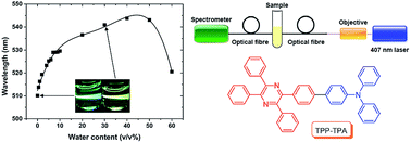
Tetraphenylpyrazine-triphenylamine (TPP-TPA) was used to detect water traces in organic solvents by monitoring the shift of the fluorescence peak wavelength. This wavelength based method avoids the intrinsic problems of fluorescence intensity change based methods. The use of optical fibres for the detection provides a remote and field-deployable sensing ability.
For instance, in the pharmaceutical and chemical industries, the presence of water inhibits not only the production of chemicals and drugs, but also the efficiency and usefulness of the products.1 Especially in organometallic chemistry, the presence of water leads to quenching of reactive organometallic compounds, such as organolithium and Grignard reagents, and inhibits the reaction or lowers the product yields. Moreover, due to the high reactivity of the organometallic reagents, under certain circumstances such as fire and explosions, the presence of water can lead to disastrous failures.2 Furthermore, water is a contaminant in refined gasoline. It affects the stability of fuel combustion, corrodes storage tanks and is strongly associated with unexpected microorganism proliferation.3 More importantly, when the temperature is low enough, emulsification and phase separation may occur, causing clogged fuel ducts and leading to engine damage and failure.2 Therefore, developing a simple, stable and accurate sensor to detect water in organic solvents is important and useful.
The Karl Fischer titration is the most widely used traditional method for the determination of water in liquid and solid samples over a large dynamic range.4,5 This method is based on the simultaneous reaction of iodine and water with a pre-made alkyl sulfite reagent.2 The consumption of a known amount of iodine, detected by either a volumetric (for relatively high contents of water, over 0.1 v/v%) or a coulometric approach (for relatively low contents of water, less than 0.1 v/v%), can thus be correlated with the content of water introduced into a system.2 However, such approaches have several unavoidable disadvantages, such as the need for specialized instruments and water-free titration cells, and the fact that they can only be performed by trained personnel and are easily disturbed by interfering compounds.5 Furthermore, such approaches require ex situ analysis, indicating their incapacity for real-time monitoring.2
Recent research has focussed on developing highly sensitive fluorescent probes that can be made using simple synthesis, and have potential applications in remote and in situ monitoring.2
Different fluorescence dyes can be categorised by the different sensing mechanisms underlying their mode of action.2 For chemosensors, the reversible association of water with a vital part of the fluorophores in their immediate environment can cause a change in either the intensity or the peak position (wavelength) of the fluorescence of the probes due to a variety of mechanisms, such as photo-induced electron transfer (PET),6 intramolecular charge transfer (ICT),7 excited state intramolecular proton transfer and water as a competitive ligand.8,9 We have summarized several florescence dyes in this field to provide a brief overview (Table 1).
# | Solvent | Sensing mechanism | Ref. |
---|---|---|---|
a Abbreviations: [aggregation-enhanced emission (AEE)]. | |||
OF-2 | 1,4-Dioxane | PET | 10 |
THF | |||
Acetonitrile | |||
Ethanol | |||
KD-F0011 (immobilized to PEGDMA) | Diethyl ether | ICT | 11 |
THF | |||
Ethyl acetate | |||
Acetonitrile | |||
TPEM | THF | ICT and AEE | 12 |
TPEBM | THF | ICT and AEE | |
ECPS | Acetonitrile | Aggregation based monomer–excimer switching | 13 |
Ethanol | |||
Methanol | |||
1,4-Dioxane |
Firstly, most of the reported fluorescent methods, shown in Table 1, are based on the change of fluorescence intensity as a function of water content.10–13 However, the change of fluorescence intensity can be caused by a range of factors in practical measurements, not only the change of water content. For the optical fibre-based detection, the power instability of the excitation source (i.e. laser) and the inconsistency of the optical alignment, i.e. the vibrations of optical components (mirrors, lenses and mounts) or drift in the laser beam or changes in environmental conditions such as temperature can lead to fluorescence intensity change.14 Furthermore, the fluorescence intensity is prone to be affected by the background noise from the scattering light of impurities, other ions and the luminescence of optical components.3 Thus, a fluorescence intensity measurement requires costly, stable laser source, consistency of the optical alignment and pure samples to diminish any possible background noise to gain high sensitivity and accuracy. These ideal conditions can hardly be achieved in practical measurements. In addition, for quantitative analysis, fluorescence intensity change-based methods require high photo-stability of the fluorescence molecules, i.e. consistency of fluorescence intensity of fluorophores over a long period, and the concentration of fluorophores must be precisely controlled in each sample. All these limitations can clearly affect the signal stability, sensitivity and reproducibility of fluorescence intensity change-based methods.
In contrast, the fluorescence peak wavelength is a unique characteristic of a particular molecule during the fluorescence process and it is not easily affected by other factors. Although previous fluorescence dyes with a twisted intramolecular charge transfer (TICT) effect, TPEM and TPEBM, developed by Yu et al., were found to shift the fluorescence peak wavelength with water content, there was no reported limit of detection (LOD) for any organic solvent.12
Secondly, laboratory-based fluorescence spectrophotometers have been used for most of the fluorescent methods, requiring transport of samples to a laboratory, making on-site and in situ measurement impossible. However, practical measurements in industry require fast detection and on-site analysis of water content in organic solvents for quality testing.
In this paper, we report a fluorescence dye, TPP-TPA (Fig. 1a), with aggregation-enhanced emission (AEE) and solvatochromic luminescence properties, for the detection of water traces by measuring the shift of the fluorescence peak wavelength to overcome the aforementioned limitations of the fluorescence intensity change-based methods.15 The detection range of our method is fairly large, from 0 to 60 v/v% in THF/water mixtures with a LOD of 0.04 v/v%. In addition, we used optical fibres and a portable spectrometer to achieve remote and in situ sensing ability.
![]() | ||
Fig. 1 (a) Molecular structure of TPP-TPA.15 (b) Setup of the optical fibre platform. |
Water detection measurements were conducted in 2 mL bulk solutions with constant TPP-TPA concentration of 10 μM. THF is completely miscible with water. The initial concentration of TPP-TPA in THF was 1 mM. A range of water concentrations was prepared by adding the initial TPP-TPA solution to water/THF mixture with a finial volume of 2 mL. Laser power of 86.4 μW and spectrometer integration time of 5.00 s were used. Each measurement comprised 3 discrete spectral scans that the device driver accumulated and averaged before the operating software (OceanView) received a spectrum. An optical shutter was synchronised with the data acquisition of the Ocean Optics spectrometer to minimise photo-bleaching (Fig. 1b). Considering that THF can dissolve PMMA, borosilicate glass tubes (Kimble) were used as containers rather than PMMA cuvettes during the experiments.
Fig. 2 shows that the fluorescence peak wavelength of TPP-TPA in THF with 0 v/v% water content increases linearly with time. Although during the experiments care was taken to prevent water absorption from the environment by sealing the glass tubes with a stopper, storing freshly dried THF in a flask with molecular sieves, using a nitrogen balloon and using the THF immediately after being decanted out, fluorescence red-shifting of TPP-TPA occurred, indicating absorption of water vapour by THF from the experiment atmosphere. The effect of THF water vapor absorption on the fluorescence peak wavelength was observed to begin after 30 seconds at the second measurement continuously. The standard deviation of all the slopes of the linear growth relationships between fluorescence peak wavelength and time for different water content samples from 0 to 40 v/v% was 0.00256 nm s−1, which indicated the ultra-sensitivity of TPP-TPA to the presence of water in the THF. Due to the unavoidable water absorption property of THF and the ultra-sensitivity of TPP-TPA to the presence of water, each data point is the result of the first measurement for quantitative analysis. Compared to a method using laboratory-based fluorescence spectrophotometers where samples are liable to changes in the water content during the transport to a laboratory, our method using optical fibres and portable spectrometers with quick response allows measuring the continuously changing water content in realtime.
Fig. 3 shows the fluorescence spectrum of TPP-TPA in THF with different water content, 0, 20, 40, 60 v/v% and using the portable Ocean Optics QE65000 spectrometer. In order to observe peak wavelength shifting with changed water content, the fluorescence intensity of each raw spectrum obtained from OceanView was normalized to the range 0 to 1 without using any smoothing algorithm. The fluorescence peak wavelength of TPP-TPA shows a continuous increase until the water content reaches 40 v/v%. When the water content exceeds 40 v/v%, it decreases dramatically. The red-shifting of the peak is due to the solvatochromic luminescence property of TPP-TPA in THF/water mixtures, which is induced by the gradually increased fraction of polar water.15,16 The blue-shifted peak that occurred after 40 v/v% is attributed to aggregate formation due to the decreasing solvating power of the THF/water mixture towards the fluorophore as a result of reduced association of water with TPP-TPA (Fig. 4).15 Concurrently, the rotation of the phenyl rings of TPP-TPA was greatly restricted in the aggregate state, and thus the unique AEE occurred.15 In addition, it is worth mentioning that the fluorescence of 0 v/v% and 30 v/v% water-in-THF solutions can be easily observed and distinguished by the naked eyes, as shown in Fig. 4.
The relationship between the fluorescence peak wavelength and water content up to 60 v/v% can be fitted with a quartic polynomial regression, which can be used to quantitatively determine the water content. The following equation was obtained by fitting the experimental data of the first measurement (y is the fluorescence peak wavelength; x is the water content):
y = 512.331 + 3.172x − 0.167x2 + 0.004x3 − 3.762 × 10−5x4 (R = 0.9863) |
The limit of detection (LOD) is defined as the lowest concentration at which an analyte can be sensed over the noise with a high degree of certainty of generally three times the standard deviation of the blank.2 The standard deviation for the first measurement data of the 10 blank samples is 0.087 nm, thus the LOD of TPP-TPA in THF is 0.08 v/v%.
An issue for quantitative analysis is that there is overlap between the fluorescence peak wavelength range of TPP-TPA before and after 40 v/v% water content. One approach to overcome this issue is to add extra water to the test samples and monitor the wavelength shifting. If red-shifting of the wavelength is observed, the authentic water content of the test sample is under 40 v/v% or vice versa. However, for most applications, the aim is to detect trace amounts of water. For such applications, the quartic polynomial regression between the fluorescence peak wavelength and water content over the range from 0 to 10 v/v% results in a lower LOD of 0.04 v/v%.
To determine the consistency of the fluorescence peak wavelength for different fluorophore concentrations, the fluorescence peak wavelength of TPP-TPA was measured in THF solutions with 1.00 v/v% water content (Fig. 5), using the same optical setup but a laboratory-based Horiba iHR320 spectrometer to gain higher sensitivity than portable Ocean Optics spectrometer for rigorous comparison. The results indicated the high signal stability of fluorescence peak wavelength for practical application regardless of fluorescence intensity change. The results also demonstrated the reduced requirements for sample preparation and measuring environment of TPP-TPA, e.g. the laser power, the position of the container and the fluorophore concentration, which do not need to be precisely controlled each time.
Furthermore, the photo-stability of TPP-TPA was investigated by a laboratory-based Horiba iHR320 spectrometer. The TPP-TPA sample was contained in a glass tube sealed with stopper and exposed to continuously high-power 405 nm laser over 5 min (Fig. 6). The results showed that the fluorescence intensity change ΔI/I was 1.34% and the peak wavelength change Δλ/λ was 0.06%, which indicated that the effect of photo-bleaching was negligible when it was used for measurements within 5 min.
In a commercial fluorescence spectrophotometer, the light for excitation and the fluorescence are propagating through free space in the large sample compartment before reaching the test sample and detector. However, in our optical fibre platform, a test sample is located closer to the tip of optical fibres for excitation along with the collection of fluorescence from fluorophores. Moreover, the laser source and fluorescence can be guided by optical fibres with high thermal, mechanical and corrosion stability, a feature which dramatically improves sensitivity and accuracy in harsh industrial environments and strong magnetic fields.17 More importantly, the setup used in our experiments can be easily assembled into a hand-held device if an optical circulator or fibre coupler is used instead of separate mirrors, lenses and filters to allow on-site and in situ measurements in the field.18 Furthermore, our optical fibre platform can be generally applied to other fluorophores to realize remote sensing ability in real time over long distances at inaccessible sites and thus opens up possibility for new applications.17
This journal is © The Royal Society of Chemistry 2016 |