Color measurement of the animal integument predicts the content of specific melanin forms

Ismael Galván*a and Kazumasa Wakamatsub
aDepartment of Evolutionary Ecology, Doñana Biological Station – CSIC, 41092 Sevilla, Spain. E-mail: galvan@ebd.csic.es
bDepartment of Chemistry, Fujita Health University School of Health Sciences, Toyoake, Aichi 470-1192, Japan

Received 7th July 2016 , Accepted 12th August 2016

First published on 15th August 2016


Abstract

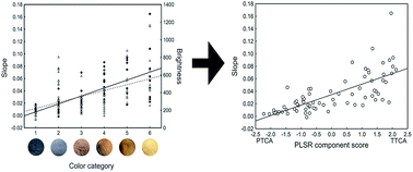
The appearance of animals largely depends on melanins present in their integument. However, it is unclear how different melanin forms create different animal color phenotypes. We used reflectance spectrophotometry to measure the color expression of feathers and hairs of 59 species of birds and 12 species of mammals, comprising a significant part of the palette of melanin-based colors, and analyzed for the first time the detailed chemical composition of melanins in the same samples by HPLC. We quantified color variation by means of the slope of percent reflectance regressed against wavelength, as this was the best predictor of a human categorization of color phenotype, increasing with the following scale: black, grey, dark brown, dark orange, light brown and light orange. Color slope variation was explained by levels of the 5,6-dihydroxyindole-2-carboxylic acid (DHICA) unit of eumelanin and the benzothiazole moiety of pheomelanin in feathers and hairs, but not by levels of the 5,6-dihydroxyindole (DHI) unit of eumelanin nor the benzothiazine moiety of pheomelanin. DHICA-eumelanin and benzothiazole-pheomelanin components explained color expression in opposite ways, decreasing and increasing, respectively, with color slope. Color slope, and also color categorization as perceived by humans, can therefore be used to infer the melanin chemical composition of feathers and hairs. Given that cytotoxic reactive oxygen species (ROS) are more abundantly formed during the synthesis of DHI than during the synthesis of DHICA in eumelanins, and in pheomelanins with higher benzothiazine/benzothiazole ratios, melanin-based colors interestingly reflect the content of the less pro-oxidant melanin forms.


Introduction

The visual appearance of most organisms depends to some extent on the presence of melanins in their integument. Melanins are thus the most extended biological pigments, and certainly the most abundant in higher vertebrates.1 Melanins are divided into eumelanins, polymers composed of indole units, and pheomelanins, composed of sulphur-containing heterocycles.2 The chemical heterogeneity of melanins gives them different optical properties in the visible spectral range, hence providing a diversity of colors to skin and its associated structures such as scales, feathers and hairs when melanosomes (i.e., specialized organelles of melanocytes where melanin synthesis takes place) are transferred to surrounding epidermal keratinocytes.3

It is known that eumelanins are darker than pheomelanins, the former conferring black, brown and grey colors and the latter conferring yellowish and reddish colors.4 However, the entire diversity of color phenotypes that can be generated by melanins is still unknown. Previous studies have investigated how different color parameters of the animal integument predict the total content of eumelanin and pheomelanin,5 but melanin diversity is greater than just eumelanin and pheomelanin.6 By using synchrotron-based photoionization mass spectrometry, Liu et al.7 associated different structural components of eumelanin and pheomelanin to different animal colors, but the lack of specific markers of those components (i.e., standards) and quantitative descriptions of colors make that additional analyses are needed to firmly infer an association between melanin chemistry and color phenotype. This will have broad implications, as a great interest exists in deciphering the color phenotype of extinct animals based on information on fossilized melanins8 and in finding potential trade-offs between physiological costs and benefits of producing different melanin structural units.6

Therefore, here we aimed at analyzing the expression of colors in natural melanins covering the entire palette of melanin-based traits in birds and mammals, and investigating how the different components of eumelanin and pheomelanin polymers explain that variability.

Melanocytes usually produce both eumelanins and pheomelanins from the common precursor dopaquinone that is formed by the oxidation of L-tyrosine. Eumelanin is formed when sulfhydryl compounds are absent or below certain levels in melanocytes, while pheomelanin is formed when sulfhydryls are above a threshold level and get incorporated to the process.2 The indole units of eumelanins, which are composed of 5,6-dihydroxyindole (DHI) and 5,6-dihydroxyindole-2-carboxylic acid (DHICA) moieties, result from the decarboxylative or nondecarboxylative rearrangement of dopachrome, a product derived from dopaquinone cyclization.9 Pheomelanin units, by contrast, are composed of benzothiazine and benzothiazole moieties.10 High-performance liquid chromatography (HPLC) allows the detection of degradation products of melanins that are specific to the different structural units of eumelanins and pheomelanins. In particular, pyrrole-2,3,5-tricarboxylic acid (PTCA) and pyrrole-2,3-dicarboxylic acid (PDCA), which are specific markers of DHICA and DHI eumelanin units, respectively, and 4-amino-3-hydroxyphenylalanine (4-AHP) and thiazole-2,4,5-tricarboxylic acid (TTCA), which are specific of benzothiazine and benzothiazole pheomelanin moieties, respectively.11,12

We used HPLC to measure levels of PTCA, PDCA, 4-AHP and TTCA in feathers of 59 species of birds and hairs of 12 species of mammals, comprising a comprehensive diversity of colors that natural melanins can generate (Fig. 1). For this, we obtained 1–2 feathers from 1–2 bird specimens deposited in museum collections for each species, complemented by samples obtained from wild populations (Table S1). Similarly, we obtained 10–15 hairs from 1–2 mammal specimens deposited in museum collections for each species (Table S1). The species were chosen on the basis of homogeneity in the color patches that were analyzed, i.e. avoiding complex plumage or pelage patterns consisting in differently perceived color hues. We avoided iridescent colorations, as these are generated by melanosome morphology and not by melanin chemistry.13


image file: c6ra17463a-f1.tif
Fig. 1 Images of species included in the study, showing the color patches that were analyzed. The names of the species are provided in Table S1. Note that some species were included in two color categories because two different color patches were analyzed in the same specimens (see Table S1). These images are only used to show the appearance of the species included the study, not to determine color categories. The photographs, with the exception of #53 which belongs to one of the authors of the study (I. G.), are covered by a CC BY license (https://creativecommons.org/licenses/by/2.0/; photographs #1–6, 10, 12, 14, 15, 17, 19, 20, 22–24, 30, 32–34, 42, 44, 46, 47, 54, 55, 57, 58, 60 and 64–70) or by a CC BY-SA license (https://creativecommons.org/licenses/by-sa/2.0/; photographs #7–9, 11, 13, 16, 18, 21, 25–29, 31, 35–41, 43, 45, 48–52, 56, 59 and 61–63). Photograph credits and links: #1 (Tony Smith; https://flic.kr/p/fq4NP7), #2 (procristination; https://flic.kr/p/5cEteB), #3 (James St. John; https://flic.kr/p/dCphEp), #4 (Peter von Bagh; https://flic.kr/p/C7A9gd), #5 (Stefan Berndtsson; https://flic.kr/p/egDf2V), #6 (Ekaterina Chernetsova (Papchinskaya); https://flic.kr/p/mVCZEz), #7 (Dornenwolf; https://flic.kr/p/kY9J7W), #8 (John Haslam; https://flic.kr/p/KnYBS), #9 (Bernard DUPONT; https://flic.kr/p/d47kkd), #10 (big-ashb; https://flic.kr/p/r2bEQV), #11 (Francesco Veronesi; https://flic.kr/p/vynehC), #12 (Hans G Bäckman; https://flic.kr/p/jqd9CJ), #13 (Larry Miller; https://flic.kr/p/eKGHsS), #14 (Joe McKenna; https://flic.kr/p/bWjLLw), #15 (Smabs Sputzer; https://flic.kr/p/fGaoLk), #16 (Harum koh; https://flic.kr/p/qAAgEe), #17 (Vibhu Prasad; https://flic.kr/p/9CeLfU), #18 (Bernard DUPONT; https://flic.kr/p/srHCWr), #19 (nociveglia; https://flic.kr/p/d9vtmu), #20 (Ingrid Taylar; https://flic.kr/p/eZhjvt), #21 (sébastien bertru; https://flic.kr/p/ePV5UK), #22 (Michele Lamberti; https://flic.kr/p/hg5mFP), #23 (Michele Lamberti; https://flic.kr/p/ea4doW), #24 (robin_24; https://flic.kr/p/93q8Nc), #25 (Sciadopitys; https://flic.kr/p/aYXDqz), #26 (Juan Emilio; https://flic.kr/p/bHtiui), #27 (Susanne Nilsson; https://flic.kr/p/DthK7w), #28 (Bernard Stam; https://flic.kr/p/dFn5Up), #29 (Ferran Pestaña; https://flic.kr/p/8RKUWq), #30 (Stefan Berndtsson; https://flic.kr/p/f3zxHq), #31 (rjime31; https://flic.kr/p/55skE2), #32 (Peter Trimming; https://flic.kr/p/a2jNtS), #33 (Peter Trimming; https://flic.kr/p/cbndLG), #34 (Noel Reynolds; https://flic.kr/p/bkGzmb), #35 (sébastien bertru; https://flic.kr/p/ePUWLe), #36 (Mark Hodgson; https://flic.kr/p/eNZ2Ta), #37 (Susanne Nilsson; https://flic.kr/p/KHd9kZ), #38 (sébastien bertru; https://flic.kr/p/84B5us), #39 (Hedera baltica; https://flic.kr/p/G9QTtZ), #40 (Bernard Stam; https://flic.kr/p/dRYGZo), #41 (Smudge 9000; https://flic.kr/p/zV4BHY), #42 (USFWS Endangered Species; https://flic.kr/p/8FgABc), #43 (Juan Emilio; https://flic.kr/p/nBU8Pn), #44 (Michele Lamberti; https://flic.kr/p/eWPGT9), #45 (Bernard DUPONT; https://flic.kr/p/kwPAX3), #46 (Jason Crotty; https://flic.kr/p/cfv8S5), #47 (Ron Kinight; https://flic.kr/p/nTru5f), #48 (Alastair Rae; https://flic.kr/p/a2mZnM), #49 (Lip Kee; https://flic.kr/p/tFUR4u), #50 (Andrej Chudý; https://flic.kr/p/4FxzSW), #51 (Francesco Veronesi; https://flic.kr/p/HmKL1U), #52 (Oona Räisänen; https://flic.kr/p/qMQXuG), #54 (Michele Lamberti; https://flic.kr/p/eBu3YZ), #55 (Peter Trimming; https://flic.kr/p/bKYqJk), #56 (sébastien bertru; https://flic.kr/p/eQ7D8j), #57 (Frank Vassen; https://flic.kr/p/9akQva), #58 (Ron Knight; https://flic.kr/p/diXcbm), #59 (Lip Kee; https://flic.kr/p/4mZKMQ), #60 (Mike Prince; https://flic.kr/p/8jvqN8), #61 (Jason and Alison; https://flic.kr/p/8YiNML), #62 (Ferran Pestaña; https://flic.kr/p/8QCbQk), #63 (Jörg Hempel; https://flic.kr/p/7spWru), #64 (Derek Keats; https://flic.kr/p/pQn429), #65 (Ron Knight; https://flic.kr/p/diXchB), #66 (Frank Vassen; https://flic.kr/p/bVSw7h), #67 (Frank Vassen; https://flic.kr/p/s9ECnq), #68 (Frank Vassen; https://flic.kr/p/aUyu1z), #69 (Francesco Veronesi; https://flic.kr/p/ob6q3w), #70 (Ron Knight; https://flic.kr/p/diXePW), #71 (Michele Lamberti; https://flic.kr/p/ecmCqj).

Methods

HPLC analyses

Feather and hair samples were first homogenized with Ten-Broeck glass homogenizer at a concentration of 10 mg ml−1 water (removing barbs and rachis parts not corresponding to the target color patch in the case of feathers), and then using alkaline H2O2 oxidation of eumelanin and pheomelanin to measure PTCA, PDCA and TTCA levels12 and reductive hydrolysis of pheomelanin with hydriodic acid (HI) to measure 4-AHP levels.11

For 4-AHP analyses, 100 μl of sample homogenate was taken in a 10 ml screw-capped conical test tube, to which 20 μl 50% H3PO2 and 500 μl 57% HI were added. The tube was heated at 130 °C for 20 h, after which the mixture was cooled. An aliquot (100 μl) of each hydrolysate was transferred to a test tube and evaporated to dryness using a vacuum pump connected to a dry ice-cooled vacuum trap and two filter flasks containing NaOH pellets. The residue was dissolved in 200 μl 0.1 M HCl. An aliquot (10–20 μl) of each solution was analysed on the HPLC system (JASCO 880-PU pump, JASCO Catecholpak C18 column and EICOM ECD-300 electrochemical detector; Eicom, Kyoto, Japan). A standard solution (10–20 μl) containing 500 ng each of 4-AHP (synthesized by K. W.) and 3-AHP (3-amino-4-hydroxyphenylalanine; 3-aminotyrosine from Sigma) in 1 ml 0.1 M HCl was injected every 10 samples (Fig. 2).


image file: c6ra17463a-f2.tif
Fig. 2 HPLC chromatograms for (A) melanin marker (PTCA, PDCA, TTCA and 4-AHP) standards and (B) feather samples from two species of birds included in the study as examples.

For PTCA, PDCA and TTCA analyses, 100 μl of sample homogenate was taken in a 10 ml screw-capped conical test tube, to which 375 μl 1 M K2CO3 and 25 μl 30% H2O2 (final concentration: 1.5%) were added. The mixture was mixed vigorously at 25 ± 1 °C for 20 h. The residual H2O2 was decomposed by adding 50 μl 10% Na2SO3 and the mixture was then acidified with 140 μl 6 M HCl. After vortex-mixing, the reaction mixture was centrifuged at 4000 g for 1 min, and an aliquot (80 μl) of the supernatant was directly injected into the HPLC system (JASCO 880-PU pump, Shiseido Capcell Pak MG C18 column and JASCO UV detector; Shiseido Co., Ltd., Tokyo, Japan). A standard solution (80 μl) containing 1 μg each of PTCA, PDCA, TTCA and TDCA (thiazole-2,3-dicarboxylic acid) in 1 ml water was injected every 10 samples. All these standards were synthesized by K. W. (Fig. 2).

Resulted values were multiplied by a conversion factor (PTCA: 25, PDCA: 50, 4-AHP: 9, TTCA: 34) to obtain absolute amounts of markers per mg of feather or hair. HPLC analyses were conducted blindly from results of spectrophotometric analyses (see below).

Spectrophotometric analyses

Before conducting HPLC analyses, we measured the color expression of feathers and hairs by reflectance spectrophotometry. Thus, these analyses were conducted without any information on the melanin contents of samples. We used an Ocean Optics Jaz spectrophotometer (range 220–1000 nm) with ultraviolet (deuterium) and visible (tungsten–halogen) lamps and a bifurcated 400 micrometer fiber optic probe. The fiber optic probe both provided illumination and obtained light reflected from the sample, with a reading area of ca. 1 mm2. Feathers were mounted on a light absorbing foil sheet (Metal Velvet coating, Edmund Optics, Barrington, NJ) to avoid any background reflectance. Measurements were taken at a 90° angle to the sample. All measurements were relative to a diffuse reflectance standard tablet (WS-1, Ocean Optics, Dunedin, FL), and reference measurements were frequently made. An average spectrum of five-six readings on different points of the target color patches in feathers or hairs was obtained for each bird, removing the probe after each measurement. The analyses were made on individual feathers separately, and mean spectra were then calculated. Given the small size of hairs, measurements were not taken on individual hairs but on the groups of 10–15 hairs from each specimen. Reflectance curves were determined by calculating the median of the percent reflectance in 10 nm intervals. As we were interested in investigating the diversity of melanin-based colors as perceived by humans, we only considered the visible spectral range (400–700 nm) in the analyses.

Spectral data were summarized as a measure of total brightness, as this is currently considered the best predictor of total levels of melanins in feathers, with lower values (i.e., darker colors) denoting higher melanin contents.5 Brightness was defined as the summed reflectance across the entire spectral range. Additionally, as the reflectance of melanins steadily increases from 300 to 700 nm and shows no spectral peaks,14 variation in the perceived color generated by melanins may be given to a large extent by variation in the slope of the reflectance curves (Fig. 3). We therefore calculated the slope of reflectance regressed against wavelength in the 400–700 nm range (Fig. 3) and used it as an additional descriptive measurement of melanin-based color expression.


image file: c6ra17463a-f3.tif
Fig. 3 Mean reflectance spectra (±s.e.) of the specimens used in the study. The colors of symbols represent the different color categories of the animals considered: black, grey, dark brown, dark orange, light brown and light orange.

Statistical analyses

The same feathers/hairs that were analyzed by reflectance spectrophotometry were then measured by HPLC as described above, with some exceptions for which we could only analyze samples from different specimens with each technique (Table S1). We thus investigated the differential contribution of PTCA, PDCA, 4-AHP and TTCA levels (predictor variables) to explain variability in color expression (brightness or slope; response variables). We used partial least squares regression (PLSR) analyses,15 as this is an appropriate statistical technique to analyzing the predictive capacity of melanin markers, which use to be highly intercorrelated.16 Indeed, in this case the degree of correlation between these variables was high (PTCA–PDCA: r = 0.44, P < 0.0001; PTCA-4-AHP: r = −0.31, P = 0.008; PTCA–TTCA: r = −0.19, P = 0.102; PDCA-4-AHP: r = 0.10, P = 0.414; PDCA–TTCA: r = 0.25, P = 0.030; 4-AHP-TTCA: r = 0.73, P < 0.0001; n = 74). Melanin markers were log10-transformed prior to analyses to achieve normality assumptions.

The significance of the extracted PLSR components was determined with two criteria. First, a cross-validation test of the parameter Q2 was carried out to determine if a component was significant. Then, we tested the significance of the correlation coefficient of the relationship between PLSR scores for the response variable and PLSR component scores, thus determining if the amount of variance explained in the response variable was significant. We also tested the statistical significance of the regression coefficients of the predictors in the PLSR analyses, to determine the degree of correlation between the response variable and these predictors. The latter test was made by bootstrapping using 100 replications. All PLSR analyses were made with the software TANAGRA 1.4.17

As our interest was to investigate the diversity of melanin-based colors as perceived by humans, we assigned the studied species to one of six color categories (on the basis of perception of the museum specimens used in the study) to determine which reflectance measurement (brightness or slope) best predicted the human perception of color. We assigned a value to these categories that increased with decreasing perceived darkness (i.e., increased from black to orange). Although this constitutes a subjective categorization of color, it was simply made as a convenient way to relate quantitative color measurements (brightness and slope) to the human perception of melanin-based color variation. Thus, color categories and their corresponding values were: black (1), grey (2), dark brown (3), dark orange (4), light brown (5) and light orange (6) (Fig. 1). We therefore regressed brightness and slope against this scale, and found that color category significantly predicted both brightness and slope, although the correlation coefficient was higher for slope (r = 0.69, n = 74, P < 0.0001; slope = −0.0071 + 0.0129x color category) than for brightness (r = 0.47, n = 74, P < 0.0001; Fig. 4). This indicates that our measurement of slope reliably explains the perceived variation in melanin-based color phenotypes, explaining a higher proportion of that variation than brightness. Thus, we used the slope as a response variable in the PLSR analyses to investigate the association between melanin chemistry and color expression. It must be noted that color categorization is a simple measurement just aiming at testing if melanin composition of feathers and hairs correlates with the general variation in color that is perceived by humans. Slope is still highly correlated with color category even if different orders of categories, for example assigning a value of 4 to grey colors as these may sometimes be perceived as darker than dark brown and dark orange colors, are considered (r = 0.50, P < 0.0001).


image file: c6ra17463a-f4.tif
Fig. 4 Relationship between melanin-based color category in 59 species of birds and 12 species of mammals and two color expression measures: color slope (left axis, solid symbols and continuous line) and brightness (right axis, open symbols and dashed line). Color category refers to a scale based on the human perception of melanin-based color variation, increasing with decreasing perceived darkness. Inserts are photographs showing examples of these categories with details of color patches for some species included in the study (from 1 to 6): Fulica atra (black), Larus argentatus (grey), Mustela erminea (dark brown), Coracias garrulus (dark orange), Gazella dorcas (light brown) and Saxicola rubetra (light orange). Complete photographs of these species are shown in Fig. 1. Slope refers to the slope of the regression between the amount of light reflectance and wavelength in the range 400–700 nm, and brightness refers to the summed reflectance in that range. The lines are the regression lines.

Results

The PLSR analysis generated one significant component that explained 44% of variance in color slope, which was significantly correlated with this component (r = 0.73, n = 74, P < 0.0001; Fig. 5). This component was negatively related to PTCA levels (predictor weight = −0.70) and to PDCA levels (predictor weight = −0.28), and positively related to 4-AHP levels (predictor weight = 0.49) and to TTCA levels (predictor weight = 0.44). As the square of predictor weights indicates the proportion of variance explained by the PLSR component (i.e., 44%) that is explained by each predictor variable,15 it follows that PTCA levels accounted for most variation in color slope, explaining 22% of variance in this variable. Bootstrapping analyses showed that the regression coefficients were significant in the case of PTCA (−0.030, P < 0.005) and TTCA (0.012, P < 0.05), but not in the case of PDCA (−0.006, 0.2 < P < 0.3) and 4-AHP (0.004, 0.4 < P < 0.5). Thus, variation in the perceived variation in melanin-based colors reflects variation in PTCA and TTCA levels, the contribution of PDCA and 4-AHP being non-significant in explaining this variation (Fig. 5).
image file: c6ra17463a-f5.tif
Fig. 5 Relationship between color phenotype (expressed as the slope of the amount of light reflectance regressed against wavelength) and the scores of a partial least-squares regression (PLSR) component related to the melanin composition of feathers and hairs. The names of significant predictors below the PLSR component indicate which side of the axis increased with increasing values. The line is the regression line. The point on the top right of the figure is not an outlier, as indicated by a Cook's distance (0.51) smaller than 2 and a leverage (0.04) smaller than 2p/n (0.05; p is the number of parameters in the model and n is the sample size).34

To corroborate the results of the PLSR model and to obtain a simple predictive equation, we conducted a general linear model (GLM) regressing color slope against the only two significant predictors that resulted from the PLSR model (i.e., PTCA and TTCA). PTCA and TTCA values (as ng mg−1) were added to the model without any transformations, neither logarithmic nor applying conversion factors. The GLM model explained a significant proportion of variance in color slope (31%, F2,73, P < 0.0001), and the resulting equation was: slope = 0.0409 + (−3.1347 × 10⁻⁵ × PTCA) + (1.4753 × 10⁻⁵ × TTCA). We used this equation to test the capacity of PTCA and TTCA to predict the color slope of animals in different datasets from other studies. We then used the equation relating color slope to color category (see Methods above) to predict the color phenotype. In particular, we used available data of melanin contents in different color forms of the hair of alpacas Vicugna pacos18 and house mice Mus musculus and humans12 (Table 1). Assuming that predicted color categories around zero correspond to the lowest color category considered here (i.e., 1 = black), and that rose grey forms correspond to brown colors and fawn forms correspond to orange colors in alpaca,18 our data predicted the color of 12 out of 20 cases (i.e., 60%; Table 1). This, however, must be taken with caution as color nomenclature in alpacas is not standardized and often confusing.18

Table 1 PTCA and TTCA levels in the hair of different color forms of alpacas, mice and humans from previously published sources,12,18 and color slope and category predicted from data in this study. Descriptions of hair color correspond to those provided by the authors of the published sources
Species Hair color PTCA (ng mg−1) TTCA (ng mg−1) Predicted slope Predicted color category Congruence
Alpaca Black 2145 200 −0.023 −1.26 Yes
Black brown 1526 329 −0.002 0.39 Yes
Grey 1041 108 0.010 1.31 No
Silver grey 988 194 0.013 1.54 Yes
Rose grey 419 490 0.035 3.26 Yes
Red/brown 179 1372 0.056 4.85 Yes
Dark brown 130 764 0.048 4.28 No
Chestnut 99.2 1084 0.054 4.72 Yes
Brown 43.2 378 0.045 4.05 No
Light brown 25.3 151 0.042 3.83 No
Fawn 14.6 70.4 0.041 3.77 Yes
Pink-skin fawn 13.1 143 0.043 3.85 Yes
Dark-skin fawn 7.6 98.3 0.042 3.81 Yes
White 4.1 7.8 0.041 3.72 No
Light fawn 2.4 21.3 0.041 3.74 Yes
Mouse Black (a/a) 2000 97 −0.020 −1.03 Yes
Yellow (e/e) 121 243 0.041 3.70 No
Human Black 340 98 0.032 3.01 No
Blonde 39 38 0.040 3.67 No
Red 62 89 0.040 3.67 Yes


Discussion

Our findings indicate that color slope, measured as percent reflectance regressed against wavelength, can be used to predict the melanin chemical composition of feathers and hairs. As slope was strongly correlated with a scale of color expression variation as perceived by humans, these color categories (black, grey, dark brown, dark orange, light brown and light orange) are equally useful to determine melanin composition. However, slope is a continuous variable, meaning that it can quantify color variation within a single color category in the scale of human perception. Therefore, slope is the most useful measurement to determine the melanin composition of feathers and hairs. However, our PLSR model explained 44% of variance in color slope, thus leaving ca. 60% of variance unexplained. This means that, although our study shows that the resulting color phenotype is clearly associated with the concentration of certain melanin forms, there may be other factors that are more relevant for explaining the expression of color than melanin concentration. Future studies should explore these factors.

Our results show that the color phenotype of birds and mammals reflects the content of the carboxylated (DHICA) unit of eumelanin and the content of the benzothiazole moiety of pheomelanin. No color phenotype is only generated by DHICA or benzothiazoles. Rather, color variation reflects different combinations of the two components: black colors contain the highest contents of DHICA and the lowest contents of benzothiazoles, while the opposite applies to light orange colors. These results differ from those previously found by Liu et al.7 They concluded that black color is generated by the two units of eumelanin (DHI and DHICA) with no contribution of pheomelanin, that brown color is mainly generated by pheomelanin with contribution of both benzothiazines and benzothiazoles and that grey color is mainly generated by pheomelanins, although standards of melanin units were not used in these analyses.

This study represents the first detailed chemical analysis of melanins in a wide range of melanin-based color phenotypes in animals. Our findings have implications to understand the evolution of animal coloration. During the final stages of eumelanogenesis, significant amounts of cytotoxic species, including reactive oxygen species (ROS) such as superoxide and hydrogen peroxide, are formed in melanocytes. The amount of ROS generated is much greater during the formation of DHI than during the formation of DHICA, as supported by a lower survival of melanocytes with no activity of the enzymes Tyrp1 and Tyrp2, which are involved in the DHICA route.19–21 This is because DHICA melanin exhibits potent hydroxyl radical-scavenging properties in the Fenton reaction while DHI melanin does not,21 and because the delocalized π-electron systems of the DHI polymer makes it generates a broader variety of free radical species than DHICA melanin.22 As selection is blind to genes and only acts on phenotypes, it is likely that individuals with color phenotypes denoting high carboxylated eumelanin contents are selected because of these protective benefits independently of other benefits that uncarboxylated eumelanin may confer (e.g., a higher protection against UV radiation).6 On the other hand, color phenotypes denoting pheomelanins with high relative benzothiazole contents would be selected for similar reasons to those suggested above for eumelanin, as, once formed, such pheomelanins produce less ROS under exposure to energetic radiation (such as UV or ionizing radiation) than pheomelanins with higher benzothiazine contents.10,23–26

Interestingly, then, melanin-based color phenotypes reflect the content of the less pro-oxidant melanin forms (i.e., DHICA-eumelanin and benzothiazole-pheomelanin), so selection may act on these phenotypes because of the same potential adaptive benefits related to the avoidance of cytotoxicity during or after melanogenesis. Whether selection pressure correlates positively or negatively with the color phenotype gradient (i.e., vertical axis in Fig. 5) will probably depend on the differential benefits that eumelanin27,28 and pheomelanin14,29 confer to individuals. These predictions for selective effects on melanin-based coloration should be valid for comparisons within color phenotypes (e.g., individuals with more intense black or grey coloration vs. others with less intense coloration) as well as comparisons between color phenotypes. They should also be useful to identify individuals or species particularly susceptible to the effects of environmental oxidative stress,24 a possibility that should be explored in humans regarding hair and skin coloration.

Lastly, given the current interest in determining the chemical composition of fossilized melanin granules to infer the color of extinct animals,8,30,31 our findings represent a key tool to elucidate the color corresponding to fossil specimens for which melanin composition can be established. The morphology of fossilized melanin granules has been used in some studies as a predictor of feather color in extinct birds, in all cases considering that granule morphology is related to melanin chemistry and then to the color being expressed.32,33 Our study provides a direct association between melanin chemistry and color with a proven predictive capacity of the melanin-based coloration of animals. It must be considered, however, that our data only predicted 60% of cases of alpaca, mouse and human hair color for which PTCA and TTCA values had been reported in other studies. Further work is necessary to get a proper understanding of all factors contributing to the expression of melanin-based coloration, including sources of variation not related to the concentration of different melanin forms. Only this comprehensive understanding will allow to make precise predictions of the color of extinct and extant animals.

Acknowledgements

We thank the staff of bird and mammal collections of Doñana Biological Station (EBD, Sevilla), the National Museum of Natural Sciences (MNCN, Madrid) and the Icelandic Institute of Natural History (IINH, Reykjavik), and particularly Josefina Barreiro and Ólafur K. Nielsen for their kind collaboration with sampling at MNCN and IINH, respectively. Domingo Rivera and Carolina Bravo provided feather samples from common cranes Grus grus and great bustards Otis tarda, respectively. Lucie Carrié helped with the spectrophotometric analyses of some samples. I. G. is supported by a Ramón y Cajal (RYC) Fellowship (RYC-2012-10237) from the Spanish Ministry of Economy and Competitiveness (MINECO) and by the project IN(15)CMA_CMA_1422 from Fundación BBVA (‘Convocatoria 2015 de ayudas Fundación BBVA a investigadores y creadores culturales’).

References

  1. F. Solano, New J. Sci., 2014, 2014, 498276 CrossRef.
  2. S. Ito and K. Wakamatsu, Photochem. Photobiol., 2008, 84, 582–592 CrossRef CAS PubMed.
  3. M. d'Ischia, K. Wakamatsu, F. Cicoira, E. Di Mauro, J. C. García-Borrón, S. Commo, I. Galván, G. Ghanem, K. Kenzo, P. Meredith, A. Pezzella, C. Santato, T. Sarna, J. D. Simon, L. Zecca, F. A. Zucca, A. Napolitano and S. Ito, Pigm. Cell Melanoma Res., 2015, 28, 520–544 CrossRef PubMed.
  4. I. Galván, J. Erritzøe, K. Wakamatsu and A. P. Møller, Comp. Biochem. Physiol., Part A: Mol. Integr. Physiol., 2012, 162, 259–264 CrossRef PubMed.
  5. K. J. McGraw, R. J. Safran and K. Wakamatsu, Funct. Ecol., 2005, 19, 816–821 CrossRef.
  6. I. Galván and F. Solano, Physiol. Biochem. Zool., 2015, 88, 352–355 CrossRef PubMed.
  7. S. Y. Liu, M. D. Shawkey, D. Parkinson, P. Tyler, T. P. Troy and M. Ahmed, RSC Adv., 2014, 4, 40396–40399 RSC.
  8. J. Vinther, BioEssays, 2015, 37, 643–656 CrossRef PubMed.
  9. M. d'Ischia, A. Napolitano, A. Pezzella, P. Meredith and T. Sarna, Angew. Chem., Int. Ed., 2009, 48, 3914–3921 CrossRef PubMed.
  10. K. Wakamatsu, K. Ohtara and S. Ito, Pigm. Cell Melanoma Res., 2009, 22, 474–486 CrossRef CAS PubMed.
  11. K. Wakamatsu, S. Ito and J. L. Rees, Pigm. Cell Res., 2002, 15, 225–232 CrossRef CAS.
  12. S. Ito, Y. Nakanishi, R. K. Valenzuela, M. H. Brilliant, L. Kolbe and K. Wakamatsu, Pigm. Cell Melanoma Res., 2011, 24, 605–613 CrossRef CAS PubMed.
  13. C. M. Eliason, P. P. Bitton and M. D. Shawkey, Proc. R. Soc. B, 2013, 280, 20131505 CrossRef PubMed.
  14. I. Galván and A. P. Møller, Physiol. Biochem. Zool., 2013, 86, 184–192 CrossRef PubMed.
  15. L. M. Carrascal, I. Galván and O. Gordo, Oikos, 2009, 118, 681–690 CrossRef.
  16. I. Galván, A. Jorge, K. Ito, K. Tabuchi, F. Solano and K. Wakamatsu, Pigm. Cell Melanoma Res., 2013, 26, 917–923 CrossRef PubMed.
  17. R. Rakotomalala, Proceedings of EGC'2005, RNTI-E-3, 2005, vol. 2, pp. 697–702 (in French) Search PubMed.
  18. R. Cransberg, K. Wakamatsu and K. Munyard, Small Ruminant Res., 2013, 114, 240–246 CrossRef.
  19. V. J. Hearing, Am. J. Hum. Genet., 1993, 52, 1–7 CAS.
  20. K. Urabe, P. Aroca, K. Tsukamoto, D. Mascagna, A. Palumbo, G. Prota and V. J. Hearing, Biochim. Biophys. Acta, 1994, 1221, 272–278 CrossRef CAS.
  21. S. Jiang, X. M. Liu, X. Dai, Q. Zhou, T. C. Lei, F. Beermann, K. Wakamatsu and S. Z. Xu, Free Radical Biol. Med., 2010, 48, 1144–1151 CrossRef CAS PubMed.
  22. R. Micillo, L. Panzella, K. Koike, G. Monfrecola, A. Napolitano and M. d'Ischia, Int. J. Mol. Sci., 2016, 17, 746 CrossRef PubMed.
  23. K. Wakamatsu, Y. Nakanishi, N. Miyazaki, L. Kolbe and S. Ito, Pigm. Cell Melanoma Res., 2012, 25, 434–445 CrossRef CAS PubMed.
  24. I. Galván, A. Bonisoli-Alquati, S. Jenkinson, G. Ghanem, K. Wakamatsu, T. A. Mousseau and A. P. Møller, Funct. Ecol., 2014, 28, 1387–1403 CrossRef.
  25. A. Napolitano, L. Panzella, G. Monfrecola and M. d'Ischia, Pigm. Cell Melanoma Res., 2014, 27, 721–733 CrossRef CAS PubMed.
  26. L. Panzella, L. Leone, G. Greco, G. Vitiello, G. D'Errico, A. Napolitano and M. d'Ischia, Pigm. Cell Melanoma Res., 2014, 27, 244–252 CrossRef CAS PubMed.
  27. G. R. Bortolotti, in Bird Coloration, Function and Evolution, ed G. E. Hill and K. J. McGraw, Harvard University Press, Cambridge, 2006, vol. II: pp. 3–35 Search PubMed.
  28. M. Brenner and V. J. Hearing, Photochem. Photobiol., 2008, 84, 539–549 CrossRef CAS PubMed.
  29. H. E. Hoekstra, R. J. Hirschmann, R. A. Bundey, P. A. Insel and J. P. Crossland, Science, 2006, 313, 101–104 CrossRef CAS PubMed.
  30. J. A. Clarke, D. T. Ksepka, R. Salas-Gismondi, A. J. Altamirano, M. D. Shawkey, L. D'Alba, J. Vinther, T. J. DeVries and P. Baby, Science, 2010, 330, 954–957 CrossRef CAS PubMed.
  31. F. Zhang, S. L. Kearns, P. J. Orr, M. J. Benton, Z. Zhou, D. Johnson, X. Xu and X. Wang, Nature, 2010, 463, 1075–1078 CrossRef CAS PubMed.
  32. R. M. Carney, J. Vinther, M. D. Shawkey, L. D'Alba and J. Ackermann, Nat. Commun., 2012, 3, 637 CrossRef PubMed.
  33. Q. Li, J. A. Clarke, K. Q. Gao, C. F. Zhou, Q. Meng, D. Li, L. D'Alba and M. D. Shawkey, Nature, 2014, 507, 350–353 CrossRef CAS PubMed.
  34. M. J. Crawley, GLIM for Ecologists, Blackwell Science, Oxford, 1993 Search PubMed.

Footnote

Electronic supplementary information (ESI) available. See DOI: 10.1039/c6ra17463a

This journal is © The Royal Society of Chemistry 2016
Click here to see how this site uses Cookies. View our privacy policy here.