Injectable and self-healing dynamic hydrogel containing bioactive glass nanoparticles as a potential biomaterial for bone regeneration

Ana Gantarab, Nataša Drnovšeka, Pablo Casusoc, Adrián Pérez-San Vicentec, Javier Rodriguezc, Damien Dupin*c, Saša Novakab and Iraida Loinazc
aJožef Stefan Institute, Department for Nanostructured Materials, Ljubljana, Slovenia
bJožef Stefan International Postgraduate School, Ljubljana, Slovenia
cIK4-CIDETEC Research Centre, Materials Division, Paseo Miramón 196, Donostia-San Sebastián 20009, Spain. E-mail: ddupin@cidetec.es

Received 6th July 2016 , Accepted 13th July 2016

First published on 13th July 2016


Abstract

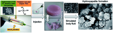
Combination of an Au-based 4-arms thiol terminated poly(ethylene glycol) [Au-(PEGSH)4] dynamic hydrogel exhibiting self-healing ability with 100 nm bioactive glass (BAG) nanoparticles agglomerated in 10 μm clusters, produced via a particulate sol–gel method, resulted in the formation of hydrogel nanocomposites [Au-(PEGSH)4–BAG] with enhanced properties. Au-(PEGSH)4–BAG hydrogel nanocomposites were prepared by injecting simultaneously an aqueous solution of a (PEGSH)4 homopolymer containing different amounts of BAG nanoparticles with an aqueous solution of HAuCl4 using a double barrel syringe. Electron microscopy studies suggested that the clusters of inorganic particles were homogeneously distributed into the polymeric matrix. Rheology studies demonstrated that stiffer hydrogels were obtained after the addition of BAG nanoparticles. The presence of the inorganic colloids appeared to affect slightly the dynamic character of the pristine hydrogel by slowing down the exchange reaction between gold–thiolate (Au–S) and disulfides (SS). Despite slower Au–S/SS exchange, the resulting hydrogel nanocomposites were still exhibiting pH-dependent properties and self-healing abilities, as judged by frequency sweep experiments. In addition, compression tests demonstrated the major drawbacks of each individual material, i.e. brittleness for BAG nanoparticles and weak consistency for Au-(PEGSH)4, were suppressed to result in a material composite with high resistance to stress and relatively large deformation ability. Moreover, the slow diffusion inside the 3D matrix allowed the degradation of the BAG nanoparticles to be delayed as well as the pH to be maintained around physiological values. For the same reason, the incorporation of those BAG nanoparticles into the dynamic hydrogel proved to reduce the cytotoxicity of the organic particles. Finally, in vitro degradation of BAG nanoparticles embedded in the dynamic hydrogel led to the formation of hydroxyapatite. As a result, osteoinductive properties could be anticipated for the Au-(PEGSH)4–BAG hydrogel nanocomposite which allows this new injectable and self-healing dynamic biomaterial to be considered as a scaffold to induce and promote bone self-repair.


Introduction

Tissue engineering (TE) is an advanced field of biomedicine for the regeneration of damaged tissues based on a triad consisting of a 3D scaffold, stimulating molecules/agents, and cells.1 Depending on the targeted application, different materials, stimuli and cells will be selected to achieve the desired properties. Various examples have been reported for regeneration of tissues such as cardiovascular-type,2 bone,3 cartilage,4,5 neuronal6,7 and skin.8 The most important features of these biomaterials for successful TE applications are biocompatibility, biodegradation, low immune response, 3D environment and mechanical support. The latter point is of crucial importance for bone regeneration where materials with appropriate mechanical properties and, preferably, with the ability to induce and promote new bone formation are required.

Injectable hydrogels, with the capacity to absorb large amount of water, are particularly attractive as vehicle and temporary scaffolds for cells in TE. Their role consists mainly in maintaining the cells in the area to treat, while offering minimally invasive procedure for the patient.9 The requirements for an ideal injectable hydrogel are (i) fast gel formation,10,11 (ii) spontaneous recovery of the mechanical and rheological properties if damaged, (iii) biocompatible and, preferably, (iv) bioactive to enhance tissue formation. Recently, dynamic hydrogels based on reversible covalent bonds, such as [phenylboronate-salicylhydroxamate] reaction,12 metal–ligand exchange,13,14 boronic ester bonds,15 dual acylhydrazone/disulfide bonds,16 radically reshuffling trithiocarbonate,17 radical-mediated disulfide fragmentation,18 and thiol–disulfide exchange19 have emerged as powerful materials with peculiar properties. More specifically, the reversible bond allows the material to rearrange permanently, resulting in self-healing properties and frequency-dependent stiffness. This latter property is also referred as shock absorbing behaviour for the material due to its increasing stiffness under high frequency stress. A 3D scaffold with such characteristics, i.e. resistance to undesired shock and even recovery of its mechanical integrity if damaged, might be of interest for TE. Unfortunately, few of these dynamic bonds occur at physiological conditions. Recently, Casuso et al. reported the preparation of dynamic hydrogels based on gold(I)–thiolate (Au–S) interactions with similar rheological properties as the synovial fluid.20 The same group prepared an injectable dynamic hydrogels by simply mixing simultaneously aqueous solutions of commercially available poly(ethylene glycol) tetrathiol [(PEGSH)4] and HAuCl4.21 The spontaneous redox reaction between Au(III) ions and thiols led to the formation of a 3D network based on (PEGSH)4 homopolymers linked via Au–S species and disulfides bonds (SS). The resulting biocompatible hydrogel exhibited self-healing property and frequency-dependent stiffness due to the permanent exchange between Au–S and SS, readily occurring at physiological conditions. In addition, nucleophilic thiolate concentration, which promotes Au–S/SS exchange, could be varied by adjusting the pH of the hydrogel. Hence, the dynamic behaviour could be tuned in function of pH. Moreover, the mechanical properties of Au-based dynamic hydrogel could be easily adjusted from a viscous liquid exhibiting a liquid-to-gel transition to a free-standing hydrogel at higher concentration.20,22 Unfortunately, this dynamic hydrogel, albeit not cytotoxic, was not able to attract cells due to the lack of bioactivity and adhesion site of the PEG homopolymer.23,24 Therefore, incorporation of bioactive compounds into Au based dynamic hydrogel would be required for TE applications.

Bioactive glass (BAG) and glass-ceramics, i.e. amorphous and semicrystalline silica-based materials, respectively, are used clinically for tissue regeneration due to their appealing properties, especially their high bioactivity.25 Not only is BAG highly osteoconductive, but it can also activate certain gene pathways enhancing cell differentiation and osteogenesis. When compared with other bioresorbable inorganic materials, such as hydroxyapatite or other CaP, BAGs degrade rapidly and promote angiogenesis of newly formed tissues. Unlike polymeric materials, BAGs can be easily linked to soft and hard tissues via formation of hydroxyapatite layer, which has been described as a bonding layer.26 However, the brittle nature of BAGs material has limited its application for load bearing applications. To tackle this problem, colloidal BAGs have been incorporated as reinforcement in biodegradable polymers.27–30 The resulting composite scaffolds, composed of inorganic particles dispersed in a polymeric network, can overcome the drawbacks of the individual materials to mimic decellularized bone tissue. Examples have been reported demonstrating that such environment promotes cells proliferation and bone regeneration.27–30

In this study, we report the preparation of Au-based dynamic hydrogels containing different amounts of BAG nanoparticles, synthesized by a sol–gel method, as depicted in Scheme 1. The microstructure and mechanical properties of the resulting hydrogels nanocomposites were studied by electron microscopy and rheological studies, using the pristine dynamic hydrogel as reference. Both characterization techniques were also used to estimate the ability of the dynamic hydrogel nanocomposites to induce mineralization, i.e. apatite formation, and to self-heal, respectively. The release of degradation by-product of BAG nanoparticles alone or incorporated into the dynamic hydrogels was monitored by Induced Coupled Plasma-Mass spectroscopy (ICP-MS) using a Franz cell diffusion chamber. Finally, preliminary in vitro studies were carried out in the presence of HeLa and human osteosarcoma (HOS) cells to demonstrate the beneficial impact of using the dynamic hydrogel to reduce the cytotoxicity of BAGs nanoparticles.


image file: c6ra17327f-s1.tif
Scheme 1 Schematic representation of the formation of the dynamic hydrogel nanocomposites based on 10 wt% (PEGSH)4 homopolymer with 20 mol% of HAuCl4 containing 10 wt% of 100 nm BAGs nanoparticles (agglomerated in clusters of 10 microns). The hydrogel was obtained by injecting simultaneously the polymeric aqueous solution containing phenol red as pH indicator and BAGs nanoparticles, previously sonicated to minimize the formation of aggregates, with an aqueous solution containing HAuCl4, using a double barrel syringe. After injection the resulting hydrogels based on (PEGSH)4 linked via Au–S species and disulfide with homogeneously distributed agglomerated BAGS nanoparticles were placed in a disc mold prior to rheological studies.

Results and discussion

Hydrogel nanocomposite preparation

Hydrogel composites containing BAG particles have already been reported.11,31–34 However, few of those examples meet all the requirements previously mentioned for an ideal injectable hydrogel for bone regeneration.32,35 Here, Au-based dynamic hydrogel was used to overcome those issues. As previously reported, in addition to instantaneous gelation the rheological properties of Au based dynamic hydrogel can be easily tuned by adjusting its concentration and the amount of Au–S species.21 However, in this study the composition of the dynamic hydrogel, denominated Au-(PEGSH)4, was kept constant at 10 wt% (PEGSH)4 and 20 mol% HAuCl4 based on theoretical amount of thiols present in the polymer (assuming that all (PEGSH)4 chain ends are functionalized with a thiol group). BAG nanoparticles were synthesized by the particulate sol–gel method, and were treated at 600 °C for 2 hours to remove any undesirable organic compounds, as previously described by Drnovšek.36 Field Emission-Scanning Electron Microscopy (FE-SEM) studies showed that particles of around 100 nm in diameter agglomerated in clusters of 10 microns were produced due to the low stability of BAG nanoparticles in aqueous media (Fig. 1, left-hand side). Transmission electron microscopy (TEM) allowed single BAG nanoparticles redispersed in ethanol to be observed (Fig. 1, right-hand side). The heterogeneous shape of the BAG nanoparticles was confirmed. The higher magnification used in TEM imaging allows smaller nanostructures to be observed within the BAG nanoparticles, confirming the nano-porous structure of the inorganic colloidal material. The amorphous structure of the inorganic particles was confirmed by X-Ray Diffraction (XRD), as shown in Fig. S1 in ESI. Such amorphous structure has been reported as crucial to confer BAG nanoparticles osteoinductive properties and promote bone growth in vivo.37
image file: c6ra17327f-f1.tif
Fig. 1 Field Emission-Scanning Electron Microscopy (FE-SEM) image of 100 nm BAG nanoparticles agglomerated in cluster of 10 microns (left) and Transmission Electron Microscopy (TEM) image of a single BAG nanoparticle redispersed in ethanol (right).

Au-(PEGSH)4 dynamic hydrogel nanocomposites were prepared by mixing an aqueous solution containing (PEGSH)4 and BAG nanoparticles with an aqueous solution of HAuCl4 (Scheme 1). Three different hydrogel nanocomposites were prepared with 5, 10 and 20 wt% of BAG nanoparticles, designated as Au-(PEGSH)4–BAG5, Au-(PEGSH)4–BAG10 and Au-(PEGSH)4–BAG20, respectively. It is important to mention that the aqueous solution containing (PEGSH)4 and BAG nanoparticles was previously sonicated to minimize the inherent agglomeration of the inorganic particles observed by FE-SEM. It is also noteworthy that the final pH of Au-(PEGSH)4–BAG hydrogel nanocomposites was around pH 10, unlike Au-(PEGSH)4 dynamic hydrogel which exhibited a pH at 3.6 due to the production of HCl during the formation of Au–S species. The low pH observed for the dynamic hydrogel alone could be overcome by adding the corresponding amount of NaOH to counterbalance the formation of HCl, either in the final hydrogel or in the precursor polymer solution. On the other hand, the high pH of the hydrogel nanocomposites is attributed to the highly alkaline conditions (pH 10) used to prepare BAG nanoparticles. Physiological pH could be achieved by adding concentrated HCl (1 M) to the precursor aqueous solution containing the polymer and BAG nanoparticles prior to injection. Then, both aqueous solutions could be injected using a double barrel syringe equipped with a mixer needle which allows a rapid mixing and the instantaneous formation of the hydrogel at physiological pH (see video and Fig. S7 in ESI). Thus, dynamic hydrogels nanocomposites represent a potential injectable biomaterial for minimally invasive procedures.

All Au-(PEGSH)4–BAG hydrogels exhibited a milky coloration due to the scattering of the large cluster based on 100 nm BAG nanoparticles. The uniformity of the coloration also indicates the homogeneous distribution of the inorganic particles within the 3D network. The microstructures of freeze-dried Au-(PEGSH)4–BAG hydrogels nanocomposites with an increasing amount of BAG nanoparticles are shown in Fig. 2. FE-SEM images suggest that clusters of BAG nanoparticles were homogeneously dispersed in Au-(PEGSH)4 matrix. However, SEM allowed only images of small areas at the surface of the samples to be observed. Therefore, the presence of larger aggregated clusters of BAGs nanoparticles with higher local concentration, which imply the heterogeneous distribution of the inorganic colloids, cannot be discarded. As expected, the density of BAG nanoparticles clusters inside the polymeric matrix is increasing with higher amount of BAG nanoparticles incorporated. It is worth mentioning that the size of the agglomerates located at the surface of the hydrogels nanocomposites corresponds to that observed for the pristine BAG nanoparticles in Fig. 1, left-hand side image.


image file: c6ra17327f-f2.tif
Fig. 2 FE-SEM images showing the microstructure of freeze-dried Au-(PEGSH)4–BAG hydrogel nanocomposites: Au-(PEGSH)4–BAG5 (A), Au-(PEGSH)4–BAG10 (B) and Au-(PEGSH)4–BAG20 (C); (D) Au-(PEGSH)4–BAG10 at higher magnification.

Viscoelastic properties

Ideally, an injectable hydrogel should behave as a low-viscosity liquid during application, but as an elastic solid when filling the defect, with a fast transition between both states. Attempts to estimate the gelation time of Au-(PEGSH)4–BAG hydrogel nanocomposites were carried out by monitoring the variation of the shear storage modulus (G′) and the shear loss modulus (G′′) at 1% deformation and constant frequency of 0.1 Hz. Unfortunately, after injecting both solutions directly on the rheometer plates G′ was already greater than G′′ which is characteristic of the hydrogel state (Fig. S2 in ESI shows the results obtained for Au-(PEGSH)4–BAG20). It is important to mention that a dead time of around 1 minute between the injection and the start of the measurement could not be avoided. The slight increase of G′ and G′′ observed in Fig. S2 confirmed that equilibrium for the hydrogel was not reached after 4 hours. Thus, the hydrogel was formed within 1 min after mixing both solutions but requires more than 4 hours to equilibrate. Such rapid gelation ensures that any encapsulated species would be retained into the 3D network avoiding any diffusion into the surrounding tissues. Frequency sweep studies of Au-(PEGSH)4 dynamic hydrogel and Au-(PEGSH)4–BAG hydrogels nanocomposites adjusted at pH 10 and pH 7 were carried out (Fig. 3). First, it is important to note that G′ is greater than G′′ at all frequencies for all samples, which confirms that free-standing hydrogels were obtained independently of the amount of BAG nanoparticles incorporated and pH (Fig. 3 and Table 1). From Fig. 3A and B, it can be seen that higher G′ was obtained when the amount of BAG nanoparticles was increased. Clearly, the hardening effect of BAG nanoparticles was more pronounced at neutral pH compared to alkaline pH. The pH-dependent rheological properties observed for Au-(PEGSH)4–BAG hydrogels nanocomposites confirmed the absence of interactions, chemicals or electrostatics, between BAG nanoparticles and the polymeric network. In the presence of such interactions, the inorganic additive would have acted as an additional cross-linker with a more pronounced increase of stiffness and without any effect of the pH on the resulting rheological properties.27 In contrast, the slight stiffening effect observed at higher amount of BAG nanoparticles is attributed to an increase of polymer entanglements. Indeed, the total volume occupied by 10 microns clusters, physically entrapped in the polymer network, resulted in a lower degree of freedom for the network. As previously reported, Au–S/SS permanent exchange allows perpetual rearrangements of the network.21
image file: c6ra17327f-f3.tif
Fig. 3 Frequency sweep studies depicting shear storage (G′, filled symbols) and shear loss modulus (G′′, open symbols) for a constant deformation (1%) applied as a function of frequency for Au-(PEGSH)4 dynamic hydrogel (stars) and Au-(PEGSH)4–BAG5 (circles), Au-(PEGSH)4–BAG10 (squares) and Au-(PEGSH)4–BAG20 (triangles) hydrogels nanocomposites adjusted at pH ∼10 (A) and at pH ∼7 (B); (C) and (D) represent the corresponding tan[thin space (1/6-em)]δ = G′′/G′ at pH 10 and pH 7, respectively.
Table 1 Summary of the shear elastic modulus (G′) at different frequencies and the relaxation time (λt), determined by frequency sweep experiments, of the dynamic hydrogel nanocomposites prepared using 10 wt% (PEGSH)4 homopolymer and 20 mol% HAuCl4 with different amounts of BAG nanoparticles
Sample name [BAG nanoparticles] [wt%] G′ at 0.1 Hz [Pa] G′ at 1 Hz [Pa] G′ at 10 Hz [Pa] λt at pH 10 (s)
Au-(PEGSH)4 0 2220 3450 8820 0.18
Au-(PEGSH)4–BAG5 5 1090 4330 11[thin space (1/6-em)]580 0.5
Au-(PEGSH)4–BAG10 10 1750 6240 15[thin space (1/6-em)]880 0.9
Au-(PEGSH)4–BAG20 20 8170 16[thin space (1/6-em)]440 35[thin space (1/6-em)]750 1.25


This exchange reaction confers frequency-dependent stiffness to the network depending on the frequency of the deformation applied. For example, Fig. 3C, which represents the loss tangent (tan[thin space (1/6-em)]δ = G′′/G′), illustrates this peculiar behaviour. When the deformation is applied at increasing frequencies, but slower than the rate of Au–S/SS exchange, the network has sufficient time to reorganize, despite the presence of BAG nanoparticles. This rearrangement of the polymer chains results in an increase of the viscous character (G′′) while the elastic behaviour (G′) slightly decrease, resulting in an increase of tan[thin space (1/6-em)]δ. On the other hand, when the deformation is applied at higher frequencies than the rate of the dynamic exchange G′ increases while G′′ decreases due to insufficient time for the network to rearrange, which led to the stiffening of the material and a decrease of tan[thin space (1/6-em)]δ. This phenomenon could be observed for Au-(PEGSH)4 at both pH values and for all Au-(PEGSH)4–BAG hydrogel nanocomposites at pH 10, characteristic of dynamic hydrogel. Interestingly, for free-standing dynamic hydrogels, the frequency, ωc, when tan[thin space (1/6-em)]δ is reaching its maximum has been used to calculate the relaxation time, λt, of the dynamic hydrogel via the relationship: λt = 1/ωc. λt can be considered as an estimation of Au–S/SS exchange rate.12,16,19–21,38 For Au-based dynamic hydrogels, λt depends on the amount of nucleophilic thiolate present in the hydrogel. From Fig. 3C, ωc occurs at lower frequency when the amount of BAG nanoparticles is increased. Thus, the incorporation of BAG nanoparticles appeared to slow down Au–S/SS exchange at pH 10 with longer λt from 0.18 s for Au-(PEGSH)4 dynamic hydrogel to 0.5 s, 0.9 s and 1.25 s for Au-(PEGSH)4–BAG5, Au-(PEGSH)4–BAG10 and Au-(PEGSH)4–BAG20, respectively, as summarized in Table 1. Obviously, Au–S/SS exchange was affected by the increase of G′ (Fig. 3A), which implied a lower mobility of the polymer chains at higher amount of BAG nanoparticles. At pH 7, slower Au–S/SS exchange was observed due to lower amount of reactive thiolate as observed for Au-(PEGSH)4 dynamic hydrogel alone with a λt increasing from 0.18 s at pH 10 to 0.5 s at pH 7. At similar pH, increasing the amount of BAG nanoparticles appeared to have a dramatic effect on the rate of Au–S/SS exchange, as the maximum of tan[thin space (1/6-em)]δ was likely to occur at frequency lower than 0.01 Hz for Au-(PEGSH)4–BAG hydrogels nanocomposites (Fig. 3D). In this case, lower quantity of nucleophilic thiolate at pH 7 combined with lower mobility of the polymer chains, due to the presence of BAG nanoparticles (Fig. 3B), resulted in a dramatic slowdown of Au–S/SS exchange. Only the maximum of tan[thin space (1/6-em)]δ for Au-(PEGSH)4–BAG5 which is likely to occur at 0.01 Hz could be extrapolated. This corresponds to λt of around 10 s at pH 7 compared to 0.5 s at pH 10, which confirms significant slower of Au–S/SS exchange rate due to the stiffening effect of BAG nanoparticles.

Interestingly, human bones exhibit shock-absorber properties. Over the frequency range associated with most daily activities, tan[thin space (1/6-em)]δ comprised between 0.01 and 0.02 were observed.39 In our case, the loss factor recorded for Au-(PEGSH)4–BAG hydrogels nanocomposites are higher, but tan[thin space (1/6-em)]δ at around 0.04 was obtained for Au-(PEGSH)4–BAG10 at pH 7 (Fig. 3D). Despite a slight difference, Au-(PEGSH)4–BAG10 could be considered as a fairly good approximation to mimic the mechanical environment of bone tissue.

Thanks to Au–S/SS exchange, Au-(PEGSH)4 dynamic hydrogels exhibit self-healing properties, as previously reported.21 Despite slower Au–S/SS exchange rate, Au-(PEGSH)4–BAG hydrogels nanocomposites exhibited self-healing abilities, as shown in Fig. 4A–C. Au-(PEGSH)4–BAG10 adjusted at pH 7 was cut into pieces with a scalpel and placed back into the same mould with light pressure. After resting overnight, one piece of hydrogel nanocomposite was obtained (Fig. 4C). The absence of cracks or fissures confirmed the recovery of a unique network, as judged by visual inspection. Independently of the amount of BAG nanoparticles, all hydrogels exhibited similar self-healing properties which confirmed that the presence of BAG nanoparticles did not impede Au–S/SS exchange.


image file: c6ra17327f-f4.tif
Fig. 4 (Left) Digital pictures of Au-(PEGSH)4–BAG10 hydrogels nanocomposite at pH 7: (A) as-prepared, (B) cut into pieces with a scalpel and (C) self-healed overnight in a disc shaped mould; (Right) frequency sweep studies for (triangles) Au-(PEGSH)4–BAG20, (squares) Au-(PEGSH)4–BAG10, and (circles) Au-(PEGSH)4–BAG5 hydrogel nanocomposites adjusted at pH 7 after being cut with a scalpel and self-healed in disc shape mould with light pressure overnight. Note that filled and open symbols represent G′ and G′′, respectively.

Fig. 4 (right-hand side) shows the frequency sweep curves obtained for all hydrogel nanocomposites, adjusted at pH 7, after self-healing. By comparing Fig. 3B and 4D, Au-(PEGSH)4–BAG hydrogels nanocomposites exhibited a decrease of G′ and G′′, which indicates a weaker hydrogel network.40 Hence, full recovery of the hydrogel microstructure was not achieved. This decrease of the rheological properties after self-healing was attributed to the breaking of non-self-healing bond during the dissection of the hydrogel. In addition, it is also worth mentioning that the degradation of BAGs nanoparticles could decrease the concentration of the inorganic particles, which also results in lower stiffness. Then, despite a significant loss of the original mechanical properties fairly good recovery were achieved, taking into account that the complete shredding of the materials carried out in this experiment is not expected to occur in relevant conditions for tissue replacement. In addition, the permanent exchange reaction allows the hydrogel nanocomposites to be shaped, as previously reported.21 Therefore, the instantaneous gelation of Au-(PEGSH)4–BAG hydrogels nanocomposites after injection might not represent an inconvenient as light pressure would allow any defects and voids to be filled with this dynamic material.

Compression test

The behaviour of freshly prepared Au-(PEGSH)4 dynamic hydrogel and Au-(PEGSH)4–BAG hydrogel nanocomposites, adjusted at pH 7, under compression stress was investigated. From Fig. 5 and S3 in ESI, Au-(PEGSH)4 dynamic hydrogel required less than 1 kPa to induce more than 30% deformation. On the other hand, the presence of BAG nanoparticles clearly increased the stress required to achieve small deformation. In more details, 30% deformation was reached after applying stresses at around 16 kPa for Au-(PEGSH)4–BAG5 and Au-(PEGSH)4–BAG20, whereas lower compression was required, at around 13 kPa for Au-(PEGSH)4–BAG10. First, those results confirm the hardening effect offered by the BAG nanoparticles, as previously observed by rheology. The discrepancy observed for Au-(PEGSH)4–BAG10 might indicate that the agglomerates of BAG nanoparticles are not homogeneously distributed throughout all the hydrogel due to their low stability in aqueous media. However, the mechanical properties of the hydrogels were reasonably constant between samples, as shown by the reproducible compression tests obtained for 3 samples of each hydrogels prepared in the same conditions (Fig. S4 in ESI). It is interesting to point out that, while the dynamic hydrogel alone only flattened without breaking, the hydrogel nanocomposites appeared to suffer fracture, as shown by the dramatic drop of the compressive stress. This fracture is somewhat occurring at larger deformation and higher compressive stress as the amount of BAG nanoparticles was increased (Fig. 5 and Table S1 in ESI). In contrast, Gantar et al.27 previously reported that despite reaching relatively high compressive stress (5 MPa) BAG nanoparticles alone could not be deformed (less than 5%). In the same study, hydrogel nanocomposites based on gellan gum hydrogel allowed the deformation to be improved. However, no more than 20% deformation was reached before rupture of the 3D network. Here, high deformations, i.e. 50, 65 and 71%, were observed for Au-(PEGSH)4–BAG5, Au-(PEGSH)4–BAG10 and Au-(PEGSH)4–BAG20, respectively. This was attributed to Au–S/SS exchange responsible for the permanent rearrangement of the network. It is interesting to point out that the maximum stress at fracture was increasing with the amount of BAG nanoparticles incorporated, which confirms its hardening effect. In summary, major inconvenient from both individual materials, i.e. brittleness for BAG nanoparticles and relative soft consistency for the dynamic hydrogel, appear to be suppressed, resulting in a composite material with high deformation and relatively high resistance to stress.
image file: c6ra17327f-f5.tif
Fig. 5 Compression stress vs. deformation curves at 10 mm min−1 for Au-(PEGSH)4–BAG5, Au-(PEGSH)4–BAG10 and Au-(PEGSH)4–BAG20 hydrogel nanocomposites and Au-(PEGSH)4 dynamic hydrogel for comparison.

In vitro degradation behaviour

As previously mentioned, BAG nanoparticles are easily degraded by dissolution of silicon and calcium ions in aqueous media, which are known to enhance cell differentiation and stimulate osteogenesis.41 However, one of the main drawback is the relatively fast degradation which could occur within 3–4 days due to the amorphous structure of the inorganic elements.26 The dissolution rate of either the BAG nanoparticles alone or embedded in the dynamic hydrogel were estimated using a Franz cell chamber filled with deionised water at 37 °C. The concentration of released ions was monitored by Inductively Coupled Plasma-Mass Spectroscopy (ICP-MS). Franz cell chamber, usually used to mimic skin penetration of active ingredients, proved to be useful for this release experiments. The samples were placed at the top while aliquots could be easily taken from the main chamber containing high volume of water to avoid any salt saturation (Fig. S4 in ESI). Clearly from Fig. 6A, ions were released much faster by BAG nanoparticles alone compared to Au(PEGSH)4–BAG10. In both cases, Ca2+ ions were released faster than Si4+ ions (Fig. 6A). This faster release was attributed to the weak ionic bonds between Ca2+, which can be easily disrupted, allowing mineral ions to dissolve more rapidly than co-bonded silica tetrahedrons. As expected, the low diffusion inside the 3D network allowed the amount of ions released to be considerably reduced for Au(PEGSH)4–BAG10 hydrogel nanocomposite at similar time-scale. In addition, it is worth mentioning that sediments of BAG nanoparticles were located on the filter due to the large size of the aggregated inorganic colloids. In contrast, BAG nanoparticles were homogenously dispersed within the polymeric matrix. One can assume that much faster ions release would have been observed with homogeneously redispersed BAG nanoparticles clusters. Therefore, despite higher surface contact area of homogenously dispersed inorganic clusters, lower diffusion inside Au(PEGSH)4–BAG10 can effectively delay the degradation/dissolution of BAG nanoparticles. The ability of the hydrogel to prevent the fast dissolution of BAG nanoparticles might promote mineralization and formation of hydroxyapatite to promote bone regeneration.
image file: c6ra17327f-f6.tif
Fig. 6 (A) Release kinetics of Ca2+ (triangles) and Si4+ (squares) ions detected by ICP-MS released during the degradation of BAG nanoparticles alone (filled symbols) and Au-(PEGSH)4–BAG10 hydrogel nanocomposite (open symbol) in ultrapure water using a Franz cell at 37 °C and (B) pH changes of BAG nanoparticles alone (squares), Au-(PEGSH)4–BAG10 hydrogel nanocomposite (triangles) and Au-(PEGSH)4 dynamic hydrogel (circles).

Under static conditions and low dilution, the pH of an aqueous solution of BAG nanoparticles increases rapidly to 10.7 within a minute due to the fast dissolution of Ca2+ ions from the SiO2 network. Such alkaline pH would be detrimental to cells survival and proliferation. From Fig. 6B, despite a slow decrease of the pH over time the pH of the dispersion of BAG nanoparticles remained higher than pH 9 after 3 days. Therefore, BAG nanoparticles alone might not be indicated for in vitro and in vivo applications as the dramatic change of pH, especially in the alkaline range, would be lethal for cells. In contrast, the pH of Au(PEGSH)4 dynamic hydrogel was almost constant during the same period. The slight decrease to pH 7.2 might be attributed to the release of HCl formed during the formation of Au–S species. However, when the hydrogel is prepared in phosphate buffer solution (PBS) as required for application as biomaterial, this decrease of pH was not observed. For Au(PEGSH)4–BAG10, a slight increase of the pH was observed during the first hour but remained at around pH 8 after 3 days. Here also, the increase of pH was attributed to the presence of BAG nanoparticles prepared at high pH. However, similarly to the release experiments from Fig. 6A, the slow diffusion inside the polymeric network allows the dramatic increase of pH to be attenuated. It is important to mention that the pH of both hydrogels, with and without BAG nanoparticles, was adjusted to physiological values with aqueous solutions of HCl and NaOH, respectively. Unfortunately, the pH of the aqueous dispersion of BAG nanoparticles could not be adjusted on a similar way due to the very fast dissolution of the particles in aqueous media, realising Ca2+ ions, which results in the increase of pH. However, the slight alkalinity of hydrogel nanocomposites was solved when the material was prepared in PBS which maintained pH at 7.4. On the other hand, redispersion of BAG nanoparticles alone in PBS was insufficient to maintain the local pH for at physiological values. Although our results cannot be used to estimate the ionic concentration or the change in pH in vivo, this experiment shows a fairly good indication of the materials behaviour under static conditions.

When Ca-releasing materials are immersed into simulated body fluid (SBF),42 mineralization could be anticipated as a result of calcium and phosphorous supersaturation, both present in SBF. This is typically not observed for polymeric materials, but does occur during the degradation of Ca-phosphates and BAG nanoparticles. Despite criticism due to the lack of resemblance with in vivo conditions,43 hydroxyapatite (HA) formation in SBF remains an indication, albeit incomplete, to anticipate bioactivity of biomaterials. As control experiment, Au-(PEGSH)4 dynamic hydrogel alone did not form HA when immersed into SBF. Moreover, the hydrogel was completely dissolved after only a week due to the permanent Au–S/SS exchange which did not allow further characterization to be carried out. In contrast, the presence of well-defined HA crystals (Ca[thin space (1/6-em)]:[thin space (1/6-em)]P = 1.67, as judged by EDS analysis) was observed after 30 days (Fig. 7), similarly to gellan gum based hydrogels nanocomposites containing 10% of BAG nanoparticles, previously reported.27 It is noteworthy that after 30 days the polymer matrix was partially degraded, but appeared to remain structured with the presence of HA crystals (Fig. 7). Thus, in addition to induce hydroxyapatite formation the presence of BAG nanoparticles also allowed the degradation of the dynamic hydrogel to be delayed. In addition to the formation of HA, release of BAGs nanoparticles was not observed during the time of the experiment, which confirms that the dynamic allowed the supersaturation of ions required for mineralisation. The maintenance of the 3D scaffold during relatively long period of time is crucial in TE as regeneration of large bone defects might require several weeks.


image file: c6ra17327f-f7.tif
Fig. 7 FE-SEM image of the formation of hydroxyapatite crystals due to the mineralization of BAG nanoparticles for Au-(PEGSH)4–BAG10 after 30 days immersion in simulated body fluid (SBF).

Effect of degradation by-products on cell viability and proliferation

Preliminary in vitro test on HeLa cells (Fig. S7 in ESI) were carried out to study the cytotoxicity of the released by-product during the degradation of BAG nanoparticles, Au-(PEGSH)4 dynamic hydrogel alone and Au-(PEGSH)4–BAG10 hydrogel nanocomposite, placed in an insert (Fig. S6 in ESI). Positive and negative control experiments consisting of only cells and in the presence of Triton X surfactant, respectively, were also carried out for comparison. For Au-(PEGSH)4 dynamic hydrogel, no cytotoxic effect was detected, as shown by the similar absorbance compared to the positive control and the large population of green cells, as judged by MTS and LIVE/DEAD assays respectively (Fig. S7A and C in ESI). More importantly, high cytotoxicity was observed for BAG nanoparticles alone after only 24 h as judged by the very low absorbance observed similar to the negative control experiment in Fig. S7A. The cytotoxicity of BAG nanoparticles was confirmed by LIVE/DEAD assay with the only presence of dead cell (red in Fig. S7D). Death of HeLa cells, which are known to be quite resistant, confirmed the cytotoxicity of BAG nanoparticles, as expected due to the inherent increase of pH previously observed in Fig. 6B. On the other hand, the quantity of HeLa cells appeared to be constant over 3 days in the presence of Au-(PEGSH)4–BAG10, despite containing the same amount of BAG nanoparticles, as judged by MTS assay. In addition, LIVE/DEAD assay confirmed that only healthy cell (green in Fig. S7E) with characteristically spread morphology were observed. Thus, despite a low proliferation, dead cells were not observed in the presence of the hydrogel nanocomposites. The slow release of the degradation by-product in the polymeric matrix, resulting in the maintenance of the overall pH, proved to be sufficient to lower the cytotoxicity of BAG nanoparticles. In addition, cytotoxicity experiments of Au-(PEGSH)4–BAG10 were carried out with osteosarcoma cell line (HOS), which are more relevant for bone application as suggested by one of the reviewer. The lower absorbance observed for the MTT assay indicates that the proliferation of HOS cells is clearly affected by the presence of the hydrogel nanocomposite. However, cells are still alive after 6 days (Fig. 8A). In addition, microscopy images confirmed the lower density of cells in the presence of the hydrogel, but the presence of dead cells (red) could not be observed from the Live/DEAD experiments. More importantly, Ki67 staining, which shows the activity of proliferating nuclei, confirmed that, albeit very slow, HOS cells are still proliferating. It is important to mention that the degradation of the hydrogel might increase the viscosity and slow down the diffusion of the nutrients which would affect the cell behaviour and proliferation. This result confirms that the combination of the dynamic hydrogel and the inorganic particles led to an improved biomaterial, although an optimization of the final formulation of the hydrogel nanocomposite might be required for TE application.
image file: c6ra17327f-f8.tif
Fig. 8 (A) Schematic representation of co-culture of Au-(PEGSH)4 dynamic hydrogel with human osteosarcoma HOS cell line. Cell proliferation measured by MTS assay of HOS cells (dark grey) compared to Au-(PEGSH)4–BAG10 treated cells (light grey) (N = 3). Standard medium was used as a control (blank). Statistical significance values (one way ANOVA) were p ≤ 0.05. Optical pictures of HOS alone and HOS in the presence of Au-(PEGSH)4–BAG10 after 1 and 6 days. (B) LIVE/DEAD assay after 4 days with healthy cells in green and dead cell in red for cells only (HOS) and treated with Au-(PEGSH)4–BAG10 hydrogel nanocomposite (scale bar 50 μm). (C) Ki67 staining of HOS cells treated with Au-(PEGSH)4–BAG10 hydrogel nanocomposite. Nuclei were counterstained with Hoechst 33258 (blue). (Scale bars 50 μm.)

Experimental

Materials & methods

Bioactive glass powder (BAG) with 70 n/n% SiO2 and 30 n/n% CaO was synthesized using a modified sol–gel synthesis under alkaline conditions.36 The preparation of BAG nanoparticles involved the reaction of stoichiometric amounts of tetraethyl orthosilicate (TEOS, Si(OC2H5)4, 99.9% purity, Alfa Aesar) and calcium nitrate tetrahydrate (CN, Ca(NO3)2·4H2O, 99% purity, Alfa Aesar). Particulate sols were prepared under basic conditions. TEOS was diluted together with 50 vol% ethanol and then added dropwise to a mixture of ethanol, water, NH4OH and nitrates while stirring vigorously (mixture 1). Calcium nitrate, was dissolved in an excess of water to avoid Ca(OH)2 precipitation and added to mixture 1 prior to addition of TEOS. During the synthesis the pH of the forming particulate dispersion was kept at pH 10 by adding NH4OH to ensure the same conditions of hydrolysis and condensation as well as to avoid the formation of Ca(OH)2. The process was performed using the Titrando 835 (Metrohm, Switzerland). After stirring for 1 h, the white precipitate was dried in a microwave oven. Finally, BAG nanoparticles were thermally treated (calcination step, 2 h at 600 °C), ensuring all toxic residue is removed from the material (evidenced by DSC, not present here).

4-arms thiol terminated poly(ethylene glycol) (PEG, (PEGSH)4, Mn ∼10[thin space (1/6-em)]000 g mol−1), gold(III) chloride hydrate (HAuCl4·2H2O) and phenol red (C19H14O5S) were purchased from Sigma-Aldrich (USA) and used as received. To prepare composite hydrogels (10 w/v%), 100 mg of the (PEGSH)4 was dissolved in 0.691 mL of deionized water. 5 μL of phenol red (0.07 M) was added as a visual pH indicator. Different amounts of bioactive glass (BAG; 50 mg, 100 mg and 200 mg) were added to the aqueous solution of (PEGSH)4 and redispersed by ultra-sonication, resulting in a ratio (PEGSH)4[thin space (1/6-em)]:[thin space (1/6-em)]BAG of 2[thin space (1/6-em)]:[thin space (1/6-em)]1, 1[thin space (1/6-em)]:[thin space (1/6-em)]1 and 1[thin space (1/6-em)]:[thin space (1/6-em)]2. 8.1 μL of 1.0 M aqueous solution of HAuCl4 dissolved in 0.3 mL of deionized water, was added to the (PEGSH)4–BAG aqueous dispersion to form a 3D network. The pH of the hydrogels was adjusted to pH 7 by addition HCl (1.0 M) (pH meter Metrohm, Switzerland). For Au-(PEGSH)4 dynamic hydrogel as control, 5 mL of NaOH (1 M) was added to the polymer solution to counterbalance the formation of HCl during the formation of Au–SH species.

Microstructure

Electron microscopy. The microstructure of the BAG nanoparticles was examined by field-emission scanning electron microscope (FE-SEM; Zeiss SUPRA 35VP, Carl Zeiss SMT, Oberkochen, Germany and JEOL JSM 7600F, Tokyo, Japan) and transmission electron microscope (TEM, JEOL 2100 FX and JEOL JEM 2010F, Jeol Inc., Tokyo Japan). The distribution of BAG nanoparticles within freeze-dried Au-(PEGSH)4–BAG hydrogel nanocomposite were observed using SEM (FE-SEM Ultra Plus, Carl Zeiss, Germany). Freeze-dried samples of Au-(PEGSH)4–BAG hydrogels nanocomposites deposited on carbon tape were sputter-coated with a thin overlayer of gold prior to inspection to prevent sample-charging effects. SEM images of the samples were taken at an accelerating voltage of 20 kV. In situ X-ray spectroscopy (EDS) (INCA – 300, Oxford) was used to estimate the elemental composition (Ca and Si) of BAG nanoparticles.

Single bioactive glass for TEM observation was prepared by putting a drop of BAG nanoparticles redispersed in ethanol at 0.5 wt% on the Cu mesh grid. In order to observe single BAG nanoparticles, aliquot from the upper non-settled part of the particle dispersion was taken and analysed.

Prior to SEM observation BAG nanoparticles samples were placed on carbon tape and sputter-coated with thin overlayer of carbon to minimize sample charging.

X-ray diffraction. X-ray diffractometry of BAG particles was performed by high-temperature PANalytical X'Pert PRO (PANalytical B.V., Netherlands) using CuKα1 radiation (1.5406 Å, with the angle 2θ scanned at 20–70 °C using a step of 0.04°). Samples were heated 10 °C min−1 in argon flow and XRD spectra were collected at 25 °C.

Mechanical properties

Compression tests. Mechanical properties of the nanocomposite hydrogels in compression mode were estimated by INSTRON (3365 model) tensiometer controlled by Bluehill Lite software. The samples were prepared in disc shape 8.88 ± 0.32 mm in diameter and 8.77 ± 1.62 mm in height. One sample for each composition was tested.
Rheology studies. Rheological measurements were carried out using AR2000Ex (TA Instruments) rheometer using a parallel plates geometry (20 mm diameter acrylic plate). The pH of Au-(PEGSH)4–BAG hydrogels nanocomposites and Au-(PEGSH)4 dynamic hydrogel was previously adjusted at pH 7 and 10 with a Crison GLP22 pH meter. The experiments were conducted at constant temperature, i.e. 20 °C. Shear storage and shear loss moduli (G′ and G′′, respectively) were obtained at constant deformation (1%) with increasing frequency (from 0.01 to 50 Hz). Gelation point of Au-(PEGSH)4–BAG10 hydrogel nanocomposite was monitored by studying the evolution of G′ and G′′ at 0.1 Hz for 600 min after mixing the two precursor solutions directly on the rheometer.

Self-healing ability of Au-(PEGSH)4–BAG hydrogels nanocomposites was examined by comparing the dynamic rheological properties of the material before and after self-healing process of the same hydrogel cut into pieces and placed in the same mould with slight pressure overnight.

Degradation and mineralization

The degradation of BAG nanoparticles and Au-(PEGSH)4–BAG hydrogels nanocomposites was studied using a Franz cell placed into a water bath at 37 °C to simulate body temperature. The samples, previously adjusted at pH 7, were placed into the upper chamber of the cell, which is separated from the lower chamber by a cellulose membrane (cut-off 10[thin space (1/6-em)]000 g mol−1). Both chambers were then filled with ultra-pure water. Over a period of 7 days, 100 μL aliquots were taken from the lower chamber. These samples were then diluted with an aqueous solution of 0.2% HNO3 and the concentration of Ca and Si (released during degradation) determined using Inductively Coupled Plasma-Mass Spectrometry (ICP-MS, ICPE-9000, Shimadzu).

The enhanced ability of Au-(PEGSH)4–BAG10 hydrogel nanocomposite to induce apatite formation was estimated by immersing the hydrogels in simulated body fluid (SBF) for 7 and 30 days at 37 °C, according to the procedure described elsewhere,42 and compared to PEG hydrogel place in the same conditions. Apatite crystals were observed by FE-SEM of the dried hydrogel nanocomposite.

In vitro cell viability

MTS assay. The effect of the degraded species from BAGS nanoparticles, Au-(PEGSH)4 dynamic hydrogels and Au-(PEGSH)4–BAG10 hydrogel nanocomposite on HeLa cell growth was evaluated using Cell Titer 96® Aqueous One Solution Cell proliferation Assay (Promega) at day 1, 2 and 3. HeLa cells were seeded at a density of 3000 cells per cm2 in 24 well plates. 200 mg of samples were placed in an insert, which was set in each well of HeLa cultures. As a proliferation control for HeLa cells, cell culture media and Triton X-100 were used. Cells were cultured at 37 °C in a humidified incubator for 2 hours with 20 μL of Cell Titer 96® Aqueous One Solution Reagent containing tetrazolium compound [3-(4,5-dimethylthiazol-2-yl)-5-(3-carboxymethoxyphenyl)-2-(4-sulfophenyl)-2H-tetrazolium)] and an electron coupling reagent, phenazine ethosulfate (PES) per 100 μL of cultured media. The absorbance per well was measured at 490 nm using a micro-plate reader (Multiscan ascent, Thermo). Cell doublings were calculated as follows: number of cell doublings = log[thin space (1/6-em)]2 (no. of cells at day 7/no. of cell seeded).
Live/Dead assay. Cell viability after BAG nanoparticles and hydrogels co-culture was assessed with Live/Dead assay (Invitrogen). HeLa cells were seeded at a density of 3000 cells per cm2 in 22 mm diameter coverslips (Menzel-Glaser). Samples were processed according to the manufacturer's recommendation. At day 3, the inserts containing BAG nanoparticles or hydrogel nanocomposite were removed from the 24 well culture plates and cells attached to the coverslip were rinsed with PBS1× (Gibco) and processed for Live-Dead assay. Cells were incubated with the ethidium–calcein mixture and incubated for 30 min at room temperature. After this treatment, the slides were prepared for microscopy on glass slides by mounting with Vectashield (Vector). A fluorescence microscope (Leica, DM 2000) and associate software were used to visualize the viability of cells. For cytotoxicity test with osteosarcoma cell line (HOSATCC® CRL-1543™), similar protocol was followed, except that only 100 mg hydrogel were used. MTT and Live/Dead assays were carried out at days 1, 4 and 6. For immunofluorescence (Ki67 staining), cells attached to coverslips at day 4 were rinsed with PBS and fixed with 4% paraformaldehyde for 10 min, PBS-washed three times, and processed for immunofluorescence. Briefly, cells were permeabilized with 0.5% Triton X-100 for 10 minutes, washed twice with PBS, and blocked with 10% FBS (in PBS) for 20 minutes at RT. Primary (Ki67 d (1:200); SIGMA) and secondary antibody (Alexa Fluor 488, Molecular Probes) were incubated with 10% FBS (in PBS) for 1 hour at RT, with three 10 minutes PBS washes in between. Cells were washed again with PBS and nuclei were counterstained with Hoechst 33258 (Sigma-Aldrich).

Conclusions

In summary, Au-(PEGSH)4–BAG hydrogels nanocomposites were easily prepared by injecting simultaneously an aqueous solution containing (PEGSH)4 homopolymer and BAG nanoparticles with an aqueous solution containing HAuCl4. Neither gelation, nor self-healing ability of the hydrogels nanocomposites was affected by the addition of BAG nanoparticles. However, the addition of BAG nanoparticles into the dynamic hydrogel resulted in a stiffer material, which slightly affects Au–S/SS exchange reaction responsible for the dynamic properties of the material, as judged by rheological and mechanical studies.

In aqueous media, the fast release of Ca2+ and Si4+ ions due to the degradation of BAG nanoparticles resulted in a drastic increase of pH, which caused cell death on exposure to non-diluted leachate. The slow diffusion of water within the hydrogel prevented this dramatic increase of pH to occur by preventing fast degradation of BAG nanoparticles. Thus, the pH of the media remained constant, resulting in favourable cell viability, albeit proliferation was much slower compared to the positive control consisting of cells only. While PEG homopolymer is biocompatible but not bioactive, the presence of BAG nanoparticles resulted in the formation of HA, which allows enhanced osteoconductivity of the injectable biocompatible PEG-based hydrogel to be anticipated. This suggests that, even if the formulation of both materials might require some optimization, Au-(PEGSH)4–BAG hydrogel nanocomposites are a potential injectable bioconductive material for bone self-repair.

Acknowledgements

The Slovenian Research Agency is acknowledged for financially supporting the Ph.D. study of the first author, Ms. Ana Gantar. This work was performed within the short-term scientific mission (STSM Reference number: COST-STSM-MP1005-15698) of Ms. Gantar at the CIDETEC, Spain, under the COST Action MP1005 “From nano to macro biomaterials (design, processing, characterization, modelling) and applications to stem cells regenerative orthopaedic and dental medicine (NAMABIO)”.

Notes and references

  1. D. Williams, Mater. Today, 2004, 7, 24–29 CrossRef CAS.
  2. C. L. McGann, E. A. Levenson and K. L. Kiick, Macromol. Chem. Phys., 2013, 214, 203–213 CrossRef CAS PubMed.
  3. D. M. R. Gibbs, C. R. M. Black, J. I. Dawson and R. O. C. Oreffo, J. Tissue Eng. Regener. Med., 2014, 10, 187–198 CrossRef PubMed.
  4. B. Sharma, S. Fermanian, M. Gibson, S. Unterman, D. A. Herzka, B. Cascio, J. Coburn, A. Y. Hui, N. Marcus, G. E. Gold and J. H. Elisseeff, Sci. Transl. Med., 2013, 5, 9 Search PubMed.
  5. K. L. Spiller, S. A. Maher and A. M. Lowman, Tissue Eng., Part B, 2011, 17, 281–299 CrossRef CAS PubMed.
  6. V. Guarino, M. A. Alvarez-Perez, A. Borriello, T. Napolitano and L. Ambrosio, Adv. Healthcare Mater., 2013, 2, 218–227 CrossRef CAS PubMed.
  7. Z. Z. Khaing, R. C. Thomas, S. A. Geissler and C. E. Schmidt, Mater. Today, 2014, 17, 332–340 CrossRef CAS.
  8. M. Madaghiele, C. Demitri, A. Sannino and L. Ambrosio, Int. J. Burns Trauma, 2014, 2, 153–161 CrossRef.
  9. H. Tan and K. G. Marra, Materials, 2010, 3, 1746–1767 CrossRef CAS.
  10. K. H. Bae, L.-S. Wang and M. Kurisawa, J. Mater. Chem. B, 2013, 1, 5371–5388 RSC.
  11. X. H. Chen, Y. B. Zhao, S. N. Geng, R. J. Miron, Q. Zhang, C. T. Wu and Y. F. Zhang, Int. J. Nanomed., 2015, 10, 839–846 CAS.
  12. M. C. Roberts, M. C. Hanson, A. P. Massey, E. A. Karren and P. F. Kiser, Adv. Mater., 2007, 19, 2503–2507 CrossRef CAS.
  13. L. H. He, D. E. Fullenkamp, J. G. Rivera and P. B. Messersmith, Chem. Commun., 2011, 47, 7497–7499 RSC.
  14. A. Skardal, J. X. Zhang, L. McCoard, S. Oottamasathien and G. D. Prestwich, Adv. Mater., 2010, 22, 4736–4740 CrossRef CAS PubMed.
  15. O. R. Cromwell, J. Chung and Z. Guan, J. Am. Chem. Soc., 2015, 137, 6492–6495 CrossRef CAS PubMed.
  16. G. Deng, F. Li, H. Yu, F. Liu, C. Liu, W. Sun, H. Jiang and Y. Chen, ACS Macro Lett., 2012, 1, 275–279 CrossRef CAS.
  17. Y. Amamoto, J. Kamada, H. Otsuka, A. Takahara and K. Matyjaszewski, Angew. Chem., Int. Ed., 2011, 50, 1660–1663 CrossRef CAS PubMed.
  18. B. D. Fairbanks, S. P. Singh, C. N. Bowman and K. S. Anseth, Macromolecules, 2011, 44, 2444–2450 CrossRef CAS PubMed.
  19. G. A. Barcan, X. Zhang and R. M. Waymouth, J. Am. Chem. Soc., 2015, 137, 5650–5653 CrossRef CAS PubMed.
  20. P. Casuso, A. Pérez-San Vicente, H. Iribar, A. Gutierrez-Rivera, A. Izeta, I. Loinaz, G. Cabañero, H.-J. Grande, I. Odriozola and D. Dupin, Chem. Commun., 2014, 50, 15199–15201 RSC.
  21. P. Casuso, I. Odriozola, A. Pérez-San Vicente, I. Loinaz, G. Cabañero, H.-J. Grande and D. Dupin, Biomacromolecules, 2015, 16, 3552–3561 CrossRef CAS PubMed.
  22. X. J. Loh, B. Choi, X. Loh, A. Tan, C. Loh, E. Ye, M. Joo and B. Jeong, in In-Situ Gelling Polymers, Springer, Singapore, 2015, pp. 5–35 Search PubMed.
  23. M. P. Lutolf, J. L. Lauer-Fields, H. G. Schmoekel, A. T. Metters, F. E. Weber, G. B. Fields and J. A. Hubbell, Proc. Natl. Acad. Sci. U. S. A., 2003, 100, 5413–5418 CrossRef CAS PubMed.
  24. J. A. Benton, B. D. Fairbanks and K. S. Anseth, Biomaterials, 2009, 30, 6593–6603 CrossRef CAS PubMed.
  25. M. N. Rahaman, D. E. Day, B. S. Bal, Q. Fu, S. B. Jung, L. F. Bonewald and A. P. Tomsia, Acta Biomater., 2011, 7, 2355–2373 CrossRef CAS PubMed.
  26. J. R. Jones, Acta Biomater., 2015, 23, S53–S82 CrossRef PubMed.
  27. A. Gantar, L. P. da Silva, J. M. Oliveira, A. P. Marques, V. M. Correlo, S. Novak and R. L. Reis, Mater. Sci. Eng., C, 2014, 43, 27–36 CrossRef CAS PubMed.
  28. Z. K. Hong, R. L. Reis and J. F. Mano, Acta Biomater., 2008, 4, 1297–1306 CrossRef CAS PubMed.
  29. S. K. Misra, T. Ansari, D. Mohn, S. P. Valappil, T. J. Brunner, W. J. Stark, I. Roy, J. C. Knowles, P. D. Sibbons, E. V. Jones, A. R. Boccaccini and V. Salih, J. R. Soc., Interface, 2010, 7, 453–465 CrossRef CAS PubMed.
  30. A. R. Boccaccini, M. Erol, W. J. Stark, D. Mohn, Z. K. Hong and J. F. Mano, Compos. Sci. Technol., 2010, 70, 1764–1776 CrossRef CAS.
  31. D. S. Couto, Z. K. Hong and J. F. Mano, Acta Biomater., 2009, 5, 115–123 CrossRef CAS PubMed.
  32. T. E. L. Douglas, W. Piwowarczyk, E. Pamula, J. Liskova, D. Schaubroeck, S. C. G. Leeuwenburgh, G. Brackman, L. Balcaen, R. Detsch, H. Declercq, K. Cholewa-Kowalska, A. Dokupil, V. Cuijpers, F. Vanhaecke, R. Cornelissen, T. Coenye, A. R. Boccaccini and P. Dubruel, Biomed. Mater., 2014, 9, 14 Search PubMed.
  33. P. R. Gabbai-Armelin, D. A. Cardoso, E. D. Zanotto, O. Peitl, S. C. G. Leeuwenburgh, J. A. Jansen, A. C. M. Renno and J. J. J. P. van den Beucken, RSC Adv., 2014, 4, 45778–45785 RSC.
  34. M. Sohrabi, S. Hesaraki, A. Kazemzadeh and M. Alizadeh, Mater. Sci. Eng., C, 2013, 33, 3730–3744 CrossRef CAS PubMed.
  35. B. Jeong, Y. H. Bae, D. S. Lee and S. W. Kim, Nature, 1997, 388, 860–862 CrossRef CAS PubMed.
  36. N. Drnovšek and S. Novak, Development of Coatings on Ti6Al4V Alloy for New Generation Implants Bone with Improved Osseointegration: Doctoral Dissertation, N. Drnovšek, 2012.
  37. N. Drnovsek, S. Novak, U. Dragin, M. Ceh, M. Gorensek and M. Gradisar, Int. Orthop., 2012, 36, 1739–1745 CrossRef PubMed.
  38. W. C. Yount, D. M. Loveless and S. L. Craig, J. Am. Chem. Soc., 2005, 127, 14488–14496 CrossRef CAS PubMed.
  39. E. Garner, R. Lakes, T. Lee, C. Swan and R. Brand, J. Biomech. Eng., 2000, 122, 166–172 CrossRef CAS PubMed.
  40. B. Yang, Y. L. Zhang, X. Y. Zhang, L. Tao, S. X. Li and Y. Wei, Polym. Chem., 2012, 3, 3235–3238 RSC.
  41. A. Hoppe, N. S. Guldal and A. R. Boccaccini, Biomaterials, 2011, 32, 2757–2774 CrossRef CAS PubMed.
  42. T. Kokubo and H. Takadama, Biomaterials, 2006, 27, 2907–2915 CrossRef CAS PubMed.
  43. H. B. Pan, X. L. Zhao, B. W. Darvell and W. W. Lu, Acta Biomater., 2010, 6, 4181–4188 CrossRef CAS PubMed.

Footnote

Electronic supplementary information (ESI) available: X-ray diffraction, gelation time, compression vs. deformation, Franz cell and insert description, optical microscope picture of HeLa cells digital picture of the injecting system. See DOI: 10.1039/c6ra17327f

This journal is © The Royal Society of Chemistry 2016
Click here to see how this site uses Cookies. View our privacy policy here.