Cisplatin and doxorubicin dual-loaded mesoporous silica nanoparticles for controlled drug delivery

Hanwen Li a, Huijuan Yua, Caiying Zhub, Jianhua Hua, Ming Du*b, Fayong Zhang*c and Dong Yang*a
aState Key Laboratory of Molecular Engineering of Polymers, Department of Macromolecular Science, Fudan University, Shanghai 200433, China. E-mail: yangdong@fudan.edu.cn
bObstetrics & Gynecology Hospital, Shanghai Medical College, Fudan University, Shanghai 200011, China. E-mail: mutudum@sina.com
cDepartment of Neurosurgery, Affiliated Huashan Hospital, Fudan University, Shanghai 200040, China. E-mail: zhangfayong66@yahoo.com

Received 5th July 2016 , Accepted 25th September 2016

First published on 28th September 2016


Abstract

Multicomponent therapeutic platforms have been proposed to minimize dosage of each drug and reduce toxicity, leading to achieving a synergistic effect and maximizing therapeutic efficacy. In this work, a novel dual drug co-delivery system based on poly(acrylic acid) modified mesoporous silica nanoparticles (PAA-MSNs) for the combination of doxorubicin (DOX) and cisplatin (Pt@PAA-MSNDOX) was designed and synthesized. The cisplatin was conjugated with carboxyl groups to form pH-responsive crosslinking shells after the encapsulation of DOX into the mesopores of MSNs. The obtained shell cross-linking nanocomposites were confirmed by various spectroscopic methods. The drug release behaviour of the dual drug loaded nanocomposites was pH sensitive. As a result, 70.0% of Pt(II) and 79.9% of DOX were released within 144 h at pH 5.5 with 0.9% NaCl, while only 15.9% of Pt(II) and 25.3% of DOX was released at pH 7.4. Confocal fluorescence microscopy revealed that Pt@PAA-MSNDOX nanocomposites effectively delivered and released DOX to the nuclei of HeLa (human cervical carcinoma) cells. An in vitro cell assay demonstrated the high biocompatibility of PAA-MSN and increased cytotoxicity of Pt@PAA-MSNDOX nanocomposites in both HeLa and A357 (human melanoma cells) tumor cells with respect to free single drug or single drug loaded nanoparticles at the same dosage. This unique drug co-delivery system using an anticancer drug as a cross-linking linkage suggests a promising application in multi-drug delivery for combination cancer therapy.

Introduction

Most of the chemotherapeutics drugs suffer from their intrinsic defects, such as poor water-solubility, severe toxicity and side effects.1–3 To overcome these limitations, various controlled drug delivery systems (CDDSs) have been developed.4–6 Furthermore, administration of one single drug often fails to achieve ideal therapeutic efficacy, due to the rapid build-up of resistance, which is called multidrug resistance (MDR).7,8 To date, the most effective and safe solution for MDR is combination chemotherapy by using multiple non-cross-resistant anticancer agents to capitalize on the potential synergistic effects.9–11 Combination therapy can be achieved by co-administration of drugs with different activities, and in principle, it can maximize cytotoxicity and reduce dosage of each drug while minimizing the cell resistance to any one drug.12–14 Nonetheless, the interaction between two drugs is dependent on the feed ratio and can be synergistic or potentially antagonistic.15 Therefore, the concept of nanoparticle-based multidrug co-delivery systems has been proposed to maintain the synergistic therapeutic efficiency and overcome the undesirable toxicity and MDR in the modern clinical cancer therapy simultaneously.

Until now, various nanoparticles, such as polymer micelles,16,17 nanocapsules,18,19 dendrimer assemblies,20–22 and inorganic nanoparticles,23–25 have been used as drug carriers for drug co-delivery systems. Among them, mesoporous silica nanoparticle (MSN) as an excellent carrier for drug or gene delivery, possesses extraordinary chemical and physical properties, such as high specific surface area, large pore volume, versatile chemistry for further functionalization, and tunable nanoparticle size, facilitating the anticancer drug loading and gene attachment.26–28 In addition, the biocompatibility of MSNs both in vitro and in vivo has been extensively studied by several groups.29–31 Particularly, the multifunctional MSNs showed a prolonged blood circulation and the passive accumulation in solid tumor through the enhanced permeability and retention (EPR). For instance, Shi et al.32 have successfully designed a new kind of hollow mesoporous silica nanoparticles to concurrently deliver doxorubicin (DOX) and P-gp-associated shRNA molecules for chemotherapy treatment of tumor, which effectively suppress drug efflux pump, and concurrently restore the drug sensitivity. Meng et al.33 have reported a gemcitabine and paclitaxel co-delivery systems based on a lipid-coated mesoporous silica nanoparticles platform, and the drug co-delivery system provided more effective tumor shrinkage than free single drug or single drug loaded nanoparticles with no evidence of local or systemic toxicity. Although these noncovalent dual-drug deliveries are efficient to reduce the adverse side effects, minimize the drug resistance, and offer the advantage of increased therapeutic efficacy, the burst release of loaded drug is still inevitable during blood circulation. Recently, polymer-caged strategy has been widely used to prevent the burst release of loaded drug and afford external stimuli-responsibility.34,35 Cisplatin, as one of the most widely used DNA-modifying chemotherapy drugs,36–42 can be conjugated with carbonyl groups of polymer chains through coordination bond,43–45 which is an effective and routine strategy for combination with other anticancer drugs. DOX shows efficacy in the treatments of diverse human cancers (e.g., breast, lung, prostate, brain, cervix, bone, and bladder cancers), with intricate features of intercalating DNAs and inhibiting nucleic acid synthesis.46,47 The combination of DOX/cisplatin free drugs has shown a good synergistic effect against a wide range of cancer cell lines because of the different mechanisms by which DOX and cisplatin act.48–50

Herein, we designed a novel type of cisplatin and doxorubicin co-delivery system based on poly(acrylic acid) modified mesoporous silica nanoparticles (PAA-MSNs) platform to advance cancer therapy. The channels of MSNs were loaded with DOX, while the covalently grafted PAA chains improved the hydrophilicity and afforded an abundance of carboxyl groups conjugated with cisplatin prodrugs to form cross-linking outer shells. The synthetical complex effectively allowed co-delivery of synergistic drug combinations with enhanced synergistic efficacy. Cisplatin and doxorubicin could be released from the complex followed an acid-triggered manner. The in vitro cell assays demonstrated that the PAA-MSNs was highly biocompatible and suitable to use as drug carrier, and the dual drug loaded complex showed more remarkable cytotoxicity to HeLa (human cervical carcinoma) and A357 (human melanoma cells) cells than any free single drug or single drug loaded nanoparticles.

Results and discussion

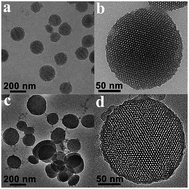
The synthesized MSNs and PAA-MSNs were both uniform spherical nanoparticles with clear porous structure, as shown in Fig. 1. The average diameter of MSNs was about 170 nm (Fig. 1a). After covalently grafted with PAA shell, the average diameter of nanoparticles increased to about 180 nm (Fig. 1c), corresponding to the polymer shell thickness of about 10 nm (Fig. 1d). The successful coating of polymer onto MSNs was also confirmed by the appearance of a strong band at 1723 cm−1 in the FTIR spectra (Fig. S1), which was the characteristic of carboxyl groups of PAA. The periodically well-organized hexagonal array of mesopores represented on the TEM images for both MSNs and PAA-MSNs (Fig. 1b and d, respectively) indicated the characteristic of MCM-41 type MSNs.51
image file: c6ra17213j-f1.tif
Fig. 1 TEM images of (a and b) MSNs and (c and d) PAA-MSNs.

The low-angle X-ray diffraction (LA-XRD) patterns of MSNs (Fig. 2a) further confirmed the hexagonal (p6m) mesoporous structure with four resolved diffraction peaks which can be exactly assigned to the (100), (110), (200) and (210) family planes. The XRD pattern of PAA-MSNs had similar diffraction peaks with those of MSNs, only with a decrease in peak intensity, arising from the polymer coating52 (Fig. 2a). According to the N2 adsorption–desorption measurements, the BET isotherms of MSNs (Fig. 2b) exhibited a type IV isotherm with a BET surface area of ∼1181 m2 g−1 and a total pore volume of ∼2.11 cm3 g−1, with the mesopore size of about 4.13 nm (Fig. 2b, inset). While, after grafted with polymer shell, PAA-MSNs exhibited reduced BET surface area of 423 m2 g−1 and pore volume of 0.675 cm3 g−1, but the shape of the hysteresis loop remained unchanged, which indicated that the pore shape was not significantly changed after polymer grafting. The mesopore size of PAA-MSNs was decreased to 3.8 nm due to the pore-filling effect (Fig. 2b, inset).


image file: c6ra17213j-f2.tif
Fig. 2 XRD patterns (a), N2 adsorption–desorption isotherms (b) and pore-size distribution (b, inset) of MSNs, PAA-MSNs and Pt@PAA-MSN.

To prevent burst release of loaded drug and achieve co-delivery of two drugs, PAA shell was cross-linked by cis-diamminediaqua platinum(II) complex. It is well-documented that the Pt(II) complex can be formed when CDDP is dissolved in water, and this process can be accelerated by AgNO3 due to the formation of insoluble AgCl.53–55 While the Pt(II) complex was stable in aqueous solution, it reacted at a reasonable pace with carboxyl groups to give Pt@PAA-MSN. The successful crosslinking of PAA shell was confirmed by various spectroscopic methods. The spherical and porous structure still remained (Fig. 3a), and the presence of Pt element could be clearly observed by line scanning analysis of chemical elements at the surface of Pt@PAA-MSN, as it shown in the EDS spectrum (Fig. 3b). As revealed by the FT-IR spectra in Fig. 3c, in comparison with PAA-MSNs, the peak at 1723 cm−1 of Pt@PAA-MSN weakened substantially. Moreover, the broad, medium intensity band at 960–875 cm−1 almost disappeared, which is characteristic for the carboxyl dimer due to out of plane OH⋯O hydrogen deformation, and there were slight differences at 1233–1184 cm−1 corresponding to the differences of C–O stretch between carboxylic acid and ester linkage, whereas the free CDDP spectrum showed no clear IR peak at 870–1240 cm−1 (Fig. 3b). These results indicated that the carboxyl group in polymer shells has reacted with Pt(II) complex. This was also confirmed by the zeta potential characterization. Due to the presence of abundant carboxyl groups, the zeta potential of PAA-MSNs was −44.1 mV. After reacting with Pt(II) complex, the zeta potential was increased to −14.2 mV, revealing the consuming of carboxyl groups. In addition, the hydrodynamic diameter and size distribution of MSN, PAA-MSNs and Pt@PAA-MSN were measured by DLS. As shown in Fig. 3d, the average diameter of MSNs was about 293 nm with a PDI of 0.254, much larger than that observed from TEM due to the hydrate layer in aqueous environment.56 While the average diameter was decreased from 325 nm (PDI 0.182) for PAA-MSNs to 282 nm (PDI 0.208) for Pt@PAA-MSN, which suggested that after crosslinking, the contracted polymer chains exhibited smaller size in aqueous environment. It was worth noting that the PAA-MSNs showed a smaller PDI than MSNs, revealing that PAA-MSNs had better dispersibility in water than MSNs, because of the graft of hydrophilic polymer chains.


image file: c6ra17213j-f3.tif
Fig. 3 TEM image (a), EDX spectrum (b) and STEM image (b, inset) of Pt@PAA-MSNDOX. (c) FT-IR spectra of CDDP, PAA-MSNs and Pt@PAA-MSN. (d) DLS curves of MSNs, PAA-MSNs and Pt@PAA-MSN in water.

To further estimate the stability of nanoparticles in physiological conditions, the colloidal stability of PAA-MSNs and Pt@PAA-MSNs were studied and confirmed in PBS (pH = 7.4) and DMEM medium containing 10 vol% of fetal bovine serum (purchased from Gibco Company, USA), respectively. As shown in Fig. S2a, the diameter of PAA-MSNs dispersed in PBS was about 303 nm with a PDI of 0.175 while after crosslinking, the diameter of Pt@PAA-MSN decreased to 291 nm in accordance with the results of analysis in deionized water. As for serum dispersion circumstance (Fig. S2b), the diameters of both nanoparticles were slightly larger than the results of test in water or PBS caused by mild agglomeration and the distribution peak smaller than 100 nm could be attribute to the substance in serum. These results accentuated the potential of the developed MSNs platform as a drug carrier for CDDSs.

Notably, the unconspicuous XRD peaks of Pt@PAA-MSN (Fig. 2a, blue curve) revealed the capping effect of the polymeric network after cross-linking,57–59 which was corroborated by N2 adsorption–desorption measurements (Fig. 2b, blue curve), as evidenced by the nonporous isotherm characteristic together with a decrease of surface area (115.09 m2 g−1) and pore size distribution. The conjugation amount of Pt(II) complex was estimated by ICP-MS and TGA. The TGA curve of PAA-MSNs yielded 56% weight loss from the grafted polymer when heated in the N2 atmosphere to 800 °C (Fig. S3, red curve) while less 7% weigh loss was observed for pure MSNs in the same temperature range (Fig. S3, black curve), implying the graft amount of polymer shell was about 50%. The weight loss of Pt@PAA-MSN (Fig. S3, blue curve) was about 45%, corresponding that the weight ratio of Pt(II) complex was 26.2%, which was in accordance with the result of 25.6% determined by ICP-MS.

The preparation procedure of cisplatin-conjugated, doxorubicin-loaded PAA-caged MSNs composite microspheres was schematically illustrated in Scheme 1. The PAA-MSNs could be utilized to achieve co-delivery of two different chemotherapeutic drugs, that is, DOX was first loaded in channel of mesopores of MSNs via diffusion effect and physical adsorption while Pt(II) was covalent loaded in PAA shell to form crosslinking network. The loading content of DOX was easily determined to be 22.9% by UV-vis measurement, and the loading content of Pt(II) complex was 20.8% by ICP-MS. The surface analysis of PAA-MSNDOX was investigated to compare with Pt@PAA-MSN. As shown in Fig. S4, the PAA-MSNDOX showed the nonporous isotherm characteristic with a decreased surface area of 181.7 m2 g−1 due to the pore filling effect.


image file: c6ra17213j-s1.tif
Scheme 1 Schematic illustration of the synthesis and drug release behavior of Pt@PAA-MSNDOX.

As previously reported,54,55,60 the formation structure of Pt(II) complex prodrug reacting with carboxyl could be broken in the presence of Cl or under acidic condition, and then the cytotoxic Pt(II) are released. The drug released behaviour of dual-drug co-delivery systems was tested and compared.

Fig. 4 showed the release behaviors of Pt(II) and DOX from the Pt@PAA-MSNDOX complex in vitro by using dialysis tubes under the selected conditions. Since the complex was stable in phosphate buffer (pH 7.4) without NaCl, the release amounts of Pt(II) and DOX was about 3.5% and 5.0%, respectively. While both drugs started to release in the presence of NaCl. The high concentration of chloride anions replaced the carboxylate ligands to destroy the crosslinking structure for release of both drugs. However, the release rate under this condition was noticeably slower and seems to level off at 15.9% for Pt(II) and 25.3% for DOX, respectively. As it has been proposed previously, the incapability to release all the conjugated CDDP can be explained by the resulting structure after release at neutral or physiological pH (7.4), in which the polymer chains had some negative charges to inhibit the attack of chloride anions. In comparison, in mildly acidic environments (pH 5.5), the Pt(II) release was relatively accelerated, and the amount of released Pt(II) reached 63.6%. In this way, as cross-linked outer shell was gradually disrupted and more of the carboxyl group was protonized leading to the dissociation of electrostatic interaction between PAA and DOX, so that more of the incorporated DOX was released and the release amount was 74.0%. Particularly, the most rapid release rate was actuated at pH 5.5 with the action of NaCl. In the acidic condition containing chloride anions, 70.0% of Pt(II) and 79.9% of DOX were released within 144 h. As a consequence, Pt(II) cross-linking shell could act as a diffusion barrier and block DOX burst release during blood circulation (pH 7.4), whereas after internalization of complex into tumor cells via endocytosis, Pt(II) complex crosslinking shell would cleave under the lower pH (∼5) of the phagolysosomal system and condition of consistently higher concentration of chloride (Scheme 1), leading to enhance cytotoxic Pt(II) and DOX releasing.


image file: c6ra17213j-f4.tif
Fig. 4 In vitro (a) Pt(II) and (b) DOX release profiles of Pt@PAA-MSNDOX in aqueous solution with or without 0.9% NaCl at pH 7.4 and pH 5.5, respectively.

The fluorescent DOX could suppress the formation of nucleic acid via DNA binding and prompts tumor cells apoptosis.61 Thus, the DOX released from DDSs should be gathered in the nucleus, which could be detected by the confocal fluorescence microscopy. The cellular uptake properties of Pt@PAA-MSNDOX complex were investigated with HeLa cells (purchased from the Institute of Biochemistry and Cell Biology, Shanghai Institutes for Biological Sciences, Chinese Academy of Sciences) via fluorescence microscopy. As seen in Fig. 5, the strong red fluorescence of DOX was observed and the position was in accordance with the cell nuclei stained with blue DAPI. The well-overlap in Fig. 5d confirmed that complex could effectively release DOX inside cells. It is speculated that due to the abundance of chloride ion and low pH in endosome/lysosome, Pt(II) complex prodrug can be released through the crosslinking cleavage after the cellular uptake of Pt@PAA-MSNDOX, thereby leading to the cellular release and prominent accumulation of DOX inside cell nuclei.


image file: c6ra17213j-f5.tif
Fig. 5 Fluorescence microscopy images of HeLa cells after incubation with Pt@PAA-MSNDOX for 6 h. (a) DOX (red), (b) cell nuclei stained by DAPI (blue), (c) bright field, (d) overlay of (a)–(c).

The in vitro cytotoxicity of PAA-MSNs at different concentrations and incubation times was estimated by CCK-8 kit assays. As shown in Fig. 6, PAA-MSNs showed no obvious cytotoxic effect on HeLa cells at a concentration ranged from 0.01 to 0.25 mg mL−1 after incubation for 24 h and 48 h. Even at the concentration of PAA-MSNs up to 0.5 mg mL−1, the cell viability was above 85% after incubation for 48 h. These results suggest PAA-MSNs exhibit a good biocompatibility, and are suitable to use as a platform for loading anticancer drugs.


image file: c6ra17213j-f6.tif
Fig. 6 Cell cytotoxicity of PAA-MSNs against HeLa cells with different incubation time of 24 and 48 h.

The antitumor activity and synergistic effect of Pt@PAA-MSNDOX complex with different drug concentrations and incubation times were studied in vitro using the CCK-8 assays. A357 and HeLa cells (purchased from the Institute of Biochemistry and Cell Biology, Shanghai Institutes for Biological Sciences, Chinese Academy of Sciences) were chosen for the cytotoxic activity test with the free DOX, free Pt(II), PAA-MSNDOX, Pt@PAA-MSN, DOX/Pt mixture and PAA-MSNDOX/Pt@PAA-MSN mixture as the control groups. The cytotoxicity experiments were based on the total molar concentrations of both drugs and the significance of differences of assays was determined with one-way ANOVA followed by a Fisher's LSD test. As shown in Fig. 7a and b, after 24 h incubation, all samples showed growth inhibition abilities against A357 and HeLa cells, and the cytotoxicity increased along with the increased concentrations of drugs. Free single drug and single drug-loaded nanoparticles induced similar cytotoxicity, demonstrating that the drug-loaded nanoparticles could efficiently release drugs under acidic pH in endosomes/lysosomes and presence of chloride anions, whereas the Pt@PAA-MSNDOX complex showed highest cytotoxicity in both cells (Fig. 7a and b). Furthermore, with the incubation time increasing to 48 h (Fig. 7c and d), Pt@PAA-MSNDOX complex showed more potent cellular growth inhibition abilities against A357 and HeLa cells, which indicated that more cytotoxic Pt(II) and DOX was released from complex over time. Significantly, in contrast with free single drugs or single drug-loaded nanoparticles, Pt@PAA-MSNDOX complex exhibited higher cytotoxicity in both A357 and HeLa cells, especially at low concentration (Fig. 7c and d). It could be found that the cell viabilities of A357 and HeLa cells were 8.8% and 25.6% at 1.0 μM of co-delivery drugs for 48 h, respectively, which was much lower than that of free single drug and single drug-loaded micelles. It was worth noting that Pt@PAA-MSNDOX complex containing 0.1 μM of drug concentration also showed an effective antitumor activity to A357 and HeLa cells, exhibiting a robust enhancement of combination potency. This could be evidenced by the statistics analysis results. As listed in Tables S1–S4, compared with single free drug or single drug loading system, the Pt@PAA-MSNDOX showed the significantly higher level of cytotoxicity to A357 and HeLa cells at incubation time of 24 h or 48 h since the drug concentration were above 0.1 μM. Meanwhile, the distinguishing antitumor activity of Pt@PAA-MSNDOX complex compared with free single drugs and single drug loaded nanoparticles could be seen from the IC50 values (Table S5). The IC50 values of Pt@PAA-MSNDOX complex against A357 and HeLa cells at 24 h were 0.66 and 0.73 μM, respectively, which could reduce to 0.13 and 0.30 μM at 48 h. The results were much lower than those of free single drugs or single drug loaded nanoparticles (P < 0.05), revealing that Pt@PAA-MSNDOX complex showed the highest antitumor activity. In addition, the synergy of Pt@PAA-MSNDOX complex was further estimated using the combination index (CI) based on the IC50 value of drugs (Table S5). It is proposed that CI < 1 indicates a synergistic effect and CI = 1 indicates an additive effect, while CI > 1 indicates an antagonism effect.62 As shown in Table 1, all the CI values of Pt@PAA-MSNDOX complex were all much lower than 1, indicating the synergistic effect for the inhibition of tumor cells. The synergistic effect could be more clearly seen in Fig. S5 and S6. Pt@PAA-MSNDOX complex, DOX/Pt mixture and PAA-MSNDOX/Pt@PAA-MSN mixture exhibited better inhibition efficacy than the single free drug alone and single drug loaded nanoparticles over the same concentrations, which was more significant at incubation time of 48 h (Fig. S5c and d and S6c and d). From the results discussed above, we can see that Pt@PAA-MSNDOX complex can significantly inhibit the proliferation of tumor cells even at very low concentrations, and outperform free single drugs and single drug loaded nanoparticles, implying that the PAA-MSNs is a promising platform for co-delivery of multiple antitumor drugs and resulting complex might have an important practical application for synergistic combination drug chemotherapy.


image file: c6ra17213j-f7.tif
Fig. 7 In vitro cell viability of A357 and HeLa cells against free single drugs, Pt@PAA-MSN, PAA-MSNDOX and Pt@PAA-MSNDOX at different concentrations and incubation times: (a) A357 cells incubated for 24 h; (b) HeLa cells incubated for 24 h; (c) A357 cells incubated for 48 h; (d) HeLa cells incubated for 48 h.
Table 1 Combination index (CI) values of Pt@PAA-MSNDOX for A357 and HeLa cells at different incubation time
Cells and incubation time CI (based on the IC50 of single free drugs) CI (based on the IC50 of single drugs loading system)
a The IC50 values of free Pt and Pt@PAA-MSN were beyond the scope of tests, thus 10 μM was chosen to calculate the limits of the corresponding CI values.
A357 24 h <0.265a <0.080a
A357 48 h 0.154 0.198
HeLa 24 h 0.166 0.208
HeLa 48 h 0.189 0.168


Conclusions

In summary, synergistic combination of cisplatin and DOX was successfully achieved by packaging both drugs into a novel co-delivery system based on the PAA-caged MSNs platform. The abundant carboxyl group from the outer polymeric shells improved the hydrophilicity and could be conjugated with the hydrated cisplatin prodrugs. The resulting crosslinking shells acted as a gate to prevent the drug burst release at neutral or physiological pH (7.4) from the MSNs, which served as a reservoir for DOX. Particularly, triggered drug-releasing characteristics of Pt@PAA-MSNDOX nanocomposites could been demonstrated in an acidic environment. The in vitro cellular cytotoxicity test revealed that the PAA-MSNs were highly biocompatible and suitable to utilize as drug carriers. Moreover, the Pt@PAA-MSNDOX nanocomposites exhibited more remarkable cytotoxicity than free single drugs or single drug loaded nanoparticles to HeLa and A357 cells, presenting the great potential as a pH-responsive co-delivery system for chemotherapy treatment of cancer.

Experimental

Materials

Tetraethyl orthosilicate (TEOS), cetyltrimethylammonium bromide (CTAB, 99%), acetonitrile, and mesitylene (TMB) were purchased from J&K Chemical Co., Ltd. Acrylic acid (AA, 99%), azodiisobutyronitrile (AIBN, 98%), and methacryloxy propyl trimethoxy silane (MPS) were purchased from Aldrich. Doxorubicin hydrochloride (DOX·HCl) and cisplatin (CDDP) were purchased from Beijing Huafeng United Technology Co., Ltd. NaOH (96%), NH4NO3 (99%), and AgNO3 (99.8%) were purchased from Sinopharm Chemical Reagent Co., Ltd. Fetal bovine serum (FBS) and DMEM were purchased from Gibco company, USA. All the reagents were analytical grade and used without further treatment.

Preparation of poly(acrylic acid) functionalized MSNs (PAA-MSNs)

Mesoporous silica nanoparticles (MSNs) were prepared according to our previous work.63 Typically, 3.5 mL of TMB, 0.5 g of CTAB and 0.14 g of NaOH were added into 250 mL of deionized water. After vigorously stirred at 80 °C for 4 h, 2.5 mL of TEOS was quickly added into the mixture, followed by continually stirring at 80 °C for another 2 h. The resultant MSNs was filtrated and dried in vacuo at 35 °C for 12 h. The surface of MSNs was functionalized with MPS by a post-grafting method. 0.1 g of MSNs was refluxed in 24 mL of ethanol containing 0.2 mL of MPS for 12 h. Then, the mixture was centrifuged, washed with copious ethanol, and dried in vacuo at 35 °C for 12 h to give double-bond functionalized MSNs (MPS-MSNs).

PAA-MSNs were prepared by distillation–precipitation polymerization of AA in acetonitrile using MBA as a cross-linker and AIBN as an initiator. Typically, 25 mg of MPS-MSNs was dispersed into 40 mL of acetonitrile under ultrasonication in a 100 mL of flask, equipped with a fractionation column, a Liebig condenser, and a receiver. Then, 0.1 mL of AA, 18 mg of MBA and 2.5 mg of AIBN were successively added into the flask, and the mixture was heated to reflux. The reaction was completed after distilling off 20 mL of acetonitrile from the mixture. The resultant PAA-MSNs were purified by centrifugation and washing with ethanol for several times. Finally, further purification was conducted to remove the structure-template CTAB and TMB. Briefly, 0.1 g of as-synthesized PAA-MSNs was refluxed in an ethanol solution of ammonium nitrate (NH4NO3/C2H5OH, 10 mg mL−1) for 6 h. The product was filtrated and dried in vacuo at 35 °C for 12 h, to give PAA-MSNs as a white powder.

Preparation of cisplatin and doxorubicin dual-loaded PAA-MSNs (Pt@PAA-MSNDOX)

Typically, PAA-MSNs and DOX·HCl were dispersed into deionized water to form 2 mg mL−1 solution, respectively. Then, 5 mL of PAA-MSNs solution was mixed with 5 mL of DOX·HCl solution, followed by stirring at room temperature for 24 h to reach the equilibrium state. At the same time, 10 mg of CDDP was dissolved in 10 mL of deionized water and mixed with 11.3 mg of silver nitrate ([AgNO3]/[CDDP] = 2) to form an aqueous complex. The solution was stirred in dark at room temperature for 4 h. After reaction, the AgCl precipitate was removed by centrifugation at 12[thin space (1/6-em)]000 rpm for 5 min, and the supernatant was purified by passing through a 0.22 μm filter. Then, the supernatant containing cis-diamminediaqua platinum(II) complex was added into the above mixture of PAA-MSNs and DOX·HCl and reacted in an incubator shaker at 37 °C for 24 h. The cisplatin and doxorubicin dual-loaded PAA-MSNs (Pt@PAA-MSNDOX) was obtained by centrifugation at 12[thin space (1/6-em)]000 rpm for 5 min. The control samples, cisplatin loaded PAA-MSNs (Pt@PAA-MSN) and doxorubicin loaded PAA-MSNs (PAA-MSNDOX), were prepared by a similar procedure, only without adding DOX·HCl or CDDP, respectively. All the obtained nanocomposites were washed three times with 2 mL of deionized water to remove the physically absorbed drugs and lyophilized to obtain solids. The loading amount of DOX was determined by a UV-vis spectrophotometer at 480 nm and the loading amount of Pt(II) was evaluated by the inductively coupled plasma-mass spectrometer (ICP-MS).

Release profiles of Pt@PAA-MSNDOX

10 mg of Pt@PAA-MSNDOX was dispersed into 5 mL of deionized water and transferred into a dialysis bag with a cut-off molecular weight of 3.5 kDa. The bag was immersed in 50 mL of PBS with or without 0.9% NaCl at 37 °C in a shaking incubator, and the pH was adjusted to either 5.5 or 7.4. At specified time intervals, 5 mL of external medium was withdrawn and replaced with the same volume of fresh buffer solution. The amount of cisplatin present in the sample was determined by ICP and the concentration of DOX was measured by a UV-vis spectrophotometer at 480 nm.

In vitro cell assay

The in vitro cytotoxicity of CDDP, DOX, Pt@PAA-MSN, PAA-MSNDOX, PAA-MSNDOX/Pt@PAA-MSN mixture, DOX/Pt mixture and Pt@PAA-MSNDOX against HeLa (human cervical carcinoma) cells and A357 (human malignant melanoma) cells was evaluated by CCK-8 kit assays. The cells were purchased from the Institute of Biochemistry and Cell Biology, Shanghai Institutes for Biological Sciences, Chinese Academy of Sciences. The cells were maintained onto 96-well plates with a density of 1 × 10⁴ cell per well and incubated at 37 °C in an atmosphere containing 5% CO2 for 24 h to allow cell attachment. Then, the medium was replaced with a fresh medium containing the indicated concentration of Pt(II), DOX, Pt@PAA-MSN, PAA-MSNDOX and Pt@PAA-MSNDOX, respectively. After incubation for 24 and 48 h, the amount of viable cells was evaluated via the CCK-8 assay. The same procedure of cytotoxicity test of PAA-MSNs against HeLa cells was implemented as mentioned above.

In vitro cellular uptake

The cellular uptake and intracellular release behaviors of Pt@PAA-MSNDOX were observed with a confocal fluorescence microscopy based on the fluorescence property of DOX. The HeLa cells, purchased from the Institute of Biochemistry and Cell Biology, Shanghai Institutes for Biological Sciences, Chinese Academy of Sciences, were seeded in glass-bottom Petri dishes using Roswell Park Memorial Institute (RPMI) 1640 medium supplemented with 10% fetal bovine serum, 2 mM L-glutamine, 100 U mL−1 penicillin and 100 U mL−1 streptomycin at 37 °C under 5% CO2 and allowed to adhere for 24 h. The medium was then replaced with 2 mL of the pre-prepared Pt@PAA-MSNDOX solutions in serum-free culture medium and cells were incubated for 6 h. After three washes with PBS, 4% paraformaldehyde was added for another 30 min at 37 °C and then the nuclei were stained with the blue dye 4′,6-diamidino-2-phenylindole (DAPI). Fluorescence images of cells were obtained using confocal microscope.

Statistical analysis

The differences between the cytotoxicity against A357 and HeLa cells were analyzed by one-way analysis of variance (ANOVA) with least significant difference (LSD) test. Statistical analysis was performed using the SPSS software (SPSS Inc, Chicago, IL). Differences were considered to be significant at a level of P < 0.05.

The combination index calculation

To investigate the combinatorial therapeutic effect resulting from co-delivery of Pt(II) and DOX, the combination index (CI) based on the IC50 value was calculated and compared. The CI value was calculated on the basis of the equation:
image file: c6ra17213j-t1.tif
where IC50 is the drug concentration of 50% cellular cytotoxicity of tumor cells, IC50(A) and IC50(B) are the IC50 values of the drug given individually, and IC50(A)pair and IC50(B)pair are the IC50 values of the drug given as an A–B pair.

Characterization

Transmission electron microscopy (TEM), and high-angle annular dark-field, scanning TEM (HAADF-STEM) images and elemental line scanning analysis were obtained using a Tecnai G2 20 TWIN microscope operated at 200 kV. Thermogravimetric analysis (TGA) was performed on a Mettler Toledo TGA instrument at a heating rate of 20 °C min−1 in N2. Fourier transform infrared (FT-IR) spectra were recorded on a Nicolet 6700 FT-IR spectrometer using the KBr pellet method. Powder low-angle X-ray diffraction (XRD) patterns were acquired on a Bruker D4 X-ray diffractometer over a scan range 1–10°. The surface analysis was performed by nitrogen sorption isotherms at 77 K with an ASAP 2020 sorptometer. The surface areas were calculated by the Brunauer–Emmett–Teller (BET) method, and the pore size distributions were calculated by the Barrett–Joyner–Halenda (BJH) method. All samples were degassed at 100 °C for 6 h prior to the analysis. The total pore volume was estimated from the amount adsorbed at a relative pressure P/P0 of 0.99. The size distribution of the nanoparticles was measured by dynamic light scattering (DLS) using a Malvern Zetasizer Nano (ZS90), and the dispersant was deionized water. The zeta potentials were measured by a Malvern Zetasizer Nano (ZS90). The UV-vis absorbance spectra were measured with a Perkin-Elmer Lambda 35 spectrophotometer. The amounts of Pt were determined by using Inductively Coupled Plasma-Atomic Emission Spectrometer (Hitachi P-4010). The cellular images were acquired with a fluorescence microscopy (BX51, Olympus).

Acknowledgements

The authors thank the financial support from the International Science & Technology Cooperation Program of China (2014DFE40130), and the National Natural Science Foundation of China (51373035, 51373040, 51573030, 51573028, and 81372796).

References

  1. I. I. Slowing, J. L. Vivero-Escoto, C.-W. Wu and V. S. Y. Lin, Adv. Drug Delivery Rev., 2008, 60, 1278–1288 CrossRef CAS PubMed.
  2. P. Yang, S. Gai and J. Lin, Chem. Soc. Rev., 2012, 41, 3679–3698 RSC.
  3. H. Yu, C. Xi, J. Hu, M. Du and D. Yang, Sci. Adv. Mater., 2015, 7, 855–860 CrossRef CAS.
  4. G.-H. Wang, G.-L. Huang, Y. Zhao, X.-X. Pu, T. Li, J.-J. Deng and J.-T. Lin, J. Mater. Chem. B, 2016, 4, 3832–3841 RSC.
  5. J. L. Wang, J. A. Kaplan, Y. L. Colson and M. W. Grinstaff, Angew. Chem., Int. Ed., 2016, 55, 2796–2800 CrossRef CAS PubMed.
  6. A. Noureddine, M. Gary-Bobo, L. Lichon, M. Garcia, J. I. Zink, M. W. C. Man and X. Cattoën, Chem.–Eur. J., 2016, 22, 9624–9630 CrossRef CAS PubMed.
  7. M. M. Gottesman, T. Fojo and S. E. Bates, Nat. Rev. Cancer, 2002, 2, 48–58 CrossRef CAS PubMed.
  8. G. Szakacs, J. K. Paterson, J. A. Ludwig, C. Booth-Genthe and M. M. Gottesman, Nat. Rev. Drug Discovery, 2006, 5, 219–234 CrossRef CAS PubMed.
  9. H. Zhu, H. Chen, X. Zeng, Z. Wang, X. Zhang, Y. Wu, Y. Gao, J. Zhang, K. Liu, R. Liu, L. Cai, L. Mei and S. Feng, Biomaterials, 2014, 35, 2391–2400 CrossRef CAS PubMed.
  10. G. Tian, X. Zheng, X. Zhang, W. Yin, J. Yu, D. Wang, Z. Zhang, X. Yang, Z. Gu and Y. Zhao, Biomaterials, 2015, 40, 107–116 CrossRef CAS PubMed.
  11. X. Chen and Z. Liu, J. Mater. Chem. B, 2016, 4, 4382–4388 RSC.
  12. K. Wang, Q. Hu, W. Zhu, M. Zhao, Y. Ping and G. Tang, Adv. Funct. Mater., 2015, 25, 3380–3392 CrossRef CAS.
  13. H. Wang, J. Hu, X. Cai, J. Xiao and Y. Cheng, Polym. Chem., 2016, 7, 2319–2322 RSC.
  14. Z. Bao, P. Gao, G. Xia, Z. Wang, M. Kong, C. Feng, X. Cheng, Y. Liu and X. Chen, J. Mater. Chem. B, 2016, 4, 3936–3944 RSC.
  15. D. Liu, H. Zhang, B. Herranz-Blanco, E. Makila, V. P. Lehto, J. Salonen, J. Hirvonen and H. A. Santos, Small, 2014, 10, 2029–2038 CrossRef CAS PubMed.
  16. L. Zhu, F. Perche, T. Wang and V. P. Torchilin, Biomaterials, 2014, 35, 4213–4222 CrossRef CAS PubMed.
  17. J. Li, Z. Li, M. Li, H. Zhang and Z. Xie, J. Appl. Polym. Sci., 2015, 132, 41440,  DOI:10.1002/app.41440.
  18. C. He, Z. Tang, H. Tian and X. Chen, Adv. Drug Delivery Rev., 2016, 98, 64–76 CrossRef CAS PubMed.
  19. S. Lu, L. Xu, E. T. Kang, R. Mahendran, E. Chiong and K. G. Neoh, Eur. J. Pharmacol., 2016, 84, 103–115 CrossRef CAS PubMed.
  20. C. Zhang, D. Pan, K. Luo, W. She, C. Guo, Y. Yang and Z. Gu, Adv. Healthcare Mater., 2014, 3, 1299–1308 CrossRef CAS PubMed.
  21. J. Gu, X. Fang, J. Hao and X. Sha, Biomaterials, 2015, 45, 99–114 CrossRef CAS PubMed.
  22. X.-Y. Hu, K. Jia, Y. Cao, Y. Li, S. Qin, F. Zhou, C. Lin, D. Zhang and L. Wang, Chem.–Eur. J., 2015, 21, 1208–1220 CrossRef CAS PubMed.
  23. P. Liang, C.-Q. Wang, H. Chen, R.-X. Zhuo and S.-X. Cheng, Polym. Int., 2015, 64, 647–653 CrossRef CAS.
  24. G.-F. Luo, W.-H. Chen, Y. Liu, Q. Lei, R.-X. Zhuo and X.-Z. Zhang, Sci. Rep., 2014, 4, 6064,  DOI:10.1038/srep06064.
  25. L. Maggini, I. Cabrera, A. Ruiz-Carretero, E. A. Prasetyanto, E. Robinet and L. De Cola, Nanoscale, 2016, 8, 7240–7247 RSC.
  26. H. Li, J. Gu, L. A. Shah, M. Siddiq, J. Hu, X. Cai and D. Yang, Mater. Sci. Eng., C, 2015, 49, 210–216 CrossRef CAS PubMed.
  27. S. R. Gayam, P. Venkatesan, Y.-M. Sung, S.-Y. Sung, S.-H. Hu, H.-Y. Hsu and S.-P. Wu, Nanoscale, 2016, 8, 12307–12317 RSC.
  28. J. Shen, H. C. Kim, H. Su, F. Wang, J. Wolfram, D. Kirui, J. Mai, C. Mu, L. N. Ji, Z. W. Mao and H. F. Shen, Theranostics, 2014, 4, 487–497 CrossRef CAS PubMed.
  29. C.-Y. Lai, B. G. Trewyn, D. M. Jeftinija, K. Jeftinija, S. Xu, S. Jeftinija and V. S. Y. Lin, J. Am. Chem. Soc., 2003, 125, 4451–4459 CrossRef CAS PubMed.
  30. I. Slowing, B. G. Trewyn and V. S. Y. Lin, J. Am. Chem. Soc., 2006, 128, 14792–14793 CrossRef CAS PubMed.
  31. J. Lu, M. Liong, J. I. Zink and F. Tamanoi, Small, 2007, 3, 1341–1346 CrossRef CAS PubMed.
  32. M. Wu, Y. Chen, L. Zhang, X. Li, X. Cai, Y. Du, L. Zhang and J. Shi, J. Mater. Chem. B, 2015, 3, 766–775 RSC.
  33. H. Meng, M. Y. Wang, H. Y. Liu, X. S. Liu, A. Situ, B. Wu, Z. X. Ji, C. H. Chang and A. E. Nel, ACS Nano, 2015, 9, 3540–3557 CrossRef CAS PubMed.
  34. X. Wang, Z. Zhou, X. Guo, Q. He, C. Hao and C. Ge, RSC Adv., 2016, 6, 35550–35558 RSC.
  35. Y. Zhan, M. Goncalves, P. Yi, D. Capelo, Y. Zhang, J. Rodrigues, C. Liu, H. Tomas, Y. Li and P. He, J. Mater. Chem. B, 2015, 3, 4221–4230 RSC.
  36. J. L. Gu, J. P. Liu, Y. S. Li, W. R. Zhao and J. L. Shi, Langmuir, 2013, 29, 403–410 CrossRef CAS PubMed.
  37. I. Munaweera, Y. Shi, B. Koneru, A. Patel, M. H. Dang, A. J. Di Pasqua and K. J. Balkus, J. Inorg. Biochem., 2015, 153, 23–31 CrossRef CAS PubMed.
  38. S. Mohapatra, S. R. Rout, R. Narayan and T. K. Maiti, Dalton Trans., 2014, 15841–15850 RSC.
  39. J. L. Vivero-Escoto and M. Elnagheeb, Nanomaterials, 2015, 5, 2302–2316 CrossRef CAS.
  40. B. Ahn, J. Park, K. Singha, H. Park and W. J. Kim, J. Mater. Chem. B, 2013, 1, 2829–2836 RSC.
  41. X. Y. Zhu, J. L. Gu, Y. S. Li, W. R. Zhao and J. L. Shi, Microporous Mesoporous Mater., 2014, 196, 115–121 CrossRef CAS.
  42. W. Zhang, J. L. Shen, H. Su, G. Mu, J. H. Sun, C. P. Tan, X. J. Liang, L. N. Ji and Z. W. Mao, ACS Appl. Mater. Interfaces, 2016, 8, 13332–13340 CAS.
  43. J. L. Gu, S. S. Su, Y. S. Li, Q. J. He, F. Y. Zhong and J. L. Shi, J. Phys. Chem. Lett., 2010, 1, 3446–3450 CrossRef CAS.
  44. M. Li, Z. Tang, S. Lv, W. Song, H. Hong, X. Jing, Y. Zhang and X. Chen, Biomaterials, 2014, 35, 3851–3864 CrossRef CAS PubMed.
  45. V. T. Huynh, G. Chen, P. de Souza and M. H. Stenzel, Biomacromolecules, 2011, 12, 1738–1751 CrossRef CAS PubMed.
  46. M. B. Zheng, C. X. Yue, Y. F. Ma, P. Gong, P. F. Zhao, C. F. Zheng, Z. H. Sheng, P. F. Zhang, Z. H. Wang and L. T. Cai, ACS Nano, 2013, 7, 2056–2067 CrossRef CAS PubMed.
  47. Y. Ma, X. Fan and L. Li, Carbohydr. Polym., 2016, 137, 19–29 CrossRef CAS PubMed.
  48. J. Lin, Z. Tang, M. Li and X. Chen, J. Controlled Release, 2015, 213, e48–49 CrossRef PubMed.
  49. H. Ma, C. He, Y. Cheng, Z. Yang, J. Zang, J. Liu and X. Chen, ACS Appl. Mater. Interfaces, 2015, 7, 27040–27048 CAS.
  50. S. Lu, L. Xu, E. T. Kang, R. Mahendran, E. Chiong and K. G. Neoh, Eur. J. Pharmacol., 2016, 84, 103–115 CrossRef CAS PubMed.
  51. M. Vallet-Regi, A. Ramila, R. P. del Real and J. Perez-Pariente, Chem. Mater., 2001, 13, 308–311 CrossRef CAS.
  52. Y. Zhao, B. G. Trewyn, I. I. Slowing and V. S. Y. Lin, J. Am. Chem. Soc., 2009, 131, 8398–8400 CrossRef CAS PubMed.
  53. N. Nishiyama, M. Yokoyama, T. Aoyagi, T. Okano, Y. Sakurai and K. Kataoka, Langmuir, 1999, 15, 377–383 CrossRef CAS.
  54. V. T. Huynh, G. Chen, P. de Souza and M. H. Stenzel, Biomacromolecules, 2011, 12, 1738–1751 CrossRef CAS PubMed.
  55. L. Ye, K. Letchford, M. Heller, R. Liggins, D. Guan, J. N. Kizhakkedathu, D. E. Brooks, J. K. Jackson and H. M. Burt, Biomacromolecules, 2011, 12, 145–155 CrossRef CAS PubMed.
  56. L. Yuan, Q. Tang, D. Yang, J. Z. Zhang, F. Zhang and J. Hu, J. Phys. Chem. C, 2011, 115, 9926–9932 CAS.
  57. C. Y. Lai, B. G. Trewyn, D. M. Jeftinija, K. Jeftinija, S. Xu, S. Jeftinija and V. S. Y. Lin, J. Am. Chem. Soc., 2003, 125, 4451–4459 CrossRef CAS PubMed.
  58. S. Giri, B. G. Trewyn, M. P. Stellmaker and V. S. Y. Lin, Angew. Chem., Int. Ed., 2005, 44, 5038–5044 CrossRef CAS PubMed.
  59. R. Liu, X. Zhao, T. Wu and P. Feng, J. Am. Chem. Soc., 2008, 130, 14418–14419 CrossRef CAS PubMed.
  60. A. Saisyo, H. Nakamura, J. Fang, K. Tsukigawa, K. Greish, H. Furukawa and H. Maeda, Colloids Surf., B, 2016, 138, 128–137 CrossRef CAS PubMed.
  61. Y.-Z. Zhao, C.-Z. Sun, C.-T. Lu, D.-D. Dai, H.-F. Lv, Y. Wu, C.-W. Wan, L.-J. Chen, M. Lin and X.-K. Li, Cancer Lett., 2011, 311, 187–194 CrossRef CAS PubMed.
  62. L. Ma, M. Kohli and A. Smith, ACS Nano, 2013, 7, 9518–9525 CrossRef CAS PubMed.
  63. H. Li, J. Z. Zhang, Q. Tang, M. Du, J. Hu and D. Yang, Mater. Sci. Eng., C, 2013, 33, 3426–3431 CrossRef CAS PubMed.

Footnotes

Electronic supplementary information (ESI) available. See DOI: 10.1039/c6ra17213j
These authors contributed equally to this work.

This journal is © The Royal Society of Chemistry 2016
Click here to see how this site uses Cookies. View our privacy policy here.