Lijuan Lianga,
Liancheng Zhaoa and
Xianshun Zeng*ab
aSchool of Materials Science and Engineering, Harbin Institute of Technology, Harbin 150001, China
bSchool of Materials Science & Engineering, Tianjin University of Technology, Tianjin 300384, China. E-mail: xshzeng@tjut.edu.cn; Fax: +86-22-60215226; Tel: +86-22-60216748
First published on 2nd September 2016
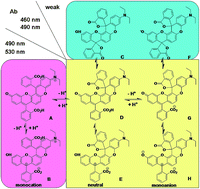
A π-extended rhodol derivative 1 with five fused six-membered rings has been synthesized. As the optical properties of the dye 1 are strictly controlled by two spirorings within the structure, its reversible structural conversions among the monocation, neutral, and monoanion forms were investigated in detail under different conditions by the electronic absorption and fluorescence emission spectroscopy. Then the chemical modification methodology of dye 1 was investigated in detail. The phenol hydroxyl could be replaced by a chlorine atom by using POCl3 as a chlorination reagent. Only one of the carboxyl groups could be esterificated by alcohol under strong acidic conditions. Finally, a thiosemicarbazide spirolactam 7 was obtained from dye 1 as a highly Hg2+-selective fluorescent chemodosimeter.
Rhodamine, fluorescein and rhodol (hybrids of a rhodamine and a fluorescein) dyes are widely utilized as fluorescent probes in the past few years. Among these derivatives, rhodols have become the interesting compound for fluorescent labels in biological detections with the inherited excellent photophysical properties from rhodamines and fluoresceins such as high extinction coefficients, quantum yields, photostability, and solubility in a variety of solvents.8 However, the characteristic electronic absorption and fluorescence emission wavelengths of these dyes are only covering a narrow range due to the small π-conjugation of the fluorophores. Recently, Kamino and co-workers developed rhodamine luminophores-xanthene derivatives ABPX via Friedel–Crafts acylation (Fig. 1).9a Compared with rhodamine, fluorescein and rhodol dyes, they presented longer absorption and emission properties. However, they presented in a neutral bispirolactone form in THF solution, and only showed a property of aggregation-induced emission enhancement (AIEE) in strong acid solutions.9a,b Such optical properties of these dyes were unfavorable for further applications. To exploit this fluorophore as a signaling reporter in neutral media, Wong et al. designed a Hg(II)-selective chemodosimer ABPX-Hg. Compared with the related rhodamine derivatives with only one spirolactam group in many cases, ABPX-Hg was found to exhibit selective Hg(II) ion sensing behaviors with successive ring-openings of two spirolactam moieties in neutral methanol (Fig. 1).9c Thus, this type of π-extended xanthene dyes with long absorption and emission properties may find potential applications in biological purposes.3a,9e,10,11
Enlightened by Kamino's work9a–d we reported herein the design, synthesis and characteristics of a π-extended rhodol-type fluorescent dye 1 with an asymmetric hybrid structure of rhodamine and fluorescein. The fluorophore of the dye 1 was composed of five fused six-membered rings and its electronic absorption and fluorescence emission spectra were strictly controlled by two spirorings (Scheme 1). As the structural conversions of the two spirorings within the dye could produce fruitful electronic absorption and fluorescence emission spectroscopy which may find smart application in probe design by carefully tuning these processes, the influences of the two spirorings on optical and chemical properties of the dye 1 under different conditions were investigated in detail. Meanwhile, to exploit the dye as a signalling fluorophore, the thiosemicarbazide spirolactam derivative 7 was prepared as a highly Hg2+-selective fluorescent chemodosimeter.
![]() | ||
Scheme 1 Synthetic route of dye 1. (a) 2-(4-Diethylamino-2-hydroxybenzoyl) benzoic acid, 98% H2SO4, 100 °C, 3 h. |
As shown in Scheme 2, the dye 1 was speculated to exist in solution in the monocation forms A and B, neutral forms C, D and E, and monoanion forms F, G and H.5a,9b,12 Forms A and B were the bispiroring-opened species with two ring-opened groups, and forms D, E, G and H were the single ring-opened species. Forms C and F were in the bisspirolactone conformations. To get a general idea of the structural conversions among these forms, the optical properties of dye 1 (cis- and trans-isomers) were investigated in detail under different conditions.
Firstly, the absorbance spectra of dye 1B (10 μM) were investigated in MeOH:
H2O (1
:
1, v/v) under different pH conditions. As shown in inset of Fig. 4, the absorption bands of dye 1 over 400 nm decreased with the increase of pH from 1 to 4. Three absorption bands at 462, 498, and 533 nm, and a shoulder at ca. 435 nm were observed in this pH range. These absorption bands were assigned to the structure of the double ring-opened form A or B. The very weak absorption over 400 nm at ca. pH 4 meant that the dye 1 was mainly converted to a bisspirolactone conformation C. However, two absorption peaks at 462 and 488 nm, and a shoulder at ca. 430 nm were observed in the pH range (5–12). The strength of the two absorption peaks increased with pH values. It was assigned to the single ring-opened form G or H. To further elucidate the structural conversions of the dye among its structural isomers, the absorbance spectra of dye 1 were measured in MeOH
:
H2O (1
:
1, v/v) under strong acidic and basic conditions (Fig. S7†). Even in strong basic media (5 M NaOH), the dye showed almost the same absorption bands (merely ca. 10 nm red shifts of the absorption peaks) as those from pH 5 to 12 except the obvious change of the absorbance ratios (A462/A488). The results indicated that the dye 1 kept in the single ring-opened form G or H in basic conditions. However, with HCl concentrations increasing from 1 M to 6 M the absorption band at 450 nm increased significantly along with a slight decrease of the absorbance at 498 and 533 nm, respectively. Based on the reported results from the carefully elucidated structural conversions of fluorescein under acidic media,13 this absorption changes can be assigned to A and B.
![]() | ||
Fig. 4 Absorption spectra of dye 1B (10 μM) in MeOH![]() ![]() ![]() ![]() ![]() ![]() ![]() ![]() |
Subsequently, the concentration-dependent absorption and emission spectra of dye 1B (5–200 μM) were investigated in freshly prepared methanol solutions in the presence of TFA (1%) (Fig. S8†). As shown in Fig. 5, the absorptions at 488 nm almost increased linearly along with the increase of the concentration of dye 1B from 5 × 10⁻⁶ M to 2 × 10⁻⁴ M. Meanwhile, there were almost no changes about the absorbance ratios (A464/A535) with the concentration increase, indicating that the concentration changes did not induce the structural conversions of the dye. However, the fluorescence changes showed a typical concentration-dependent inner filter effect. Its emission intensities at ca. 575 nm increased with the concentration from 5 × 10⁻⁶ M to 5 × 10⁻⁵ M but decreased slowly with the increase of the concentration above 5 × 10⁻⁵ M (Fig. S9†). Unlike ABPX,9a no aggregate spectra at long wavelength were observed which indicated that the AIEE behaviour was not existed in this asymmetric structure.
![]() | ||
Fig. 5 Absorption spectra of dye 1B at various concentrations (5–200 μM) in MeOH in the presence of TFA (1%). Inset: absorption spectra at 495 nm. |
Finally, the absorption and fluorescence spectra of 1B were then measured in various solvents with different polarity in the presence and absence of TFA (1%) (Fig. 6 and S10†). Almost no absorbances over 400 nm of the dye 1B were observed in neutral organic solvents (inset of Fig. 6), indicated that the dye 1 was mainly in a bisspirolactone conformation C in neutral organic solvents. As shown in Fig. 6, very weak absorptions bands over 400 nm were observed in 1,4-dioxane, THF, DMF and DMSO solutions in the presence of TFA, which indicated that dye 1B was mainly in a bisspirolactone conformation C in these solvents. It also showed weak absorptions over 400 nm in acidic water solutions. But it exhibited moderate to strong absorptions in acidic toluene, CH2Cl2, CHCl3, acetonitrile, methanol and ethanol solutions. However, the absorbance ratios (A464/A535) in toluene, CH2Cl2, CHCl3, and H2O were obviously higher than other ones, indicating that related higher ratios (D/A) existed in these solvent systems. Then, the fluorescence spectra further proved these conclusions (Fig. S10†). Upon excitation at 450 nm, only very weak fluorescence intensities at ca. 515 nm (being assigned to the emission of the single ring-opened form D) were observed in acidic 1,4-dioxane, THF, DMF, toluene and DMSO solutions. Moderate to strong fluorescence intensities at ca. 515 nm or ca. 575 nm (the emission at 575 nm was assigned to the monocationic double ring-opened form A) were observed in other solvents. However, the fluorescence ratios (I515/I575) were quite different. Strong fluorescence emission intensities at ca. 515 nm and higher I515/I575 ratios were observed in CHCl3 and H2O than those in others, meaning that the higher ratios of D/A in these solvent systems were obtained.
![]() | ||
Fig. 6 Absorption spectra of dye 1B (10 μM) in various solvents in the presence of TFA (1%). Inset: absorption spectra of dye 1B (10 μM) in neutral organic solvents. |
Then the optical properties of the trans-isomer 1A were also investigated in detail under these conditions (Fig. S11–S15†).
Both the cis- and trans-isomer of 1 exhibited essentially the same spectroscopic responses under the same condition, indicating that structural conversions among these forms of the trans-isomer 1A and the cis-isomer 1B were almost the same.
It is well known that rhodamine derivatives can be easily converted to acyl chloride derivatives by the treatment of phosphorus oxychloride for further reactions.4e,f,14 Thus, the dye 1 were firstly treated with POCl3 in dry 1,2-dichloroethane at 110 °C for 24 h under nitrogen (N2), and then the ‘acyl chloride derivative’ were condensed with anhydrous ethanol in the presence of triethylamine to prepare the double ring-opened ester derivative. However, merely the colourless chlorinated bisspirolactone product 2 was obtained (Scheme 3). Then, SOCl2 was used to prepare the ‘acyl chloride derivative’ of dye 1. The dye 1 was mixed with SOCl2 in EtOH:
THF
:
pyridine (2
:
5
:
2, v/v/v), and only the monoester 3 was obtained. The results suggested that both POCl3 and SOCl2 couldn't convert dye 1 to a bisacyl chloride intermediate.
Then, protonic acid was used as catalyst for the preparation of the double ring-opened ester derivatives. The dye 1 was refluxed with anhydrous methanol in the presence of concentrated H2SO4 (98%) overnight, and only the single ring-opened monoester 4 was achieved in 79% yield. The reaction of trans-isomer 1A with HCl-saturated methanol also produced the only single ring-opened monoester 4A in 31% yields. Finally, the attempt to convert the monoester 4 to the double ring-opened product 5 was carried out under basic condition by using ethyl bromide in acetonitrile in the presence of sodium bicarbonate. After refluxing for 24 h under nitrogen atmosphere, however, only the starting material 4 was recovered. The structure of dye 2, 3 and 4 was characterized by high-resolution mass spectrometry (HRMS), 1H- and 13C-nuclear magnetic resonance (NMR) (Fig. S16–S26†).
Next, the fluorescence spectra of these derivatives were investigated in ethanol in the presence and absence of hydrochloric acid (Fig. 7). The compound 2 exhibited almost no emission in EtOH which was basically similar to that of the dye 1B, indicating that they were in forms of the bispirolactone. However, the ester derivatives 3 and 4 showed a characteristic single ring-opened emission band at ca. 520 nm. Meanwhile, the double ring-opened characteristic emission band at ca. 570 nm of 3 and 4 were also observed, indicating that there was a structural equilibrium conversion between single ring-opened ester form and the double ring-opened form. However, compounds 1–4 showed the same emission bands at ca. 570 nm upon addition of hydrochloric acid (100 equivalents), meaning that they were all converted to double ring-opened structures in strong acidic media.
![]() | ||
Fig. 7 Fluorescence spectra of compound 1B, 2, 3, and 4 (10 μM) in the presence and absence of hydrochloric acid (100 equivalents) in EtOH. λex = 480 nm, slit: 10 nm; 10 nm. |
Then, the absorbance spectra of 1A, 2, 3 and 4 (10 μM) were investigated in MeOH:
H2O (1
:
1, v/v) under different pH conditions (Fig. S27–S34†). The trans-isomer 1A showed almost the same absorption bands and emission wavelength as those of cis-isomer 1B. The compounds 2 and 4 exhibited almost the same absorption and emission bands in MeOH
:
H2O under different pH conditions which were basically similar to that of the dye 1, indicating that the three absorption bands at 468, 500, and 535 nm and emission band at 570 nm were assigned to the structure of the double ring-opened form in the pH range (1–3). However, two absorption peaks with low absorbance at 440 nm, 458 and 485 nm, and emission band at ca. 525 nm were observed in the pH range (4–12) which was assigned to the single ring-opened form. Compared with 1, 2 and 4, the strength of the two absorption peaks of compound 3 at 458 and 488 nm increased with pH values and the absorption band at 535 nm decreased. These absorption bands were assigned to the structure conversion from the double ring-opened form in acidic conditions to single ring-opened form in basic conditions.
![]() | ||
Scheme 4 Preparation of chemodosimeter 7. (a) Hydrazine monohydrate, ethanol, reflux, overnight. (b) Methyl 2-isothiocyanatobenzoate, dimethylformamide, room temperature, N2, 24 h. |
Firstly, the optical titrations of the chemodosimeter 7 with various concentrations of Hg2+ in aqueous solution EtOH:
H2O (1
:
1, v/v) were performed to investigate the profiles of the spectroscopic changes. Theoretically, the reaction of 7 with Hg2+ should be carried out step by step for the ring-opening processes. Thus, the absorption bands were speculated to appear at ca. 460 and 490 nm at first stage (as discussed for dye 1, Fig. 4–6) which should correspond to the single ring-opened form M or N (Scheme 5). However, synchronous enhancement of the main absorption bands at 470, 505 and 540 nm were assigned to the double ring-opened product 8, suggesting that the Hg2+-promoted ring-opening processes were carried out simultaneously (Fig. 8a). Meanwhile, the fluorescence of 7 showed a weak emission at apo 520 nm upon excitation at 460 nm, which indicated a minor portion of single ring-opened form of 7 in EtOH
:
H2O (1
:
1, v/v) (Fig. S41†). The emission intensity at 520 nm diminished quickly with the addition of small amount of Hg2+ along with a rapid increase at ca. 575 nm which agreed well with the simultaneous ring-opening processes as the same as the conclusion got from the absorption spectra.
To get readily increased fluorescence signalling, the optimized excitation wavelength at 520 nm was chosen. Then, the steadily enhanced fluorescence spectra of the chemodosimeter 7 were obtained upon addition of Hg2+, and ca. 26-fold enhancement of fluorescence intensity at 580 nm was observed under saturated condition (Fig. 8b and S42†). Based on the discussed electronic absorption and fluorescence emission spectra, the Hg2+-promoted ring-opening processes were hypothesized as shown in Scheme 5. At first stage, the Hg2+-promoted single ring-opened products M or N were formed in the presence of Hg2+ ions. Then, M or N underwent a ring-opening process with the subsequent Hg2+ ions to form the double ring-opened product 8. Normally, the intermediates M or N should be observed. However, the fact that the spectra only showed a double ring-opened product 8 meant that the conversions from M or N to 8 carried out very quickly. So the conclusion was that the double spirorings within the chemodosimeter 7 were opened simultaneously by Hg2+-promoted irreversible desulfurization reaction. This was in contrast to the two successive ring-opening processes of the proposed symmetric rhodamine derivatives in the presence of various concentration of Hg(II) ion.9c Thus, the emission band at 580 nm (Fig. 8b) indicated the formation of a 1,3,4-oxadiazole derivative 8 from thiosemicarbazide moieties. The HRMS analysis of the reaction mixture of compound 7 with Hg(ClO4)2 in acetonitrile provided direct evidence for the formation of compound 8 via single ring-opened intermediates M or N. The peaks (m/z) at 974.3141 corresponded to the oxadiazole form 8·H2O. Meanwhile, the peaks (m/z) of the intermediate M or N were also observed at 992.3193, which corresponded to the mass of the monoring opened product (M + H)+ (Fig. S43†).
Subsequently, the response of the chemodosimeter 7 to other metal ions was evaluated. As shown in Fig. 9, the addition of 10 equivalents of Ag+, Al3+, Ca2+, Cd2+, Co2+, Cr3+, Cu2+, Fe2+, Fe3+, K+, Mg2+, Na+, NH4+, Ni2+, Pb2+, and Zn2+ in EtOH:
H2O (1
:
1, v/v) had no obvious effect on the fluorescence emission. Among the metal ions examined, the selectivity observed for Hg2+ over other ions was remarkably high. In addition, the colorless to pink color change associated with the reaction of 7 with Hg2+ was readily detectable visually, and no color changes were promoted by other metal ions. Addition of Hg2+ to the solution of 7 caused instantaneous development of a strong yellow fluorescence at 580 nm, which corresponded to the double ring-opened product 8. The selectivity of 7 was almost identical to that of RhOH towards Hg2+.9e Furthermore, the time-dependent fluorescence intensity changes of 7 with Hg2+ (2 equiv.) were studied and the results were shown in Fig. S44.† It can be seen that fluorescence signal of the system with Hg2+ increased for a few minutes, and leveled off within 5 min. Hence, the chemodosimeter 7 can be considered as an effective fluorescent probe for Hg2+.
To further explore the utility of chemodosimeter 7 as an ion-selective probe for Hg2+, the competition experiments were conducted. The chemodosimeter 7 (10 μM) was first mixed with 10 equivalents of various metal ions, and then 10 equivalents of Hg2+ was added. The absorption spectra were exploited to monitor the competition events. As can be seen from Fig. 10, no strong interference was observed in the presence of 10 equivalents of a series of metal ions. Upon addition of Ag+, Al3+, Ca2+, Cd2+, Co2+, Cr3+, Cu2+, Fe2+, Fe3+, K+, Mg2+, Na+, NH4+, Ni2+, Pb2+, and Zn2+, the absorption spectra were almost identical to the presence of Hg2+ alone and these metal ions did not inhibit the absorption response of 7 to Hg2+. The results demonstrated that the chemodosimeter 7 was able to discriminate between Hg2+ and chemically close ions, especially Co2+, Cu2+, Fe2+, Ni2+, and Zn2+ which are common interfering ions in many cases.16 The detection limit was estimated to be 1.3 × 10⁻⁷ M (Fig. S45†),17 which was much lower than that of the reported compound RhOH (detection limits in the range of 2.1–5.7 × 10⁻⁵ M). Compared to RhOH, 7 also had the advantage of wide linear response concentration (0.13–25 μM).9e Finally, the emission spectra of compound 7 upon the addition of Hg2+ in different pH were studied, which indicated that the appropriate pH range was from 2 to 11 (Fig. S46†).
For practical applicability, compound 7 was used to the analog detecting of mercury ions in real samples. Thus, we chose different water sources, such as running water, soil water, lake water and spring water to detect mercury ions (Fig. S47†). The fluorescence spectra of 7 were then measured in EtOH:
H2O (1
:
1, v/v) solvents with various water samples in the presence and absence of Hg2+. As shown in Fig. S47,† the emission intensity of 7 had a weak enhancement in running water, soil water, lake water and spring water because of the complex composition in real samples. Upon addition of 4 equiv. Hg2+, 7 showed prominent fluorescence turn-on effect of the emission band at 580 nm, and ca. 3-fold enhancement of fluorescence intensity were observed within 5 min. The results indicated that chemodosimeter 7 can test Hg2+ in real samples with significantly more complex composition than laboratory conditions. Thus chemodosimeter 7 can function as the practical fluorescent detection for Hg2+ with the advantage of reliable fluorescent response and technical simplicity.
Footnote |
† Electronic supplementary information (ESI) available: Experimental details; spectra or other electronic format. CCDC 1433402. For ESI and crystallographic data in CIF or other electronic format see DOI: 10.1039/c6ra17024b |
This journal is © The Royal Society of Chemistry 2016 |