Gold nanocluster formation using morpholino oligomer as template and assembly agent within hybrid bio-nanomaterials

Saumen Chakraborty*a, Reginaldo C. Rocha*a, Anil Desireddya, Kateryna Artyushkovab, Timothy C. Sanchezc, Albert T. Perryb, Plamen Atanassovb and Jennifer S. Martinez*ad
aCenter for Integrated Nanotechnologies, Materials Physics and Applications Division, Los Alamos National Laboratory, Los Alamos, NM 87545, USA. E-mail: saumenc@olemiss.edu; rcrocha@lanl.gov
bCenter for Micro-Engineered Materials, Department of Chemical & Biological Engineering, University of New Mexico, Advanced Materials Laboratory, Albuquerque, NM 87106, USA
cBioenergy and Biome Sciences, Bioscience Division, Los Alamos National Laboratory, Los Alamos, NM 87545, USA
dInstitute for Materials Science, Los Alamos National Laboratory, Los Alamos, NM 87545, USA. E-mail: jenm@lanl.gov

Received 1st July 2016 , Accepted 8th September 2016

First published on 9th September 2016


Abstract

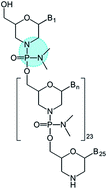
We report the use of a phosphorodiamidate morpholino oligomer as a DNA analogue to template the assembly of a gold nanocluster possessing 6 Au atoms and a diameter of ∼0.9 nm. Spectroscopic characterization indicates that Au preferentially binds to the phosphorodiamidate backbone as opposed to the nucleobases. A high fraction of Au(I) relative to Au(0) is observed by X-ray photoelectron spectroscopy and electrochemistry. This new nanocluster was co-assembled with carbon nanotubes and a fuel-cell enzyme, bilirubin oxidase. The hybrid bio-nanoassembly can reduce O2 to H2O without changing the overpotential of the enzyme.


Introduction

Metal nanoclusters (NCs) having a diameter of <2 nm and consisting of ∼2–144 atoms assembled in well-defined geometric and electronic states are showing unique applications in many areas, including catalysis, sensing, detection, photovoltaics and luminescence.1 Owing to the small size of NCs, their electronic and redox states become discrete just like molecular complexes. Combined with specific assembly of atoms,2 these features lead to manifestation of size-dependent properties of NCs.3

Ligands play an important role in the synthesis, stability and assembly of NCs.4 Although more complex than small ligand molecules in monolayer protected NCs, DNA has increasingly been used as a ligand in the synthesis of AgNCs,5 AuNCs,6 CuNCs,7 and PtNCs8 yielding nanoclusters with interesting optical and catalytic properties. As a natural nanoscale material with precise base-pairing rules and high affinity for metal ions, DNA is an attractive choice of ligand for the assembly of nanoparticles (NPs) and NCs into well-defined and stable structures.9

Phosphorodiamidate morpholino oligomers (PMO) are analogues of DNA where the negatively charged phosphate backbone of DNA is replaced with a charge neutral phosphorodiamidate backbone (Fig. 1). Morpholino antisense oligomers are used as research tools to selectively modify cellular activities.10 Exploiting this role of morpholino oligomers, various cellular processes can be blocked including translation, splicing, and ribozyme activity.10 Recently, Bao et al. have reported new luminescent AuNCs using small molecules containing morpholino rings as ligands.11 Motivated by this finding, we questioned whether morpholino oligomers, as analogues of DNA, could be used as ligands in the synthesis of AuNCs. Such morpholino oligomers present the advantage of having morpholino as the ligand for Au binding, as well as the DNA bases for assembly. We also questioned if the additional P,N sites of the phosphorodiamidate morpholine backbone could act as Au binding sites for nanocluster formation (vs. the traditional use of nucleobases as ligands within DNA templated nanoclusters) and whether morpholino-protected AuNC could be assembled with carbon support and enzymes for potential use in hybrid biofuel cells.6c As a result, here we report the synthesis and characterization of a AuNC protected by this PMO ligand (see Fig. S1 for full structure showing nucleobases), as well as its co-assembly with single walled carbon nanotubes (SWNTs) and a multicopper oxidase.


image file: c6ra16891d-f1.tif
Fig. 1 Chemical structure of phosphorodiamidate morpholino oligomer (PMO) with the Au-binding pocket shown in the cyan dotted circle. B represents nucleobase.

Results and discussion

The synthesis of PMO-templated gold nanocluster (PMO–AuNC) was performed similar to that of DNA-templated AuNC (see materials and methods for details). To investigate whether any secondary structure change of PMO was occurring upon cluster formation, we used circular dichroism (CD) spectroscopy. PMO alone showed four primary CD bands (Fig. 2, red curve), with two positive bands at λmax = 274 and 221 nm, and two negative bands at λmin = 240 and 212 nm, respectively. Although the exact nature of the secondary structure adopted by PMO could not be determined, in DNA the presence of these four bands is characteristic of well-folded structures.12 Upon cluster formation, a red shift in the spectral features are observed with λmax = 278 and 225 nm and with λmin = 214 and 246 nm, respectively (Fig. 2, blue curve). These spectral changes suggest that the overall well-folded secondary structure of the PMO ligand is maintained within the PMO-cluster, with spectral shifts reflecting some conformational changes in the base stacking interactions upon cluster formation. Similar red shifts have been observed upon formation of DNA-templated gold6c and silver nanoclusters.5d
image file: c6ra16891d-f2.tif
Fig. 2 Formation of PMO–AuNC causes secondary structural changes in the morpholino oligomer. CD spectra of 67 μM PMO (red trace) and PMO–AuNC (blue trace) showing changes in the secondary structure.

We analyzed the cluster size by TEM. The micrograph of PMO–AuNC (Fig. 3) shows multiple small clusters with an average diameter of ∼0.9 nm, indicating that the Au material is a true nanocluster in nature, and not large plasmonic particles (>2 nm in diameter). Although some polydispersity is observed in the TEM micrograph, this observation is a common phenomenon due to sintering caused by the electron beam in TEM imaging of such small gold clusters.13


image file: c6ra16891d-f3.tif
Fig. 3 TEM micrograph of PMO–AuNC showing the clusters with a diameter of ∼0.9 nm.

To determine the number of Au atoms in the PMO–AuNC, we employed MALDI mass spectrometry. After screening several MALDI matrices (see materials and methods), satisfactory results were obtained with 3-indoleacrylic acid (IAA) as the matrix in negative ionization mode without observing laser-induced fragmentation of the sample. The molecular mass of PMO–AuNC was obtained at m/z 9406 Da (Fig. S2), suggesting that the PMO–AuNC is a 6-atom Au cluster protected by a single morpholino oligomer (the molecular weight of PMO is 8328 Da). Energy dispersive X-ray (EDX) spectroscopy was also used to estimate the cluster size (Fig. S3). The atomic percentages of P and Au were obtained from the P Kα (2.013 keV) and Au Lα (9.712 keV) lines, which were used to find the relative ratio of Au to PMO. From this analysis, the number of Au atoms present in the PMO–AuNC was also calculated to be 6 (see materials and methods). This assignment was also corroborated (see materials and methods) by X-ray photoelectron spectroscopy (XPS). Taken together, the data indicate that the PMO–AuNC is a 6-atom nanocluster of ∼0.9 nm in diameter.

X-ray photoelectron spectroscopy (XPS) was used to probe the oxidation state of Au in the nanocluster. The Au 4f XPS spectrum of PMO–AuNC (Fig. 4, black line) shows two intense peaks at 84.6 and 88.3 eV corresponding to the Au 4f7/2 and Au 4f5/2 components, respectively. Spectral analysis (Fig. 4, green line) yielded a Au(0) species at 83.3 eV (Fig. 4, blue line; 4f7/2) and a Au(I) species at 84.6 eV (Fig. 4, red line; 4f7/2) with a relative population of the two species of 6% and 94%, respectively. A high fraction of Au(I) oxidation state has been observed in DNA-templated,6c as well as glutathione-protected AuNC.14


image file: c6ra16891d-f4.tif
Fig. 4 The PMO–AuNC is a cluster with primarily Au(I) oxidation state (∼94%). XPS spectra of PMO–AuNC showing overall spectral envelope (black trace), fitting (green trace), deconvolution of the Au 4f7/2 Au(0) (blue trace) and Au(I) (red trace) components at 83.3 eV and 84.6 eV, respectively. The corresponding Au 4f5/2 components are shown as grey solid lines.

We next attempted to characterize the Au–PMO binding site. In addition to the nucleobases, where silver and gold are ligated in DNA-templated nanoclusters,5d,6c,15 and phosphorus, PMO has additional nitrogen atoms in both the morpholino ring and the N(CH3)2 moiety of the backbone. Therefore, we used XPS and FTIR spectroscopy to distinguish between peripheral nucleobases and the phosphorodiamidate as potential Au binding sites. First, the P 2p XPS spectrum of PMO alone shows a single species with a peak at 133.6 eV (Fig. S4, red curve) corresponding to the P 2p3/2 component of the phosphorodiamidate backbone. When the cluster is formed, a new P 2p3/2 species appears at 132.7 eV (Fig. S4, blue curve). The decrease in binding energy suggests weakening of the P–N bonds of phosphorodiamidate. Upon inspection of the N 1s spectrum of PMO alone, we found two species at 393.3 and 400.7 eV (Fig. S5) corresponding to the amine and amide groups in the ligand. In PMO–AuNC, a new peak at 402.4 eV (Fig. S5, magenta curve) appears. This shift of the binding energy to higher energy suggests a reduced electron density at the N site. This observation is consistent with Au binding to the N atoms in the PMO pocket (Fig. 1) whose electron donation to Au(I) lowers their electron density and causes the upshift in the XPS binding energy. Therefore, it is unlikely that metal binding involves the peripheral nucleobases, which are too remote to cause the combined P and N spectral changes associated with the phosphorodiamidate backbone.

Analysis by FTIR spectroscopy provided further evidence for Au binding within the phosphorodiamidate moiety. In the absence of Au, PMO shows the characteristic bands for the asymmetric P–O–C stretch at 1000 cm−1, P–N stretch at 1107 cm−1, asymmetric P[double bond, length as m-dash]O stretch at 1214 cm−1, N(CH3)2 at 1386 cm−1, and ring stretching (C[double bond, length as m-dash]O, C[double bond, length as m-dash]N) of the nucleobases in the region of 1455–1693 cm−1 (Fig. 5, blue trace).16 The spectrum of PMO–AuNC shows very similar features (Fig. 5, red trace), but with changes observed in the regions corresponding to P–N and N(CH3)2 vibrations. In particular, the peak for the P–N stretch is observed at 1090 cm−1 in PMO–AuNC (Fig. 5, dotted line b), which is a downshift of 17 cm−1 from the corresponding peak at 1107 cm−1 for PMO alone (Fig. 5, dotted line a). Additionally, the N(CH3)2 peak also shifts by 22 cm−1, from 1386 cm−1 (Fig. 5, dotted line c) to 1364 cm−1 (Fig. 5, dotted line d) upon cluster formation. In both cases, a fraction of the original peak remains in the cluster, indicating the presence of a small amount of residual uncomplexed oligomer. Since no noticeable shifts in the ring vibrations of the nucleobases were observed upon cluster formation, these observations also suggest that the Au–N binding occurs within the phosphorodiamidate pocket of PMO (Fig. 1). This binding mode contrasts that of metal–DNA nanoclusters, in which metal ions bind to DNA through the nucleobases.5d,6c,15 By using PMO as a DNA replacement, the electron-rich chemical environment in the phosphorodiamidate backbone presumably favors this metal binding mode compared to that involving the nucleobases.


image file: c6ra16891d-f5.tif
Fig. 5 Au binds to the phosphorodiamidate backbone. FTIR spectra of PMO (blue trace) and PMO–AuNC (red trace) showing shifts of the P–N stretching [1107 cm−1 (line a) → 1090 cm−1 (line b)], and N(CH3)2 vibrations [1386 cm−1 (line c) → 1364 cm−1 (line d)] upon cluster formation.

We studied the electrochemical properties of PMO–AuNC using cyclic voltammetry (CV) and differential pulse voltammetry (DPV). CV shows two broad features (Fig. 6, purple trace), while DPV shows a broad peak (Fig. 6, black trace) indicating coalescence of the two closely spaced electrochemical processes observed by CV. Deconvolution of the DPV trace (Fig. 6, red and blue dotted lines) yielded two peaks at −118 and 5 mV (vs. Ag/AgCl). While many thiolate-protected AuNCs [e.g. Au25(SC6)18, Au38(SC2H4Ph)24, Au67(SR)35] show multiple DPV peaks corresponding to sequential one electron oxidation of various charge states of the cluster,17 we assign the two peaks observed here as two stepwise Au(0)/Au(I) processes in the cluster.6c Compared to our previous report on a DNA-protected AuNC, where two analogous oxidations were observed at 155 and 210 mV,6c the relatively more negative potentials observed here indicate that the PMO–AuNC can undergo oxidation more easily. This is consistent with the chemical environment of the phosphorodiamidate backbone which yields the higher content of Au(I) observed in the Au 4f XPS of PMO–AuNC compared to the DNA-protected AuNC.


image file: c6ra16891d-f6.tif
Fig. 6 Redox potentials of PMO–AuNC in 20 mM phosphate buffer solution at pH 7. Broad features are observed in CV (purple line) and DPV (black line). Deconvolution of the DPV peak shows two processes at approximately −118 mV (blue line) and 5 mV (red line). The overall peak fitting is also shown (green line).

Finally, inspired by our previous findings for the DNA-protected AuNC,6c we tested whether the PMO–AuNC can also be co-assembled with enzymes and carbon support materials for preparing cathodes in enzymatic fuel cells. In a procedure similar to that used for the DNA–AuNC,6c we used the multi-copper bilirubin oxidase (BOD) from Myrothecium verrucaria as the enzyme of choice because BOD is a widely used fuel cell enzyme for oxygen reduction reaction (ORR).18 First, we dispersed the PMO–AuNC with single-walled carbon nanotubes (SWNT) and tetrabutylammonium bromide (TBAB). While the TBAB modification lowers the acidity of Nafion, the nucleobases of PMO–AuNC allows for stacking by π–π interaction with the SWNT. In a final step, BOD was covalently functionalized on PMO–AuNC/SWNT using succinimidyl ester–amine cross-linking chemistry, where the surface exposed Lys groups of BOD covalently attach to the succinimidyl ester group of 1-pyrenebutanoic acid succinimidyl ester (PBSE) while the pyrene group of PBSE stacks by non-covalent π–π interaction with the SWNT. The final composites consisting of (1) PMO–AuNC, SWNT and BOD (BOD–PMO–AuNC/SWNT), (2) plasmonic Au particles (PMO–AuNP), SWNT and BOD (BOD–PMO–AuNP/SWNT), (3) PMO, SWNT and BOD (BOD-PMO/SWNT), and (4) SWNT and BOD (BOD/SWNT) were then drop cast on a rotating disk electrode, air dried, and their electrochemical performance for ORR was performed using linear sweep voltammetry (LSV).

We tested the ORR activity of BOD–PMO–AuNC/SWNT, BOD–PMO–AuNP/SWNT and BOD-PMO/SWNT and compared the onset potential (Eonset) and electrocatalytic current density with the enzyme alone (BOD/SWNT) in O2-saturated buffer solution (100 mM phosphate buffer; pH 7.5). The BOD/SWNT (Fig. 7, black trace) shows Eonset of 542 mV vs. Ag/AgCl and catalytic current density of −406 μA cm−2 at −100 mV, while the BOD–PMO/SWNT composite shows a lower Eonset of 529 mV and significantly diminished current density of −76 μA cm−2 (Fig. 7, blue trace), suggesting that PMO hinders both the kinetics and thermodynamics of enzymatic ORR catalyzed by BOD. Next, the BOD–PMO–AuNC/SWNT composite shows a very similar Eonset of 541 mV compared to that of BOD alone and a lower current density of −276 μA cm−2 (Fig. 7, red curve). Similar value of onset potential in the presence of PMO–AuNC suggests that the presence of the cluster is not affecting the thermodynamics of enzymatic ORR, while the lower current density suggests that the presence of cluster is impeding the ORR kinetics by lowering the efficiency of electron transfer (ET) from the electrode to the enzyme active site. In contrast to the DNA–AuNC reported previously,6c which significantly lowered the overpotential and improved the kinetics of ORR, our results here show that the presence of PMO–AuNC does not improve the overpotential and kinetics of ORR. The oxidizing nature of the PMO–AuNC compared to DNA-protected AuNC may hinder ET, leading to unchanged Eonset and poor kinetics. Finally, the BOD–PMO–AuNP/SWNT composite shows an Eonset of 553 mV, an increase of 11 mV from the enzyme alone, and a current density of −392 μA cm−2 (Fig. 7, purple trace). We then tested whether the product of ORR is formed from 4-e reduction (H2O) or 2-e reduction (H2O2) via rotating ring disk electrode (RRDE) experiments. The BOD/SWNT, BOD–PMO–AuNC/SWNT and BOD–PMO–AuNP/SWNT composites showed minimal production of H2O2, with 6–9% H2O2 at midpoint potentials and 1–2% H2O2 at −100 mV (Fig. S6). This result shows that, although not as effective as the DNA–AuNC for facilitated ORR, these PMO–AuNC composites are selective toward the 4-e reduction of O2 to H2O.


image file: c6ra16891d-f7.tif
Fig. 7 LSV showing O2 reduction for the composites BOD/SWNT (black trace), BOD–PMO/SWNT (blue trace), BOD–PMO–AuNP/SWNT (purple trace) and BOD–PMO–AuNC/SWNT (red trace) measured in O2-saturated 100 mM phosphate buffer (pH 7.5) with electrode rotation rate of 800 rpm and scan rate of 10 mV s−1. Shaded regions represent standard deviations from three independent measurements. The data were collected against Ag/AgCl and converted to RHE [ERHE = EAg/AgCl + (0.059 × pH) + E0Ag/AgCl; where E0Ag/AgCl = 0.197 V].

Conclusion

Here we presented the successful synthesis and characterization of a 6-atom gold nanocluster templated by the DNA analogue phosphorodiamidate morpholino oligomer. Combined spectroscopic data indicate a preferential Au binding mode involving the phosphorodiamidate pocket of the PMO backbone as opposed to the peripheral nucleobases. Compared to DNA–AuNC, the PMO–AuNC is more easily oxidized. When the PMO–AuNC is co-assembled with SWNT and a multi-copper oxidase enzyme the PMO–AuNC does not improve the thermodynamics and kinetics of enzymatic ORR, unlike the DNA–AuNC. Several factors, including the high fraction of Au(I), low redox potential and the ligand nature may have contributed to the lack of electrocatalytic enhancement by PMO–AuNC. Learning from these principles is important to the design of better ET catalysts for efficient enzymatic fuel cells.

Experimental section

Synthesis and purification of PMO–AuNC

15 μM of PMO (Gene Tools LLC) CCTCTTACCTCAGTTACAATTTATA was incubated with 225 μM HAuCl4·3H2O (Sigma-Aldrich, >99% trace metal basis) in 20 mM phosphate buffer at pH 7, 1 mM Mg(OAc)2, for 12 h at 22 °C. The initially colorless solution turned light yellow upon incubation. Reduction was performed for 12 h at 22 °C with addition of 2.25 mM dimethylamine borane (Sigma-Aldrich). A light purple solution was obtained upon completion of reduction indicating the presence of plasmonic Au particles. The solution was then centrifuged with 30 kDa MWCO membranes (Millipore) to separate plasmonic Au particles from nanoclusters. The large particles were retained in the membrane, while the nanoclusters were obtained as a yellow filtrate, which was concentrated with 10 kDa MWCO membranes and stored at 4 °C until further use. A molar extinction coefficient (ε260) of 2.59 × 10⁵ M−1 cm−1 as provided by Gene Tools LLC, was used to determine the concentrations of PMO in respective samples.

CD spectroscopy

Three scans of CD data were collected for each sample at 22 °C on a JASCO spectrometer using a quartz cuvette with a path length of 1 mm.

Transmission electron microscopy

TEM images of PMO–AuNC sample were collected at 200 keV acceleration voltage using a FEI Tecnai F30 instrument. Approximately 1 μL of an as-prepared PMO–AuNC sample was drop-cast on Cu grids coated with a < 10 nm thin carbon film (Pacific Grid-Tech, 300 mesh, OD: 3.05 mm, hole size: ∼63 μm) and air-dried before measurements.

MALDI mass spectrometry

MS spectra of PMO–AuNC (prepared at 10[thin space (1/6-em)]:[thin space (1/6-em)]1 matrix to sample ratio) were collected on a ABSciex 4800 Plus TOF/TOF MALDI spectrometer equipped with a 200 Hz frequency Nd:YAG laser, and operating at a wavelength of 355 nm. The spectrometer was externally calibrated with a TOF/TOF calibration peptide mixture of des-Arg–bradykinin (1.0 pmol μL−1), angiotensin I (2.0 pmol μL−1), glu-fibrinopeptide B (1.3 pmol μL−1), and adrenocorticotropic hormone (1–17 clip-2.0 pmol μL−1), (18–39 clip-1.5 pmol μL−1), (7–38 clip-3.0 pmol μL−1). The following matrices were tested in both positive and negative mode of ionization: 2,5 dihydroxy benzoic acid, 3-hydroxy picolinic acid, α-cyano-4-hydroxy cinnamic acid, and 3-indoleacrylic acid. The 3-indoleacrylic acid matrix gave satisfactory results, without fragmentation of the cluster.

Energy dispersive X-ray spectroscopy

EDX spectra were collected at 30 kV acceleration voltage using a FEI Quanta 400 FEG-E-SEM instrument equipped with an EDX system (EDAX Inc.). Data processing was done using Genesis software. For EDX analysis, the PMO–AuNC was synthesized in 50 mM NH4OAc (pH 6.9), 1 mM Mg(OAc)2 buffer to avoid error in calculating atomic% of P 2p from phosphate buffer. The atomic% of P 2p (79.5) using P Kα (2.013 keV) obtained from PMO–AuNC was divided by the number of phosphorodiamidate groups (24) in PMO so as to obtain a normalization factor 3.3 (79.5/24) corresponding to a single piece of PMO. The atomic% of Au (20.4) from Lα (9.712 keV) was then used against the normalization factor to obtain the total number of Au atoms bound to a single morpholino oligomer. The number of Au atoms obtained from this analysis is 6.2 ± 0.2.

X-ray photoelectron spectroscopy

PMO–AuNC samples were drop cast on mica surface. A Kratos Axis Ultra DLD spectrometer was used with a monochromatic Al Kα source operating at 225 W and standard operating conditions of charge neutralizer (bias voltage of 3.1 V, filament voltage of −1.0 V, and filament current of 2.1) for charge compensation. Three different areas on the samples were analyzed. Survey and high resolution C 1s, O 1s, N 1s, S 2p, P 2p and Au 4f spectra were acquired at 80 and 20 eV pass energy, respectively. Data analysis and quantification were performed using the CasaXPS software. A linear background was used for C 1s, N 1s, O 1s, P 2p and S 2p and Shirley background for Au 4f spectra. Quantification utilized sensitivity factors that were provided by the manufacturer. All the spectra were charge referenced to the C 1s at 284.7 eV. A 70% Gaussian/30% Lorentzian (GL (30)) line shape was used for the curve fittings.

Atomic% of P (3.5%) obtained from P 2p XPS spectrum of PMO–AuNC was normalized to one PMO ligand, yielding a normalization factor of 0.14 (3.5/24). This normalization factor was then divided by the atomic% of Au (0.9%) to obtain the number of Au atoms in the PMO–AuNC. From this analysis the number of Au atoms in PMO–AuNC was calculated to be 6.1 ± 0.2, indicating the PMO–AuNC is a 6 atom cluster.

Fourier-transform infrared spectroscopy

IR absorption spectra were obtained with a Bruker Equinox 55 FTIR instrument at a nominal resolution of 2 cm−1 and an average of 16 scans. The spectra were collected using dry films from aqueous solution samples drop cast and vacuum dried on polyethylene IR cards.

Electrochemistry

Cyclic voltammetry (CV) and differential pulse voltammetry (DPV) experiments were performed on a CH Instruments CHI760E potentiostat equipped with a three-electrode system using a glassy carbon working electrode (disk diameter = 3 mm), Pt wire as counter electrode and Ag/AgCl as reference electrode. In CV, six full scans were run at a scan rate of 50 mV s−1. DPV scans were run with a pulse period of 250 ms, pulse width of 25 ms, amplitude of 25 mV, and increment of 2 mV. Solutions containing approximately 200 μL of M-AuNC in 20 mM phosphate buffer at pH 7, 1 mM Mg(OAc)2 were deoxygenated with Ar in an electrochemical cell (BASi) prior to measurements.

Preparation of composite materials for ORR experiments

A suspension of 0.17% SWNT (Raymor) in 0.3[thin space (1/6-em)]:[thin space (1/6-em)]1 methanol[thin space (1/6-em)]:[thin space (1/6-em)]water solution was prepared and bath sonicated with 0.27% tetrabutylammonium bromide (TBAB)-modified Nafion (provided by Prof. Shelley Minteer, University of Utah) in absolute ethanol for 30 min at room temperature (RT) to disperse the SWNT. A PMO–AuNC solution (5 μL) was incubated with 40 μL of the TBAB-Nafion/SWNT suspension for 1 h at RT to allow for stacking of the nucleobases of PMO to the SWNT by π–π stacking. Next, 2 μL (4 mg mL−1) of 1-pyrenebutanoic acid succinimidyl ester (PBSE, Sigma-Aldrich) suspended in ethanol was added to TBAB-Nafion/PMO-AuNC/SWNT and incubated for additional 1 h at RT to allow for adsorption of PBSE on the SWNT again by π–π stacking between the SWNT and the pyrene ring of PBSE. Subsequently, 2 μL of a 200 mg mL−1 BOD (Amano Enzyme Inc.) solution in 100 mM phosphate buffer at pH 7.5 was added and the composite mixture was further incubated for 16–18 h at 4 °C to conjugate the PBSE with surface Lys groups of BOD via succinimidyl ester–amine chemistry. Control composites were prepared using the same procedure.

Preparation of modified electrodes for ORR measurements

A glassy carbon rotating disk electrode (RDE) (disk area of 0.2475 cm2, Pine Instruments) was cleaned with alumina of increasingly fine grits of 1, 0.3, and 0.05 mm, and rinsed with deionized water. A SWNT/TBAB–Nafion suspension (10 μL) was drop cast on the electrode surface and dried under a flow of N2 gas. Next, 10 μL of the composite materials prepared as described above, (1) PMO–AuNC, SWNT and BOD (BOD–PMO–AuNC/SWNT), (2) plasmonic Au particles (PMO–AuNP), SWNT and BOD (BOD–PMO–AuNP/SWNT), (3) PMO, SWNT and BOD (BOD-PMO/SWNT), and (4) SWNT and BOD (BOD/SWNT) were drop cast on the RDE and allowed to air-dry before experiments. The modified electrodes were then used for ORR experiments.

ORR catalysis

ORR measurements were performed with a WEB30 Pine bi-potentiostat and a rotator (Pine Instruments) using a three-electrode setup (glassy carbon working electrode, Pt wire auxiliary electrode, Ag/AgCl reference electrode) with O2-saturated 100 mM phosphate buffer solution at pH 7.5 as the electrolyte. Open circuit potential was allowed to reach equilibrium before ORR measurements. For linear sweep voltammetry (LSV) experiments, the disk potential was swept from 0.8 to 0 V at a scan rate of 10 mV s−1. At least three sets of independent ORR data were collected from three different preparations of composite samples. Conversion from Ag/AgCl to RHE potential was performed using the expression:19 ERHE = EAg/AgCl + (0.059 × pH) + 0.197.

Mass and charge balance analysis

For the mass and charge balance analysis using rotating ring disk electrode (RRDE) the disk current was swept from 0.8 to 0 V at a scan rate of 10 mV s−1 while the ring was held at a constant potential of 0.8 V. Data analysis was performed according to eqn (1).
 
image file: c6ra16891d-t1.tif(1)
where n is the number of electrons consumed in O2 reduction, iR is the ring current, iD is the disk current, and η is the collection efficiency at the electrode.20 For RRDE, the collection efficiency is known to be 37%.

Acknowledgements

We acknowledge financial support by the Laboratory Directed Research and Development program at LANL (A. D.), the Basic Energy Sciences, Biomolecular Materials Program, Division of Materials Science & Engineering (S. C., R. C. R., J. S. M.). P. A. acknowledges the Air Force Office of Scientific Research (grant FA9550-12-1-0112) and ARO-Multi-University Research Initiative (grant W911NF-14-1-0263 to the University of Utah). This work was performed, in part, at the Center for Integrated Nanotechnologies (CINT), a User Facility operated for the U.S. Department of Energy (DOE), Office of Science. Los Alamos National Laboratory is operated by Los Alamos National Security, LLC, for the National Nuclear Security Administration of the U.S. DOE under contract DE-AC52-06NA25396. The authors thank Dr Darrick Williams (CINT) for assistance with EDX data collection.

References

  1. (a) L. Shang, S. Dong and G. U. Nienhaus, Nano Today, 2011, 6, 401–418 CrossRef CAS; (b) C.-A. J. Lin, C.-H. Lee, J.-T. Hsieh, H.-H. Wang, J. K. Li, J.-L. Shen, W.-H. Chan, H.-I. Yeh and W. H. Chang, J. Med. Biol. Eng., 2009, 29, 276–283 Search PubMed; (c) G. Li and R. Jin, Acc. Chem. Res., 2013, 46, 1749–1758 CrossRef CAS PubMed; (d) W. S. Compel, O. A. Wong, X. Chen, C. Yi, R. Geiss, H. Häkkinen, K. L. Knappenberger and C. J. Ackerson, ACS Nano, 2015, 9, 11690–11698 CrossRef CAS PubMed; (e) R. Ho-Wu, S. H. Yau and T. Goodson, ACS Nano, 2016, 10, 562–572 CrossRef CAS PubMed; (f) Y. Liu, Q. Yao, X. Wu, T. Chen, Y. Ma, C. N. Ong and J. Xie, Nanoscale, 2016, 8, 10145–10151 RSC; (g) Y.-T. Wu, C. Shanmugam, W.-B. Tseng, M.-M. Hiseh and W.-L. Tseng, Nanoscale, 2016, 8, 11210–11216 RSC.
  2. (a) A. Das, C. Liu, H. Y. Byun, K. Nobusada, S. Zhao, N. Rosi and R. Jin, Angew. Chem., Int. Ed., 2015 Search PubMed; (b) R. Jin, C. Liu, S. Zhao, A. Das, H. Xing, C. Gayathri, Y. Xing, N. L. Rosi, R. R. Gil and R. Jin, ACS Nano, 2015, 9, 8530–8536 CrossRef CAS PubMed; (c) C. Liu, T. Li, G. Li, K. Nobusada, C. Zeng, G. Pang, N. L. Rosi and R. Jin, Angew. Chem., Int. Ed., 2015, 54, 9826–9829 CrossRef CAS PubMed; (d) S. Yamazoe, S. Takano, W. Kurashige, T. Yokoyama, K. Nitta, Y. Negishi and T. Tsukuda, Nat. Commun., 2016, 7, 10414 CrossRef CAS PubMed; (e) A. Dass, S. Theivendran, P. R. Nimmala, C. Kumara, V. R. Jupally, A. Fortunelli, L. Sementa, G. Barcaro, X. Zuo and B. C. Noll, J. Am. Chem. Soc., 2015, 137, 4610–4613 CrossRef CAS PubMed; (f) A. Dass, T. Jones, M. Rambukwella, D. Crasto, K. J. Gagnon, L. Sementa, M. De Vetta, O. Baseggio, E. Aprà and M. Stener, J. Phys. Chem. C, 2016, 120, 6256–6261 CrossRef CAS.
  3. (a) R. W. Murray, Chem. Rev., 2008, 108, 2688–2720 CrossRef CAS PubMed; (b) P. D. Jadzinsky, G. Calero, C. J. Ackerson, D. A. Bushnell and R. D. Kornberg, Science, 2007, 318, 430–433 CrossRef CAS PubMed; (c) M. Walter, J. Akola, O. Lopez-Acevedo, P. D. Jadzinsky, G. Calero, C. J. Ackerson, R. L. Whetten, H. Grönbeck and H. Häkkinen, Proc. Natl. Acad. Sci. U. S. A., 2008, 105, 9157–9162 CrossRef CAS PubMed; (d) Y. Negishi, K. Nobusada and T. Tsukuda, J. Am. Chem. Soc., 2005, 127, 5261–5270 CrossRef CAS PubMed; (e) D. R. Kauffman, D. Alfonso, C. Matranga, H. Qian and R. Jin, J. Am. Chem. Soc., 2012, 134, 10237–10243 CrossRef CAS PubMed.
  4. (a) Z. Wu and R. Jin, Nano Lett., 2010, 10, 2568–2573 CrossRef CAS PubMed; (b) J. Xie, Y. Zheng and J. Y. Ying, J. Am. Chem. Soc., 2009, 31, 888–889 CrossRef PubMed.
  5. (a) J. Sharma, H. C. Yeh, H. Yoo, J. H. Werner and J. S. Martinez, Chem. Commun., 2010, 46, 3280–3282 RSC; (b) H. C. Yeh, J. Sharma, J. J. Han, J. S. Martinez and J. H. Werner, Nano Lett., 2010, 10, 3106–3110 CrossRef CAS PubMed; (c) J. Sharma, R. C. Rocha, M. L. Phipps, H.-C. Yeh, K. A. Balatsky, D. M. Vu, A. P. Shreve, J. H. Werner and J. S. Martinez, Nanoscale, 2012, 4, 4107–4110 RSC; (d) J. T. Petty, J. Zheng, N. V. Hud and R. M. Dickson, J. Am. Chem. Soc., 2004, 126, 5207–5212 CrossRef CAS PubMed; (e) E. G. Gwinn, P. O'Neill, A. J. Guerrero, D. Bouwmeester and D. K. Fygenson, Adv. Mater., 2008, 20, 279–283 CrossRef CAS.
  6. (a) T. A. Kennedy, J. L. MacLean and J. Liu, Chem. Commun., 2012, 48, 6845–6847 RSC; (b) G. Liu, Y. Shao, K. Ma, Q. Cui, F. Wu and S. Xu, Gold Bull., 2012, 45, 69–74 CrossRef CAS; (c) S. Chakraborty, S. Babanova, R. C. Rocha, A. Desireddy, K. Artyushkova, A. E. Boncella, P. Atanassov and J. S. Martinez, J. Am. Chem. Soc., 2015, 137, 11678–11687 CrossRef CAS PubMed; (d) Y. Chen, G. Tao, R. Lin, X. Pei, F. Liu and N. Li, Chem.–Asian J., 2016, 11, 1677–1681 CrossRef CAS PubMed.
  7. X. Jia, J. Li, L. Han, J. Ren, X. Yang and E. Wang, ACS Nano, 2012, 6, 3311–3317 CrossRef CAS PubMed.
  8. (a) R. Seidel, L. Colombi Ciacchi, M. Weigel, W. Pompe and M. Mertig, J. Phys. Chem. B, 2004, 108, 10801–10811 CrossRef CAS; (b) J. N. Tiwari, K. Nath, S. Kumar, R. N. Tiwari, K. C. Kemp, N. H. Le, D. H. Youn, J. S. Lee and K. S. Kim, Nat. Commun., 2013, 4, 2221 Search PubMed.
  9. M. Mertig, L. Colombi Ciacchi, R. Seidel, W. Pompe and A. De Vita, Nano Lett., 2002, 2, 841–844 CrossRef CAS.
  10. J. Summerton, J. Drug. Discov. Dev. Deliv., 2016, 3, 1019 Search PubMed.
  11. Y. Bao, H.-C. Yeh, C. Zhong, S. A. Ivanov, J. K. Sharma, M. L. Neidig, D. M. Vu, A. P. Shreve, B. S. Dyer, J. H. Werner and J. S. Martinez, J. Phys. Chem. C, 2010, 114, 15879–15882 CAS.
  12. J. Kypr, I. Kejnovská, D. Renčiuk and M. Vorlíčková, Nucleic Acids Res., 2009, 37, 1713–1725 CrossRef CAS PubMed.
  13. (a) Y. Levi-Kalisman, P. D. Jadzinsky, N. Kalisman, H. Tsunoyama, T. Tsukuda, D. A. Bushnell and R. D. Kornberg, J. Am. Chem. Soc., 2011, 133, 2976–2982 CrossRef CAS PubMed; (b) P. Ramasamy, S. Guha, E. S. Shibu, T. S. Sreeprasad, S. Bag, A. Banerjee and T. Pradeep, J. Mater. Chem., 2009, 19, 8456–8462 RSC.
  14. Z. Luo, X. Yuan, Y. Yu, Q. Zhang, D. T. Leong, J. Y. Lee and J. Xie, J. Am. Chem. Soc., 2012, 134, 16662–16670 CrossRef CAS PubMed.
  15. (a) M. L. Neidig, J. Sharma, H.-C. Yeh, J. S. Martinez, S. D. Conradson and A. P. Shreve, J. Am. Chem. Soc., 2011, 133, 11837–11839 CrossRef CAS PubMed; (b) B. Sengupta, C. M. Ritchie, J. G. Buckman, K. R. Johnsen, P. M. Goodwin and J. T. Petty, J. Phys. Chem. C, 2008, 112, 18776–18782 CrossRef CAS; (c) J. Duguid, V. A. Bloomfield, J. Benevides and G. J. Thomas, Biophys. J., 1993, 65, 1916–1928 CrossRef CAS PubMed.
  16. (a) E. Taillandier and J. Liquier, Methods Enzymol., 1991, 211, 307–335 Search PubMed; (b) A. A. Ouameur and H.-A. Tajmir-Riahi, J. Biol. Chem., 2004, 279, 42041–42054 CrossRef CAS PubMed; (c) N. Tercero, K. Wang, P. Gong and R. Levicky, J. Am. Chem. Soc., 2009, 131, 4953–4961 CrossRef CAS PubMed; (d) G. Socrates, Infrared Characteristic Frequencies, Wiley&Sons, 2nd edn, 1994 Search PubMed; (e) M. Bolboaca, T. Stey, A. Murso, D. Stalke and W. Kiefer, Appl. Spectrosc., 2003, 57, 970–976 CrossRef CAS PubMed.
  17. (a) D. Garcia-Raya, R. Madueno, M. Blazquez and T. Pineda, J. Phys. Chem. C, 2009, 113, 8756–8761 CrossRef CAS; (b) P. R. Nimmala, B. Yoon, R. L. Whetten, U. Landman and A. Dass, J. Phys. Chem. A, 2013, 117, 504–517 CrossRef CAS PubMed; (c) H. Qian, Y. Zhu and R. Jin, ACS Nano, 2009, 3, 3795–3803 CrossRef CAS PubMed.
  18. (a) D. M. Ivnitski, C. Khripin, H. R. Luckarift, G. R. Johnson and P. Atanassov, Electrochim. Acta, 2010, 55, 7385–7393 CrossRef CAS; (b) J. Liu, S. Chakraborty, P. Hosseinzadeh, Y. Yu, S. Tian, I. Petrik, A. Bhagi and Y. Lu, Chem. Rev., 2014, 114, 4366–4469 CrossRef CAS PubMed.
  19. A. J. Bard and L. R. Faulkner, Electrochemical methods: fundamentals and applications, Wiley, New York, 1980 Search PubMed.
  20. (a) K. L. Hsueh, D. T. Chin and S. Srinivasan, J. Electroanal. Chem. Interfacial Electrochem., 1983, 153, 79–95 CrossRef CAS; (b) C. M. A. Brett and A. M. O. Brett, Electrochemistry Principles, Methods and Applications, Oxford University Press Inc., New York, NY, 1993 Search PubMed.

Footnotes

Electronic supplementary information (ESI) available: MALDI-MS, EDX, P 2p and N 1s XPS, H2O2% from RRDE data. See DOI: 10.1039/c6ra16891d
Department of Chemistry and Biochemistry University of Mississippi, University, MS 38677, USA

This journal is © The Royal Society of Chemistry 2016
Click here to see how this site uses Cookies. View our privacy policy here.