DOI:
10.1039/C6RA16848E
(Paper)
RSC Adv., 2016,
6, 94815-94825
Biobased waterborne hyperbranched polyurethane/NiFe2O4@rGO nanocomposite with multi-stimuli responsive shape memory attributes†
Received
30th June 2016
, Accepted 26th September 2016
First published on 28th September 2016
Abstract
A biobased waterborne hyperbranched polyurethane nanocomposite was fabricated with nickel ferrite/reduced graphene oxide nanohybrid (NiFe2O4@rGO) by following an in situ polymerization technique. NiFe2O4@rGO was prepared through a hydrothermal method. The prepared nanohybrid and nanocomposite were characterized by using different spectroscopic and analytical techniques. Various properties of the nanocomposite were also evaluated. The nanocomposite demonstrated excellent thermal stability (initial degradation temperature up to 335 °C) and good mechanical properties (tensile strength 24.23 MPa, elongation at break 280% and toughness 28.35 MJ m−3). The nanocomposite also exhibited outstanding multi-stimuli responsive shape memory behavior under sunlight, thermal and microwave (300 W) irradiation. Both the polymer matrix and nano-filler acted complementarily towards an efficient shape recovery behavior. The performance of the nanocomposite was found to be dependent on the nanomaterial loading. Thus, the present study showed that the developed nanocomposite can be used as a high performing, non-contact triggered smart material for advanced shape memory application.
Introduction
Shape memory polymers (SMP) belong to the “smart” class of materials, which are attracting copious interest for many advanced applications.1 This type of material possesses the ability to switch into a fixed temporary deformed state, which can be switched off to the original shape by using certain stimuli.2 SMPs have found numerous applications in different domains, such as functional textiles, smart customer products, active aircraft equipment, adaptive biomedical devices, interactive electronic apparatus etc.3–6 Among various polymeric materials, polyurethane (PU) has been considered as one of the most promising candidates for shape memory application due to its high recoverable strain, wide range of transition temperatures for shape recovery, high control of retraction and softening temperature etc.7–9 PU consists of soft and hard domains, in which the former and latter constitute the reversible and frozen phase, respectively. The reversible phase is responsible for holding the temporary deformation. On the counter side, the frozen phase, which contains physical or chemical crosslinked points is responsible for memorizing the permanent shape. In real practice, there exists a transition temperature (Ttrans), with respect to which shape fixation and recovery can be achieved.
In conventional SMP, shape recovery is generally achieved by direct heating the polymer above Ttrans. However, in recent time, stress has been given to use indirect heating devices by using non-contact and remote triggers for shape recovery. In this perspective, PU nanocomposites have been studied extensively.10–14 Nanomaterials which respond specifically against certain stimuli can be used to develop such smart shape memory PU nanocomposite. Nanomaterials like carbon nanotube, carbon fiber, graphene, iron oxide, silicon carbide etc. are reportedly utilized in stimuli responsive shape memory polymers using triggers of electrical field, magnetic field, microwave, light etc.15–23 In this regard, graphene based nanomaterials are attractive. Graphene nano-sheets (GNs) are composed of extremely thin 2D honeycomb lattices of carbon atoms. GNs possess ultrahigh mechanical properties and excellent electrical and thermal conductivities. Thus, incorporation of GNs within a PU matrix can bring shape recovery at a lower heating through the employment of remote heating devices like microwave, sunlight or other electromagnetic radiations. However, the major impediment faced in the fabrication of graphene-based PU nanocomposite is the poor compatibility of graphene with PU.24 Hence, in many occasions, covalent modification of graphene has been performed in order to bring a strong interaction between PU and nanomaterial. However, such modification of GN surface typically destroys the graphitic structure and restricts reinforcement of the thermal conductivity of the nanocomposite. Therefore, a more convenient route is desirable, which ensures a strong PU/GNs interaction without disturbing the graphitic structure of GNs.
In this context, fabrication of waterborne hyperbranched polyurethane (WPU) nanocomposite with nickel ferrite/reduced graphene oxide nanohybrid (NiFe2O4@rGO) may form an effective tactic. WPU contains a large number of ionic groups along the polymeric backbone. Thus, the metal system (NiFe2O4) of the nanohybrid can interact with the ionic centers of WPU chains and there by forming a uniformly dispersed NiFe2O4@rGO/WPU nanocomposite system. NiFe2O4 being a magnetic nanomaterial may additionally help to achieve enhanced shape recovery ability by using non-contact mode. At this juncture it is pertinent to mention that the ionic groups of WPU may play a significant role in the shape recovery ability. Because, these groups impart high degree of crystallinity within the hard segments through strong electrostatic and secondary interactions, which can influence the shape memory property.25
Considering these facts, the present study made an attempt to prepare NiFe2O4@rGO nanohybrid by using a hydrothermal method. Different weight percentages of this nanohybrid were used to prepare nanocomposites of WPU by following an in situ technique. Both the nanohybrid and nanocomposite were characterized by using different spectroscopic and analytical techniques. Different properties, such as thermal, mechanical etc. were also evaluated. The developed system was tested as a shape memory material using different external stimuli like microwave, thermal and sunlight.
Experimental
Materials
Isophorone diisocyanate (IPDI, Aldrich, Germany), poly(ethylene glycol) with Mn 600 g mol−1 (PEG 600, Merck, India), 1,4 butane diol (BD, Merck, India), tannic acid (TA, Sigma Aldrich, Belgium), 2,2-bis(hydroxymethyl)propionic acid (BMPA, Aldrich, Germany), triethylamine (TEA, Merck, India) and tetrahydrofuran (THF, Merck, India) were used in the polymer preparation. Glycerol based hyperbranched epoxy (HE) used in the modification of WPU was prepared according to the method described elsewhere.26 Poly(amido amine) (PAA) was procured from Huntsman Corporation, USA with amine value of 5–7 eq. kg−1. On the other hand, graphene powder (Sigma Aldrich, Germany), iron(III)chloride hexahydrate (FeCl3·6H2O, Aldrich, Germany), nickel(II)chloride hexahydrate (NiCl2·6H2O, Sigma Aldrich, Germany), trisodium acetate (Na3C6H5O7, Merck, India), 98% sulphuric acid (H2SO4, Merck, India), potassium permanganate (KMnO4, Merck, India) and 30% hydrogen peroxide (H2O2, Merck, India) were used in the preparation of the nanohybrid.
Characterization
Fourier Transform Infrared (FTIR) spectra of NiFe2O4@rGO nanohybrid and nanocomposite were recorded on a PerkinElmer, USA, Model: Frontier MIR-FIR, spectrometer using KBr pallets. X-ray diffraction (XRD) patterns of the nanohybrid and nanocomposite were obtained by using a Bruker AXS, Germany, Model: D8 Focus instrument (from 2θ = 10–70° at a scanning rate of 2° min−1). Graphitic structure of rGO sheets was studied with the help of Raman spectrometer, Renishaw, UK, Model: Renishaw Basis Series with 514 nm Laser. The shape, size and decoration of the nanohybrid as well as its distribution over the polymer matrix were visually studied by using High Resolution Transmission Electron Microscopic (HRTEM) study. TEM images were obtained from JEOL, JEMCXII, Japan, microscope at an operating voltage of 200 kV using Cu grid of TED PELLA INC, Ultrathin C, Type A, 400 mesh. The microscopic data were analyzed for Fast Fourier Transform (FFT) and Inverse Fast Fourier Transform (IFFT) images by using Gatan Digital Micrograph software. The elemental composition of the nanohybrid was confirmed through Electron Dispersive X-ray (EDX) technique using Scanning Electron Microscope (SEM) of JEOL, Model: JSM 6390LV, Japan. Various mechanical properties like tensile strength and elongation break of the nanocomposite were evaluated by using Universal Testing Machine (UTM, Zwick, Germany), maintaining an operational cross-head speed of 100 mm min−1. For mechanical performance evaluation, polymeric nanocomposites films of dimension 5 cm × 1 cm × 0.04 cm (length × breath × thickness) were used following standard protocol of ASTM D882-12. The preparation of polymeric films is provided in the nanocomposite preparation section. Scratch hardness was measured with a scratch hardness tester (Sheen Instruments, Model no. 705, UK) equipped with stylus accessories and operating at a travel speed of 30 mm min−1. Impact test was performed according to the standard protocol of ASTM D1037-16 a (falling weight method) using impact tester provided by S.C. Dey & Co., India. On the other hand, thermal degradation profile was recorded by thermal gravimetric analyzer (PerkinElmer, USA, Model: TGA 4000) under the inert atmosphere of nitrogen (scan rate of 5 °C min−1, gas flow rate of 30 mL min−1). Similarly, dynamic scanning calorimetric (DSC) measurements were made with the help of PerkinElmer, USA Model: DSC 6000, thermal instrument. All the tests were done under the inert atmosphere of nitrogen at a flow rate of 30 mL min−1 and scanning rate of 10 °C min−1. A cycle of heating–cooling–heating was followed in the temperature range of −60 °C to 120 °C.
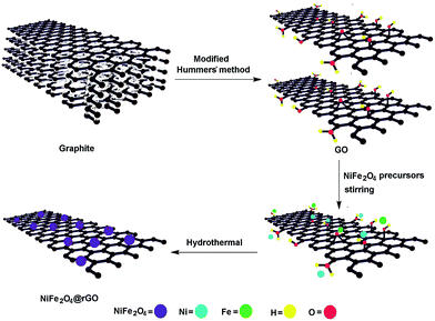
Preparation of NiFe2O4@rGO
Graphene oxide (GO) was prepared by the oxidation of graphitic powder according to modified Hummers' method.27 Briefly, 2 g of graphitic powder was subjected to acid treatment with 35 mL of 98% H2SO4 under constant stirring for 1 h. It was followed by addition of 6 g of KMnO4 to this mixture very slowly and cautiously at temperature below 20 °C. When evolved heat subsides, the mixture was vigorously stirred at 35 °C for 4 h. Then it was diluted with 90 mL of water under cool and stirring condition. After 1 h, a dark brown colored solution was observed. To this solution, 30% H2O2 was added dropwise until the color turned into bright yellow. From this solution, GO was separated by centrifuge technique, which was purified by subsequent washing with 5% HCl solution and water for several cycles. This GO was dried inside an oven. In order to prepare NiFe2O4@rGO nanohybrid, an aqueous dispersion of GO (1.0 g) was prepared by using ultrasonication to which 1.04 g of FeCl3·6H2O and 2.31 g of NiCl2·6H2O were added. 100 mg of trisodium acetate was also added as chelating agent. This mixture was stirred for 1 h and then poured inside a Teflon lined hydrothermal reactor. It was subjected to a temperature of 180 °C for 10 h.28 After the completion of the process, NiFe2O4@rGO nanohybrid was separated by centrifuge, washed with ethanol (to remove trisodium acetate) and water several times and then dried inside an oven at 50 °C.
Fabrication of NiFe2O4@rGO/WPU
NiFe2O4@rGO/WPU nanocomposite was prepared by following an in situ technique. Briefly IPDI, PEG 600 and BMPA were allowed to react under nitrogen flow at 85 ± 2 °C for 1.5 h inside a four necked glass reactor equipped with a mechanical stirrer, a nitrogen inlet and a condenser. The –NCO/–OH ratio was maintained at 1.5. In the next step, BD and TA were added to the reaction mixture at room temperature using THF as solvent (maintaining –NCO/–OH = 1.1). Then the reaction was continued at 70 ± 2 °C for 3.5 h. After completion of this step, BD and NiFe2O4@rGO were introduced into the reaction mixture by maintaining –NCO/–OH ratio one. Further, reaction was carried out for another 1.5 h at 70 ± 2 °C. Then, the polymeric solution was allowed to cool. When temperature felt below 25 °C, TEA was introduced very slowly with constant mechanical stirring for 30 min. Consequently, water was added at a very slow rate maintaining strong mechanical agitation for about 45 min. Then THF was removed under reduced pressure. Different weight percentages of NiFe2O4@rGO, viz. 0.5, 1.0, 1.5 and 2.0 wt% were used to prepare different compositions of nanocomposites, which were coded as PNC0.5, PNC1.0, PNC1.5 and PNC2.0, respectively. Likewise, WPU was prepared following the same method, except no nanomaterial was added. To prepare the films, these compositions were mixed with 10 wt% of HE and PAA by ultrasonication, which were then cast on glass plates, dried and heated at 100 °C for 45 min. Films were finally pilled out and used for various testing.
Shape memory behavior
The study of shape memory effect of polymeric nanocomposite consists of two processes, viz. shape fixation and shape recovery. The shape fixation of the tested films was carried out through a series of thermal cyclic experiments. Briefly, the films were given a spiral shape by folding them at temperature Tm + 20 °C (55 °C). This deformed state was fixed by subjecting the spiral shaped films instantaneously into an ice cool bath (temperature 5 ± 1 °C) for 15 min. Then, the temporarily fixed samples were dried inside a vacuum oven at room temperature (25 °C) and kept for 30 min. Thereafter, shape recovery of the temporarily folded polymeric films was carried out by using different stimuli; viz. thermal, microwave, and sunlight. For, thermally induced shape recovery, the samples were directly heated in a water bath at 40 °C temperature and time required to retain the original shape was recorded. Similarly, microwave triggered shape recovery effect was studied by putting the sample inside a domestic microwave oven (LG, India) at a power of 300 W. For sunlight induced shape recovery, the samples were irradiated directly under sunlight in a thermostat maintaining a constant temperature of 25 °C. The light intensity was measured as 80![[thin space (1/6-em)]](https://www.rsc.org/images/entities/char_2009.gif) 000–100000 lux. In all the experiments, time of shape recovery was recorded. Also, in order to express effectiveness of these polymeric nanocomposites as shape memory material, quantitative evaluation was made in terms of percentage shape fixity and recovery using following equations:
000–100000 lux. In all the experiments, time of shape recovery was recorded. Also, in order to express effectiveness of these polymeric nanocomposites as shape memory material, quantitative evaluation was made in terms of percentage shape fixity and recovery using following equations:
where, θ in degrees denotes the angle between the tangential line at the midpoint of the sample and the line connecting the midpoint and the end of the curved sample. Shape memory test was performed in triplicate for all the compositions.
Results and discussion
Preparation of NiFe2O4@rGO nanohybrid
Hydrothermal method was found fruitful to prepare NiFe2O4@rGO nanohybrid system. Initial ultrasonication of GO solution resulted exfoliation of the stacked graphitic structure. This helped to bring a proper interaction between GO, NiFe2O4 precursors and chelating agent during the mixing process. Generally, the basal plane of GO contains hydroxyl and epoxy groups, whereas the carboxylic groups are present on the edges. The oxygen functional groups of GO played the anchoring role in the formation of NiFe2O4 containing rGO nano-sheets. In a plausible mechanistic route as depicted in Scheme 1, it was assumed that GO undergoes an interaction with the metallic precursor of NiFe2O4 through these functional groups. This happened either through dative or ionic interactions. These absorbed Fe/Ni precursors on GO acted as the sites for nucleation. On the other hand, trisodium citrate which was used as chelating agent, bound rest of Fe(III) and Ni(II) present in the system forming citrate complexes. Upon subjecting into a hydrothermal condition, [Fe(citrate)] or [Ni(citrate)]− complexes got weaken and gradually released Fe(III)/Ni(II) to the nucleation site. This helped to achieve a controlled nucleation and crystal growth process over the GO sheets. At the final stage of the hydrothermal process, Ni(II) and Fe(III) integrated at the nucleation site to form NiFe2O4 phase. In addition to act as binder, trisodium acetate also served as the reducing agent for GO. Literature showed that under hydrothermal condition trisodium acetate oxidized into dicarboxy acetone.29 Electron released in this process was accepted by GO and utilized in the reduction of various functional groups. Moreover, hydrothermal method involves thermal reduction of GO as well. Under hydrothermal condition extensive decarboxylation, dehydration took place at the defect sites.30 This resulted in aromatization and thus helped in the formation of a nearly perfect graphitic structure in the form of rGO. Thus, the overall process was found effective to obtain NiFe2O4@rGO nanohybrid.
 |
| | Scheme 1 Plausible mechanism for the formation of NiFe2O4@rGO. | |
Characterization of NiFe2O4@rGO
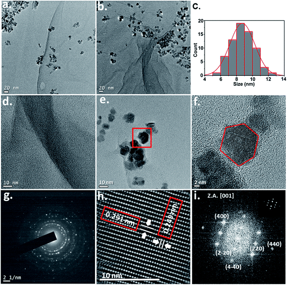
The prepared NiFe2O4@rGO nanohybrid was characterized for structural confirmation. FTIR study revealed the presence of different chemical functionalities in the nanohybrid system (Fig. 1a). The band near 3400 cm−1 is assigned to the –O–H stretching vibration of absorbed moisture entrapped within NiFe2O4 crystal structure. On the other hand, peaks appeared at frequencies 488 and 670 cm−1 are attributed to Fe–O deformation vibration. Appearance of these two peaks is significant. They are considered as the major metal–oxygen vibrations that occurred in NiFe2O4 crystal. On the other hand, peak at 1604 cm−1 is assigned for –C![[double bond, length as m-dash]](https://www.rsc.org/images/entities/char_e001.gif) C– stretching of rGO structure. Likewise, very weakly intense peaks near 2900 cm−1 and 1720 cm−1 region can be attributed to –C–H and –CO stretching frequencies, respectively. These peaks were absent in the FTIR spectrum of pristine NiFe2O4 (Fig. S1a†) and originated from the few defect sites that exist within the graphitic structure of rGO. Weak peak intensities of these peaks confirmed the presence of GNs in reduced form. On the other hand, X-ray diffraction study was carried out to confirm the formation of NiFe2O4 phase in the nanohybrid system. XRD technique is useful to investigate phase purity of the system. The XRD pattern as shown in Fig. 1b, confirmed the inorganic phase present in the nanohybrid as NiFe2O4. XRD diffraction peaks at 2θ values 18.4°, 30.3°, 35.7°, 43.4°, 53.54°, 57.4° and 63.1° are indexed as (111), (220), (311), (400), (422), (511) and (440) crystal planes of NiFe2O4, respectively. The crystallographic data were confirmed by comparing with data obtained from ICDD-PDF-2 data (data card no. 86-2267). Further, data are compared with the XRD pattern of pristine NiFe2O4 (Fig. S1b†), and the peak positions confirmed the formation of NiFe2O4 phase during the hydrothermal process. The (011) and (022) peaks of rGO almost disappeared in the XRD diffractogram. This may happen due to intercalation of NiFe2O4 within the graphitic layers during crystal growth process. This results in rupturing of regular graphitic stacking. Further, high intensity of NiFe2O4 crystal diffraction may also results diminishing of rGO diffraction intensity. Raman analysis was performed in order to detect the presence of rGO phase in the nanohybrid system. Raman is quite helpful for the study of disorder present in the graphitic structure. From the Raman spectrum of NiFe2O4@rGO nanohybrid, two distinct bands, viz. D and G are identified. D band at 1342 cm−1 corresponds to defect in the graphitic structure, while G band at 1592 cm−1 represents graphitic sp2 symmetric carbon stretching (Fig. 1c). Moreover, compared to the band positions of GO, D and G band in rGO suffers a blue-shift. This is an indicative of reduction of GO into rGO. The relative intensity ratio of D and G band supports the fact. The ratio of ID/IG was found to be decreased in the nanohybrid system compared to GO. Hence, it indicates an effective reduction of GO during the hydrothermal process as described in the earlier section. Further, a 2D band centered around at 2850 cm−1 was also observed in the Raman spectrum of NiFe2O4@rGO, which was not present in GO. This has risen due to the formation of graphene structures. Besides, confirming the presence of rGO phase in the nanohybrid, Raman analysis also revealed the formation of NiFe2O4 phase. The peaks appeared in 200–800 cm−1 region are assigned for metal vibration of NiFe2O4 phase (compared to the Raman spectrum of pure NiFe2O4 phase as shown Fig. S1c†). Thus, Raman spectrum of NiFe2O4@rGO confirmed the formation of both the inorganic and carbon based phase during the hydrothermal preparation. SEM/EDX study confirmed the presence of Ni, Fe, O and C in the nanohybrid system (Fig. 1d). Morphological study of the nanohybrid including shape, size and distribution was carried out through visual analysis by using TEM as the tool. TEM images depicted in Fig. 2a and b confirmed the presence of NiFe2O4 over GNs. However, small agglomeration of NiFe2O4 can be observed due to its paramagnetic behavior. But, still a stable nanohybrid system with controlled morphology was perceived through the hydrothermal preparation as evidenced by TEM pictures. Statistical analysis revealed that size distribution lies in between 5–13 nm (Fig. 2c). The largest fraction of NiFe2O4 particles possesses size in the range of 7–10 nm. TEM image also showed edge of graphene sheets (Fig. 2d). HRTEM image showed that the magnetic nanoparticles seemed to possess near hexagonal shape (Fig. 2e and f). HRTEM image of NiFe2O4 further showed presence of lattice fringes. Conversion of selected area of Fig. 2f into corresponding IFFT image revealed two different inter-planar distances (Fig. 2h). Here, d value of 0.291 nm can be ascribed for (220) lattice plane, while that of 0.249 nm is assigned for (311) crystallographic plane of NiFe2O4. Selected area diffraction (SAED) pattern of the nanohybrid showed concentric circles with bright spots indicating the presence of crystalline structure (Fig. 2g). FFT image also provides valuable information about the crystal phase of the nanohybrid system (Fig. 2i). From the FFT image (220), (400) and (440) planes of NiFe2O4 are identified. On the other hand, Fig. 3 shows the magnetic behavior of the nanohybrid under the influence of an ordinary magnet.
C– stretching of rGO structure. Likewise, very weakly intense peaks near 2900 cm−1 and 1720 cm−1 region can be attributed to –C–H and –CO stretching frequencies, respectively. These peaks were absent in the FTIR spectrum of pristine NiFe2O4 (Fig. S1a†) and originated from the few defect sites that exist within the graphitic structure of rGO. Weak peak intensities of these peaks confirmed the presence of GNs in reduced form. On the other hand, X-ray diffraction study was carried out to confirm the formation of NiFe2O4 phase in the nanohybrid system. XRD technique is useful to investigate phase purity of the system. The XRD pattern as shown in Fig. 1b, confirmed the inorganic phase present in the nanohybrid as NiFe2O4. XRD diffraction peaks at 2θ values 18.4°, 30.3°, 35.7°, 43.4°, 53.54°, 57.4° and 63.1° are indexed as (111), (220), (311), (400), (422), (511) and (440) crystal planes of NiFe2O4, respectively. The crystallographic data were confirmed by comparing with data obtained from ICDD-PDF-2 data (data card no. 86-2267). Further, data are compared with the XRD pattern of pristine NiFe2O4 (Fig. S1b†), and the peak positions confirmed the formation of NiFe2O4 phase during the hydrothermal process. The (011) and (022) peaks of rGO almost disappeared in the XRD diffractogram. This may happen due to intercalation of NiFe2O4 within the graphitic layers during crystal growth process. This results in rupturing of regular graphitic stacking. Further, high intensity of NiFe2O4 crystal diffraction may also results diminishing of rGO diffraction intensity. Raman analysis was performed in order to detect the presence of rGO phase in the nanohybrid system. Raman is quite helpful for the study of disorder present in the graphitic structure. From the Raman spectrum of NiFe2O4@rGO nanohybrid, two distinct bands, viz. D and G are identified. D band at 1342 cm−1 corresponds to defect in the graphitic structure, while G band at 1592 cm−1 represents graphitic sp2 symmetric carbon stretching (Fig. 1c). Moreover, compared to the band positions of GO, D and G band in rGO suffers a blue-shift. This is an indicative of reduction of GO into rGO. The relative intensity ratio of D and G band supports the fact. The ratio of ID/IG was found to be decreased in the nanohybrid system compared to GO. Hence, it indicates an effective reduction of GO during the hydrothermal process as described in the earlier section. Further, a 2D band centered around at 2850 cm−1 was also observed in the Raman spectrum of NiFe2O4@rGO, which was not present in GO. This has risen due to the formation of graphene structures. Besides, confirming the presence of rGO phase in the nanohybrid, Raman analysis also revealed the formation of NiFe2O4 phase. The peaks appeared in 200–800 cm−1 region are assigned for metal vibration of NiFe2O4 phase (compared to the Raman spectrum of pure NiFe2O4 phase as shown Fig. S1c†). Thus, Raman spectrum of NiFe2O4@rGO confirmed the formation of both the inorganic and carbon based phase during the hydrothermal preparation. SEM/EDX study confirmed the presence of Ni, Fe, O and C in the nanohybrid system (Fig. 1d). Morphological study of the nanohybrid including shape, size and distribution was carried out through visual analysis by using TEM as the tool. TEM images depicted in Fig. 2a and b confirmed the presence of NiFe2O4 over GNs. However, small agglomeration of NiFe2O4 can be observed due to its paramagnetic behavior. But, still a stable nanohybrid system with controlled morphology was perceived through the hydrothermal preparation as evidenced by TEM pictures. Statistical analysis revealed that size distribution lies in between 5–13 nm (Fig. 2c). The largest fraction of NiFe2O4 particles possesses size in the range of 7–10 nm. TEM image also showed edge of graphene sheets (Fig. 2d). HRTEM image showed that the magnetic nanoparticles seemed to possess near hexagonal shape (Fig. 2e and f). HRTEM image of NiFe2O4 further showed presence of lattice fringes. Conversion of selected area of Fig. 2f into corresponding IFFT image revealed two different inter-planar distances (Fig. 2h). Here, d value of 0.291 nm can be ascribed for (220) lattice plane, while that of 0.249 nm is assigned for (311) crystallographic plane of NiFe2O4. Selected area diffraction (SAED) pattern of the nanohybrid showed concentric circles with bright spots indicating the presence of crystalline structure (Fig. 2g). FFT image also provides valuable information about the crystal phase of the nanohybrid system (Fig. 2i). From the FFT image (220), (400) and (440) planes of NiFe2O4 are identified. On the other hand, Fig. 3 shows the magnetic behavior of the nanohybrid under the influence of an ordinary magnet.
 |
| | Fig. 1 (a) FTIR spectrum of NiFe2O4@rGO; (b) XRD pattern of NiFe2O4@rGO; (c) Raman spectra of GO and NiFe2O4@rGO; (d) EDX spectrum of NiFe2O4@rGO. | |
 |
| | Fig. 2 (a) and (b) TEM images of NiFe2O4@rGO; (c) size distribution of NiFe2O4; (d) edge of rGO sheets; (e) and (f) HRTEM of NiFe2O4 phase; (g) SAED pattern of NiFe2O4@rGO; (h) IFFT image of NiFe2O4 phase; (i) FFT image of NiFe2O4 phase (inset FTT after masking). | |
 |
| | Fig. 3 Magnetic behavior of NiFe2O4@rGO. | |
Fabrication of NiFe2O4@rGO/WPU
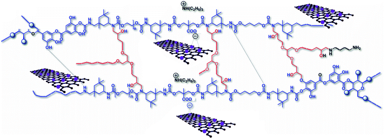
WPU was fabricated with the prepared nanohybrid by following an in situ polymerization technique. Generally, compatibility and state of dispersibility of nanomaterial greatly influence the properties of polymeric nanocomposite. Hence, in the fabrication process maximum care was taken to ensure the formation of a homogenously distributed NiFe2O4@rGO/WPU nanocomposite. In the present study, we found both the nanomaterial and polymer matrix complementing each other to achieve such a highly compatible and uniformly distributed system. The common problem associated with the fabrication of conventional PU/graphene system is poor compatibility as discussed in the Introduction section. However, we found WPU matrix suitable for this purpose. WPU contains a large number of –COOH groups along the polymeric chains. These were incorporated as ionic centers along with the internal emulsifier BMPA. In the neutralization step (as mentioned in preparative method), by adding tertiary amine, these groups were converted into carboxylate ions. These ionic centers played a key role to bring a homogeneous dispersion of NiFe2O4@rGO over the polymer matrix. It is assumed that metallic part of NiFe2O4 went into a strong interaction with the negatively charged carboxylate groups of the polymer chains as shown in Scheme 2. At this juncture, it is pertinent to mention that BMPA was reacted in the very first step, which ensures a uniform distribution of –COOH groups along the polymeric arms. Similarly, NiFe2O4 were found to be distributed uniformly over graphene sheets. Thus, absorption of NiFe2O4@rGO over the polymer matrix through metal/COO− interaction leads to a uniformly distributed and highly compatible nanocomposite system. Further, NiFe2O4@rGO was incorporated during the chain extension process. Such tactic seemed to be effective as nanomaterial can rather easily penetrate low viscous growing polymeric chains under the used reaction condition. Moreover, incorporation of the nanohybrid during the polymerization reaction helped to achieve a strong interaction in the neutralization step through an in situ mechanism. Likewise, in situ polymerization technique further helped to achieve covalent interactions with the polymer matrix through the surface functional groups. TEM image of PNC2.0 confirmed a uniform distribution even at a high nanomaterial loading (Fig. 4a and b). FTIR spectra as shown in Fig. 5 confirmed various functional groups present in the nanocomposite system (e.g. 3400 cm−1 for –O–H stretching, 2900–3000 cm−1 for –C–H stretching, 1710–1740 cm−1 for –CO stretching, 1615 cm−1 for –CC– stretching and 1470 cm−1 for –C–H bending, 1120 cm−1 –C–O stretching and 488 cm−1 for Fe–O deformed vibration). On the other hand, modification of these nanocomposites with HE and PAA helped to create additional cross-linking points within the system, which contributed significantly to form a dimensionally stable structure. In context of shape memory application, such system may prove worthy to memorize the permanent shape of the material.
 |
| | Scheme 2 In situ fabricated NiFe2O4@rGO/WPU nanocomposite. | |
 |
| | Fig. 4 (a) and (b) TEM images of PNC2.0. | |
 |
| | Fig. 5 FTIR spectra of PNCs. | |
Mechanical properties
Mechanical properties of the polymeric nanocomposite were evaluated and results are given in Table 1. It has been found that incorporation of NiFe2O4@rGO to WPU reinforced most of the mechanical properties; such as tensile strength (σ), Young's modulus (E), toughness (T), impact resistance, scratch hardness etc. Further, such improvement was found to be dependent on nanomaterial loading. The mechanical properties of the graphene based polymeric nanocomposites depend on several factors, such as state of dispersion of nanomaterial, its geometry, aspect ratio, orientation of nano-sheets, interfacial interaction between the polymer and the nanomaterial, nanomaterial concentration etc. As a nanomaterial, rGO possesses several favorable attributes, like high mechanical strength (tensile strength 125 GPa, tensile modulus 1100 GPa), high aspect ratio, two dimensional geometry etc.31 These factors contributed significantly towards improved performance of NiFe2O4@rGO/WPU nanocomposite. NiFe2O4 containing rGO sheets provided a better adhesion of two dimensional graphene sheets into the WPU matrix as described earlier. This facilitates efficient load transfer ability between graphene sheets and the WPU matrix. Further, very strong secondary interactions exists within the polymeric structure in the form of H-bonding, π–π stacking, van-der-Waals' interaction etc. These forces also played an important role towards improved mechanical properties by providing structural stiffness. Thus, as a consequence of all these factors, very good mechanical performance of NiFe2O4@rGO/PU nanocomposite was perceived. High tensile properties of the nanocomposite like σ and E originate from the strong interfacial adhesion and good compatibility between the nanohybrid and WPU matrix. Multiple interactions as described earlier make the hard segment stiff which enhanced these properties.32 Furthermore, high strength shows orientation of rGO sheets at high strain causing a synergic effect which leads to an improved modulus upon the formation of the nanocomposite.33 Such results are in agreement with most of the earlier studies on polyurethane/graphene nanocomposites by various groups.32,34,35 Although, incorporation of graphene often reinforces the E and σ, but elongation at break (ε%) demonstrates a complex behavior. Wu et al. found decrease in ε% values with increase in graphene content.36 On the other hand, Chen et al. showed almost unchanged values of ε% of both pristine and nanocomposite systems.37 Thakur et al. showed increase in the ε% values up to 2.0 wt% of graphene content.32 Zhang et al. found an irregular trend of ε% with graphene content.38 They found an increasing trend up to an optimized loading after which ε% started decreasing. Similarly, Bian et al. recorded an initial decrease of ε% which then reinforced at higher graphene content.39 In the present study, ε% values were found to follow an irregular trend. Up to 1.5 wt% of the nanomaterial loading, ε% values were increased, though the increment was not very substantial. At 2.0 wt% of NiFe2O4@rGO, ε% was found to be decreased. At low loading of the nanohybrid, polymer chains are assumed to be aligned in the initial stage of tensile loading. As a consequence, rGO sheets orient themselves along the loading direction. At high stress, layers of rGO are sliding past each other as they are strongly bound to the adjacent WPU chains through covalent and multiple secondary interactions. This further helps to overcome the π–π stacking interactions among rGO sheets. Since, rGO preferentially enhanced the hard segments compared to soft segments, unusual stiffness of soft segments was avoided. Such combined effect results enhancement of ε% values. But, at high loading (>1.5 wt%), such effect has only nominal contribution. The crowded nano-filler imparts very high structural rigidity to the polymer matrix. Under such circumstances, slipping of graphene layers no longer overcome the stiffness of the system. As a result, ε% values start to decrease. However, all the compositions of NiFe2O4@rGO/WPU possess very good flexibility as proved by the bending values. Similarly, gloss values were found good for all the compositions.
Table 1 Mechanical properties of PNCs
| Properties |
Composition |
| WPU |
PNC0.5 |
PNC1.0 |
PNC1.5 |
PNC2.0 |
| Obtained from the slopes of the elastic region in the stress–strain curves. Calculated by integrating the area under stress–strain curve. Limit of the instrument for scratch hardness was 10 kg (maximum). Limit of the instrument for impact resistance was 8.3 kJ m−1 (maximum) for film of thickness 1 mm. Limit of the mandrel diameter was 1 mm (minimum). |
| σ (MPa) |
11.45 ± 0.9 |
14.74 ± 0.7 |
16.94 ± 0.8 |
19.05 ± 0.4 |
24.23 ± 0.5 |
| Ea (MPa) |
53.08 |
75.12 |
79.47 |
88.76 |
92.47 |
| ε (%) |
285 ± 3 |
289 ± 2 |
291 ± 4 |
294 ± 2 |
280 ± 3 |
| Tb (MJ m−3) |
19.36 |
22.42 |
24.74 |
27.42 |
28.35 |
| Scratch hardnessc (kg) |
7.5 ± 0.2 |
8.5 ± 0.3 |
9.0 ± 0.2 |
10 ± 0.1 |
>10 |
| Impact resistanced (kJ m−1) |
7.5 |
7.9 |
8.1 |
>8.3 |
>8.3 |
| Bendinge (m) |
<0.001 |
<0.001 |
<0.001 |
<0.001 |
<0.001 |
| Gloss (60°) |
90.3 ± 3 |
88.47 ± 1 |
87.7 ± 1 |
87.1 ± 1 |
86.84 ± 2 |
Thermal properties
Various thermal properties of NiFe2O4@rGO have been evaluated. Thermogravimetric analysis showed improvement of thermal stability of the pristine system by the formation of nanocomposite. TGA thermograms undergo a shift towards higher temperature with increase in NiFe2O4@rGO content in the polymer matrix (Fig. 6a). Such improvement can be attributed to the presence of metal containing graphene sheets in the nanocomposite systems. The nano-filler in the polymer occupied the free space and made the system more rigid. This delayed the molecular thermal chain motion leading to the enhancement of thermal stability. Further, metallic NiFe2O4 possesses very high thermal stability. They act as a thermal barrier against the volatile decomposed products that are escaping from the polymer system and there by delaying the degradation process. Likewise, char produced as a result of thermal decomposition of rGO served as thermal insulator, which tends to protect the non-degraded polymer part from thermal degradation. Besides, the role of nano-filler, the secondary interactions like H-bonding, π–π interaction, van der waals' attraction etc. also played crucial role towards improved thermal stability of NiFe2O4@rGO/WPU nanocomposite. In earlier works, various groups have demonstrated similar type of enhancement in the thermal stability of polyurethane/graphene nanocomposites.40–43 On the other hand, glass transition temperature (Tg) of the nanocomposite was found to be increased with increase in nanomaterial content (Fig. 6b). Single value of Tg confirms the formation of single phase and highly compatible system. This confirms the restriction imparted by NiFe2O4@rGO on the polymer chain mobility. Ramanathan et al. have made a thorough study on the effect of graphene on Tg of various polymeric nanocomposites.44 They have shown that good dispersion as well as strong interaction between the nano-sheets and the polymer matrix leads to formation of interphase zone around each sheet in which polymer mobility is restricted. Similarly, melting point (Tm) was also found increasing by the incorporation of NiFe2O4@rGO nanohybrid (Fig. 6c). Pokharel et al. demonstrated similar increase in Tm for graphene based polymeric nanocomposite.45 Thus, a positive improvement of thermal properties was perceived. The details of characteristics thermal temperatures are provided in Table S1.†
 |
| | Fig. 6 (a) TGA thermograms of PNCs; (b) DSC curves (cooling) of PNCs showing Tg; (c) DSC curves (heating) of PNCs showing Tm. | |
Shape memory
Shape memory behavior of polymeric material can be considered as an entropic effect. Under normal condition, polymer resides a permanent macroscopic shape in which polymeric chains tend to orient themselves in a random coil formation. Such state corresponds to the maximum entropy of the polymeric chains leading to a thermodynamically stable stage. However, upon heating the polymeric system above shape memory transition temperature (Ttrans + 20 °C in the present case) the chain mobility increases significantly. Application of external force under such condition leads to a conformational change, which results a temporary deformed state. The whole process is accompanied by lowering of entropy. This lower entropy state is kinetically entrapped by freezing the polymeric chain segments in an ice cool condition. This ultimately results in fixation of the temporary shape. This temporary shape was successfully retrieved by heating above the transition temperature i.e. Tm. All the compositions of polymeric nanocomposite found to possess good shape memory behaviour. It was found that with increase in NiFe2O4@rGO content, shape recovery time decreases (Table 2). This can be ascribed to the high thermal conductivity of rGO, which facilitates an effective heating throughout the polymer matrix resulting random orientation of the polymeric chain with maximum entropy. On the other hand, all the compositions responded effectively towards the non-contact external stimuli like sunlight and microwave. It is mainly due to the presence of NiFe2O4@rGO nano-filler within the polymer matrix. NiFe2O4@rGO contains both magnetic and electrically active components. Hence, it is not surprising that NiFe2O4@rGO/WPU undergo interaction with electromagnetic radiation like microwave or even normal sunlight. It is a known fact that NiFe2O4/graphene based nanohybrid system can absorb microwave energy.46 It is also very active under normal sunlight condition.47 As a result, it absorbs energy, which can stimulate an indirect but, effective heating within the polymer matrix. The overall study showed that microwave irradiation of the deformed nanocomposite film at 300 W is good enough to retrieve the original shape within a minute (Fig. 7). However, under normal sunlight with flux density of 80000–100000 lux, NiFe2O4@rGO/WPU took little longer time for shape recovery. However, in both the cases effective shape recovery time was recorded. All the shape memory parameters are shown in Table 2. Similar, to the thermally induced shape recovery, a dose dependent lowering of shape recovery time was witnessed in case of microwave and sunlight triggers. The change in temperature with time under sunlight irradiation and consequence shape memory activity is summarized in Table S2.† It has been found that shape recovery is achieved when temperature crosses the transition temperature.
Table 2 Shape memory parameters of PNCs
| Stimulus |
Shape memory parameter |
PNC0.5 |
PNC1.0 |
PNC1.5 |
PNC2.0 |
| Microwave |
Recovery time (s) |
72 |
60 |
53 |
40 |
| Shape recovery (%) |
96.9 |
97.3 |
97.6 |
98.4 |
| Shape fixity (%) |
94.2 |
95.3 |
96.4 |
96.9 |
| Sunlight |
Recovery time (s) |
170 |
150 |
135 |
115 |
| Shape recovery (%) |
93.2 |
93.5 |
94.2 |
95.3 |
| Shape fixity (%) |
94.2 |
95.3 |
96.4 |
96.9 |
| Thermal |
Recovery time (s) |
90 |
82 |
75 |
60 |
| Shape recovery (%) |
96.5 |
97.1 |
97.8 |
98.6 |
| Shape fixity (%) |
94.2 |
95.3 |
96.4 |
96.9 |
 |
| | Fig. 7 Shape memory behavior of PNCs under microwave (300 W). | |
Conclusions
From the study inference can be made that NiFe2O4@rGO nanohybrid can be effectively used in the in situ fabrication of water borne polyurethane system. Such an approach may be effective to overcome poor compatibility problem of polyurethane with graphene-based materials. The study showed that all the compositions of the polymeric nanocomposite possess high mechanical properties and thermal stability. Such high performance is attributed to the uniform distribution of nanomaterial over the matrix. In addition to that, developed nanocomposite demonstrated multi-stimuli responsive shape memory behavior. Microwave and sunlight irradiation are successfully utilized as nano-contact and remote trigger in the shape recovery process. Thus, such a system can be used as low VOC containing, eco-friendly material with high performance and shape memory ability for their allied applications.
Acknowledgements
The authors acknowledge SAIF, NEHU, Shillong, India for TEM imaging.
Notes and references
- Q. Zhao, H. J. Qi and T. Xie, Prog. Polym. Sci., 2015, 49, 79–120 CrossRef.
- J. Hu, Y. Zhu, H. Huang and J. Lu, Prog. Polym. Sci., 2012, 37, 1720–1763 CrossRef CAS.
- Y. Liu, H. Du, L. Liu and J. Leng, Smart Mater. Struct., 2014, 23, 023001 CrossRef.
- P. Singhal, W. Small, E. Cosgriff-Hernandez, D. J. Maitland and T. S. Wilson, Acta Biomater., 2014, 10, 67–76 CrossRef CAS PubMed.
- J. Hu, Shape memory polymers and textiles, Elsevier, 2007 Search PubMed.
- M. Behl and A. Lendlein, Mater. Today, 2007, 10, 20–28 CrossRef CAS.
- S. Chen, H. Yuan, Z. Ge, S. Chen, H. Zhuo and J. Liu, J. Mater. Chem. C, 2014, 2, 1041–1049 RSC.
- T. Takahashi, N. Hayashi and S. Hayashi, J. Appl. Polym. Sci., 1996, 60, 1061–1069 CrossRef CAS.
- B. S. Lee, B. C. Chun, Y. C. Chung, K. I. Sul and J. W. Cho, Macromolecules, 2001, 34, 6431–6437 CrossRef CAS.
- H. Meng and G. Li, Polymer, 2013, 54, 2199–2221 CrossRef CAS.
- F. Liu and M. W. Urban, Prog. Polym. Sci., 2010, 35, 3–23 CrossRef CAS.
- X. Yan, F. Wang, B. Zheng and F. Huang, Chem. Soc. Rev., 2012, 41, 6042–6065 RSC.
- A. Lendlein and S. Kelch, Clin. Hemorheol. Microcirc., 2005, 32, 105–116 CAS.
- S. Dong, B. Zheng, D. Xu, X. Yan, M. Zhang and F. Huang, Adv. Mater., 2012, 24, 3191–3195 CrossRef CAS PubMed.
- H. Kalita and N. Karak, Polym. Bull., 2013, 70, 2953–2965 CrossRef CAS.
- J. W. Cho, J. W. Kim, Y. C. Jung and N. S. Goo, Macromol. Rapid Commun., 2005, 26, 412–416 CrossRef CAS.
- J. Leng, H. Lv, Y. Liu and S. Du, Electroactivate shape-memory polymer filled with nanocarbon particles and short carbon fibers, Appl. Phys. Lett., 2007, 91(14), 144105 CrossRef.
- S. Thakur and N. Karak, RSC Adv., 2013, 3, 9476–9482 RSC.
- Y. Liu, H. Lv, X. Lan, J. Leng and S. Du, Compos. Sci. Technol., 2009, 69, 2064–2068 CrossRef CAS.
- I. S. Gunes, F. Cao and S. C. Jana, Polymer, 2008, 49, 2223–2234 CrossRef CAS.
- Z. Fan, J. Wang, Z. Wang, H. Ran, Y. Li, L. Niu and S. Yang, Carbon, 2014, 66, 407–416 CrossRef CAS.
- X. Liu, H. Li, Q. Zeng, Y. Zhang, H. Kang, H. Duan, Y. Guo and H. Liu, J. Mater. Chem. A, 2015, 3, 11641–11649 CAS.
- S. Thakur and N. Karak, J. Mater. Chem. A, 2014, 2, 14867–14875 CAS.
- Z. Fan, J. Wang, Z. Wang, H. Ran, Y. Li, L. Niu, P. Gong and S. Yang, Carbon, 2014, 66, 407–416 CrossRef CAS.
- M. Momtaz, M. Barikani and M. Razavi-Nouri, Iran. Polym. J., 2015, 24, 505–513 CrossRef CAS.
- S. Barua, G. Dutta and N. Karak, Chem. Eng. Sci., 2013, 95, 138–147 CrossRef CAS.
- T. Chen, B. Zeng, J. L. Liu, J. H. Dong, X. Q. Liu, Z. Wu and Z. M. Li, J. Phys.: Conf. Ser., 2009, 188, 012051 CrossRef.
- D. Chen, D. Chen, X. Jiao, Y. Zhao and M. He, Powder Technol., 2003, 133, 247–250 CrossRef CAS.
- S. Kumar, K. S. Gandhi and R. Kumar, Ind. Eng. Chem. Res., 2007, 46, 3128–3136 CrossRef CAS.
- R. Larciprete, S. Fabris, T. Sun, P. Lacovig, A. Baraldi and S. Lizzit, J. Am. Chem. Soc., 2011, 133, 17315–17321 CrossRef CAS PubMed.
- I. A. Ovid'ko, Adv. Mater. Sci., 2013, 34, 1–11 Search PubMed.
- S. Thakur and N. Karak, ACS Sustainable Chem. Eng., 2014, 2, 1195–1202 CrossRef CAS.
- O. K. Park, J. Y. Hwang, M. Goh, J. H. Lee and B. C. Ku, Macromolecules, 2013, 46, 3505–3511 CrossRef CAS.
- X. Wang, Y. Hu, L. Song, H. Yang, W. Xing and H. Lu, J. Mater. Chem., 2011, 21, 4222–4227 RSC.
- J. T. Kim, B. K. Kim, E. Y. Kim, S. H. Kwon and H. M. Jeong, Eur. Polym. J., 2013, 49, 3889–3896 CrossRef CAS.
- C. Wu, X. Huang, G. Wang, X. Wu, K. Yang, H. Li and P. Jiang, J. Mater. Chem., 2012, 22, 7010–7019 RSC.
- Z. Chen and H. Lu, J. Mater. Chem., 2012, 22, 12479–12490 RSC.
- J. Zhang, C. Zhang and S. A. Madbouly, J. Appl. Polym. Sci., 2015, 132, 41751 Search PubMed.
- J. Bian, H. L. Lin, F. X. He, X. W. Wei, I.-T. Chang and E. Sancaktar, Composites, Part A, 2013, 47, 72–82 CrossRef CAS.
- M. Fu, Q. Jiao and Y. Zhao, J. Mater. Chem. A, 2013, 1, 5577–5586 CAS.
- S. K. Yadav and J. W. Cho, Appl. Surf. Sci., 2013, 266, 360–367 CrossRef CAS.
- P. Song, Z. Cao, Y. Cai, L. Zhao, Z. Fang and S. Fu, Polymer, 2011, 52, 4001–4010 CrossRef CAS.
- X. Wang, Y. Hu, L. Song, H. Yang, W. Xing and H. Lu, J. Mater. Chem., 2011, 21, 4222–4227 RSC.
- T. Ramanathan, A. A. Abdala, S. Stankovich, D. A. Dikin, M. Herrera-Alonso, R. D. Piner, D. H. Adamson, H. C. Schniepp, X. Chen, R. S. Ruoff, S. T. Nguyen, I. A. Aksay, R. K. Prud'homme and L. C. Brinson, Nat. Nanotechnol., 2008, 3, 327–331 CrossRef CAS PubMed.
- P. Pokharel, B. Pant, K. Pokhrel, H. R. Pant, J. G. Lim, H.-Y. Kim and S. Choi, Composites, Part B, 2015, 78, 192–201 CrossRef CAS.
- M. Fu, Q. Jiao and Y. Zhao, J. Mater. Chem. A, 2013, 1, 5577–5586 CAS.
- Y. Fu, H. Chen, X. Sun and X. Wang, AIChE J., 2012, 58, 3298–3305 CrossRef CAS.
Footnote |
| † Electronic supplementary information (ESI) available: FTIR, Raman and XRD pattern of NiFe2O4, thermal characteristics temperature and change in temperature with time under sunlight irradiation. See DOI: 10.1039/c6ra16848e |
|
| This journal is © The Royal Society of Chemistry 2016 |
Click here to see how this site uses Cookies. View our privacy policy here. 


