
Plasmonic-ELISA: expanding horizons
Jitendra Satija
*a,
Nirmal Punjabi
b,
Debasish Mishra
ac and
Soumyo Mukherji
*bde
aSchool of BioSciences and Technology (SBST), VIT University, Vellore, 632 014, India. E-mail: jsatija11@gmail.com; jsatija@vit.ac.in; Tel: +91 416 220 2151
bDepartment of Biosciences and Bioengineering, IIT Bombay, Mumbai 400 076, India. E-mail: mukherji@iitb.ac.in; Fax: +91 22 2572 3480; Tel: +91 22 2576 7767
cCentre for Biomaterials, Cellular and Molecular Theranostics (CBCMT), VIT University, Vellore, 632 014, India
dCentre of Excellence for Nanoelectronics, IIT Bombay, Mumbai 400 076, India
eCentre for Research in Nanotechnology and Science, IIT Bombay, Mumbai 400 076, India
First published on 30th August 2016
Abstract
The enzyme-linked immunosorbent assay (ELISA) has been an integral part of in vitro diagnostic tests due to its high specificity, standard configuration, and convenient readout. The convergence of the plasmonic property, i.e. localized surface plasmon resonance (LSPR), of noble metal nanoparticles with the ELISA technique has established a novel category of immunoassay, known as “plasmonic ELISA” (pELISA). It has enabled the naked eye detection of various disease biomarkers down to attomolar concentration. The color development relies on the biocatalytic reaction mediated modulation of LSPR properties. Through this review we present various current state-of-art pELISA strategies adopted for naked-eye quantification of disease biomarkers along with their advantages, limitations and applicability. These strategies are broadly classified into following four categories based on the different means of LSPR modulation: (i) aggregation, (ii) controlled growth kinetics, (iii) metallization, and (iv) etching of metal nanoparticles. Furthermore, the paper highlights the different biocatalytic reactions and their role in plasmonic color development as a function of analyte concentration. The review ends with an elucidation of current challenges and future perspectives of pELISA precisely focusing on the need for development of next generation low cost point-of-care diagnostic kits.
 Jitendra Satija | Jitendra Satija received his B. Pharm. from Rajasthan University, India, in 2005 and M. Pharm. with a specialization in Pharmaceutics from Dr. H. S. Gour University, Sagar, India, in 2008. He completed his PhD at IIT Bombay in 2013, India on optical biosensors involving development of dendrimer based labeled and label-free plasmonic immunoassays. Currently, he is an Associate Professor at School of BioSciences and Technology (SBST) at VIT University, Vellore, India. His research interests include plasmonic biosensors, nanomaterials in theranostics, fiber optic biosensors, and sensor matrices design. |
 Nirmal Punjabi | Nirmal Punjabi did his B.E. in biomedical engineering from University of Mumbai, in 2009. Since then he is pursuing his M.Tech. + Ph.D. dual degree in biomedical engineering from Indian Institute of Technology, Bombay. His research interests include design, fabrication, and characterization of optical waveguide sensors for biosensing applications and development of plasmonic bioassays for point-of-care diagnostic devices. |
 Debasish Mishra | Debasish Mishra is currently working in the capacity of associate professor in School of BioSciences and Technology (SBST) and is also associated with Centre for Biomaterials Cellular and Molecular Theranostics (CBCMT) in VIT University, India. He obtained his PhD from Indian Institute of Technology (IIT) Kharagpur, India in 2014. His research interest lies in hard tissue engineering, nano/bio-materials, immunotherapeutics for cancer and BioMEMS. He is also interested in understanding the science of cell–cell and cell–environment communication. He has above 15 publications in peer reviewed journals, 3 conference papers, 4 invited talks and 1 Indian patent. |
 Soumyo Mukherji | Soumyo Mukherji did his B.Tech. in Instrumentation Engineering from IIT Kharagpur, M.S. in Mechanical Engineering from Colorado State University, and Ph.D. from University of North Carolina (Chapel Hill, USA). Thereafter, he joined IIT Bombay, where he holds the position of Institute Chair Professor in the Department of Biosciences and Bioengineering. He is interested in the area of sensor development for widescale deployment. |
Introduction
The enzyme-linked immunosorbent assay (ELISA) is one of the most popular used immunoassay platforms for specific and quantitative detection of target analytes in complex samples. Due to its simplicity, high specificity, ease of readability and acceptability, the impact of ELISA is reflected in the overwhelmingly large number of applications. Today, this technique is routinely used in clinical diagnosis,1–6 and various other research domains including food quality control,7–12 environmental monitoring,13–18 biotechnology,19–22 and defence.23–26 Although, the conventional ELISA techniques (Fig. 1) have offered many advantages, there are certain limitations too. These include (i) need for expensive and large amounts of antibodies, (ii) requirement for long and multiple incubations for diffusion-limited reactions, (iii) need for multiple steps for reagent incubation and washing, (iv) lack of established multiplexing, (v) use of expensive instruments to readout the color intensity, and (vi) laborious procedure.27–30 More importantly, the detection limit of conventional ELISA is barely at nanomolar concentration level, which is inadequate to reach the clinical threshold of many protein biomarkers, especially in the early stage of diseases.31–33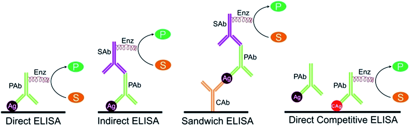 | ||
| Fig. 1 Various types of conventional ELISA formats. In direct ELISA, first the antigen (Ag) is allowed to adsorb on a substrate and then it is detected by an enzyme (Enz)-linked primary antibody (PAb). In indirect ELISA, the adsorbed Ag is detected in two-steps. First, an unlabeled PAb is applied which specifically binds with Ag. Thereafter, an Enz-linked-secondary antibody (SAb) is bound to the PAb. In sandwich ELISA, the Ag is captured specifically by capture antibodies (CAb) which are immobilized on the functionalized substrate. Thereafter, the detection antibody (PAb in figure) recognizes Ag and binds to it, forming an antibody–antigen–antibody sandwich type structure. If the detection antibody is conjugated to an enzyme, like direct ELISA, then the assay is called a direct sandwich ELISA. If the detection antibody is unlabeled, then a secondary detection antibody will be required resulting in an indirect sandwich ELISA (shown in figure). In competitive ELISA, the main event is the competitive binding between the sample antigen and known competitor antigen (CAg), which is proportional to the concentration of the sample antigen. Various types of competitive ELISA are available but in the simplest procedure, first sample antigen is incubated with unlabeled PAb. Thereafter, admixture of this antibody–antigen complex and known CAg is added to the substrate. Subsequently, Enz–PAb is introduced which binds specifically with known CAg and followed by washing to remove unbound immunocomplex (PAb–Ag), CAg and free Enz–PAb. In the final step, color is developed in all ELISA by biocatalytic conversion of enzyme-specific substrate (S) into a color product (P). | ||
In order to address the problems associated with classical ELISA, numerous modifications have been suggested. These are broadly either nanotechnology based or non-nanotechnology based. It is noteworthy to mention some recent yet promising non-nanotechnological based approaches. Paper based ELISA kits have shown their capability as point-of-care (POC) diagnostic tool having good sensitivity, short response time, and affordability.34–38 Recent advances in microfluidics technology and their fusion with optical and electrochemical transducers has further miniaturized ELISA kits, significantly improving the detection limit, and ensured wide applicability with substantial reduction in assay time.39–44 For instance, Wang et al. (2012) developed chemiluminescence ELISA (CL-ELISA) based microfluidic paper analytical devices (μPADs) for high-throughput, rapid, stable, and reusable POC testing.45 Efforts have also been made to improve the standard protocol by making use of some alternate reagents, so that false positive or false negative detection can be minimized or completely avoided.46,47 For example, Liberelle et al. (2013) devised a chemically grafted, low-fouling, carboxymethylated dextran (CMD) layer on ELISA wells which reduced the capture antibody adsorption step to 15 min compared to the standard ELISA which usually takes up to 15 h.48
In the past decade, the convergence of nanotechnology and ELISA has shown tremendous improvement in the sensitivity, cost of analysis, and ease of operation.49–53 By capitalizing on the properties of nanoparticles (NPs), such as tuneable optoelectronic properties, large surface area to volume ratio, availability in wide range (size, shape, and composition), facile synthesis, and ease of bioconjugation, a broad range of nanoparticles-based immunoassays have been evolved. Due to their noteworthy advantages, this class of immunoassays is now categorized as “nano-ELISA”.54–56 In this class, localized surface plasmon resonance (LSPR) property of metallic nanoparticles attracts most of the research because of its ability to develop an ultrasensitive assay with a convenient readout through bare eyes. Utilizing the LSPR properties of plasmonic metal nanoparticles, this special class of nano-ELISA is gaining increasing popularity, and is known as “plasmonic ELISA (pELISA)”.57–60 Compared with the conventional ELISA technique, in which the color is developed by enzymatic catalysis of organic dye/chromogenic molecules and followed by intensity measurement using expensive instrument, pELISA performs enzyme-assisted change in color tonality of nanoparticle solution as a function of analyte concentration that can be observed conveniently with naked eyes.
Noble metal nanoparticles (e.g. gold and silver) exhibit optical absorption and scattering properties in the visible-NIR range, due to light-induced collective oscillation of surface electrons.61–64 This photon-driven coherent oscillation of localized plasmon with a resonant frequency is known as localized surface plasmon resonance. The LSPR spectral position strongly depends on metal nanoparticle size, shape, composition, agglomerate state (or interparticle spacing), and the refractive index of the dielectric medium in their surroundings.65–67 Therefore, a minor change in the morphology, assembly or local environment of metallic nanoparticles leads to modulation in their LSPR peak which can be controlled quantitatively. These include (i) direct or indirect triggering of the nanoparticles aggregation,68–70 (ii) alteration of the refractive index in the micro-/nano-surroundings of nanoparticles,71–73 (iii) transformation in nanoparticles shape and/or composition,74–77 and (iv) increase in nanoparticle size (by means of homoepitaxial or heteroepitaxial growth) (Fig. 2).78–80 These mechanisms result in LSPR wavelength shift and broadening of absorption spectra displaying a substantial color change. Selected mechanisms have been integrated with conventional ELISA resulting in development of highly sensitive naked eye detection techniques that can detect biomarkers in clinically relevant biological samples down to attomolar concentration level.81,82 This review discusses the current state-of-art of various pELISA approaches and their potential advantages, limitations, and clinical relevance with the main focus on the fundamentals of enzyme-triggered plasmonic signal generation step. Although, catalytic properties of nanoparticles have also been realized with ELISA to devise an enzyme-free immunoassay,56,83,84 but in this article only ‘true’ pELISA techniques are discussed which essentially require an enzyme-linked antibody viz. recognition molecule and unmodified antigen, absorbed/adsorbed on a functionalized substrate.
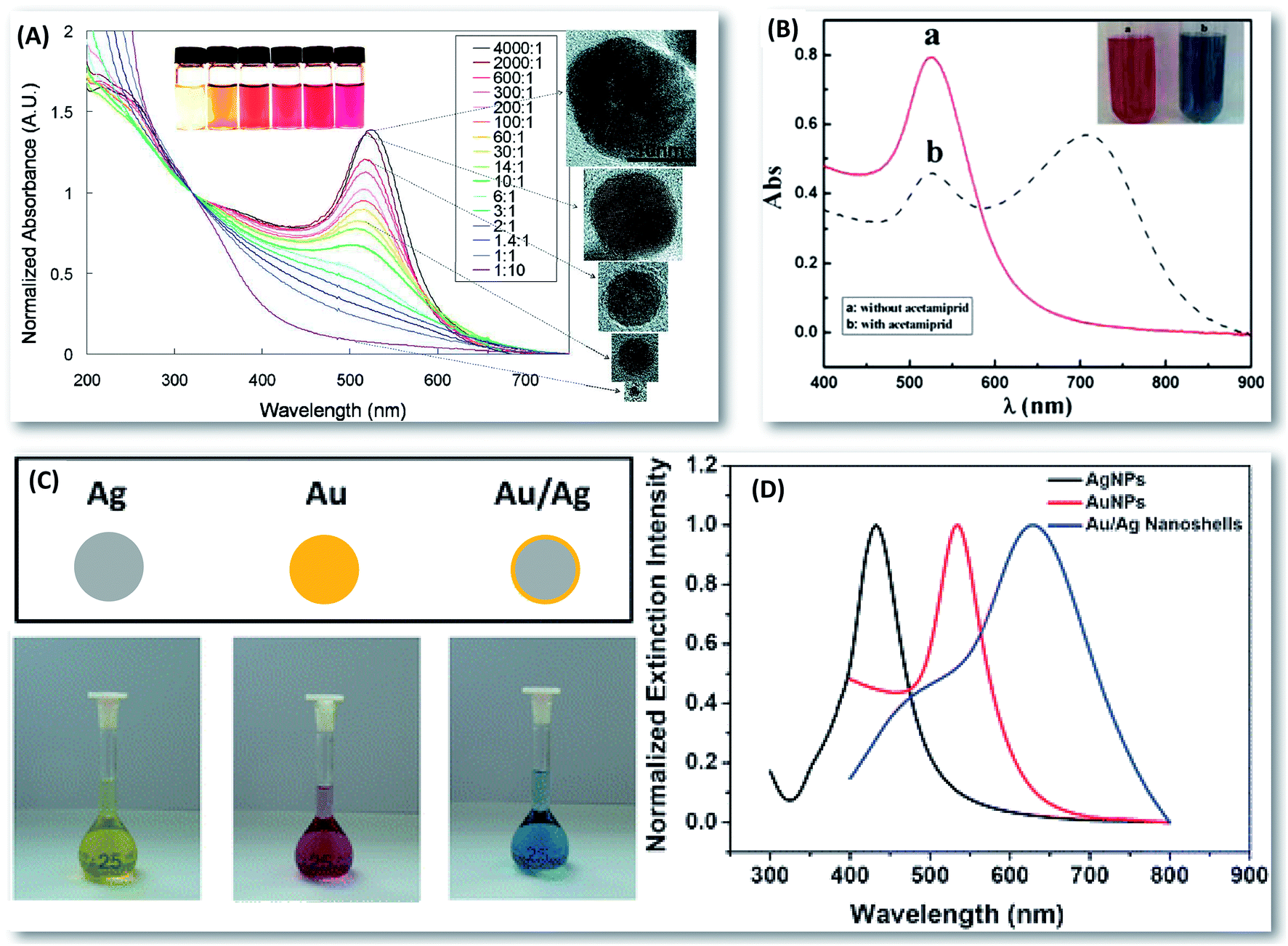 | ||
| Fig. 2 Factors affecting the localized surface plasmon resonance of metal nanostructures. (A) Normalized UV-Vis absorption spectra showing the effect of gold nanoparticles size on their characteristic LSPR peak along with a few representative TEM images. Changes in solution color from yellow-brown to red reflect increase in gold nanoparticles (AuNPs) size and red-shift in LSPR wavelength (reprinted with permission from ref. 106. Copyright 2010 American Chemical Society); (B) UV-Vis absorption spectra depicting a characteristic difference between aggregated and non-aggregated form of AuNPs. Aggregation of gold nanoparticles leads to color change from red to blue (reproduced from ref. 107 with permission of Springer); (C) photographs of different metal nanoparticles (AgNPs = silver nanoparticles; AuNPs and Au/Ag = gold silver nanoshells) showing the effect of nanoparticles composition on the colloidal solution color and (D) their characteristic extinction spectra (reproduced from ref. 108 with permission of The Royal Society of Chemistry). | ||
Mechanisms for plasmonic color development in pELISA
The pELISA approaches are typically based on optical properties of metal nanoparticles, especially of gold and silver. A very high extinction coefficient of gold nanoparticles (AuNPs) (>108 to 109 M−1 cm−1) compared to conventionally used color producing dyes (e.g. the extinction coefficient of oxidized product of 3,3′,5,5′-tetramethylbenzidine (TMB) = 5.9 × 10⁴ M−1 cm−1) makes them amenable to produce a characteristic hue even at very low concentration level.85,86 In addition, upon modulation in size/morphology/aggregation state of gold nanostructures, a prominent color change occurs (e.g. red to blue in case of aggregation of AuNPs) that can be easily differentiated through naked eyes. Therefore, pELISA eliminates the need for sophisticated instruments and thus reduces the detection expenses, making it a viable alternative for resource-constrained regions. This feature is absolutely lacking in conventional ELISA, where only yellow color solutions of different intensity are produced, which are practically difficult to discriminate by naked eyes in field scenarios. The fundamental detection principle of pELISA is to alter the size/morphology/composition/charge distribution of metal nanoparticles or refractive index in their surroundings by enzyme-assisted reaction, which eventually affects the LSPR property resulting in color change. Till date, to the best of our knowledge, at least four variants of ‘true’ plasmonic ELISA have been evolved as illustrated in Fig. 3.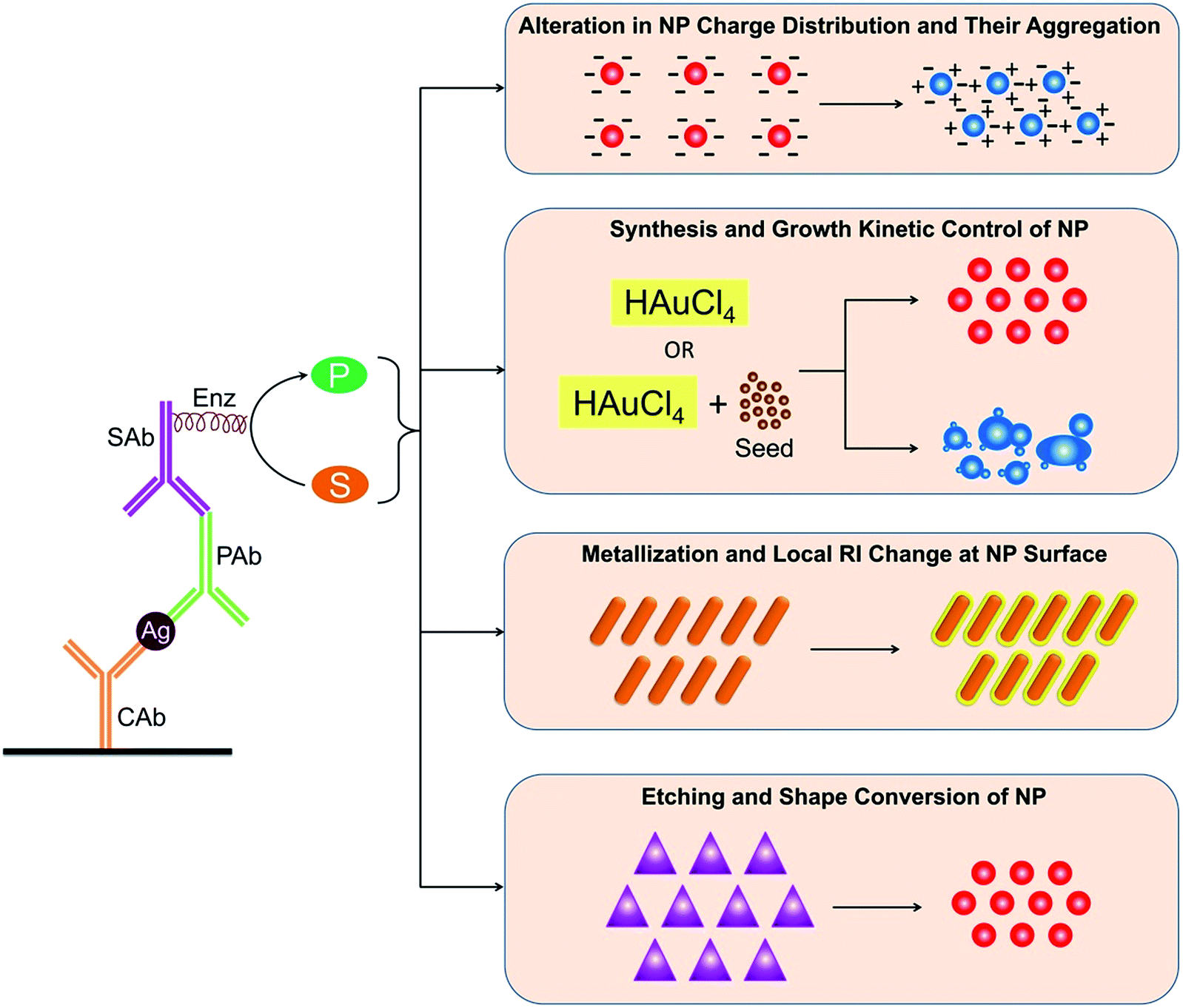 | ||
| Fig. 3 Schematic representation of different formats of plasmonic ELISA (CAb = capture antibody, PAb = primary antibody, SAb = secondary antibody, Ag = antigen, Enz = enzyme, S = enzyme substrate, P = product, HAuCl4 = gold chloride salt, NP = nanoparticles, RI = refractive index). | ||
The first strategy is based on induced aggregation of nanoparticles by tuning the attractive or repulsive forces (and thereby distance) among the nanoparticles, which can be stimulated by different events. These include (i) the bridging of the nanoparticles by complementary biorecognition complexes87–90 (ii) host–guest interactions,91–93 (iii) the use of complementary hydrogen bonds,94–96 (iv) donor–acceptor interactions,97,98 and (v) decrease of the surface charge associated with NPs.99 Since, the plasmonic property of metal NPs is dependent on interparticle distance, a decrease in the interparticle spacing leads to a stronger overlap between the plasmon fields of the adjacent particles, causing a red-shift in the LSPR band, and an easily differentiable color change.100–103 Among the noble metal nanoparticles, gold and silver nanoparticles offer excellent LSPR properties exhibiting strong and well-defined colors, and easy visualization of color change between well-spaced NPs and aggregate ones; red to blue for AuNPs and yellow to brown (or colorless) for AgNPs, respectively.104,105
The second strategy is based on formation and growth kinetics of nanoparticles by reduction of gold salt in the presence or absence of seed nanoparticles. Typical mechanistic steps involved in nanoparticle synthesis are nucleation which is followed by growth.109,110 Ideally, the nucleation event must be well separated in time from the growth step to achieve a narrow size distribution (and possible shape distribution) of nanoparticles. In the first step, rapid conversion of the metal ion precursor into its metallic state results in nuclei (or seeds) consisting of few atoms. In the next step, these nuclei grow through further deposition of metal atoms or by coalescence of nuclei.109 The formation and growth kinetics of metal nanoparticles are quite sensitive to solution conditions (i.e. pH, temperature, ionic strength, agitation, ambient light, etc.), order of addition of reagents, reaction scale and reaction vessel.111,112 A very minute change in any of these reaction parameters can affect the resultant nanoparticles geometry, and thus their LSPR property and color of the solution. For instance, in the preparation of AuNPs by citrate reduction, citrate acts as both reducing and stabilizing agent, and the molar ratio of citrate to gold dictates the size and morphology of the nanoparticles. A study by Kimling et al. (2006) reported that a high citrate concentration allows the fast and complete synthesis and stabilization of small size AuNPs resulting in sharp absorption band with a characteristic red color. On the other hand, at low citrate concentrations, the coverage of nanoparticles is incomplete during growth phase due to limited supply of reduced gold atoms. This results in ill-defined larger AuNPs formation or their aggregates displaying a broad absorption peak with blue color.113
The third strategy is based on thin film formation (usually 1–5 nm) (or metallization) on template nanoparticles by means of metal salt reduction. Typically, the metallization is carried out in a heteroepitaxial manner producing a core–shell type nanostructure.77,114–116 However, for uniform growth of metal on template nanoparticles, the lattice mismatch (defined as the difference in lattice constant of deposited metal relative to the substrate) should be very low between both the metal types. For example, the lattice mismatch between gold and silver is only 0.25%, thus gold nanoparticles can be used as template to grow silver on their surface.117 Samal et al. (2013) reported the synthesis of Au@Ag core–shell NPs by gradual reduction of Ag+ ions, through stepwise addition of silver nitrate to a solution containing preformed citrate-stabilized AuNPs and reducing agent (e.g. ascorbic acid).118 This causes the growth of silver shells onto preformed AuNPs as a function of silver ions concentration. The shell formation causes narrowing of the plasmon line width of the resultant nanostructures implying long plasmon lifetime and large field enhancement.119 This eventually shows a significant change in LSPR properties along with resultant color of nanoparticle solution. For example, the LSPR absorption of gold nanorods (AuNRs) can be precisely tuned by depositing silver nano-thin film on their surface, which can lead to apparent multicolor change (from green to red to orange) as a function of shell thickness.119,120
The fourth strategy is based on the etching of anisotropic metal nanoparticles, such as nanorods, nanotriangles, etc., by using specific oxidising agents. In anisotropic nanoparticles, the sharply curved edges and tips often have high surface energies, and thus can easily be degraded at these points.93 Upon exposure to some specific conditions, like exposure to oxidising agents, elevated temperatures, etc., the etching is initiated at specific sites of the nanoparticles ending up with a shape-conversion. Eventually, this results in a characteristic change in peak LSPR wavelength and color of the nanoparticle solution. For example, chloride ions-mediated etching of triangular silver nanoprisms (AgNPRs) results in rounding of the nanoparticle structure, producing disk-like nanoparticles with blue-shifted spectra.121 In addition, the etching site also depends upon the capping material and its density at nanoparticle surface. For instance, etching of gold nanorods preferentially occurs in the longitudinal direction when exposed to etchants like copper ions, hydrogen peroxide (H2O2), etc.74,122 This reaction results in nanorods of lower aspect ratio (AR = length/breadth) or spherical-shaped gold nanoparticles displaying a prominent blue shift in LSPR wavelength and visual color change. The preferential etching of Au ions from the terminal ends occur due to the lesser density of the capping agent, i.e. cetyltrimethylammonium bromide (CTAB), and higher surface energy. In contrast, the lower surface energy and compact CTAB layer on the lateral facets of AuNRs limits the diffusion of gold from the lateral surface of AuNRs, and thereby reduces the reactivity of this surface.
Strategies for plasmonic ELISA
Dispersion to aggregation based pELISA
In this approach, aggregation of metallic nanoparticles is induced by altering the charge distribution at their surface using an enzyme-catalysed reaction. Eventually, it shows a prominent change in color viz. UV-Vis absorption spectral response of nanoparticles solution. For instance, Nie et al. (2014) developed AuNPs aggregation based pELISA for detection of syphilis.59 In this method, after solid phase (i.e. substrate-linked) immune complex formation, acetylcholinesterase (AChE) enzyme anchored secondary antibodies were added. Thereafter, successive introduction of acetylthiocholine (ATCh) and citrate-capped AuNPs in the microtiter plates resulted in color change of colloidal AuNPs solution as a function of analyte concentration. The authors explained that AChE hydrolysed the ATCh to produce cationic thiocholine (TCh), which covalently attached to the surface gold atoms of citrate-stabilized AuNPs, contributing some positive charges (Fig. 4). This, in turn, furnished the electrostatic interaction among the nanoparticles, and initiated the aggregation of AuNPs and displayed a visible color change from red to blue. The developed AuNPs aggregation based pELISA exhibited a quasilinear response to the logarithmic concentrations of antibodies, raised against T. pallidum antigen, in the range of 1 pg mL−1 to 10 ng mL−1. The detection limit of the pELISA was observed to be 0.98 pg mL−1, which was 1000-fold more sensitive than the conventional ELISA. In an independent study, similar AChE and ATCh based biocatalytic system was used to develop pELISA strategy for the detection of enterovirus 71 (EV71, a causative agent of hand, foot, and mouth disease).123 In this study, the aggregation based pELISA could detect 104 copies per mL of EV71 in human throat swab which was four-orders of magnitude better than the horseradish peroxidase (HRP)-based conventional ELISA. | ||
| Fig. 4 Schematic illustration of pELISA based on enzyme induced aggregation of citrate-stabilized gold nanoparticles for T. pallidum antibody detection by means of generating cationic thiocholine and thereby affecting the charge distribution at nanoparticle surface (reprinted from ref. 59, Copyright 2014, with permission from Elsevier). | ||
Owing to the unprecedented sensitivity and convenient readout, several other enzyme–substrate combinations have also been successfully developed (Table 1).124,125 Xianyu et al. (2014) employed alkaline phosphatase (ALP) triggered click chemistry between azide/alkyne functionalized AuNPs to develop pELISA for the detection of immunoglobulin G (IgG) (Fig. 5).125 In this approach, first one molecule of ALP generates many molecules of ascorbic acid through dephosphorylation of ascorbic acid-phosphate. Subsequently, the generated ascorbic acid reduces the Cu(II) ions into Cu(I) ions which activates the Cu(I)-catalyzed azide/alkyne cycloaddition (CuAAC) reaction between the azide/alkyne-functionalized AuNPs. This resulted in aggregation of AuNPs and transformed the color of the nanoparticle solution from red to blue. The CuAAC-mediated pELISA could achieve naked-eye detection limit down to 80 ng mL−1 for rabbit antihuman IgG, which was 12.5-fold higher compared to the conventional ELISA.
| Type of ELISA | Enz used | Analyte | Mechanism of plasmonic color development | Characteristic features of pELISA | Reference |
|---|---|---|---|---|---|
| a AChE = acetylcholinesterase; ADH = alcohol dehydrogenase; ALP = alkaline phosphatase; ATCh = acetylthiocholine; AuNPs = gold nanoparticles; AuNSs = gold nanostars; CAT = catalase; Enz = enzyme; GOx = glucose oxidase; H2O2 = hydrogen peroxide; HCV = hepatitis C virus; HRP = horseradish peroxidase; IgG = immunoglobulin G; IgM = immunoglobulin M; LOD = limit of detection; NADH = nicotinamide adenine dinucleotide; TCh = thiocholine. | |||||
| Dispersion to aggregation based pELISA | |||||
![[thin space (1/6-em)]](https://www.rsc.org/images/entities/char_2009.gif) |
|||||
| Sandwich | AChE | Treponema pallidum antibodies | Aggregation of citrate capped AuNPs by altering the charge distribution at their surfaces by biocatalytic control on ATCh to cationic TCh conversion | LOD = 0.98 × 10⁻¹² g mL−1 | 59 |
| Quasilinear analytical range = 1 × 10⁻¹² to 1 × 10⁻⁸ g mL−1 | |||||
| Naked eye detection limit = 1 × 10⁻¹¹ g mL−1 | |||||
| Clinical sample detection = yes (syphilis could be diagnosed by measuring the Treponema pallidum antibody in patient's sera) | |||||
|
|||||
| Sandwich | AChE | Enterovirus 71 | Aggregation of citrate capped AuNPs by altering the charge distribution at their surfaces by biocatalytic control on ATCh to cationic TCh conversion | LOD = 104 copies per mL | 123 |
| Naked eye detection limit = 104 copies per mL | |||||
| Clinical sample detection = yes (hand foot and mouth disease could be detected by measuring enterovirus 71 in human throat swab solutions) | |||||
|
|||||
| Sandwich | ALP | Rabbit antihuman IgG and mycoplasma pneumonia | Aggregation of azide/alkyne-functionalized AuNPs by Cu(I)-catalyzed azide/alkyne cycloaddition click chemistry | LOD = 2 × 10⁻⁹ g mL−1 | 125 |
| Linear analytical range = 2 × 10⁻⁹ to 2 × 10⁻⁶ g mL−1 | |||||
| Naked eye detection limit = 80 × 10−9 g mL−1 | |||||
| Clinical sample detection = yes (Mycoplasma pneumonia, MP, sensing could be done by measuring anti-MP IgM in serum sample) | |||||
|
|||||
| Sandwich | HRP | Rabbit antihuman IgG and HCV | Cysteine stimulated aggregation of AuNPs by means of zwitterionic electrostatic interactions | LOD = 1 × 10⁻⁹ g mL−1 | 124 |
| Naked eye detection limit = 1 × 10⁻⁹ g mL−1 | |||||
| Clinical sample detection = yes (hepatitis C could be detected by measuring anti-HCV antibodies in serum) | |||||
|
|||||
| Formation and growth kinetics based pELISA | |||||
| Competitive | CAT | Ochratoxin A | Formation kinetics of AuNPs by enzymatic control on gold salt reduction via modulating H2O2 concentration | LOD = 5 × 10⁻²⁰ g mL−1 | 60 |
| Linear analytical range = 1 × 10⁻¹⁸ to 5 × 10⁻²⁰ g mL−1 | |||||
| Naked eye detection limit = 1 × 10⁻¹⁸ g mL−1 | |||||
|
|||||
| Competitive | CAT | Methyltestosterone | Growth kinetics of AuNPs in the presence of gold seeds by enzyme-triggered modulation of H2O2 concentration | LOD = 10 × 10−12 g mL−1 | 58 |
| Linear analytical range = 30 × 10−12 to 30 × 10−9 g mL−1 | |||||
| Naked eye detection limit = 30 × 10−12 g mL−1 | |||||
|
|||||
| Sandwich | CAT | PSA and HIV-1 capsid antigen p24 | Formation kinetics of AuNPs by enzymatic control on gold salt reduction via modulating H2O2 concentration | LOD = 1 × 10⁻¹⁸ g mL−1 | 57 |
| Naked eye detection limit = 1 × 10⁻¹⁸ g mL−1 | |||||
| Clinical sample detection = yes (HIV could be detected by measuring the HIV-1 capsid antigen p24 in the sera of HIV-infected patients) | |||||
|
|||||
| Sandwich | CAT | HIV-1 protein gp120 | Formation and growth kinetics of AuNPs by enzymatic control on gold salt reduction via modulating H2O2 concentration | LOD = 1 × 10⁻¹⁷ g mL−1 | 81 |
| Naked eye detection limit = 1 × 10⁻¹⁷ g mL−1 | |||||
|
|||||
| Sandwich | ADH | Hepatitis B surface antigen and α-fetoprotein | Seed-mediated growth kinetics of AuNPs by enzyme-triggered modulation in NADH level via catalysis reaction between NAD+ and ethanol | LOD = 1 × 10⁻¹² g mL−1 | 132 |
| Linear analytical range = 1 × 10⁻¹² to 1 × 10⁻⁶ g mL−1 | |||||
| Naked eye detection limit = 1 × 10⁻¹² g mL−1 | |||||
| Clinical sample detection = yes (hepatitis B virus could be detected by quantitative determination of hepatitis B surface antigen in sera of clinical patients) | |||||
| Sandwich | HRP | Serum asialohaptoglobin | Formation kinetics of AuNPs by enzyme-guided control on gold salt reduction via modulating H2O2 concentration | Naked eye detection = yes | 133 |
| Clinical sample detection = yes (liver cirrhosis could be detected by measuring the extent of α-2,6 sialylation of serum haptoglobin) | |||||
|
|||||
| Sandwich | CAT | Listeria monocytogenes | Formation kinetics of AuNPs by enzyme-mediated control on gold salt reduction via modulating the H2O2 concentration | LOD = 8 CFU mL−1 | 134 |
| Linear analytical range = 8 × 100 to 8 × 10⁸ CFU mL−1 | |||||
| Naked eye detection limit = 80 CFU mL−1 | |||||
|
|||||
| Sandwich | GOx | PSA | Growth kinetics of AuNPs in the presence of gold seeds by enzyme-assisted control on gold salt reduction via tuning H2O2 concentration | LOD = 3.1 × 10⁻¹⁵ g mL−1 | 135 |
| Linear analytical range = 1 × 10⁻¹⁴ to 1 × 10⁻⁹ g mL−1 | |||||
| Naked eye detection limit = 1 × 10⁻¹⁴ g mL−1 | |||||
| Clinical sample detection = yes (prostate cancer could be diagnosed by measuring the PSA level in patients serum) | |||||
|
|||||
| Metallization based pELISA | |||||
| Sandwich | GOx | PSA | Silver film formation on AuNSs by enzyme-mediated control of silver ions reduction via modulating the H2O2 concentration | LOD = 1 × 10⁻¹⁸ g mL−1 | 82 |
| Linear analytical range = 1 × 10⁻¹⁸ to 1 × 10⁻¹³ g mL−1 | |||||
| Naked eye detection limit = 1 × 10⁻¹⁸ g mL−1 | |||||
|
|||||
| Sandwich | ALP | PSA | Silver film formation on AuNRs by enzyme-mediated control on silver ions reduction via modulating the H2O2 concentration | LOD = 1 × 10⁻¹⁴ g mL−1 | 32 |
| Linear analytical range = 1 × 10⁻¹⁴ to 1 × 10⁻¹¹ g mL−1 | |||||
| Naked eye detection limit = 1 × 10⁻¹³ g mL−1 | |||||
| Clinical sample detection = yes (prostate cancer detection by measuring the PSA level in patients serum) | |||||
|
|||||
| Sandwich | ALP | Rabbit IgG | Silver film formation on AuNRs by enzyme-mediated control of silver ions reduction via modulating the ascorbic acid 2-phosphate concentration | LOD = 1.5 × 10⁻⁹ g mL−1 | 136 |
| Naked eye detection limit = 1.5 × 10⁻⁹ g mL−1 | |||||
| Linear analytical range = 1.5 × 10⁻⁹ to 20 × 10−9 g mL−1 | |||||
|
|||||
| Sandwich | ALP | H9N2 virus | Silver film formation on AuNPs by enzyme-mediated control of silver ions reduction via modulating the p-aminophenyl phosphate monohydrate concentration | LOD = 17.5 × 10⁻¹² g mL−1 | 137 |
| Linear analytical range = 20 × 10−12 to 1 × 10⁻⁹ g mL−1 | |||||
| Naked eye detection limit = 17.5 × 10⁻¹² g mL−1 | |||||
|
|||||
| Etching based pELISA | |||||
| Sandwich | ALP | Human IgG | Etching of AuNRs by enzyme-mediated generation of etchant, iodine, by controlling the iodate to iodine conversion | LOD = 100 × 10−12 g mL−1 | 138 |
| Linear analytical range = 0.1 × 10⁻⁹ to 10 × 10−9 g mL−1 | |||||
| Naked eye detection limit = 3 × 10⁻⁹ g mL−1 | |||||
| Detection is possible in complex biological samples | |||||
| Sandwich | GOx | PSA | Etching of triangular silver nanoprisms by enzyme-mediated control on H2O2 concentration | LOD = 4.1 × 10⁻¹⁵ g mL−1 | 139 |
| Linear analytical range = 10 × 10−15 to 100 × 10−12 g mL−1 | |||||
| Naked eye detection limit = 10 × 10−15 g mL−1 | |||||
| Clinical sample detection = yes (prostate cancer could be diagnosed by measuring the prostate specific antigen level in patients serum) | |||||
 | ||
| Fig. 5 Schematic illustration of pELISA based on alkaline phosphatase (ALP) triggered Cu(I)-catalyzed azide/alkyne cycloaddition (CuAAC) click chemistry between azide- and alkyne-functionalized AuNPs resulting in aggregation of gold nanoparticles (reprinted with permission from ref. 125. Copyright 2014 American Chemical Society). | ||
The key advantage of enzyme-triggered dispersion to aggregation pELISA is a visible change in color from red to blue, rather than just color intensity change, and that can be read easily through naked eyes making it suitable for POC diagnostics. Although the system is very sensitive and convenient but does not come without vices. One of the major limitation is spontaneous aggregation, also called as ‘autoaggregation’, of nanoparticles due to the presence of various external factors that are associated with complicated application environments.126 These include the existence of impurities, change in pH, extreme temperature, high ionic strength and other factors which may induce undesirable aggregation of metal nanoparticles, resulting in high backgrounds or false positive results.127–130 In addition, the colloidal suspension of the aggregates is unstable and tends to precipitate, due to increased particle size and reduced surface repelling forces, leading to reduction of color in the solution.131 Therefore, dispersion to aggregation based pELISA has been limited to a certain range and cannot be realised for wide-spread real-world applications.
Nanoparticle formation and growth kinetics based pELISA
It is a well-known fact that synthesis and growth of metal nanoparticles are very sensitive to reaction conditions and reagents concentrations. A very minute change in the concentration of reducing agent or its rate of availability can significantly affect the outcome of the nanoparticle size and shape distribution which may results in easily noticeable plasmonic color change. For example, gold nanoparticles are synthesized by chemical reduction of gold chloride salt (HAuCl4) by means of H2O2 (reaction (1) and (2)).| 2Au3+ + 3H2O2 ⇌ 2Au + 3O2 + 6H+ | (1) |
| 2H2O2 → 2H2O + O2 | (2) |
The outcome of the nanoparticle synthesis process is extremely sensitive to the concentration of H2O2, which is acting as a reducing agent in this case (Fig. 6). A report by Cecchin et al. (2014) suggests that the gold salt solution (100 μL of 0.2 mM, pH 6.5) containing H2O2 with the final concentration of 119.95 μM, appears clearly blue whereas the solution containing H2O2 with the final concentration of 120 μM develops red color.81 These results demonstrated that in situ generation and growth of colloidal nanoparticles with characteristic tonality can be achieved with extremely small changes in concentration of the reducing agent. Furthermore, the role of the reducing agent is not only limited to stoichiometric reduction of gold ions but also to tune the kinetics of AuNPs growth to yield nanoparticles with different morphologies and optical properties.
 | ||
| Fig. 6 Generation of nanoparticle solutions with different colors as a function of hydrogen peroxide concentration. Different concentrations of hydrogen peroxide were added to a solution containing gold ions (0.1 mM) in MES buffer (1 mM, pH 6.5); (a) photograph showing the generation of nanoparticle solutions with different colors and intensities after 15 min. The tonality of the solution changes from red to blue between 120 and 100 mM; (b) UV-Vis spectra for different hydrogen peroxide concentrations. The localized surface plasmon resonance peak red shifts when the concentration of hydrogen peroxide is 100 mM or lower; (c) and (d) transmission electron microscopy (TEM) images of nanoparticles grown with hydrogen peroxide at concentrations of 100 mM (c) and 120 mM (d). Scale bars = 50 nm (c) and 100 nm (d); (e) graph showing that the absorbance of the solutions at 550 nm varies with the concentration of hydrogen peroxide (reprinted by permission from Macmillan Publishers Ltd.: Nature Nanotechnology ref. 57, Copyright 2012). | ||
The amount of reducing agents, like H2O2, varies as a function of analyte concentration in ELISA techniques. The reaction between the immunocomplex containing a specific enzyme and added reagents generates and/or controls the reducing agent level, which eventually forms nanoparticles by reduction of metal salt. As a result, a characteristic color change is observed as a function of analyte concentration, e.g. from colorless to red or blue in case of gold nanoparticles. Unlike the aggregation based pELISA, in this strategy the color change occurs due to control on nanoparticles formation and growth kinetics. Therefore, this plasmonic signalling strategy does not suffer much from the interferents present in the application environment.
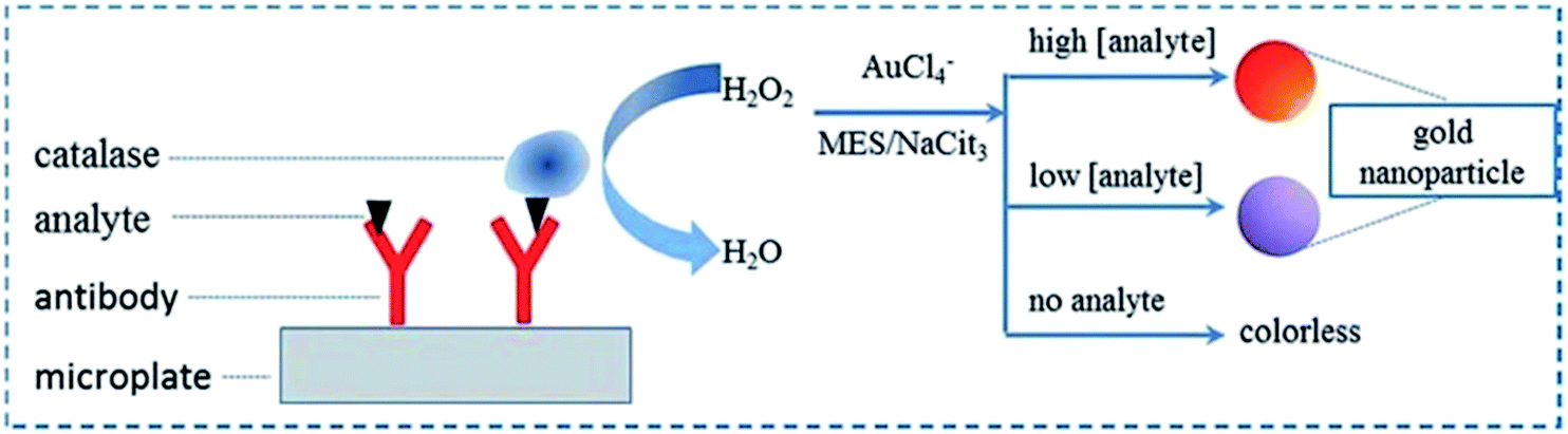
de la Rica and Stevens (2012) developed AuNPs formation and growth kinetics to quantify prostate specific antigen (PSA) and HIV-1 capsid antigen p24 in patients serum samples.57 In this method, first target molecule was captured with specific antibodies on a disposable substrate which was subsequently sandwiched using detector antibody complex, tagged with catalase (CAT). Thereafter, solution of H2O2 and gold salt were introduced sequentially that resulted in colored solutions of AuNPs, having characteristic tonality. The color change was attributed to biocatalytic cycle of CAT enzyme which controlled the level of H2O2 in the reaction mixture as a function of analyte concentration. Accordingly, synthesis and growth kinetics of AuNPs was tuned resulting in a blue- or red-colored solution in the presence or absence of the analyte, respectively. Authors discussed that in the absence of the analyte, H2O2 reduced the gold ions at a fast rate, yielding quasi-spherical, non-aggregated red-colored colloidal solution of AuNPs. On the other hand, in the presence of the analyte, the enzyme CAT consumed H2O2 resulting in decreased supply. This slowed down the kinetics of crystal growth, resulting in nanocrystals with ill-defined morphology comprising aggregated nanoparticles displaying blue color solution. Since the blue and red colors can be easily differentiated, therefore this method facilitated the detection of the analyte with the naked eye at 1 × 10⁻¹⁸ g mL−1 concentration for both PSA and HIV-1 capsid antigen p24 in whole serum.
Owing to the major advantage of simplicity in color development and its readout, many other researchers also attempted the nanoparticle synthesis and growth kinetic based plasmonic signalling scheme for various biochemical assays (Table 1).60,81,133,134 Bose et al. (2016) developed an enzyme-linked lectinosorbent assay for visual detection of serum asialohaptoglobin (Hp).133 This method employed Sambucus nigra agglutinin (SNA) to detect the extent of α-2,6 sialylation of serum haptoglobin, a biomarker for liver cirrhosis. In this assay, first the target acute phase protein Hp was captured by the anti-Hp antibody on the 96-well polystyrene plates. Thereafter, tracer biomolecule, i.e. SNA-tagged HRP enzyme, was introduced which specifically binds with sialic acid present on the Hp. Following this, H2O2 and gold(III) solution were added that consequently generated the AuNPs. The morphology of produced AuNPs was dictated by the leftover concentration of H2O2 which varied with the amount of HRP enzyme bound with sialic acid present on Hp. The higher the sialylation, the greater was the amount of SNA as well as HRP enzyme, and thus a smaller amount of peroxide. The low concentration of available peroxide for the reduction of gold(III) affected the formation kinetics and thus the aggregation propensity of AuNPs, which appeared as blue colored due to non-spherical, aggregated particles.
Recently, the use of seed-mediated growth kinetics of nanoparticles has also been investigated to expand the horizon of pELISA. Since the seed-mediated approach offers rapid synthesis of nanoparticles with precise control on their size and shape distribution, it substantially reduces the sample analysis time.78 Peng et al. (2015) developed seed-mediated nanoparticle growth kinetics based pELISA using catalytic activity of alcohol dehydrogenase (ADH) enzyme for detecting various disease biomarkers.132 In a typical sandwich ELISA format, after immunocomplex formation, ethanol, nicotinamide adenine dinucleotide (NAD+), gold seeds (size = 5 nm) and gold chloride salt were successively introduced in the microtiter plate. In the presence of antigen, the labeled ADH catalyzed the reaction between NAD+ and ethanol to generate nicotinamide adenine dinucleotide (NADH) and acetaldehyde (Fig. 7). Eventually, the NADH reduced gold ions (Au3+) into their metallic form (Au0), which gets deposited on the gold seeds. This resulted in growth of gold seeds, and apparent color change from yellow to purple was observed as a function of analyte concentration. By employing this strategy, authors validated the detection of hepatitis B surface antigen (HBsAg) and α-fetoprotein (AFP) with the lowest concentration down to 1.0 × 10⁻¹² g mL−1 through naked eyes. In addition, the experiments with real serum samples obtained from HBsAg-infected patients, demonstrated the visual detection potential of the developed pELISA for clinical analysis.
 | ||
| Fig. 7 Schematic representation of pELISA based on seed-mediated formation and growth kinetics of gold nanoparticles (reprinted with permission from ref. 132. Copyright 2015 American Chemical Society). | ||
In a different approach, Peng et al. (2014) developed a direct competitive “signal on” pELISA by exploiting the H2O2 mediated reduction of gold salt and simultaneous growth of gold seeds as a colorimetric signalling step.58 In this competitive assay, methyltestosterone (MT) was used as an analyte, while the CAT conjugated-MT was used as competitor. During the procedure, MT and competitor were introduced which bound to the primary antibodies present on polystyrene microtiter plate. Subsequent addition of H2O2, gold seeds (size = 15 nm), gold chloride salt and stabilizer caused the reduction of gold salt which resulted in color change, depending upon the concentration of MT analyte. No color change was observed in the absence of analyte because of complete decomposition of H2O2 into water and oxygen by CAT enzyme, present in the competitor. On the other hand, in the presence of analyte, the number of CAT molecules bound on the plate decreased, thereby decreasing the decomposition of H2O2 and thereby increased the availability of H2O2. This resulted in reduction of gold salt causing a change in color tonality from blue to red when the MT concentration was increased from 0.03–1 ng mL−1 (Fig. 8). The sensitivity of this pELISA (based on naked eye detection) was found to be 50 times higher than the traditional ELISA. Compared with classical direct competitive ELISA, in this pELISA, the color change was observed only in the presence of analyte and the absorbance intensity of the generated AuNPs solution increased with increasing MT concentration and therefore, authors regarded this strategy as a “signal on” pELISA method.
 | ||
| Fig. 8 Schematic illustration of direct competitive pELISA displaying enzyme-controlled formation and growth kinetics of gold nanoparticles as a function of analyte concentration (reproduced from ref. 58 with permission of The Royal Society of Chemistry). | ||
Although the formation and growth kinetics of nanoparticle is very selective, sensitive and nearly free from autoaggregation, but many other factors can interfere and result in false positive or negative plasmonic color change. These include ageing of the solutions containing nanoparticle growth precursors, the reaction scale, the type of reaction vessel (e.g. shape and material properties), the presence of convection, etc. Moreover, operator/user-induced variations can introduce subtle differences in the synthesis and growth process. For example, different ways of mixing solutions (e.g. speed or direction of motion) can affect the outcome of the nanoparticle growth step.140–142
Metallization based pELISA
This method relies on heteroepitaxial growth of plasmonic metal nanoparticles that result in core–shell type nanostructures with a noticeable change in LSPR properties and visual color change.77,114–116 For example, the LSPR absorption peak of AuNRs can be precisely tuned by depositing silver nano-thin film on their surface, which is usually manifested as apparent multicolor change.119 Since this plasmonic color development does not involve any aggregation or in situ formation of metallic nanoparticles, it is minimally affected by environmental factors and reaction conditions and thus making it quite reliable pELISA technique.Yang and Gao 2015 formulated a high-resolution pELISA based on gold/silver core/shell nanorods for easy visual readout of PSA.32 Similar to conventional ELISA, first ALP enzyme was brought in through immunoreaction with PSA target antigen and followed by addition of p-aminophenol phosphate, and silver ion precursors. The ALP enzyme produced p-aminophenol by dephosphorylating p-aminophenol phosphate, which subsequently reduced the silver ions into metallic silver. Immediately, the reduced silver gets deposited as a nano-thin film on the surface of AuNRs resulting in a significant change in the local refractive index around the AuNRs. This caused a substantial change of the longitudinal LSPR signal and solution color (Fig. 9). With an increase of PSA concentration from 0 to 1.0 ng mL−1, the color of the AuNRs solution was observed to change from pale red to green, purple and brown, which could be easily differentiated by the naked eyes. As low as 10 fg mL−1 PSA could be detected using silver shell growth on AuNRs, four orders of magnitude better than that of conventional ELISA.
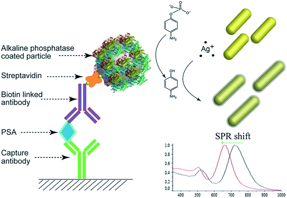 | ||
| Fig. 9 Schematic illustration of the working principle of metallization based pELISA using gold nanorods as template nanostructures. The alkaline phosphatase catalyses the production of 4-aminophenol that reduces the silver ions into metallic silver. Consequently, the metallic silver gets deposited on the gold nanorods causing a blue shift of the LSPR peak (reproduced from ref. 32 with permission of The Royal Society of Chemistry). | ||
Rodríguez-Lorenzo et al. (2012) conceptualized the idea of metallization of plasmonic transducer-based pELISA using enzyme-guided in situ formation of silver shell on gold nanostars (AuNSs).82 In this approach, the LSPR of AuNSs was tuned by glucose oxidase (GOx)-assisted crystal growth to favour either the formation of a silver coating around the AuNSs or the nucleation of silver nanocrystals in solution. Using the sandwich ELISA format, first the immunocomplex formation between PSA antigen and PSA specific GOx-labeled anti-mouse IgG antibodies was achieved. Afterwards, glucose and silver salt were introduced sequentially in the microtiter plate. The GOx oxidizes the glucose and forms gluconic acid and H2O2 as a function of analyte concentration (Fig. 10). At low concentrations of target analyte, the H2O2 concentration becomes very low, thereby nucleation rate is impeded. As a result, the growth of a conformal silver coating on AuNSs is favoured inducing a large blue shift in the LSPR peak. On the other hand, when target analyte concentration is high, large numbers of H2O2 molecules are generated resulting in stimulation of faster nucleation and growth of silver nanocrystals and less silver deposition on AuNRs. Therefore, at high concentration a very small change in the LSPR was observed. The authors experimentally demonstrated the outstanding ability of the metallization of AuNSs based pELISA to detect the cancer biomarker, PSA, down to 10−18 g mL−1 (4 × 10⁻²⁰ M) in whole serum.
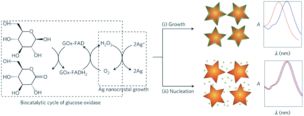 | ||
| Fig. 10 Scheme of the plasmonic signal-generation in metallization based pELISA by depositing the enzyme-guided silver film on gold nanostars as a function of analyte concentration (GOx = glucose oxidase enzyme; H2O2 = hydrogen peroxide, FADH2 = flavin adenine dinucleotide, FADH2 = reduced form of FAD) (reprinted by permission from Macmillan Publishers Ltd.: Nature Materials ref. 82, Copyright 2012). | ||
Nanoparticle etching based pELISA
In past decade, anisotropic noble metal nanomaterials, such as gold nanorods, nanocubes, nanocages, nanoprisms, and nanoflowers, have attracted considerable attention.143–147 Their unique shape-dependent optical properties have established them as ideal candidates for developing colorimetric/visual read-out based biosensors.148,149 A very minute geometrical change of anisotropic nanostructures, by means of etching, causes a significant modulation of the LSPR peak and solution color. For example, in a study by Alex et al. (2015), it was reported that chromium (Cr) mediated etching of CTAB-capped gold nanorods caused a substantial change in the solution color from violet to blue to pink, as a function of Cr(VI) concentration.75 The longitudinal LSPR peak was observed to be blue-shifted with a decrease in average AR from 2.66 to 1.58 when the concentration of Cr(VI) was varied in the range from 0–1000 μM. Recently, the etching-mediated plasmonic signal generation strategy has been combined with ELISA and has shown its potential for overcoming the problems associated with reagents, reaction scale, vessels and handling (Table 1).138,139 This is due to the fact that the plasmonic signal generation depends exclusively on the reaction between anisotropic nanostructures and etchant concentration which is controlled by enzyme, resulting in geometrical transformation of nanostructures and thereby a predefined color change. However, for this assay, nanoparticles of highly uniform size and shape are essentially required, which are usually produced by a controlled multistep process that makes this assay relatively expensive.Zhang et al. (2015) developed pELISA based on iodine-mediated etching of gold nanorods to detect PSA antigen.138 In this approach, after sandwich immunocomplex formation between goat anti-human IgG as antigen and human IgG linked with ALP enzyme as detector antibody, the ascorbic acid-2-phosphate (AA-p) was introduced in the microtiter plate. This was followed by the addition of a mixture of iodate and CTAB-capped AuNRs. In this biochemical reaction, the ALP converts AA-p into ascorbic acid that subsequently reduces iodate into iodine. The iodine, being a moderate oxidant, etches AuNRs in the presence of cetyltrimethylammonium (CTA+) ions and results in shape-conversion from nanorods to nanospheres. This shape conversion results from preferential etching of AuNRs along the axial direction compared to the lateral surface because of less surface passivation by CTAB at both the end terminals. This resulted in blue-shift of longitudinal LSPR peak and the solution color changed from blue to red. The gold nanorods etching-based pELISA strategy could visually detect human IgG at 3.0 ng mL−1 concentration. More importantly, the method could detect human IgG in fetal bovine serum at 100 pg mL−1 concentration.
In another recent study conducted by Liang et al. (2015), triangular silver nanoprisms (AgNPRs) etching-based pELISA was reported for the detection of cancer biomarkers. In this assay, the etchant, H2O2, was generated by a biocatalytic reaction between glucose oxidase enzyme and glucose (reaction (3) and (4)). The produced H2O2 directed the etching of AgNPRs into smaller spherical silver nanoparticles by means of oxidation–reduction reaction (Fig. 11).
| Glucose + O2 → gluconic acid + H2O2 | (3) |
| Metallic Ag + H2O2 → Ag+ + H2O | (4) |
 | ||
| Fig. 11 Schematic showing pELISA for prostate specific antigen detection. The plasmonic signal generation is based on glucose oxidase (GOx)-catalysed etching of triangular silver nanoprisms (AgNPRs) into smaller spherical silver nanoparticles by means of hydrogen peroxide (H2O2) as an oxidising agent (reprinted from ref. 139, Copyright 2015, with permission from Elsevier). | ||
The GOx-catalysed oxidation of glucose produces gluconic acid and H2O2, which effectively lowers the local pH. The decrease in local pH augments the oxidising power of peroxide because the standard potential of H2O2/H2O both at acidic pH (1.763 V) and at alkaline pH (0.867 V) is greater than the Ag+/Ag (0.7996 V). As a result, the silver ions are etched away from the sharp edges of AgNPRs due to high surface energy vertices and nanoprisms are transformed into spherical shaped nanoparticles. This is accompanied with a substantial blue shift of the LSPR peak and a colorimetric blue to purple change. The triangular AgNPRs-based pELISA exhibited a quasi-linear response to logarithmic concentration of PSA in the range of 10 fg mL−1 to 100 pg mL−1 with a limit of detection of 4.1 fg mL−1. The LOD of PSA detection in this pELISA exceeded that of the conventional HRP-based ELISA (1.25 ng mL−1) by more than five-orders of magnitude.
Summary and future perspective
The ELISA technique is fundamental tool of clinical practice, and is widely used from initial screening of various diseases to their therapeutic monitoring. However, the limited sensitivity, and need of an expensive instrument along with a trained professional has limited its wide applicability especially in resource-constrained countries. The merging of properties of nanoparticles, especially the localized surface plasmon resonance of noble metal nanoparticles, with ELISA has significantly improved the bioassay performance and established as plasmonic ELISA.Many exciting pELISA strategies have been proposed, which are based on plasmonic color change induced by enzymatic reaction. The color development relies on (i) dispersion to aggregation state, (ii) formation and growth kinetics, (iii) metallization, and (iv) etching of noble metal nanoparticles. These modulations in nanostructures results in significant change in colloidal solution color as a function of analyte concentration. As discussed in the review, it can be surmised that that pELISA has enabled the naked eye detection at very low concentration of various clinically relevant biomarkers and pathogens (Table 1). These pELISA strategies have offered number of advantages in terms of sensitivity, moderately low-cost, reliability, user-friendliness, and relatively easy and quick semi-quantitative determination without any use of expensive instruments. The naked eye detection capability of pELISA is due to the ease in detecting the changes in hue compared to fine changes in the color intensity. In addition, the hue differentiation is relatively less affected by the ambient light compared to the latter, which further reduces the chances of false-positive or negative response.
Among the four established strategies, in our opinion, the etching based pELISA strategy is the most robust to field conditions, due to the fact that the color development is absolutely based on shape-transformation of the nanostructure resulting in a pre-determined color change. On the other hand, rest of the three approaches, i.e. dispersion to aggregation state, formation and growth kinetics, and metallization of nanoparticles, suffers from the possibility of autoaggregation which is due to alteration in reaction conditions and environmental factors during the assay. This can give rise to false positive or negative results along with large variations in response. According to one report, human eye can detect color/hue change upto 150 steps between 390 nm to 710 nm, i.e. ∼3 nm wavelength change, a detectable color-change in the field scenarios (having different ambient lighting) will be way above this value.150 So, it can be inferred that the color generation in the range from violet to green to red (i.e. complete visible spectrum) will provide us maximum detectable range with small step size.151 Both metallization and etching strategies fulfils these criteria as the color development occurs gradually due to composition and morphological changes respectively. On the other hand, in case of aggregation and growth kinetics approaches the color development is relatively fast resulting in hue development either from red to blue or colourless to red to blue.
From the diagnostics point of view, usually quantitative information delivers trust and qualitative information provides convenience. The pELISA establishes the balance between these two aspects due to the fact that the color change occurs at very low concentrations which are close to the clinical threshold level of many disease biomarkers. Thus, pELISA can expedite the decisions to be made, facilitating the immediate administration of follow-up procedures. Depending on the application environment viz. hospital, home, remote settings, etc., and the accuracy/purpose of detection, like epidemics where infection present or not present needs to be checked, or in case of critical parameter monitoring where slight change in concentration can be fatal, a particular pELISA technique can be selected. Since, all the above explained strategies have exhibited the potential to go way below the critical detectable analyte level, the parameters that determine the choice of any pELISA technique will be the resolution limit (i.e. the minimum detectable change in analyte concentration) and linear detection range. So, we have to make some adjustments in the choice of these parameters depending on intended applications. In case of critical parameter monitoring, the resolution limit can be reduced by tuning the enzymatic reactions to limit the detection range. Similarly, in case of coarse monitoring, the resolution limit can be escalated to increase the detectable concentration range. In case of epidemics, where rapid qualitative detection is needed, a high resolution pELISA with ON and OFF response, like a litmus paper test, can be used. This creates a trade-off between the dynamic/detectable concentration range of the analyte and the detectable step size (limit of resolution).
Although pELISA has potential for ultrasensitive, semi-quantitative, naked-eye detection, for various applications, there are still several challenges especially in the translation of this technology from the laboratory to the field. To unleash the full potential of pELISA, in our opinion, future research should be focused on the following directions:
(1) Successful realization of pELISA requires proper attention to the false positive or negative response. This is particularly important under field conditions where the environmental factors, such as humidity, temperature, ambient light, etc., can significantly affect the stability of nanoparticles. Thus, systematic studies must be carried out to find out the potential interferents and appropriate modifications should be adopted in the protocol, if needed, to avoid such issues.
(2) Despite the unsurpassed sensitivity and naked eye detection capabilities of pELISA, these assays are yet to be demonstrated in portable devices linked to microfluidics. Efforts are needed to design and fabricate micro-chip technology so that the use of reagents can be minimized to make it more cost-effective and user-friendly.152–155 Also these microfluidics chips can help in regulating the flow and mixing speed, to compensate for errors due to user handling.
(3) Like the classical ELISA protocol, in pELISA the color development is based on a biocatalytic reaction. This response can be amplified by attaching multiple enzymes on particles having larger surface area like silica spheres156,157 or reactive groups such as dendrimers.158,159 In addition, the naked eye detection limit can also be improved by using magnetic beads as the substrate, which can tune the color by using magnets.55,160 Further enhancements can be achieved by increasing the dimension of vessel parallel to the vision which will increase the optical path length and thereby color intensity. Other molecular amplification strategies can also be incorporated in enzymatic reaction or with color development strategy, resulting in development of strong color or color change for a single binding event.83,161
(4) In immunoassay, typically the enzyme is brought into the solution by following a laborious and time-consuming multistep procedure. To expedite the detection process, alternative protocols should be adopted so that pELISA could become a rapid and cost-effective tool for high throughput screening.162 For example, Vashist et al. (2014) developed a simple one-step antibody immobilization by using admixture of antibody and 3-aminopropyltriethoxysilane which was based on ionic and hydrophobic interactions between the amine groups of APTES and carboxyl groups of antibody.163
(5) Typically, ELISA relies on enzymes and antibodies which are very costly and sensitive to field conditions. To circumvent this, now-a-days research is focused on the use of artificial enzymes (or nanozymes) of higher catalytic efficiency compared to conventional enzymes164 and recognition molecules, i.e. aptamers,165–170 or alternative specific reactions such as copper based plasmonic color development.56,171 The work should be directed to develop and optimize the pELISA strategies by making use of such molecules/reactions to further improve the efficiency of pELISA.
In conclusion, future advances in creating pELISA-based point of care diagnostic platforms should involve close collaborations between biologists, chemists, engineers, and clinicians. Only their combination work can bridge the gap between laboratory and field use.
Acknowledgements
We would like to thank Dr Sumana Das and Dr Gyan Prakash Modi for their constructive comments.References
- K. A. Bruggink, W. Jongbloed, E. A. L. M. Biemans, R. Veerhuis, J. A. H. R. Claassen, H. B. Kuiperij and M. M. Verbeek, Anal. Biochem., 2013, 433, 112–120 CrossRef CAS PubMed.
- S. Bystryak and C. Acharya, Clin. Chim. Acta, 2016, 456, 128–136 CrossRef CAS PubMed.
- D. Kanigicherla, J. Gummadova, E. A. McKenzie, S. A. Roberts, S. Harris, M. Nikam, K. Poulton, L. McWilliam, C. D. Short, M. Venning and P. E. Brenchley, Kidney Int., 2013, 83, 940–948 CrossRef CAS PubMed.
- A. V. Marzano, E. Cozzani, D. Fanoni, O. De Pità, C. Vassallo, E. Berti, A. Parodi, C. Crosti and M. Cugno, Br. J. Dermatol., 2013, 168, 80–84 CrossRef CAS PubMed.
- M. C. Machen, M. A. Oyama, S. G. Gordon, J. E. Rush, S. E. Achen, R. L. Stepien, P. R. Fox, A. B. Saunders, S. M. Cunningham, P. M. Lee and H. B. Kellihan, J. Vet. Cardiol., 2014, 16, 245–255 CrossRef PubMed.
- M. S. Duthie, R. Raychaudhuri, Y. L. Tutterrow, A. Misquith, J. Bowman, A. Casey, M. F. Balagon, A. Maghanoy, J. C. Beltran-Alzate, M. Romero-Alzate, N. Cardona-Castro and S. G. Reed, Diagn. Microbiol. Infect. Dis., 2014, 79, 233–239 CrossRef CAS PubMed.
- M. Tampoia, D. Giavarina, C. Di Giorgio and N. Bizzaro, Autoimmun. Rev., 2012, 12, 121–126 CrossRef CAS PubMed.
- L. Zhu, J. He, X. Cao, K. Huang, Y. Luo and W. Xu, Sci. Rep., 2016, 6, 16092 CrossRef CAS PubMed.
- A. Gomaa and J. Boye, Food Chem., 2015, 175, 585–592 CrossRef CAS PubMed.
- J. Peng, G. Cheng, L. Huang, Y. Wang, H. Hao, D. Peng, Z. Liu and Z. Yuan, Anal. Bioanal. Chem., 2013, 405, 8925–8933 CrossRef CAS PubMed.
- M. Zhang, X. Yu, Y. Wang, Y. Hu and S. Liu, Food Anal. Methods, 2012, 6, 1223–1228 CrossRef.
- Y. Zhou, C. Y. Li, Y. S. Li, H. L. Ren, S. Y. Lu, X. L. Tian, Y. M. Hao, Y. Y. Zhang, Q. F. Shen, Z. S. Liu, X. M. Meng and J. H. Zhang, Food Chem., 2012, 135, 2681–2686 CrossRef CAS PubMed.
- J. D. Byer, J. Struger, P. Klawunn, A. Todd and E. Sverko, Environ. Sci. Technol., 2008, 42, 6052–6057 CrossRef CAS PubMed.
- C. Pu, Y. F. Wu, H. Yang and A. P. Deng, Anal. Chim. Acta, 2008, 628, 73–79 CrossRef CAS.
- Y. Wang, H. Yang, M. Pschenitza, R. Niessner, Y. Li, D. Knopp and A. Deng, Anal. Bioanal. Chem., 2012, 403, 2519–2528 CrossRef CAS PubMed.
- M. Zhang, S. Liu, H. Zhuang and Y. Hu, Appl. Biochem. Biotechnol., 2011, 166, 436–445 CrossRef PubMed.
- J. D. Byer, J. Struger, E. Sverko, P. Klawunn and A. Todd, Chemosphere, 2011, 82, 1155–1160 CrossRef CAS PubMed.
- C. Desmet, L. J. Blum and C. A. Marquette, Environ. Sci.: Processes Impacts, 2013, 15, 1876–1882 CAS.
- G. Wu, S. Wang, X. Wang, X. Li, X. Deng, Z. Shen and T. Xi, Peptides, 2011, 32, 1484–1487 CrossRef CAS PubMed.
- B. Kele, G. Lengyel and J. Deak, Diagn. Microbiol. Infect. Dis., 2011, 70, 475–478 CrossRef CAS PubMed.
- D. Schlöndorff, Kidney Int., 2014, 85, 499–501 CrossRef PubMed.
- S. S. Nielsen and N. Toft, Vet. Microbiol., 2008, 129, 217–235 CrossRef CAS PubMed.
- S. Girotti, S. Eremin, A. Montoya, M. J. Moreno, P. Caputo, M. D'Elia, L. Ripani, F. S. Romolo and E. Maiolini, Anal. Bioanal. Chem., 2009, 396, 687–695 CrossRef PubMed.
- P. Marcos, C. Lili, M. G. Ravi, K. Evgeny and S. Igor, Nanotechnology, 2008, 19, 375502 CrossRef PubMed.
- K. B. Spurgers, C. R. Hurt, J. W. Cohen, L. T. Eccelston, C. M. Lind, V. R. Lingappa and P. J. Glass, J. Virol. Methods, 2013, 193, 226–231 CrossRef CAS PubMed.
- J. D. Mancuso, M. R. Krauss, S. Audet and J. A. Beeler, Vaccine, 2008, 26, 4877–4878 CrossRef CAS PubMed.
- M. F. Elshal and J. P. McCoy, Methods, 2006, 38, 317–323 CrossRef CAS PubMed.
- E. Eteshola and D. Leckband, Sens. Actuators, B, 2001, 72, 129–133 CrossRef CAS.
- S. X. Leng, J. E. McElhaney, J. D. Walston, D. Xie, N. S. Fedarko and G. A. Kuchel, J. Gerontol., Ser. A, 2008, 63, 879–884 CrossRef.
- P. J. Tighe, R. R. Ryder, I. Todd and L. C. Fairclough, Proteomics: Clin. Appl., 2015, 9, 406–422 CrossRef CAS PubMed.
- D. M. Rissin, C. W. Kan, T. G. Campbell, S. C. Howes, D. R. Fournier, L. Song, T. Piech, P. P. Patel, L. Chang, A. J. Rivnak, E. P. Ferrell, J. D. Randall, G. K. Provuncher, D. R. Walt and D. C. Duffy, Nat. Biotechnol., 2010, 28, 595–599 CrossRef CAS PubMed.
- X. Yang and Z. Gao, Chem. Commun., 2015, 51, 6928–6931 RSC.
- J. Wu, Z. Fu, F. Yan and H. Ju, Trends Anal. Chem., 2007, 26, 679–688 CrossRef CAS.
- S. K. Sia and G. M. Whitesides, Electrophoresis, 2003, 24, 3563–3576 CrossRef CAS PubMed.
- S. K. Sia, V. Linder, B. A. Parviz, A. Siegel and G. M. Whitesides, Angew. Chem., Int. Ed., 2004, 43, 498–502 CrossRef CAS PubMed.
- A. W. Martinez, S. T. Phillips, G. M. Whitesides and E. Carrilho, Anal. Chem., 2010, 82, 3–10 CrossRef CAS PubMed.
- C. M. Cheng, A. W. Martinez, J. Gong, C. R. Mace, S. T. Phillips, E. Carrilho, K. A. Mirica and G. M. Whitesides, Angew. Chem., Int. Ed., 2010, 49, 4771–4774 CrossRef CAS PubMed.
- J. Hu, S. Wang, L. Wang, F. Li, B. Pingguan-Murphy, T. J. Lu and F. Xu, Biosens. Bioelectron., 2014, 54, 585–597 CrossRef CAS PubMed.
- S. D. Kim, J. W. Chung, J. T. Kim, H. Krause and J. C. Pyun, Sens. Actuators, B, 2005, 111–112, 463–469 CrossRef CAS.
- Y. Liu, H. Wang, J. Huang, J. Yang, B. Liu and P. Yang, Anal. Chim. Acta, 2009, 650, 77–82 CrossRef CAS PubMed.
- F. Valentini, D. Compagnone, A. Gentili and G. Palleschi, Analyst, 2002, 127, 1333–1337 RSC.
- J. Albers, T. Grunwald, E. Nebling, G. Piechotta and R. Hintsche, Anal. Bioanal. Chem., 2003, 377, 521–527 CrossRef CAS PubMed.
- T. Aytur, J. Foley, M. Anwar, B. Boser, E. Harris and P. R. Beatty, J. Immunol. Methods, 2006, 314, 21–29 CrossRef CAS PubMed.
- N. Christodoulides, P. N. Floriano, C. S. Miller, J. L. Ebersole, S. Mohanty, P. Dharshan, M. Griffin, A. Lennart, K. L. M. Ballard, C. P. King, M. C. Langub, R. J. Kryscio, M. V. Thomas and J. T. McDevitt, Ann. N. Y. Acad. Sci., 2007, 1098, 411–428 CrossRef CAS PubMed.
- S. Wang, L. Ge, X. Song, J. Yu, S. Ge, J. Huang and F. Zeng, Biosens. Bioelectron., 2012, 31, 212–218 CrossRef CAS PubMed.
- K. Terato, C. T. Do, D. Cutler, T. Waritani and H. Shionoya, J. Immunol. Methods, 2014, 407, 15–25 CrossRef CAS PubMed.
- M. Lambert, M. Calamel, P. Dufour, E. Cabasse, C. Vitu and M. Pépin, J. Vet. Diagn. Invest., 1998, 10, 326–330 CrossRef CAS PubMed.
- B. Liberelle, A. Merzouki and G. D. Crescenzo, J. Immunol. Methods, 2013, 389, 38–44 CrossRef CAS PubMed.
- N. Guarrotxena, B. Liu, L. Fabris and G. C. Bazan, Adv. Mater., 2010, 22, 4954–4958 CrossRef CAS PubMed.
- A. Ambrosi, F. Airò and A. Merkoçi, Anal. Chem., 2010, 82, 1151–1156 CrossRef CAS PubMed.
- L. Wang, W. Chen, D. Xu, B. S. Shim, Y. Zhu, F. Sun, L. Liu, C. Peng, Z. Jin, C. Xu and N. A. Kotov, Nano Lett., 2009, 9, 4147–4152 CrossRef CAS PubMed.
- A. Radoi, M. Targa, B. Prieto-Simon and J. L. Marty, Talanta, 2008, 77, 138–143 CrossRef CAS PubMed.
- I. Chianella, A. Guerreiro, E. Moczko, J. S. Caygill, E. V. Piletska, I. M. P. De Vargas Sansalvador, M. J. Whitcombe and S. A. Piletsky, Anal. Chem., 2013, 85, 8462–8468 CrossRef CAS PubMed.
- C. P. Jia, X. Q. Zhong, B. Hua, M. Y. Liu, F. X. Jing, X. H. Lou, S. H. Yao, J. Q. Xiang, Q. H. Jin and J. L. Zhao, Biosens. Bioelectron., 2009, 24, 2836–2841 CrossRef CAS PubMed.
- P. Valentini, R. Fiammengo, S. Sabella, M. Gariboldi, G. Maiorano, R. Cingolani and P. P. Pompa, ACS Nano, 2013, 7, 5530–5538 CrossRef CAS PubMed.
- W. Qu, Y. Liu, D. Liu, Z. Wang and X. Jiang, Angew. Chem., Int. Ed., 2011, 50, 3442–3445 CrossRef CAS PubMed.
- R. de la Rica and M. M. Stevens, Nat. Nanotechnol., 2012, 7, 821–824 CrossRef CAS PubMed.
- C. Peng, X. Duan, G. W. Khamba and Z. Xie, Anal. Methods, 2014, 6, 9616–9621 RSC.
- X. M. Nie, R. Huang, C. X. Dong, L. J. Tang, R. Gui and J. H. Jiang, Biosens. Bioelectron., 2014, 58, 314–319 CrossRef CAS PubMed.
- X. Huang, R. Chen, H. Xu, W. Lai and Y. Xiong, Anal. Chem., 2016, 88, 1951–1958 CrossRef CAS PubMed.
- K. A. Willets and R. P. V. Duyne, Annu. Rev. Phys. Chem., 2007, 58, 267–297 CrossRef CAS PubMed.
- K. M. Mayer and J. H. Hafner, Chem. Rev., 2011, 111, 3828–3857 CrossRef CAS PubMed.
- E. Hutter and J. H. Fendler, Adv. Mater., 2004, 16, 1685–1706 CrossRef CAS.
- N. S. Punjabi, J. Satija and S. Mukherji, Adv. Photonics, 2014 Search PubMed , SeTh2C.6.
- E. Petryayeva and U. J. Krull, Anal. Chim. Acta, 2011, 706, 8–24 CrossRef CAS PubMed.
- J. N. Anker, W. P. Hall, O. Lyandres, N. C. Shah, J. Zhao and R. P. Van Duyne, Nat. Mater., 2008, 7, 442–453 CrossRef CAS PubMed.
- J. Satija, R. Bharadwaj, V. V. R. Sai and S. Mukherji, Nanotechnol., Sci. Appl., 2010, 2010, 171–188 Search PubMed.
- K. Sato, K. Hosokawa and M. Maeda, J. Am. Chem. Soc., 2003, 125, 8102–8103 CrossRef CAS PubMed.
- N. T. K. Thanh and Z. Rosenzweig, Anal. Chem., 2002, 74, 1624–1628 CrossRef CAS PubMed.
- J. Liao, Y. Zhang, W. Yu, L. Xu, C. Ge, J. Liu and N. Gu, Colloids Surf., A, 2003, 223, 177–183 CrossRef CAS.
- J. Satija, N. S. Punjabi, V. V. R. Sai and S. Mukherji, Plasmonics, 2013, 9, 251–260 CrossRef.
- E. Martinsson, M. M. Shahjamali, N. Large, N. Zaraee, Y. Zhou, G. C. Schatz, C. A. Mirkin and D. Aili, Small, 2016, 12, 330–342 CrossRef CAS PubMed.
- P. Dong, Y. Lin, J. Deng and J. Di, ACS Appl. Mater. Interfaces, 2013, 5, 2392–2399 CAS.
- T. Wen, H. Zhang, X. Tang, W. Chu, W. Liu, Y. Ji, Z. Hu, S. Hou, X. Hu and X. Wu, J. Phys. Chem. C, 2013, 117, 25769–25777 CAS.
- S. A. Alex, J. Satija, M. A. Khan, G. M. Bhalerao, S. Chakravarty, B. Kasilingam, A. Sivakumar, N. Chandrasekaran and A. Mukherjee, Anal. Methods, 2015, 7, 5583–5592 RSC.
- R. Zou, X. Guo, J. Yang, D. Li, F. Peng, L. Zhang, H. Wang and H. Yu, CrystEngComm, 2009, 11, 2797–2803 RSC.
- J. Satija, J. Tharion and S. Mukherji, RSC Adv., 2015, 5, 69970–69979 RSC.
- J. Tharion, J. Satija and S. Mukherji, Plasmonics, 2014, 10, 753–763 CrossRef.
- A. Gole and C. J. Murphy, Chem. Mater., 2004, 16, 3633–3640 CrossRef CAS.
- J. Pérez-Juste, I. Pastoriza-Santos, L. M. Liz-Marzán and P. Mulvaney, Coord. Chem. Rev., 2005, 249, 1870–1901 CrossRef.
- D. Cecchin, R. de la Rica, R. E. S. Bain, M. W. Finnis, M. M. Stevens and G. Battaglia, Nanoscale, 2014, 6, 9559–9562 RSC.
- L. Rodríguez-Lorenzo, R. de la Rica, R. A. Álvarez-Puebla, L. M. Liz-Marzán and M. M. Stevens, Nat. Mater., 2012, 11, 604–607 CrossRef PubMed.
- M. P. N. Bui, S. Ahmed and A. Abbas, Nano Lett., 2015, 15, 6239–6246 CrossRef CAS PubMed.
- S. Y. Hou, H. K. Chen, H. C. Cheng and C. Y. Huang, Anal. Chem., 2007, 79, 980–985 CrossRef CAS PubMed.
- P. D. Josephy, T. Eling and R. P. Mason, J. Biol. Chem., 1982, 257, 3669–3675 CAS.
- P. K. Jain, K. S. Lee, I. H. El-Sayed and M. A. El-Sayed, J. Phys. Chem. B, 2006, 110, 7238–7248 CrossRef CAS PubMed.
- J. Liu and Y. Lu, Nat. Protoc., 2006, 1, 246–252 CrossRef CAS PubMed.
- F. Xia, X. Zuo, R. Yang, Y. Xiao, D. Kang, A. Vallée-Bélisle, X. Gong, J. D. Yuen, B. B. Y. Hsu, A. J. Heeger and K. W. Plaxco, Proc. Natl. Acad. Sci. U. S. A., 2010, 107, 10837–10841 CrossRef CAS PubMed.
- K. Aslan, J. R. Lakowicz and C. D. Geddes, Anal. Biochem., 2004, 330, 145–155 CrossRef CAS PubMed.
- W. Shenton, S. A. Davis and S. Mann, Adv. Mater., 1999, 11, 449–452 CrossRef CAS.
- S. Y. Lin, S. W. Liu, C. M. Lin and C. H. Chen, Anal. Chem., 2002, 74, 330–335 CrossRef CAS PubMed.
- O. Crespo-Biel, A. Jukovic, M. Karlsson, D. N. Reinhoudt and J. Huskens, Isr. J. Chem., 2005, 45, 353–362 CrossRef CAS.
- S. A. Alex, N. Chandrasekaran and A. Mukherjee, Anal. Methods, 2016, 8, 2131–2137 RSC.
- K. Heo, C. Miesch, T. Emrick and R. C. Hayward, Nano Lett., 2013, 13, 5297–5302 CrossRef CAS PubMed.
- C. R. v. d. Brom, P. Rudolf, T. T. M. Palstra and B. Hessen, Chem. Commun., 2007, 4922–4924 RSC.
- G. Liu, X. Yang, T. Li, H. Yu, X. Du, Y. She, J. Wang, S. Wang, F. Jin, M. Jin, H. Shao, L. Zheng, Y. Zhang and P. Zhou, Microchim. Acta, 2015, 182, 1983–1989 CrossRef CAS.
- D. Balogh, Z. Zhang, A. Cecconello, J. Vavra, L. Severa, F. Teply and I. Willner, Nano Lett., 2012, 12, 5835–5839 CrossRef CAS PubMed.
- Y. Jiang, H. Zhao, N. Zhu, Y. Lin, P. Yu and L. Mao, Angew. Chem., Int. Ed., 2008, 47, 8601–8604 CrossRef CAS PubMed.
- C. H. Lu, Y. W. Wang, S. L. Ye, G. N. Chen and H. H. Yang, NPG Asia Mater., 2012, 4, e10 CrossRef.
- K. H. Su, Q. H. Wei, X. Zhang, J. J. Mock, D. R. Smith and S. Schultz, Nano Lett., 2003, 3, 1087–1090 CrossRef CAS.
- S. K. Ghosh and T. Pal, Chem. Rev., 2007, 107, 4797–4862 CrossRef CAS PubMed.
- D. Vilela, M. C. González and A. Escarpa, Anal. Chim. Acta, 2012, 751, 24–43 CrossRef CAS PubMed.
- P. K. Jain, W. Huang and M. A. El-Sayed, Nano Lett., 2007, 7, 2080–2088 CrossRef CAS.
- T. Shimada, K. Ookubo, N. Komuro, T. Shimizu and N. Uehara, Langmuir, 2007, 23, 11225–11232 CrossRef CAS PubMed.
- Y. Bhattacharjee and A. Chakraborty, ACS Sustainable Chem. Eng., 2014, 2, 2149–2154 CrossRef CAS.
- E. Oh, K. Susumu, R. Goswami and H. Mattoussi, Langmuir, 2010, 26, 7604–7613 CrossRef CAS PubMed.
- Q. Xu, S. Du, G. D. Jin, H. Li and X. Y. Hu, Microchim. Acta, 2011, 173, 323–329 CrossRef CAS.
- Y. Wang and S. Schlucker, Analyst, 2013, 138, 2224–2238 RSC.
- N. T. K. Thanh, N. Maclean and S. Mahiddine, Chem. Rev., 2014, 114, 7610–7630 CrossRef CAS PubMed.
- J. Polte, T. T. Ahner, F. Delissen, S. Sokolov, F. Emmerling, A. F. Thünemann and R. Kraehnert, J. Am. Chem. Soc., 2010, 132, 1296–1301 CrossRef CAS PubMed.
- C. Li, D. Li, G. Wan, J. Xu and W. Hou, Nanoscale Res. Lett., 2011, 6, 1–10 Search PubMed.
- S. Bhattacharya, S. Narasimha, A. Roy and S. Banerjee, Sci. Rep., 2014, 4, 5213 CAS.
- J. Kimling, M. Maier, B. Okenve, V. Kotaidis, H. Ballot and A. Plech, J. Phys. Chem. B, 2006, 110, 15700–15707 CrossRef CAS PubMed.
- N. Toshima and T. Yonezawa, New J. Chem., 1998, 22, 1179–1201 RSC.
- S. S. Shankar, A. Rai, A. Ahmad and M. Sastry, J. Colloid Interface Sci., 2004, 275, 496–502 CrossRef CAS PubMed.
- Z. P. Li, C. H. Liu, Y. S. Fan, Y. C. Wang and X. R. Duan, Anal. Biochem., 2006, 359, 247–252 CrossRef CAS PubMed.
- M. Rycenga, C. M. Cobley, J. Zeng, W. Li, C. H. Moran, Q. Zhang, D. Qin and Y. Xia, Chem. Rev., 2011, 111, 3669–3712 CrossRef CAS PubMed.
- A. K. Samal, L. Polavarapu, S. Rodal-Cedeira, L. M. Liz-Marzán, J. Pérez-Juste and I. Pastoriza-Santos, Langmuir, 2013, 29, 15076–15082 CrossRef CAS PubMed.
- J. Becker, I. Zins, A. Jakab, Y. Khalavka, O. Schubert and C. Sönnichsen, Nano Lett., 2008, 8, 1719–1723 CrossRef CAS PubMed.
- B. N. Khlebtsov, V. A. Khanadeev, M. Y. Tsvetkov, V. N. Bagratashvili and N. G. Khlebtsov, J. Phys. Chem. C, 2013, 117, 23162–23171 CAS.
- J. An, B. Tang, X. Zheng, J. Zhou, F. Dong, S. Xu, Y. Wang, B. Zhao and W. Xu, J. Phys. Chem. C, 2008, 112, 15176–15182 CAS.
- L. Saa, M. Coronado-Puchau, V. Pavlov and L. M. Liz-Marzan, Nanoscale, 2014, 6, 7405–7409 RSC.
- D. Liu, Z. Wang, A. Jin, X. Huang, X. Sun, F. Wang, Q. Yan, S. Ge, N. Xia, G. Niu, G. Liu, A. R. Hight Walker and X. Chen, Angew. Chem., Int. Ed., 2013, 52, 14065–14069 CrossRef CAS PubMed.
- Y. Xianyu, Y. Chen and X. Jiang, Anal. Chem., 2015, 87, 10688–10692 CrossRef CAS PubMed.
- Y. Xianyu, Z. Wang and X. Jiang, ACS Nano, 2014, 8, 12741–12747 CrossRef CAS PubMed.
- S. Lin, Y. Cheng, J. Liu and M. R. Wiesner, Langmuir, 2012, 28, 4178–4186 CrossRef CAS PubMed.
- M. R. Ivanov, H. R. Bednar and A. J. Haes, ACS Nano, 2009, 3, 386–394 CrossRef CAS PubMed.
- X. Li, J. J. Lenhart and H. W. Walker, Langmuir, 2012, 28, 1095–1104 CrossRef CAS PubMed.
- R. Pamies, J. G. H. Cifre, V. F. Espín, M. Collado-González, F. G. D. Baños and J. G. Torre, J. Nanopart. Res., 2014, 16, 1–11 CrossRef.
- N. Bizmark and M. A. Ioannidis, Langmuir, 2015, 31, 9282–9289 CrossRef CAS PubMed.
- V. Chegel, O. Rachkov, A. Lopatynskyi, S. Ishihara, I. Yanchuk, Y. Nemoto, J. P. Hill and K. Ariga, J. Phys. Chem. C, 2012, 116, 2683–2690 CAS.
- M. P. Peng, W. Ma and Y. T. Long, Anal. Chem., 2015, 87, 5891–5896 CrossRef CAS PubMed.
- P. P. Bose, G. Mandal, D. Kumar, A. Duseja and B. P. Chatterjee, Analyst, 2016, 141, 76–84 RSC.
- R. Chen, X. Huang, H. Xu, Y. Xiong and Y. Li, ACS Appl. Mater. Interfaces, 2015, 7, 28632–28639 Search PubMed.
- D. Liu, J. Yang, H.-F. Wang, Z. Wang, X. Huang, Z. Wang, G. Niu, A. R. Hight Walker and X. Chen, Anal. Chem., 2014, 86, 5800–5806 CrossRef CAS PubMed.
- Z. Gao, K. Deng, X.-D. Wang, M. Miró and D. Tang, ACS Appl. Mater. Interfaces, 2014, 6, 18243–18250 CAS.
- C. H. Zhou, J. Y. Zhao, D. W. Pang and Z. L. Zhang, Anal. Chem., 2014, 86, 2752–2759 CrossRef CAS PubMed.
- Z. Zhang, Z. Chen, S. Wang, F. Cheng and L. Chen, ACS Appl. Mater. Interfaces, 2015, 7, 27639–27645 CAS.
- J. Liang, C. Yao, X. Li, Z. Wu, C. Huang, Q. Fu, C. Lan, D. Cao and Y. Tang, Biosens. Bioelectron., 2015, 69, 128–134 CrossRef CAS PubMed.
- R. de la Rica and M. M. Stevens, Nat. Protoc., 2013, 8, 1759–1764 CrossRef PubMed.
- M. Quinn and G. Mills, J. Phys. Chem., 1994, 98, 9840–9844 CrossRef CAS.
- N. R. Jana, L. Gearheart and C. J. Murphy, Chem. Mater., 2001, 13, 2313–2322 CrossRef CAS.
- N. Li, P. Zhao and D. Astruc, Angew. Chem., Int. Ed., 2014, 53, 1756–1789 CrossRef CAS PubMed.
- C. J. Murphy, T. K. Sau, A. M. Gole, C. J. Orendorff, J. Gao, L. Gou, S. E. Hunyadi and T. Li, J. Phys. Chem. B, 2005, 109, 13857–13870 CrossRef CAS PubMed.
- E. Hao, G. C. Schatz and J. T. Hupp, J. Fluoresc., 2004, 14, 331–341 CrossRef CAS PubMed.
- N. R. Jana and T. Pal, Adv. Mater., 2007, 19, 1761–1765 CrossRef CAS.
- B. Tang, J. Wang, S. Xu, T. Afrin, W. Xu, L. Sun and X. Wang, J. Colloid Interface Sci., 2011, 356, 513–518 CrossRef CAS PubMed.
- C. Yu and J. Irudayaraj, Anal. Chem., 2007, 79, 572–579 CrossRef CAS PubMed.
- K. M. Mayer, S. Lee, H. Liao, B. C. Rostro, A. Fuentes, P. T. Scully, C. L. Nehl and J. H. Hafner, ACS Nano, 2008, 2, 687–692 CrossRef CAS PubMed.
- D. Um, in Solid Modeling and Applications: Rapid Prototyping, CAD and CAE Theory, Springer International Publishing, Cham, 2016, pp. 171–190 Search PubMed.
- T. Lin, Z. Li, Z. Song, H. Chen, L. Guo, F. Fu and Z. Wu, Talanta, 2016, 148, 62–68 CrossRef CAS PubMed.
- A. K. Yetisen, M. S. Akram and C. R. Lowe, Lab Chip, 2013, 13, 2210–2251 RSC.
- C. D. Chin, V. Linder and S. K. Sia, Lab Chip, 2007, 7, 41–57 RSC.
- D. J. Beebe, G. A. Mensing and G. M. Walker, Annu. Rev. Biomed. Eng., 2002, 4, 261–286 CrossRef CAS PubMed.
- A. G. Brolo, Nat. Photonics, 2012, 6, 709–713 CrossRef CAS.
- Y. Wang and F. Caruso, Chem. Mater., 2005, 17, 953–961 CrossRef CAS.
- X. Yang, Z. Cai, Z. Ye, S. Chen, Y. Yang, H. Wang, Y. Liu and A. Cao, Nanoscale, 2012, 4, 414–416 RSC.
- O. Yemul and T. Imae, Biomacromolecules, 2005, 6, 2809–2814 CrossRef CAS PubMed.
- J. Satija, V. V. R. Sai and S. Mukherji, J. Mater. Chem., 2011, 21, 14367–14386 RSC.
- E. F. O'Connor, S. Paterson and R. de la Rica, Anal. Bioanal. Chem., 2016, 408, 3389–3393 CrossRef PubMed.
- S. Goggins and C. G. Frost, Analyst, 2016, 141, 3157–3218 RSC.
- T. G. Henares, E. Tsutsumi, H. Yoshimura, K. Kawamura, T. Yao and H. Hisamoto, Sens. Actuators, B, 2010, 149, 319–324 CrossRef CAS.
- S. K. Vashist, E. Marion Schneider, E. Lam, S. Hrapovic and J. H. T. Luong, Sci. Rep., 2014, 4, 4407 Search PubMed.
- Q. Zhao, H. Huang, L. Zhang, L. Wang, Y. Zeng, X. Xia, F. Liu and Y. Chen, Anal. Chem., 2016, 88, 1412–1418 CrossRef CAS PubMed.
- C. S. M. Ferreira, K. Papamichael, G. Guilbault, T. Schwarzacher, J. Gariepy and S. Missailidis, Anal. Bioanal. Chem., 2008, 390, 1039–1050 CrossRef CAS PubMed.
- E. Baldrich, A. Restrepo and C. K. O'Sullivan, Anal. Chem., 2004, 76, 7053–7063 CrossRef CAS PubMed.
- Y. H. Tennico, D. Hutanu, M. T. Koesdjojo, C. M. Bartel and V. T. Remcho, Anal. Chem., 2010, 82, 5591–5597 CrossRef CAS PubMed.
- P. Weerathunge, R. Ramanathan, R. Shukla, T. K. Sharma and V. Bansal, Anal. Chem., 2014, 86, 11937–11941 CrossRef CAS PubMed.
- T. K. Sharma, R. Ramanathan, P. Weerathunge, M. Mohammadtaheri, H. K. Daima, R. Shukla and V. Bansal, Chem. Commun., 2014, 50, 15856–15859 RSC.
- D. Duan, K. Fan, D. Zhang, S. Tan, M. Liang, Y. Liu, J. Zhang, P. Zhang, W. Liu, X. Qiu, G. P. Kobinger, G. Fu Gao and X. Yan, Biosens. Bioelectron., 2015, 74, 134–141 CrossRef CAS PubMed.
- W. Liu, S. Hou, J. Yan, H. Zhang, Y. Ji and X. Wu, Nanoscale, 2016, 8, 780–784 RSC.
| This journal is © The Royal Society of Chemistry 2016 |