DOI:
10.1039/C6RA16558C
(Paper)
RSC Adv., 2016,
6, 97546-97554
Interaction between dendrimers and regulatory proteins. Comparison of effects of carbosilane and carbosilane–viologen–phosphorus dendrimers
Received
27th June 2016
, Accepted 7th October 2016
First published on 7th October 2016
Abstract
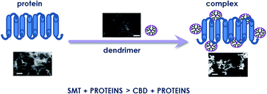
For nanoparticles to be used successfully in biomedical application, their interactions with biological fluids need to be investigated, in which they will react with proteins and other macromolecules. In this case, dendrimers might change their biological properties. Thus, the interaction of 2 generations of carbosilane–viologen–phosphorus and 2 types of carbosilane dendrimers with the 3 proteins – alkaline phosphatase from E. coli, human aspartate transaminase and L-lactate dehydrogenase from rabbit muscles, will be discussed. The techniques used included circular dichroism, zeta potential measurement, fluorescence quenching and transmission electron microscopy. The results show that positively charged hybrid and carbosilane dendrimers can interact with negatively charged regulatory proteins, and are able to form complexes. However, the spatial structure and the flexibility of dendrimer are as important as the protein configuration in complex formation.
Introduction
Since the 1980's,1–3 dendrimers have given scientists an almost unlimited field to explore, and new classes of them are constantly synthesized. Consequently, each class of dendrimers is now characterized by a range of properties. Dendrimers, due to its unique characteristics, have been examined for application as drug or genes carriers,4,5 contrasts in MRI,6,7 photosensitizers in photodynamic therapy8,9 and as antibacterial, antifungal10–12 or antiviral13 agents.
Evaluation of the biological properties should be the first and most important step in establishing the possible use of nanoparticles in biomedicine. Specifying the size, shape, architecture, surface groups, charge and other physico-chemical parameters is as important as understanding the interaction of nanostructures with living systems. Each foreign nanoparticle elicits a complex biological response. Nanostructures that appear in the biological fluid are immediately coated with proteins, creating a so-called “protein corona”.14–17 The division of nanoparticles into “hard” and “soft” groups showing their different models of interaction with proteins has been proposed. According to Tomalia,18 dendrimers are classified as soft nanostructures.18–20 Well-defined size and structure, flexibility and monodispersity are the main reasons why the dendrimers are examined for biomedical application.21
Interaction between 2 generations of hybrid dendrimers, 2 types of carbosilane dendrimers and 3 selected regulatory proteins – alkaline phosphatase (AP) from E. coli, lactate dehydrogenase (LDH) from rabbit muscles and human aspartate aminotransferase (AST) – has been investigated. The hybrid dendrimers, SMT1 and SMT2, were synthesized using a new “onion peel” approach. They have 2 kinds of cationic groups, those located at the branches due to viologen quaternized units, and those related to the ammonium groups at the surface of carbosilane wedges (Fig. 1).22 The cationic carbosilane dendrimers included here contain branches made of carbon–silicon (CBD-CS) or oxygen–silicon bonds (CBD-OS) and amino groups on the surface (Fig. 2). The CBD-CS are stable in water, whereas CBD-OS are not, being slowly hydrolyzed in aqueous environment.23,24 The enzymes have been chosen because of the different positions of their active sites, the rigidity of their structure (Table 1) and their important regulatory functions.25 They are involved in many metabolic pathways necessary for the proper bodily function.
 |
| Fig. 1 Chemical structures of dendrimers SMT1 and SMT2.22 | |
 |
| Fig. 2 Chemical structures of dendrimers CBD-CS and CBD-OS.24 | |
Table 1 Alkaline phosphatase, L-lactate dehydrogenase and aspartate transaminase characteristics
Protein |
Type |
Position of active site |
Rigidity of dimensional structure |
Molecular weight [kDa] |
Net charge |
AP |
Dimer |
Buried |
Rigid |
89 |
−18 |
LDH |
Tetramer |
Buried |
Flexible |
140 |
−5 |
AST |
Dimer |
Surface |
Flexible |
94 |
−16 |
AP belongs to the hydrolases group and is responsible for the dephosphorylation of a range of molecules, preferably in an alkaline environment. As AP is commonly found in many living species, including man, our studies were carried out on a large scale. Human AP is localized throughout the body, particularly in liver, bones, placenta and kidney. Therefore, there are a few classes of isoenzymes – API (intestinal alkaline phosphatase), APL (tissue-nonspecific alkaline phosphatase) and APP (placental alkaline phosphatase). AP is localized in the cell membrane as a GPI-anchor. Serum ALP levels are used as diagnostic markers of, e.g. osteomalacia, leukemia, lymphoma and Wilson's disease.26,27
L-Lactate dehydrogenase (LDH) is a tetrameric enzyme composed of 2 subunits M and H, encoded by the genes of LDHA and LDHB. Combining the 2 subunits gives 5 isoforms that are specifically distributed in tissues. Isoenzymes, of which LDHB predominates, are present in tissues where oxygen metabolism occurs (e.g. the heart). In contrast, isoenzymes containing predominantly LDHA occur in tissues where the conditions are anaerobic, such as skeletal muscle and the liver.28,29 Lactate dehydrogenase catalyzes the reversible conversion of pyruvate to lactate with the oxidation of NADH to NAD+. Under anaerobic conditions, LDH is an important enzyme for its ability to regenerate NAD+, and thereby allows for continued flow of carbon in the glycolysis pathway to assist anaerobic synthesis of ATP.30 Increase in the level of LDH occurs in diseases associated with tissue necrosis, and is also raised in the acute phase of viral hepatitis.28 LDH is an enzyme associated with membrane, found in most tissues of the body, and is released into the extracellular environment, when cells associated with inflammation are damaged. Due to this property, LDH is often used as a biomarker of inflammation in infections, such as bacterial meningitis, empyema and arthritis.31 A high activity of LDH can indicate increased tumor size and lead to a poor prognosis.32
Aspartate transaminase (AST) is an enzyme of the class of transferase. This enzyme catalyzes the reversible reaction of transferring an amino group from aspartate to α-ketoglutarate to produce oxaloacetate and glutamate. AST exists in 2 forms: cytosolic and mitochondrial.33,34 This protein is a homodimer, each string being made up of large and small domains between which the cofactor pyridoxal phosphate is hidden in twisted α-harmonica sandwiched between 8 α-helices from both domains. The N-terminal domain interacts with a large domain of the neighboring subunit and stabilizes the dimer.35 Aspartyl transaminase is present mainly in the liver and myocardium, and is not secreted directly into the bloodstream. Thus increase in its plasma activity is the result of cardiac myocyte or liver cell damage.33
Determining whether carbosilane–viologen–phosphorus and carbosilane dendrimers interact with these proteins is essential for their use in biomedicine.
Materials and methods
Dendrimers
Cationic dendrimers SMT1, SMT2, CBD-CS and CBD-OS were synthesized in Departamento de Quimica Inorgánica, Universidad de Alcalá (Spain);22,23 their properties are shown in Table 2.
Table 2 Parameters of studied dendrimers22–24
Dendrimer |
Generation |
Surface groups |
Inner charge |
Outer charge |
Molecular weight [g mol−1] |
SMT1 |
1 |
–NH3+ |
+12 |
+12 |
5303.73 |
SMT2 |
2 |
–NH3+ |
+12 |
+24 |
8660.73 |
CBD-CS |
2 |
–NMe3+ |
0 |
+16 |
4699.99 |
CBD-OS |
2 |
–NMe3+ |
0 |
+16 |
4603.56 |
Proteins
The regulatory proteins (enzymes) chosen were alkaline phosphatase from E. coli (AP) [EC 3.1.3.1, AP], lactate dehydrogenase from rabbit muscle (LDH) [EC 1.1.1.27, LDH] and human aspartate transaminase (AST) [ERMAD457IFCC, AST], purchased from Sigma-Aldrich (USA).
Fluorescence quenching of AP, AST and LDH
Fluorescence quenching of the proteins in the presence of dendrimers was measured with a Perkin Elmer LS-55 spectrofluorometer, using a 5 mm quartz cuvette in the range of 305–400 nm, with excitation at 296 nm. AP, AST and LDH were held 1 μM, but the dendrimers were used in increasing concentration, the levels being experiment-dependent. Samples were dissolved in 10 mM phosphate buffer, having previously shown that carbosilane–viologen–phosphorus and carbosilane dendrimers are not excited by 296 nm wavelength and do not emit fluorescence. Each spectrum is the result of at least 3 independent repetitions. The fluorescence intensities were normalized as F/F0, where F is the intensity of protein fluorescence in the presence of a dendrimer, whereas F0 is the fluorescence intensity of a protein without dendrimers.
Zeta potential measurement
Zeta potentials measured with a Zetasizer Nano ZS (Malvern, UK) based on the electrophoretic mobility of a solution of molecules in disposable plastic cells. The Zetasizer Nano ZS uses a combination of 2 techniques, electrophoresis and laser Doppler velocity measurement (LDV) to conduct phase analysis light scattering, and potentials were calculated using the Malvern software based on the Helmholtz–Smoluchowski equation.26,36 The electrophoretic mobility of the samples was determined from the average of 10 cycles. Measurements were made at 37 °C in the phosphate buffer (10 mM, pH 7.4). The concentration of enzymes was kept constant at 0.5 μM, whereas dendrimers SMT1, SMT2, CBD-CS and CBD-OS were added up to 50 μM.
Circular dichroism (CD) measurement
A Jacso J-815 CD spectropolarimeter was used to measure CD spectra of AP, AST and LDH in the absence or presence of 4 types of dendrimers: SMT1, SMT2, CBD-CS and CBD-OS. Spectra were measured in 5 mm path-length quartz cells from 195 to 260 nm, with the following parameters: scan speed – 20 nm min−1, step wavelength – 1 nm, response time – 4 s. Each spectrum is a result of at least 3 independent measurements. The enzymes were used at 1 μM. Dendrimers concentrations increased over the experiment, the final concentration depending on the enzyme being used. Measurements were made in 10 mM phosphate buffer. Dendrimers SMT1, SMT2, CBD-CS and CBD-OS dissolved in phosphate buffer (at concentrations used experimentally) were used as the baseline for the protein/dendrimers complexes spectral measurements.
Transmission electron microscopy
A JEOL-10 transmission electron microscopy was used for image capture. To form the complexes, the dendrimers were added to a protein solution at a molar ratio of 50
:
1. The samples were vortexed and incubated for 10 min at room temperature. Ten microliters of prepared solution was placed on a 200 mesh copper grid with a carbon-coated surface for 10 min and dried with filter paper. To samples were stained with 2% (m/v) uranyl acetate.
Statistical analysis
The results were subjected to statistical analysis with the use of t-test (in case of normal distribution) or Mann–Whitney rank sum test (in the case of lack of normal distribution). To assess the significance of differences between particular dendrimers ANOVA test was conducted. In case of differences, further analysis was performed using the Luke's method. Significance was chosen at p = 0.05 or less.
Results
Fluorescence quenching
In fluorescence measurements, there were significant differences between carbosilane–viologen–phosphorus and carbosilane dendrimers for all 3 proteins. Hybrid dendrimers of both generations quenched the fluorescence in concentration dependent manner. The effect of both carbosilane dendrimers was slight, the least effect being with AP. An analogous situation occurred with the carbosilane–viologen–phosphorus dendrimers, although the general effect of these nanoparticles was significant even at low molar ratios. Decrease in fluorescence intensity below 20% was found at a molar ratio (dendrimer/protein) of 5
:
1 for AST, 20
:
1 for AP and 10
:
1 for LDH for both generations of hybrid dendrimers. For carbosilane dendrimers, the F/F0 has fallen below 70% in a molar ratio of 50
:
1 only for LDH (Fig. 3). Examples of the fluorescence spectra of LDH in the presence of all dendrimers are shown in Fig. 4.
 |
| Fig. 3 Quenching of AST, AP and LDH fluorescence by dendrimers SMT1, SMT2, CBD-OS and CBD-CS. The dependence of F/F0 at the maximum fluorescence for each protein (AP λ = 330, AST λ = 340, LDH λ = 350) (n ≥ 3). | |
 |
| Fig. 4 Fluorescence quenching spectra of LDH in the presence of dendrimer SMT1, SMT2, CBD-CS, CBD-OS (n ≥ 3). | |
Zeta potential
With the enzymes kept constant at 0.5 μM, dendrimers SMT1, SMT2, CBD-CS and CBD-OS were added at increasing concentrations from 0 to 50 μM. All 4 dendrimers affected the zeta potential of the 3 enzymes. Each protein studied before dendrimer was added had negative zeta potential values (AST −12 mV, AP −17 mV, LDH −5 mV). Dendrimers changed the baseline values from negative to positive. The plateaus in most cases were reached below <+15 mV. An example of the changes of AST zeta potential with dendrimers is shown in Fig. 7. Analysis of zeta potential plots vs. dendrimer/protein molar ratio gave an estimate of the number of dendrimer molecules bound by the enzyme; the numbers of dendrimers per molecule of each of the enzymes are given in Table 3. For AST and LDH the dendrimer bound in the lowest molar ratio was CBD-CS (AST
:
CBD-CS – 1
:
11; LDH
:
CBD-CS – 1
:
4), whereas it was CBD-OS (AP
:
CBD-OS – 1
:
7) for AP. The highest molar ratio of dendrimer/enzyme was with CBD-OS and AST (18
:
1); but for dendrimer SMT2 with AP it was 12
:
1, and dendrimer SMT1 with LDH – 11
:
1.
Table 3 The number of dendrimers bound to one molecule of protein calculated based on the zeta potential measurement
|
SMT1 |
SMT2 |
CBD-CS |
CBD-OS |
AST |
14 |
12 |
11 |
18 |
AP |
11 |
12 |
8 |
7 |
LDH |
11 |
8 |
4 |
9 |
Circular dichroism
The CD spectra for AST, AP and LDH were typical of the secondary structure of α-helical proteins, with characteristic peaks at 208 nm and 222 nm.37 Adding carbosilane–viologen–phosphorus dendrimers usually changed the spectra of all 3 enzymes in a concentration-dependent manner. Unlike hybrid dendrimers, the effect of carbosilane dendrimers, both CBD-OS and CBD-CS, was slight. Changes in the spectra imply binding of nanoparticles to protein. The LDH CD spectra at increasing dendrimer concentrations are shown in Fig. 5. The mean residue ellipticity at 2 minima, 208 and 222 nm, are shown for 3 enzymes in the presence of our 4 dendrimers (Fig. 6). There were no significant changes in θ/θo for λ = 208 for all 3 enzymes after adding 2 type of carbosilane dendrimers. At λ = 222, significant changes occurred for CBD-CS/LDH at a molar ratio in excess of 10
:
1. In contrast, the hybrid dendrimers significantly changed ellipticity of AP, AST and LDH in a concentration-dependent manner at λ = 208 nm. Although SMT1 or SMT2 led to changes at λ = 222 nm for only AST and LDH, the former also affected AP structure.
 |
| Fig. 5 CD spectra of LDH in the presence of dendrimer SMT1, SMT2, CBD-CS, CBD-OS (n ≥ 3). | |
 |
| Fig. 6 Changes in mean residue ellipticity at λ = 208 nm (left panel) and λ = 222 nm (right panel) (n ≥ 3). | |
The greatest modifications in mean residue ellipticity of LDH and AST at 208 and 222 nm have previously been seen at low molar ratios from 0.5
:
1 to 5
:
1 (dendrimer/protein). With AP, the effect of carbosilane–viologen–phosphorus dendrimers was minor compared to AST and LDH; however, the influence of SMT1 was higher than SMT2 and refers to both minima.
TEM
As an example of hybrid dendrimers, SMT1 was chosen (Fig. 8), whereas it was CBD-CS for carbosilane (Fig. 9). Differences in the formation of the complexes by CBD-CS and SMT1 were found. Shapes and sizes of complexes were dependent on both the type of dendrimer and enzyme. Because TEM samples must be dried, the true nature of complexes cannot be found. However, the images confirm that complexes are indeed formed.
 |
| Fig. 7 Alteration of AST zeta potential in the presence of dendrimers SMT1, SMT2, CBD-CS and CBD-OS (n ≥ 3). | |
 |
| Fig. 8 Electron microphotographs of AST, AP and LDH proteins, alone (left panel) and complexed with dendrimer SMT1 (right panel). Bar = 100 nm. | |
 |
| Fig. 9 Electron microphotographs of AST, AP and LDH proteins, alone (left panel) and complexed with dendrimer CBD-CS (right panel). Bar = 100 nm. | |
Discussion
For nanoparticles to be considered safe and efficient in biomedical applications they must first be examined for their interaction with biological material, i.e. their interaction with different types of proteins.16,38 Nevertheless, for those in biological fluid that will bind dendrimers, the association depends on the surface properties of nanoparticle, e.g. its charge.39 Interactions of this kind have been described by Ciolkowski et al.40 who investigated the influence of positive, neutral and negative PAMAM dendrimers on porcine pepsine. Wasiak et al.41 have shown the effect of cationic phosphorus-containing dendrimers on acetylcholinesterase, and Milowska et al.42–45 have classified different types of cationic dendrimers based on their interaction with α-synuclein.
The results of our present work show that dendrimers can interact the 3 enzymes used. Analysis of zeta potential changes showed that the number of binding centers was dependent on the nature of proteins as well as on the type and generation of dendrimer.
Each of the 3 enzymes has a different position of its active site, a different rigidity of its structure and different functions in organisms.25 AST and AP are dimers, whereas LDH is a tetrameric enzyme. AP has a stable, rigid structure and active site 11 Å deep inside the molecule. Compared to E. coli AP, human phosphatases are more active but less stable.25,26,46 The next enzyme, characterized by an active center hidden inside it (10 Å), and with a flexible structure compared to AP, is LDH. Another important feature of LDH is its so-called mobile loop, which extends into a solution to enclose the bound substrate in the presence of cofactor.25,47 In AST, the active site is superficial and the structure is comparatively flexible. The conformation of the aspartate transaminase in the presence of its substrate is closed.25,48 The results with hybrid dendrimers seem to confirm those differences in the structures of these enzymes. The results in the case of carbosilane dendrimers are inconclusive.
The data from fluorescence measurements indicate that hybrid dendrimers strongly quenched the fluorescence of AP, AST and LDH. AP gave a slightly weaker emission, but this may be due to its rigid structure. In contrast for carbosilane dendrimers, slight changes occurred only with LDH (Fig. 3). The fluorescence measurements indicate the impact of dendrimers on tryptophan (Trp) residues which are located on the surface of a protein globule. In means that dendrimers can influence Trp when they are located in a vicinity of Trp. Thus, if a dendrimer binds far from Trp residue it cannot affect protein fluorescence.
These results have been confirmed by CD measurements, with one exception; the effect of hybrid dendrimers on the secondary structure of AP was relatively minor compared to AST and LDH (Fig. 6). Data from Shcharbin et al.25 on PAMAM dendrimers of generation 3 and 4 and Ionov et al.49 on phosphorus dendrimers of generations 3 and 4 slightly varied from the ones we obtained. The effect of hybrid dendrimers on the fluorescence of AST and LDH is stronger than of PAMAM dendrimers of both generations, and similar to the effect of CPD of both generations. In contrast, carbosilane dendrimers quenched the fluorescence to a lesser extent than PAMAM and phosphorus dendrimers. Nevertheless, the amount of dendrimers significantly changes AP structure increases in the following order: PAMAM G3/PAMAM G4/CBD-OS/CBD-CS, SMT2, SMT1, CPD G4/CPD G3. Both zeta potential measurements and TEM imaging confirmed the formation of dendrimers/protein complexes. Differences in the morphology of the complexes are evident in the electron micrographs (Fig. 8 and 9). The electron micrographs of complexes between enzymes and dendrimer SMT1 are similar to the CPD G4-enzyme complexes generated by Ionov et al.,49 whereas complexes with CBD-CS resemble PAMAM G4-enzyme complexes.25 Not only the structure of proteins themselves, but the structure and charge of the dendrimers affect the nature of the complexes. The SMT1 and SMT2 hybrid dendrimers have a rigid inner structure containing 12 positive charges, and there are surface charge of +12 for SMT1 and +24 for SMT2. For hybrid dendrimers the surface flexibility increases with increasing dendrimer generation. However, CBD-OS and CBD-CS dendrimers are flexible systems throughout their structure with positive charges only at the surface (+16). Because interactions between proteins and dendrimers are mostly electrostatic, the bigger the positive charge on the surface of dendrimer, the higher the efficiency should be. However, there appeared to be no relationship between any induced effect and the surface charge itself. Even though charge is important, the elasticity of the nanoparticle structure is also a significant factor.50,51 More flexible systems may be allow better adjustment of protein interactions, and therefore fewer dendrimer molecules may be necessary for binding one single protein molecule than can be envisaged by the zeta potential. The strong interaction with enzymes in hybrid systems and measured by different techniques could be due to the presence of a large number of positive charges, including the inner ones. However, the effect of hybrid dendrimers on the structures of AP, AST and LDH is comparable or even slightly lower for SMT2. For higher dendrimer generations, the core and the wedges become increasingly shielded from their surroundings by increasing surface density. Therefore, there is reduced availability of the inner positive charges in interactions with SMT2 dendrimers.
Conclusions
Positively charged hybrid and carbosilane dendrimers can interact with the negatively charged proteins, forming complexes. The spatial structure of the dendrimer is equally important to protein structure in determining the formation and nature of the complexes. This information should be useful in creating new nanoparticles for use in biomedical applications.
Acknowledgements
This work was supported by the National Science Centre of Poland, Project No. DEC-2012/04/M/NZ1/00059, partly by Belarusian Republican Foundation for Fundamental Research, Project No. B15MS-001 and by grant from MINECO CTQ-2014-54004-P to the University of Alcala. CIBER-BBN is an initiative funded by the VI National R&D&i Plan 2008–2011, Iniciativa Ingenio 2010, Consolider Program, CIBER Actions and financed by the Instituto de Salud Carlos III with assistance from the European Regional Development Fund.
References
- E. Buhleier, W. Wehner and F. Vögtle, Synthesis, 1978, 155 CrossRef CAS.
- D. A. Tomalia, H. Baker, J. Dewald, M. Hall, G. Kallos, S. Martin, J. Roeck, J. Ryder and P. Smith, Polym. J., 1985, 17, 117 CrossRef CAS.
- G. R. Newkome, Z. Yao, G. R. Baker and V. K. Gupta, J. Org. Chem., 1985, 50, 2003 CrossRef CAS.
- A. Gothwal, P. Kesharwani, U. Gupta, I. Khan, M. C. Iqbal Mohd Amin, S. Banerjee and A. K. Iyer, Curr. Pharm. Des., 2015, 30, 4519 CrossRef.
- V. Dzmitruk, A. Szulc, D. Shcharbin, A. Janaszewska, N. Shcharbina, J. Lazniewska, D. Novopashina, M. Buyanova, M. Ionov, B. Klajnert-Maculewicz, R. Gómez-Ramirez, S. Mignani, J. P. Majoral, M. A. Muñoz-Fernández and M. Bryszewska, Int. J. Pharm., 2015, 1–2, 288 CrossRef PubMed.
- É. Tóth, D. Pubanz, S. Vauthey, L. Helm and A. E. Merbach, Chem.–Eur. J., 1996, 2, 1607 CrossRef.
- H. Kobayashi and M. W. Brechbiel, Adv. Drug Delivery Rev., 2005, 15, 2271 CrossRef PubMed.
- M. Dabrzalska, M. Zablocka, S. Mignani, J. P. Majoral and B. Klajnert-Maculewicz, Int. J. Pharm., 2015, 1–2, 266 CrossRef PubMed.
- M. Dabrzalska, N. Benseny-Cases, R. Barnadas-Rodríguez, S. Mignani, M. Zablocka, J. P. Majoral, M. Bryszewska, B. Klajnert-Maculewicz and J. Cladera, Anal. Bioanal. Chem., 2016, 408, 535 CrossRef CAS PubMed.
- J. Janiszewska and Z. Urbańczyk-Lipkowska, Acta Biochim. Pol., 2006, 1, 77 Search PubMed.
- P. Zielińska, M. Staniszewska, M. Bondaryk, M. Koronkiewicz and Z. Urbańczyk-Lipkowska, Eur. J. Med. Chem., 2015, 105, 106 CrossRef PubMed.
- T. K. Lind, P. Polcyn, P. Zielinska, M. Cárdenas and Z. Urbanczyk-Lipkowska, Molecules, 2015, 1, 738 CrossRef PubMed.
- R. Roy, D. Zanini, S. J. Meunier and A. Romanowska, J. Chem. Soc., Chem. Commun., 1993, 1869–1872 RSC.
- T. Cedervall, I. Lynch, S. Lindman, T. Berggård, E. Thulin, H. Nilsson, K. A. Dawson and S. Linse, Proc. Natl. Acad. Sci. U. S. A., 2007, 13, 2050 CrossRef PubMed.
- P. Aggarwal, J. B. Hall, C. B. McLeland, M. A. Dobrovolskaia and S. E. McNeil, Adv. Drug Delivery Rev., 2009, 21, 428 CrossRef PubMed.
- M. Rahman, S. Laurent, N. Tawil, L. Yahia and M. Mahmoud, Protein-Nanoparticle Interactions, Springer, Berlin, Heidelberg, 2013 Search PubMed.
- W. Jiang, K. Lai, Y. Wu and Z. Gu, Arch. Pharmacal Res., 2014, 37, 129 CrossRef CAS PubMed.
- D. A. Tomalia, J. Nanopart. Res., 2009, 11, 1251 CrossRef CAS PubMed.
- D. A. Tomalia, New J. Chem., 2012, 36, 264 RSC.
- R. M. Kannan, E. Nance, S. Kannan and D. A. Tomalia, J. Intern. Med., 2014, 276, 579 CrossRef CAS PubMed.
- S. Svenson and D. A. Tomalia, Adv. Drug Delivery Rev., 2005, 14, 2106 CrossRef PubMed.
- S. Moreno, A. Szwed, N. El Brahmi, K. Milowska, J. Kurowska, E. Fuentes-Paniagua, E. Pedziwiatr-Werbicka, T. Gabryelak, N. Katir, J. de la Mata, M. A. Muñoz-Fernández, R. Gomez-Ramirez, A. M. Caminade, J. P. Majoral and M. Bryszewska, RSC Adv., 2015, 5, 25942 RSC.
- J. F. Bermejo, P. Ortega, L. Chonco, R. Eritja, R. Samaniego, M. Müllner, E. de Jesus, F. J. de la Mata, J. C. Flores, R. Gomez and A. Muñoz-Fernandez, Chemistry, 2007, 13, 483 CrossRef CAS PubMed.
- M. Ionov, K. Ciepluch, B. Klajnert, S. Glińska, R. Gomez-Ramirez, F. de la Mata, M. A. Munoz-Fernandez and M. Bryszewska, Colloids Surf., B, 2013, 1, 236 CrossRef PubMed.
- D. Shcharbin, M. Ionov, V. Abashkin, S. Loznikova, V. Dzmitruk, N. Shcharbina, L. Matusevich, K. Milowska, K. Gałęcki, S. Wysocki and M. Bryszewska, Colloids Surf., B, 2015, 1, 377 CrossRef PubMed.
- J. E. Coleman, Annu. Rev. Biophys. Biomol. Struct., 1992, 21, 441 CrossRef CAS PubMed.
- K. Poelstra, W. W. Bakker, P. A. Klok, M. J. Hardonk and D. K. Meijer, Lab. Invest., 1997, 76, 319 CAS.
- R. K. Murray, D. K. Granner, P. A. Mayes and V. W. Rodwell, Biochemia Harpera, 1995, 90–91, 917 Search PubMed.
- D. Mirebeau-Prunier, S. Le Pennec, C. Jacques, J. Fontaine, N. Gueguen, N. Boutet-Bouzamondo, A. Donnart, Y. Malthiery and F. Savagner, PLoS One, 2013, 8, 58683 Search PubMed.
- A. Shahriari, N. J. Dawson, R. A. V. Bell and K. B. Storey, Enzyme Res., 2013, 2013, 461374 CrossRef PubMed.
- L. C. Ede, J. O'Brien, T. Chonmaitree, Y. Han and J. A. Patel, Pediatr. Res., 2013, 73, 349 CrossRef CAS PubMed.
- B. M. William, N. R. Bongu, M. Bast, R. G. Bociek, P. J. Bierman, J. M. Vose and J. O. Armitage, Rev. Bras. Hematol. Hemoter., 2013, 35, 189 Search PubMed.
- S. Petrovic, B. Ozertic and M. Krajnovic-Ozertiv, Biochim. Biophys. Acta, 1994, 17, 201 CrossRef.
- E. Bańkowski, Biochemia, MedPharm, Polska, 2006 Search PubMed.
- F. J. Fernandez, D. Vries, E. Peña-Soler, M. Coll, P. Christen, H. Gehring and M. C. Vega, Biochim. Biophys. Acta, 2012, 1824, 339 CrossRef CAS PubMed.
- A. Sze, D. Erickson, L. Ren and D. Li, J. Colloid Interface Sci., 2003, 15, 402 CrossRef.
- N. J. Greenfield, Nat. Protoc., 2006, 1, 2876 CrossRef CAS PubMed.
- M. Lundqvist, J. Stigler, G. Elia, I. Lynch, T. Cedervall and K. A. Dawson, Proc. Natl. Acad. Sci. U. S. A., 2008, 23, 14265 CrossRef PubMed.
- A. Akesson, M. Cardenas, G. Elia, M. P. Monopoli and K. A. Dawson, RSC Adv., 2012, 2, 11245 RSC.
- M. Ciolkowski, M. Rozanek, M. Bryszewska and B. Klajnert, Biochim. Biophys. Acta, 2013, 1834, 1982 CrossRef CAS PubMed.
- T. Wasiak, M. Marcinkowska, I. Pieszynski, M. Zablocka, A. M. Caminade, J. P. Majoral and B. Klajnert-Maculewicz, New J. Chem., 2015, 39, 4852 RSC.
- K. Milowska, M. Malachowska and T. Gabryelak, Int. J. Biol. Macromol., 2011, 1, 742 CrossRef PubMed.
- K. Milowska, T. Gabryelak, M. Bryszewska, A. M. Caminade and J. P. Majoral, Int. J. Biol. Macromol., 2012, 1, 1138 CrossRef PubMed.
- K. Milowska, J. Grochowina, N. Katir, A. El Kadib, J. P. Majoral, M. Bryszewska and T. Gabryelak, Mol. Pharm., 2013, 4, 1131 CrossRef PubMed.
- K. Milowska, A. Szwed, M. Mutrynowska, R. Gomez-Ramirez, F. J. de la Mata, T. Gabryelak and M. Bryszewska, Int. J. Pharm., 2015, 30, 268 CrossRef PubMed.
- J. L. Millán, Purinergic Signalling, 2006, 2, 335 CrossRef PubMed.
- L. Qiu, M. Gulotta and R. Callender, Biophys. J., 2007, 1, 1677 CrossRef PubMed.
- J. F. Kirsch, G. Eichele, G. C. Ford, M. G. Vincent, J. N. Jansonius, H. Gehring and P. Christen, J. Mol. Biol., 1984, 15, 497 CrossRef.
- M. Ionov, A. Ihnatsyeu-Kachan, S. Michlewska, N. Shcharbina, D. Shcharbin, J. P. Majoral and M. Bryszewska, Int. J. Pharm., 2016, 29, 247 CrossRef PubMed.
- D. A. Tomalia, J. B. Christensen and U. Boas, Dendrimers, Dendrons, and Dendritic Polymers Discovery, Applications, and the Future, Cambridge University Press, Cambridge, 2012 Search PubMed.
- O. Nowacka, K. Milowska, S. Belica-Pacha, B. Palecz, K. Šipošová, Z. Gazova and M. Bryszewska, Int. J. Biol. Macromol., 2016, 82, 54 CrossRef CAS PubMed.
|
This journal is © The Royal Society of Chemistry 2016 |
Click here to see how this site uses Cookies. View our privacy policy here.