
Monitoring quality consistency of Ixeris sonchifolia (Bunge) Hance injection by integrating UV spectroscopic fingerprints, a multi-wavelength fusion fingerprint method, antioxidant activities and UHPLC/Q-TOF-MS
Lanping Yang,
Xiuman Xie,
Liu Yang,
Jing Zhang and
Guoxiang Sun*
School of Pharmacy, Shenyang Pharmaceutical University, Shenyang, P. R. China. E-mail: gxswmwys@163.com; Fax: +86-024-2398628
First published on 8th September 2016
Abstract
Traditional Chinese medicines/herbal medicines (TCM/HM) are too complicated mixture systems to investigate their quality consistency effectively using a single detection technique. Hence, finding an integrated, rapid and effective quality control method is of great importance. In the present study, a novel strategy integrating ultra violet (UV) spectroscopic fingerprints and multi-wavelength fusion fingerprints has been proposed for quality consistency of the popular patent medicine Ixeris sonchifolia (Bge.) Hance Injection (ISHI). The quality grades of 28 batches of the ISHI samples were evaluated by the systematic quantitative fingerprint method (SQFM) from qualitative and quantitative perspectives. As a result, UV spectroscopic fingerprints agree well with multi-wavelength fusion fingerprints. In order to investigate the chemical components of the ISHI sample, ultra-high performance liquid chromatography coupled with quadrupole-time-of-flight mass spectrometry (UHPLC/Q-TOF-MS) has been exploited in the negative ion mode, providing an important chemical structure foundation for further bioactivity and quality control studies. In addition, the orthogonal projection to latent structures (OPLS) was established to investigate the fingerprint-efficacy relationship between high performance liquid chromatography (HPLC) fingerprints and antioxidant activities. These results indicate that integrating UV spectroscopic fingerprints and multi-wavelength fusion fingerprints coupled with antioxidant activities and UHPLC/Q-TOF-MS provide a rapid and effective approach to monitor the quality consistency of TCM/HM.
1. Introduction
TCM/HM usually contain a great number of components, which contribute to the therapeutic effects all together with multiple targets.1 Obviously, it is inadequate to control the quality of the TCM/HM by quantifying only one or a few marker substances. Fingerprint techniques, especially chromatographic fingerprints, as a rational and powerful method, systematically characterize the complicated TCM/HM, and have been widely accepted and adopted for quality control of TCM/HM in recent years.2,3 However, single wavelength fingerprints cannot reflect the sample chemical information systematically. In this situation, a multi-wavelength fusion fingerprints method4,5 has been adopted to make up for this limitation. By combining the macroscopical fingerprint characteristics and computer technology, the fusion fingerprints technique could realize the overall evaluation of the samples.6Although chromatographic fingerprints performed on HPLC,3 UHPLC,7 gas chromatograph (GC),8 capillary electrophoresis (CE),9 etc. are frequently used for quality control of TCM/HM, cannot offer information about all the chemical components in the TCM/HM system, which limit the overall quality control of multi-constituents synergistically. In this situation, spectroscopic techniques, such as UV, near-infrared (NIR) and mid-infrared (MIR) have been proposed;10,11 and UV spectroscopic fingerprint is preferred using flow injection analysis (FIA)12 to monitor the quality consistency of TCM/HM, owing to its apparent advantages, such as short measurement time, simple sample preparation and low experimental expenses. As we all know, UV spectra can reflect the chemical bond information of π–π*, n–π* and n–σ*.13 Since UV spectra reflects the features of unsaturated bonds of components qualitatively and quantitatively, it seems to be a straightforward and effective method for TCM/HM quality control.
TCM/HM possess complex components and variety of therapeutic efficacies, thus, it is necessary to carry out the chemical profiling of TCM/HM for further bioactivity and quality control studies. In recent years, UHPLC/Q-TOF-MS has been proved to be an efficient analytical tool for the chemical components of TCM/HM.14,15 Compared with the conventional HPLC, UHPLC provides lower consumption of samples and higher sensitivity in a shorter analysis time.7 Meanwhile, Q-TOF-MS offers both valuable fragment ions and reliable formulas, which is benefited by the high resolution for structural characterization.16 Accordingly, UHPLC/Q-TOF-MS supplies a rapid and powerful method to investigate TCM/HM components.
Antioxidants can scavenge free radicals to reduce the risk of numerous diseases, which is closely related to the harmful effects, such as lipid peroxidation, protein peroxidation, and DNA damage of free radicals.17 Accordingly, research on natural antioxidants and their antioxidant properties in foods, plant materials and TCM/HM is attracting increasing attention.2,18 2,2-Diphenyl-1-picrylhydrazyl (DPPH) is a stable and commercially available radical, and the DPPH radical scavenging assay providing simple, rapid, sensitive and reproducible procedures, has been employed to evaluate the antioxidant activity of TCM/HM.3,19,20 These information encourage us to investigate the antioxidant activities of TCM/HM and correlate them with chromatographic fingerprints.
ISHI has been widely used for its anti-inflammatory and haemostatic effects, its influence on improving blood circulation, and its potential protection against ischemia brain injury with relatively low side effects.21 The compositions of ISHI mainly including organic acids, flavonoids, sesquiterpene lactones, nucleosides and other compounds.22 Previous studies on chemical components of Ixeris sonchifolia and its injection (i.e., ISHI) have been reported. For example, Liu et al. characterized a total of 33 constituents from Ixeris sonchifolia and its injection, including 16 organic acids, 7 flavonoids, 7 sesquiterpene lactones, and 3 nucleosides;23 A total of 6 sesquiterpene lactones, 6 phenolic acids and 7 flavonoids were identified or tentatively characterized in Ixeris sonchifolia by Shi et al.;24 Wang et al. characterized a total of 25 phenolic acids in ISHI;25 A total of 14 chlorogenic acids and 16 flavonoids have been screened and identified in ISHI by Wang et al.26 After a thorough literature search, 7 compounds including 2 organic acids (vanillic acid and (E)-2,5-dihydroxy cinnamic acid), 3 sesquiterpene lactones (sonchifolactone E, 3-hydroxydehydroleucodin and 11,13-dihydroixerinoside) and 2 phenols (3-(4′-hydroxy-3′-methoxyphenyl)-1-propanol and vanillin) were first identified in ISHI in the present study. In addition, published studies have shown that the antioxidant components, especially organic acids and flavonoids in Ixeris sonchifolia and its injection, can provide neuro-protective effects against ischemia-induced cellular injury.21,27 Accordingly, the antioxidant activity of the ISHI was performed by the DPPH assay in this study, and the predictive model was established using the OPLS method.28
In the present study, UV spectroscopic fingerprints and a multi-wavelength fusion fingerprints method were developed for evaluating the quality consistency of 28 batches of the ISHI samples, where the fusion fingerprint is based on the three wavelengths that displayed strong UV absorption bands. In fingerprint assessments, SQFM was established for quality analysis of the ISHI from qualitative and quantitative perspectives. In addition, the fingerprint-efficacy relationship between HPLC fingerprints and antioxidant activities was established utilizing OPLS. UHPLC/Q-TOF-MS has been exploited to systematically investigate the chemical components in ISHI in the negative ion mode, which provided information on chemical substances for bioactivity and quality control studies. It has been demonstrated that integrating UV spectroscopic fingerprints and multi-wavelength fusion fingerprints method coupled with antioxidant activities and UHPLC/Q-TOF-MS offers an effective and powerful method for quality control of ISHI.
2. Experimental
2.1 Chemicals and reagents
Reference standards, caffeic acid, 5-caffeoylquinic acid and ferulic acid were acquired from the National Institute for the Control of Pharmaceutical and Biological Products (Beijing, China); chicoric acid and luteolin-7-O-β-D-glucuronide were supplied by Chengdu Puri France Science and Technology Development Co., Ltd. (Chengdu, China); luteolin-7-O-β-D-glucoside and luteolin were provided by Shanghai Winherb Medical Technology Co., Ltd. (Shanghai, China); uridine, guanosine and DPPH were purchased from Sigma Chemical Co. (St. Louis, MO, USA). A total of 28 batches of the ISHI samples (numbered S1–S28) were obtained from Shenyang Shuangding Pharmaceutical Co., Ltd. (Shenyang, China; Manufacturer A, including S1–S23) and Tonghua Huaxia Pharmaceutical Co., Ltd. (Jilin, China; Manufacturer B, including S24–S28), respectively. In the UHPLC/Q-TOF-MS analysis, acetonitrile (MS grade) was purchased from Merck (Darmstadt, Germany); formic acid (HPLC grade, purity >95%) was obtained from Sigma (Saint Louis, MO, USA). In the HPLC and UV spectroscopic analysis, methanol (HPLC grade), acetonitrile (HPLC grade) and glacial acetic acid (HPLC grade) were purchased from Yuwang Industry Co., Ltd. (Shandong, China). High-purity deionized water was prepared by a Milli-Q system (Bedford, MA, USA). Other reagents were all of analytical grade.2.2 Preparation of standard and sample solutions
The reference standards of caffeic acid, 5-caffeoylquinic acid, ferulic acid, chicoric acid, luteolin-7-β-D-glucuronide, luteolin-7-glucoside, luteolin, uridine and guanosine were accurately weighed separately and dissolved in methanol, and stored at 4 °C prior to use.Each of the tested ISHI samples was filtered through a 0.45 μm Millipore filter (Beijing Sunrise T&D Company, China) prior to use.
2.3 Experimental conditions
![[thin space (1/6-em)]](https://www.rsc.org/images/entities/char_2009.gif) :50, v/v) was adopted as the carrier. The temperature of the PTFE tube was maintained at 35 °C. The flow rate, sample injection volume and wavelength interval was set at 0.5 mL min−1, 1 μL and 1 nm, respectively. Online UV spectra was obtained over a wavelength range of 200–400 nm with a slit width of 1 nm.
:50, v/v) was adopted as the carrier. The temperature of the PTFE tube was maintained at 35 °C. The flow rate, sample injection volume and wavelength interval was set at 0.5 mL min−1, 1 μL and 1 nm, respectively. Online UV spectra was obtained over a wavelength range of 200–400 nm with a slit width of 1 nm.2.4 Theory of SQFM
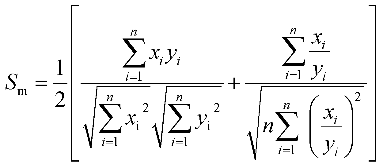
We assume that the sample fingerprint (SFP) and reference fingerprint (RFP) vector are![[x with combining right harpoon above (vector)]](https://www.rsc.org/images/entities/i_char_0078_20d1.gif) = (x1, x2,…, xn) and
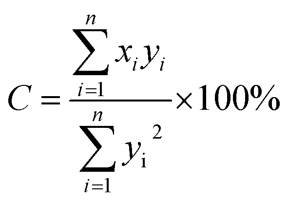
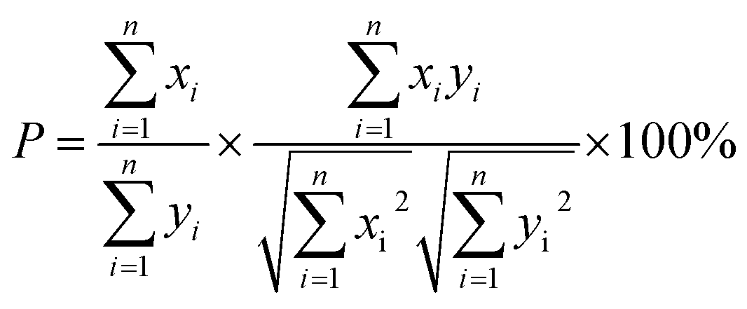
= (x1, x2,…, xn) and ![[y with combining right harpoon above (vector)]](https://www.rsc.org/images/entities/i_char_0079_20d1.gif) = (y1, y2,…yn), respectively, where xi and yi are the peak area of the co-possessing peak in the SFP and RFP, respectively. Macro qualitative similarity (Sm), a qualitative parameter as shown in eqn (1), can accurately describe the resemblance in terms of the distribution and number of fingerprints between SFP and RFP. Furthermore, projection content similarity (as defined in eqn (2)) and quantitative similarity (P, as shown in eqn (3)) were calculated for quantitative assessment of the fingerprints. Combining the above two quantitative properties yields macro quantitative similarity (Pm, as defined in eqn (4)), which is a measure to monitor the overall content of chemical components in SFP and RFP. Finally, a fingerprint leveling coefficient (α), as defined in eqn (5), is another quantitative parameter that can detect the difference between SFP and RFP.
= (y1, y2,…yn), respectively, where xi and yi are the peak area of the co-possessing peak in the SFP and RFP, respectively. Macro qualitative similarity (Sm), a qualitative parameter as shown in eqn (1), can accurately describe the resemblance in terms of the distribution and number of fingerprints between SFP and RFP. Furthermore, projection content similarity (as defined in eqn (2)) and quantitative similarity (P, as shown in eqn (3)) were calculated for quantitative assessment of the fingerprints. Combining the above two quantitative properties yields macro quantitative similarity (Pm, as defined in eqn (4)), which is a measure to monitor the overall content of chemical components in SFP and RFP. Finally, a fingerprint leveling coefficient (α), as defined in eqn (5), is another quantitative parameter that can detect the difference between SFP and RFP.
Accordingly, the quality evaluation method in terms of Sm, Pm and α is named as SQFM,3 by which TCM/HM quality can be classified into 8 grades. The evaluation criteria by SQFM are listed in Table 1. Accordingly, grade 1 belongs to the highest quality and grade 8 to the lowest one, and grades 1–5 are recognized as qualified.
 | (1) |
 | (2) |
 | (3) |
 | (4) |
 | (5) |
| Grade | 1 | 2 | 3 | 4 | 5 | 6 | 7 | 8 |
|---|---|---|---|---|---|---|---|---|
| Sm ≥ | 0.95 | 0.90 | 0.85 | 0.80 | 0.70 | 0.60 | 0.50 | <0.50 |
| Pm (%) ∈ | 95–105 | 90–110 | 80–120 | 75–125 | 70–130 | 60–140 | 50–150 | 0–∞ |
| α ≤ | 0.05 | 0.10 | 0.15 | 0.20 | 0.30 | 0.40 | 0.50 | >0.50 |
| Quality | Best | Better | Good | Fine | Moderate | Common | Defective | Inferior |
2.5 Data analysis
MassLynx 4.1 software (Waters, USA) was used for UHPLC/Q-TOF/MS data processing. An in-house developed software “Digitized Evaluation System for Super-Information Characteristics of TCM Fingerprints 4.0” (software certificate no. 0407573, China) was used to evaluate the fingerprints. SIMCA-P+ software (Version 13.0, Umetrics, Umea, Sweden) was used for chemometric analysis.3. Results and discussion
3.1 UHPLC/Q-TOF-MS analysis of the ISHI sample
Under the UHPLC/Q-TOF-MS conditions described in Section 2.3.1, totally 38 compounds (Fig. 1A), including 16 organic acids, 10 flavonoids, 8 sesquiterpene lactones, 2 nucleosides and 2 phenols, were identified in ISHI. Structures of them are summarized in Fig. 2. Information on the tR (min), observed mass (m/z), calculated mass (m/z), mass error (in ppm), molecular formula, fragment ions, identification and maximum UV absorption wavelength (UV λmax) are summarized in Table 2. Components with reference standards (compounds 3, 4, 16, 17, 18, 28, 29, 31 and 38) were unambiguously identified by comparing the retention time, accurate mass (within 5 ppm error) and MS/MS data. Components without reference standards were tentatively characterized based on accurate mass measurements (within 5 ppm error), tandem MS behavior and related literature. | ||
| Fig. 1 Total ion chromatogram of ISHI in negative ion mode (A), the MS/MS spectra and proposed fragmentation pathways of chicoric acid (B), luteolin-7-o-β-D-glucuronide (C), ixerin Z (D) and guanosine (E) in the negative ion mode. | ||
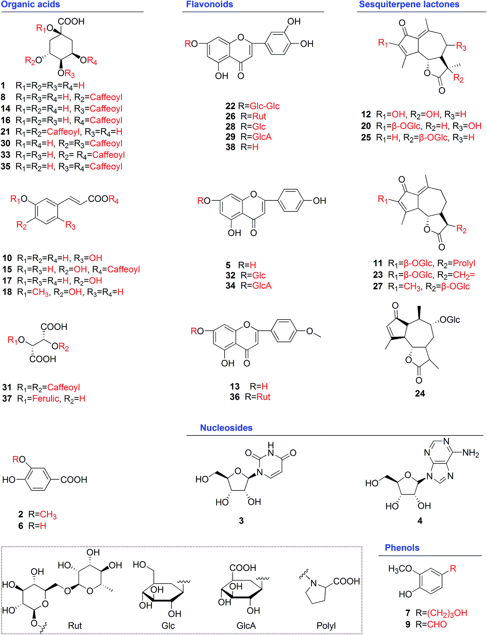 | ||
| Fig. 2 Chemical structures of 38 identified compounds. | ||
| No. | tR (min) | Observed mass (m/z) | Calculated mass (m/z) | Error (ppm) | Formula | Fragment ions | Identification | UV λmax (nm) |
|---|---|---|---|---|---|---|---|---|
| a [M + HCOO]−.b Compounds be identified in ISHI for the first time. | ||||||||
| 1 | 0.92 | 191.0559 | 191.0556 | 1.57 | C7H12O6 | 191, 173, 127 | Quinic acid | 250, 280sh |
| 2 | 1.26 | 167.0345 | 167.0344 | 0.60 | C8H8O4 | 167, 123 | Vanillic acidb | 235, 284 |
| 3 | 1.37 | 243.0617 | 243.0617 | 0.00 | C9H12N2O6 | 243, 200, 111 | Uridine | 200, 260 |
| 4 | 1.94 | 282.0836 | 282.0838 | −0.71 | C10H13N5O5 | 282, 150, 133 | Guanosine | 254, 280sh |
| 5 | 4.54 | 315.0509a | 315.0505 | 1.27 | C15H10O5 | 315, 269, 241, 151 | Apigenin | 268, 336 |
| 6 | 5.19 | 153.0187 | 153.0188 | −0.65 | C7H6O4 | 153, 109 | 3,4-Dihydroxybenzoic acid | 221, 259, 295 |
| 7 | 5.83 | 181.0867 | 181.0865 | 1.10 | C10H14O3 | 181, 163, 135 | 3-(4′-Hydroxy-3′-methoxyphenyl)-1-propanolb | 225, 275 |
| 8 | 7.08 | 353.0869 | 353.0873 | −1.13 | C16H18O9 | 353, 191, 179, 135 | 3-Caffeoylquinic acid | 246, 290sh, 324 |
| 9 | 7.36 | 151.0399 | 151.0395 | 2.65 | C8H8O3 | 151, 123 | Vanillinb | 215, 260, 290 |
| 10 | 7.67 | 179.0339 | 179.0344 | −2.79 | C9H8O4 | 179, 161, 135 | (E)-2,5-Dihydroxy cinnamic acidb | 245, 300sh, 328 |
| 11 | 8.08 | 536.2138 | 536.2132 | 1.12 | C26H35NO11 | 536, 421, 259 | 11,13-Dihydro-13-prolyl-ixerin Z | — |
| 12 | 8.33 | 277.1085 | 277.1076 | 3.25 | C15H18O5 | 277, 259, 249, 233 | Sonchifolactone Eb | 270 |
| 13 | 8.57 | 329.0669a | 329.0661 | 2.43 | C16H12O5 | 329, 283, 268, 151 | Acacetin | 255, 350 |
| 14 | 9.40 | 353.0873 | 353.0873 | 0.00 | C16H18O9 | 353, 191, 179, 173, 135 | 4-Caffeoylquinic acid | 246, 294sh, 323 |
| 15 | 9.46 | 341.0671 | 341.0661 | 2.93 | C18H14O7 | 341, 179, 151, 135 | Caffeic anhydride | 230, 288sh, 328 |
| 16 | 9.81 | 353.0873 | 353.0873 | 0.00 | C16H18O9 | 353, 191, 179, 135 | 5-Caffeoylquinic acid | 242, 292sh, 323 |
| 17 | 10.02 | 179.0345 | 179.0344 | 0.56 | C9H8O4 | 179, 135 | Caffeic acid | 244, 294sh, 325 |
| 18 | 10.21 | 193.0505 | 193.0501 | 2.07 | C10H10O4 | 193, 179, 149, 133 | Ferulic acid | 251, 290sh, 327 |
| 19 | 11.45 | 305.1029a | 305.1025 | 1.31 | C15H16O4 | 305, 215, 197 | 3-Hydroxydehydroleucodinb | 273 |
| 20 | 11.86 | 439.1610 | 439.1604 | 1.37 | C21H28O10 | 439, 421, 259 | Sonchifolactone D | 276 |
| 21 | 12.26 | 515.1185 | 515.1190 | −0.97 | C25H24O12 | 515, 353, 335, 191, 179 | 1,3-Dicaffeoylquinic acid | 244, 300sh, 328 |
| 22 | 13.05 | 609.1469 | 609.1456 | 2.13 | C27H30O16 | 609, 447, 285, 151 | Luteolin-7-O-β-D-gentiobioside | 254346 |
| 23 | 13.66 | 421.1504 | 421.1499 | 1.19 | C21H26O9 | 421, 259, 241, 215, 197 | Ixerin Z | 272 |
| 24 | 14.21 | 425.1822 | 425.1812 | 2.35 | C21H30O9 | 425, 263, 245, 201 | 11,13-Dihydroixerinosideb | 275 |
| 25 | 14.37 | 423.1643 | 423.1655 | −2.84 | C21H28O9 | 423, 261, 217 | 11,13α-Dihydroixerin Z | 274 |
| 26 | 14.51 | 593.1505 | 593.1506 | −0.17 | C27H30O15 | 593, 285, 151 | Luteolin-7-O-rutinoside | 252, 339 |
| 27 | 14.65 | 423.1645 | 423.1655 | −2.36 | C21H28O9 | 423, 243, 199 | 11β-Hydroxyleucodin-11-O-β-glucopyranoside isomer | 274 |
| 28 | 14.94 | 447.0939 | 447.0927 | 2.68 | C21H20O11 | 447, 285, 151 | Luteolin-7-O-β-D-glucoside | 254, 347 |
| 29 | 15.54 | 461.0726 | 461.0720 | 1.30 | C21H18O12 | 461, 285, 151 | Luteolin-7-O-β-D-glucuronide | 254, 349 |
| 30 | 16.64 | 515.1182 | 515.1190 | −1.55 | C25H24O12 | 515, 353, 335, 191, 173 | 3,4-Dicaffeoylquinic acid | 240, 290sh, 325 |
| 31 | 16.84 | 473.0733 | 473.0720 | 2.75 | C22H18O12 | 473, 311, 293, 179, 149 | Chicoric acid | 252, 300sh, 327 |
| 32 | 17.02 | 431.0967 | 431.0978 | −2.55 | C21H20O10 | 431, 269, 151 | Apigenin-7-O-β-D-glucoside | 253, 325 |
| 33 | 17.25 | 515.1198 | 515.1190 | 1.55 | C25H24O12 | 515, 353, 191, 179 | 3,5-Dicaffeoylquinic acid | 240, 300sh, 330 |
| 34 | 17.50 | 445.0781 | 445.0771 | 2.25 | C21H18O11 | 445, 269, 151 | Apigenin-7-O-β-D-glucuronide | 266, 326 |
| 35 | 17.99 | 515.1197 | 515.1190 | 1.36 | C25H24O12 | 515, 353, 335, 191, 173 | 4,5-Dicaffeoylquinic acid | 240, 290sh, 330 |
| 36 | 18.51 | 591.1723 | 591.1714 | 1.52 | C28H32O14 | 591, 283, 268, 151 | Acacetin-7-O-β-D-rutinoside | 252, 326 |
| 37 | 18.71 | 325.0562 | 325.0560 | 0.62 | C14H14O9 | 325, 193, 163, 149 | Feruloyltartaric acid | 252, 326 |
| 38 | 20.50 | 285.0393 | 285.0399 | −2.11 | C15H10O6 | 285, 241, 151 | Luteolin | 253, 346 |
Based on the analysis of the standards above and related literature,15,25 simple organic acids were found easily lose neutral units of CH3 (15 Da), H2O (18 Da), CO (28 Da) and CO2 (44 Da) under the negative ion mode. According to these fragmentation patterns, compounds 1, 2, 6 and 10 were identified as quinic acid, vanillic acid, 3,4-dihydroxybenzoic acid and (E)-2,5-dihydroxy cinnamic acid, respectively. After a thorough literature search, vanillic acid (compound 2) and (E)-2,5-dihydroxy cinnamic acid (compound 10) were identified in ISHI for the first time. Briefly, vanillic acid contained an [M − H]− at m/z 167.0345 (0.60 ppm, C8H7O4) in the negative ion mode, and fragment at m/z 123.0449 (2.44 ppm, C7H7O2) corresponding to [M − H − CO2]−; (E)-2,5-dihydroxy cinnamic acid exhibited an [M − H]− at m/z 179.0339 (−2.79 ppm, C9H7O4) in the negative ion mode, and fragments at m/z 161.0236 (−1.86 ppm, C9H5O3) and m/z 135.0445 (−0.74 ppm, C8H7O2) corresponding to [M − H − H2O]− and [M − H − CO2]−, respectively.
Acylated organic acids are important organic acids in the ISHI samples. Compounds 16 and 31 were unambiguously attributed to 5-caffeoylquinic acid and chicoric acid by comparison with the reference compounds. Chicoric acid, a typical acylated organic acid in the ISHI samples, was used to characterize the fragmentation pathway (Fig. 1B). An [M − H]− at m/z 473.0733 (2.75 ppm, C22H17O12) was found in the negative ion mode, which could successively lose one caffeoyl group (162 Da) or two caffeoyl groups (2 × 162 Da) to form fragments at m/z 311.0415 (3.86 ppm, C13H11O9) and m/z 149.0091 (3.36 ppm, C4H5O6), respectively. Fragment at m/z 311.0415 further lose one H2O (18 Da) or one tartaric acid (132 Da) to form fragments at m/z 293.0301 (1.37 ppm, C13H9O8) and m/z 179.0349 (2.79 ppm, C9H7O4), respectively. 5-Caffeoylquinic acid exhibited an [M − H]− at m/z 353.0873 (0.00 ppm, C16H17O9) in the negative ion mode, and fragments at m/z 191.0552 (−2.09 ppm, C7H11O6), m/z 179.0343 (−0.56 ppm, C9H7O4) and m/z 135.0440 (−4.44 ppm, C8H7O2), corresponding to [M − H − caffeoyl]−, [M − H − quinic]−, [M − H − quinic − CO2]−, respectively.
Based on the analysis of the standards above and related literature,15,23,25,30 acylated organic acids in ISHI exhibited specific diagnostic fragments, such as tartaric acid ([C4H5O6]− at m/z 149), caffeic acid ([C9H7O4]− at m/z 179) and quinic acid ([C7H11O6]− at m/z 191), under the negative ion mode. According to these fragmentation patterns, compounds 8, 14, 15, 21, 30, 33, 35 and 37 were identified as 3-caffeoylquinic acid, 4-caffeoylquinic acid, caffeic anhydride, 1,3-dicaffeoylquinic acid, 3,4-dicaffeoylquinic acid, 3,5-dicaffeoylquinic acid, 4,5-dicaffeoylquinic acid and feruloyltartaric acid, respectively.
Based on the analysis of the standards and related literature,15,23,30 flavonoids in the ISHI samples could be divided into glycoside and aglycone. Glycoside compounds in the ISHI samples easily lose glycosyl (Glc, 162 Da), gluconic acid (GlcA, 176 Da) and rutinose (Rut, 308 Da) and converted to aglycone, while aglycone in the ISHI samples containing luteolin ([C15H9O6]− at m/z 285), apigenin ([C15H9O5]− at m/z 269) and acacetin ([C16H11O5]− at m/z 283), and usually underwent the RDA reaction and produced the same fragment ion at m/z 151 [1,3A−, C7H3O4]− in the negative ion mode. According to these fragmentation patterns, compounds 5, 13, 22, 26, 32, 34 and 36 were identified as apigenin, acacetin, luteolin-7-O-β-D-gentiobioside, luteolin-7-O-rutinoside, apigenin-7-O-β-D-glucoside, apigenin-7-O-β-D-glucuronide and acacetin-7-O-β-D-rutinoside, respectively.
Based on the analysis of related literature,24,31–33 sesquiterpene lactones in the ISHI samples were found easily lose glycosyl (Glc, 162 Da) and neutral units, such as H2O (18 Da) and CO2 (44 Da) under the negative ion mode. According to these fragmentation patterns, compounds 11, 12, 19, 20, 24, 25 and 27 were identified as 11,13-dihydro-13-prolyl-ixerin Z, sonchifolactone E, 3-hydroxydehydroleucodin, sonchifolactone D, 11,13-dihydroixerinoside, 11,13α-dihydroixerin Z and 11β-hydroxyleucodin-11-O-β-glucopyranoside isomer, respectively.
To our knowledge, this is the first report on the occurrence of sonchifolactone E (compound 12), 3-hydroxydehydroleucodin (compound 19) and 11,13-dihydroixerinoside (compound 24) in ISHI. Briefly, sonchifolactone E contained an [M − H]− at m/z 277.1085 (3.25 ppm, C15H17O5) in the negative ion mode, and fragments at m/z 259.0975 (1.93 ppm, C15H15O4), m/z 249.1131 (2.49 ppm, C14H17O4) and m/z 233.1183 (2.15 ppm, C14H17O3) corresponding to [M–H–H2O]−, [M − H − CO]− and [M − H − CO2]−, respectively; 3-hydroxydehydroleucodin contained an [M + HCOO]− at m/z 305.1029 (1.31 ppm, C16H17O6) in the negative ion mode, and fragments at m/z 215.1077 (2.33 ppm, C14H15O2) and m/z 197.0970 (2.03 ppm, C14H13O) corresponding to [M − H − CO2]− and [M − H − CO2 − H2O]−, respectively; 11,13-dihydroixerinoside contained an [M − H]− at m/z 425.1822 (2.35 ppm, C21H29O9) in the negative ion mode, and fragments at m/z 263.1501 (2.28 ppm, C12H23O6), m/z 245.1396 (2.86 ppm, C12H21O5) and m/z 201.1499 (3.98 ppm, C11H21O3) corresponding to [M − H − Glc]−, [M − H − Glc − H2O]− and [M − H–Glc − H2O − CO2]−, respectively.
According to above studies, it was concluded that chemical constituents in the ISHI samples mainly consist of organic acids, flavonoids, sesquiterpene lactones, nucleosides and phenols. This is in agreement with previous studies.15,22,23,32 The information on the structures of chemical constituents in ISHI will help in developing research strategies for bioactivity and quality control studies.
3.2 UV spectroscopic/fusion fingerprint analyses
In the UV fingerprint analysis, unseparated chromatograms (namely single chromatogram peak within 1 minute. Figures were not shown here) at 326 nm and UV spectra of samples in the region 200–400 nm were recorded at the same time. The analytical principle using FIA is shown in Fig. 3A, and the typical 3D chromatogram plot of one ISHI sample (S1) is shown in Fig. 3B. The stability, repeatability and precision were estimated by the retention time and peak area of S1 with the unseparated chromatogram at 326 nm. The obtained results showed that, for stability, the relative standard deviations (RSD) were less than 1.1% and 2.1%, respectively; for repeatability, the obtained values did not exceed 0.9% and 1.8%, respectively; for precision, the obtained values were not exceed 0.5% and 1.4%, respectively.
 | ||
| Fig. 3 FIA analytical principle plot for UV spectra (A), 3D chromatogram plot (min × nm × absorbance) for one ISHI sample (B), typical UV spectra for 28 ISHI samples (C) and PCA score plot for 28 ISHI samples (D). | ||
In the fusion fingerprint analysis, the macro qualitative similarity (Sm, as shown in eqn (1)) of the fusion fingerprints was used to estimate the stability, repeatability and precision. The obtained results showed that, for stability, the RSD was less than 0.9%; for repeatability, the obtained value did not exceed 0.7%; and for precision, the RSD was less than 0.3%. These results demonstrated that the developed UV spectroscopic and fusion methods met the fingerprint analysis requirements for the ISHI samples.
Based on the maximum UV absorption (Table 2) and the structural analysis (Fig. 2) obtained by UHPLC/Q-TOF-MS, the main UV absorption bands of the ISHI samples were preliminarily assigned as follows. Organic acids in ISHI usually have a strong absorption in the region 220–250 nm, accompanied with a shoulder peak (sh) in the region 280–300 nm, as described in Table 2. Therefore, it is supposed that the absorption band at around 236 nm (Fig. 3B and C) are mainly due to π–π* electronic transitions of aromatic rings in the organic acids (Fig. 2); the absorption band at around 286 nm (Fig. 3B and C) might be attributed to n–π* electronic transitions of conjugated systems involving aromatic rings and C![[double bond, length as m-dash]](https://www.rsc.org/images/entities/char_e001.gif) O or CC and CO in the organic acids (Fig. 2). In addition, organic acids and flavonoids in ISHI showed strong absorption in the region 320–330 nm and 320–350 nm (Table 2), respectively. Hence, the absorption band at around 326 nm (Fig. 3B and C) are presumably due to n–π* electronic transitions of conjugated systems involving aromatic rings and CO in the organic acids and flavonoids (Fig. 2).
O or CC and CO in the organic acids (Fig. 2). In addition, organic acids and flavonoids in ISHI showed strong absorption in the region 320–330 nm and 320–350 nm (Table 2), respectively. Hence, the absorption band at around 326 nm (Fig. 3B and C) are presumably due to n–π* electronic transitions of conjugated systems involving aromatic rings and CO in the organic acids and flavonoids (Fig. 2).
Principal component analysis (PCA), a well known chemometrics method,3,35,36 was carried out to evaluate the discriminating ability of the three main UV absorption bands (236 nm, 286 nm and 326 nm in Fig. 3B and C). It constructed a two-dimensional matrices (28 × 3), where 28 and 3 representing the sample number and wavelength type, respectively. A two-component PCA model was performed with a total variance of 99.1% explained (PC1 = 88.4% and PC2 = 10.7%). In the PCA score plot (Fig. 3D), 28 samples except two outliers (S21 and S23) were clearly divided into two clusters marked as group 1 and 2, respectively. Group 1 (S1–S20 and S22, from manufacturer A) was obviously different from group 2 (S24–S28, from manufacturer B). Accordingly, it could conclude that the products from the same manufacturer, qualitatively, had relatively good consistency.
| Sample | UV | Fusion | ||||||
|---|---|---|---|---|---|---|---|---|
| Sm | Pm (%) | α | Grade | Sm | Pm (%) | α | Grade | |
| S1 | 0.991 | 94.6 | 0.007 | 2 | 0.950 | 98.1 | 0.017 | 1 |
| S2 | 0.995 | 96.1 | 0.003 | 1 | 0.953 | 94.2 | 0.025 | 2 |
| S3 | 0.997 | 93.9 | 0.003 | 2 | 0.962 | 98.1 | 0.041 | 2 |
| S4 | 0.999 | 93.4 | 0.001 | 2 | 0.965 | 96.1 | 0.026 | 1 |
| S5 | 0.999 | 98.0 | 0.003 | 1 | 0.961 | 92.3 | 0.027 | 2 |
| S6 | 1.000 | 95.5 | 0.001 | 1 | 0.967 | 96.4 | 0.049 | 2 |
| S7 | 0.993 | 102.0 | 0.003 | 1 | 0.979 | 98.5 | 0.006 | 1 |
| S8 | 0.953 | 106.8 | 0.010 | 2 | 0.973 | 110.9 | 0.013 | 3 |
| S9 | 0.992 | 91.5 | 0.003 | 2 | 0.968 | 93.0 | 0.021 | 2 |
| S10 | 0.996 | 107.9 | 0.007 | 2 | 0.956 | 100.2 | 0.027 | 1 |
| S11 | 0.961 | 96.7 | 0.003 | 1 | 0.953 | 99.1 | 0.002 | 1 |
| S12 | 0.988 | 95.7 | 0.006 | 1 | 0.975 | 96.5 | 0.007 | 1 |
| S13 | 0.995 | 102.7 | 0.000 | 1 | 0.978 | 99.3 | 0.001 | 1 |
| S14 | 0.998 | 111.8 | 0.003 | 3 | 0.955 | 98.2 | 0.002 | 1 |
| S15 | 0.997 | 98.2 | 0.002 | 1 | 0.973 | 103.6 | 0.006 | 1 |
| S16 | 0.978 | 110.0 | 0.003 | 3 | 0.962 | 115.3 | 0.025 | 3 |
| S17 | 0.997 | 107.7 | 0.000 | 2 | 0.974 | 106.3 | 0.015 | 2 |
| S18 | 0.997 | 103.1 | 0.004 | 1 | 0.974 | 106.8 | 0.028 | 2 |
| S19 | 0.999 | 86.5 | 0.000 | 3 | 0.966 | 112.3 | 0.020 | 3 |
| S20 | 0.997 | 98.7 | 0.000 | 1 | 0.968 | 107.1 | 0.002 | 2 |
| S21 | 0.990 | 78.4 | 0.026 | 4 | 0.952 | 78.8 | 0.033 | 4 |
| S22 | 0.994 | 112.9 | 0.004 | 3 | 0.964 | 112.3 | 0.005 | 3 |
| S23 | 0.983 | 84.2 | 0.024 | 4 | 0.975 | 78.1 | 0.029 | 4 |
| S24 | 0.992 | 101.0 | 0.003 | 1 | 0.961 | 101.3 | 0.020 | 1 |
| S25 | 0.988 | 104.8 | 0.002 | 1 | 0.970 | 104.8 | 0.003 | 1 |
| S26 | 0.992 | 103.2 | 0.003 | 1 | 0.975 | 102.9 | 0.028 | 1 |
| S27 | 0.986 | 98.9 | 0.001 | 1 | 0.970 | 96.5 | 0.017 | 1 |
| S28 | 0.988 | 102.9 | 0.000 | 1 | 1.000 | 100.0 | 0.000 | 1 |
For the UV fingerprints, the Sm and α values of the 28 ISHI samples were above 0.961 and below 0.026, respectively, indicating that all samples were similar to RFP. Based on qualitative parameters Sm and α, the quality grades of the 28 samples should be in grade 1. However, only 15 samples (S2, S5, S6, S7, S11, S12, S13, S15, S18, S20, S24, S25, S26, S27 and S28) met the grade, the remaining ones were in range grade 2–4 in combination with the quantitative similarity parameter Pm. For example, 7 samples (S1, S3, S4, S8, S9, S10 and S17) were judged as grade 2 with Pm in the range 91.5–107.9%; 4 samples (S14, S16, S19 and S22) were judged as grade 3 with Pm in the range 86.5–112.9%; 2 samples (S21 and S23) were judged as grade 4 with Pm 78.4% and 84.2%, respectively. These results indicated that although qualitative evaluation (Sm and α) was important, quantitative assessment (Pm) should not be ignored, because Pm as a quantitative parameter, describing the overall ingredient content in the samples, thus, has a great potential to be associated with medicinal efficacy in clinical situations. Generally, samples with grade ≤5 were recommended as qualified ones. Accordingly, in this study, the qualities of the 28 samples were all judged as qualified with the UV fingerprints.
For the fusion fingerprints, the Sm and α values of the 28 ISHI samples were above 0.950 and below 0.049, respectively, demonstrating that all samples had similar chemical compositions. The quality grades of the 28 samples should be judged as grade 1 based on the qualitative parameters Sm and α. However, only 14 samples (S1, S4, S7, S10, S11, S12, S13, S14, S15, S24, S25, S26, S27 and S28) were judged as grade 1, the remaining ones were in range grade 2–4 in combination with the quantitative similarity parameter Pm. For example, 8 samples (S2, S3, S5, S6, S9, S17, S18 and S20) were judged as grade 2 with Pm in the range 92.3–107.1%; 4 samples (S8, S16, S19 and S22) were judged as grade 3 with Pm in the range 110.9–115.3%; 2 samples (S21 and S23) were judged as grade 4 with Pm 78.8% and 78.1%, respectively. Based on the criteria described in Table 1 and Section 3.2.3, the qualities of all the 28 samples were judged as qualified with the fusion fingerprints.
Compared with the UV fingerprint method, the fusion fingerprints exhibited almost the same results. For example, all of the 28 samples were judged as qualified in range grade 1–4; S24–S28 were judged as grade 1 in the method of UV fingerprints as well as fusion fingerprints; S21 and S23 were judged as grade 4 in both UV and fusion fingerprints method. However, acceptable slight differences were found between the two methods. For example, S1, S4 and S10 were judged as grade 1 in the fusion fingerprints but grade 2 in the UV fingerprints; S2, S5, S6, S18 and S20 were grade 2 in the fusion fingerprints but grade 1 in the UV fingerprints; S8 was judged as grade 3 in the fusion fingerprints but grade 2 in the UV fingerprints. All in all, the fusion of the three wavelengths (236 nm, 286 nm and 326 nm) HPLC fingerprints combined with UV fingerprints provide a reliable and feasible means to control the quality consistency of the ISHI samples, which was basically consistent with our previous study using microemulsion electrokinetic chromatography method.9
3.3 Relationship between HPLC fingerprints and antioxidant activities in vitro
According to the reported literature, flavonoids and organic acids in the ISHI samples possess excellent antioxidant abilities.21,27 From the chemical components in Table 2, it appeared that the ISHI samples have excellent antioxidant abilities due to the presence of a quantity of flavonoids and organic acids. Furthermore, both flavonoids and organic acids in ISHI showed strong UV absorbance at 326 nm (as described in Table 2, Fig. 3B and C and Section 3.2.2). These information encourage us to assay the fingerprint-efficacy relationship between HPLC fingerprints at 326 nm and the antioxidant activities.The HPLC fingerprints and antioxidant activities in vitro were measured under the conditions described in Section 2.3.2 and 2.3.4, respectively. Typical HPLC chromatograms at 326 nm with 40 co-possessing fingerprints for the 28 ISHI samples are shown in Fig. 4A. Correlation analysis between the HPLC fingerprints (as the descriptor matrix X) and the 1/IC50 values (as the response matrix Y) was performed by the OPLS method.
 | ||
| Fig. 4 Typical HPLC chromatograms for 28 ISHI samples at 326 nm (A), OPLS model of standardized regression coefficient plot (B), regression coefficients plot of the calibration model (C) and validation model (D). | ||
After excluding the two outliers (S21 and S23) based on the t[1]–t[2] score plot, the remaining samples were randomly divided into two sets: one is the a training set to establish the OPLS model; another is the test set to validate the model (Table 4). As shown by the regression coefficients plot of the calibration model (Fig. 4B), 28 peaks (1, 2, 3, 4, 5, 7, 10, 11, 12, 13, 14, 15, 16, 17, 18, 19, 22, 23, 24, 26, 28, 29, 33, 34, 35, 38, 39 and 40) out of 40 fingerprints in the HPLC chromatogram were positively correlated, while the remaining peaks were negatively correlated with 1/IC50, indicating that the majority of the chemical components in ISHI possess strong antioxidant abilities. Furthermore, the peak 16, 17, 18, 22, 23, 28 and 35 in HPLC fingerprints were identified as 5-caffeoylquinic, caffeic acid, ferulic acid, luteolin-7-O-β-D-glucoside, luteolin-7-O-β-D-glucuronide, chicoric acid and luteolin, respectively, by comparing the retention time and on-line UV spectra with reference standards. The established calibration model (Fig. 4C) achieved an explained variance (R2) of 99.62%, a predictive ability (Q2) of 91.03%, a root mean square error of estimation and a cross-validation procedure value of 0.0012 and 0.0045, respectively, indicating that the obtained model was optimal.
| Obs ID (primary) | Ypred (1/IC50) | Yvar (1/IC50) | REc (%) |
|---|---|---|---|
| a Used for the calibration model.b Used for the validation model.c RE: relative error. | |||
| S1a | 0.202 | 0.202 | 0.42 |
| S2a | 0.197 | 0.197 | 0.13 |
| S3a | 0.212 | 0.213 | 0.32 |
| S4a | 0.221 | 0.221 | −0.22 |
| S6a | 0.224 | 0.224 | 0.18 |
| S7a | 0.229 | 0.230 | 0.44 |
| S8a | 0.245 | 0.245 | −0.15 |
| S9a | 0.226 | 0.225 | −0.48 |
| S12a | 0.227 | 0.227 | −0.20 |
| S13a | 0.230 | 0.230 | 0.00 |
| S14a | 0.219 | 0.219 | −0.01 |
| S15a | 0.239 | 0.237 | −0.63 |
| S16a | 0.248 | 0.251 | 1.14 |
| S19a | 0.238 | 0.238 | −0.07 |
| S20a | 0.216 | 0.216 | −0.30 |
| S22a | 0.229 | 0.228 | −0.35 |
| S24a | 0.256 | 0.256 | 0.08 |
| S25a | 0.244 | 0.243 | −0.26 |
| S5b | 0.221 | 0.216 | −2.61 |
| S10b | 0.229 | 0.221 | −3.24 |
| S11b | 0.230 | 0.230 | 0.06 |
| S17b | 0.241 | 0.238 | −1.41 |
| S18b | 0.241 | 0.239 | −0.85 |
| S26b | 0.245 | 0.243 | −0.76 |
| S27b | 0.241 | 0.238 | −1.41 |
| S28b | 0.243 | 0.243 | 0.04 |
The antioxidant activity prediction was performed on the established validation model (Fig. 4D). As a satisfactory result, an explained variance (R2) of 95.15% and a root mean square error of prediction value of 0.0039 were obtained, indicating that the established OPLS model possessed a well predictive ability. From Table 4, no significant difference was observed between the measured and predicted 1/IC50 values of all the samples.
4. Conclusions
In the present study, UHPLC/Q-TOF-MS was performed to investigate the chemical profiling of the ISHI sample. As a result, totally 38 compounds, including 16 organic acids, 10 flavonoids, 8 sesquiterpene lactones, 2 nucleosides and 2 phenols, were identified or tentatively characterized, providing important information on chemical substances for further bioactivity and quality control studies. It should be noted that 7 compounds, including 2 organic acids (vanillic acid and (E)-2,5-dihydroxy cinnamic acid), 3 sesquiterpene lactones (sonchifolactone E, 3-hydroxydehydroleucodin and 11,13-dihydroixerinoside) and 2 phenols (3-(4′-hydroxy-3′-methoxyphenyl)-1-propanol and vanillin) were identified in ISHI for the first time. Based on the structural analysis and the maximum UV absorption obtained by UHPLC/Q-TOF-MS, the three main UV absorption bands (236 nm, 286 nm and 326 nm) of the ISHI samples were preliminarily assigned from organic acids and flavonoids, which have been regarded as the major active substances in ISHI. Accordingly, UV spectroscopic fingerprints and multi-wavelength (236 nm, 286 nm and 326 nm) fusion fingerprints realized the overall evaluation of the ISHI samples. In fingerprint assessments, SQFM was established for scientific quality evaluation from qualitative and quantitative perspectives, by which the quality grades of the 28 samples from the two fingerprints methods exhibited almost the same results, and basically consistent with our previous study using microemulsion electrokinetic chromatography method, indicating that the fusion HPLC fingerprints combined with UV fingerprints provide a reliable and feasible means to control the quality consistency of the ISHI samples. Moreover, PCA has been applied to evaluate the discriminating ability of the three main UV absorption bands (236 nm, 286 nm and 326 nm), and results demonstrating that samples from the same manufacturer, qualitatively, had relatively good consistency. In addition, the fingerprint-efficacy relationship between HPLC fingerprints (at 326 nm: both flavonoids and organic acids in ISHI showed strong UV absorbance at 326 nm based on UHPLC/Q-TOF-MS) and antioxidant activities was established utilizing OPLS, providing important medicinal efficacy information for quality control. This study offered a scientific, rapid and effective analytical strategy for TCM/HM quality control, which would play an important role in ISHI factory production and clinical applications.Acknowledgements
This work was supported by National Natural Science Foundations of China (81403094 and 81560695) and the general education department of Scientific Research Project of Liaoning Province (L2013393).References
- J. X. Wang, T. Hou, L. Wei, L. Y. Shi, J. He, N. Zhou, G. W. Sun, X. L. Zhang and X. M. Liang, RSC Adv., 2015, 5, 25768–25776 RSC.
- S. T. Wang, W. Gao, Y. X. Fan, X. G. Liu, K. Liu, Y. Du, L. L. Wang, H. J. Li, P. Li and H. Yang, RSC Adv., 2016, 6, 27320–27328 RSC.
- L. P. Yang, G. X. Sun, Y. Guo, Z. F. Hou and S. Chen, PLoS One, 2016, 11, e0148878 Search PubMed.
- G. X. Sun and J. X. Zhang, Chin. J. Chromatogr., 2009, 27, 318–322 CAS.
- D. Yao, X. S. Meng, S. Wang, Y. R. Bao, Y. Pan and L. Han, China J. Chin. Mater. Med., 2013, 38, 1513–1517 CAS.
- P. P. Ren, G. X. Sun and L. N. Sun, Chin. J. Pharm. Anal., 2009, 29, 411–415 CAS.
- H. B. Zhu, C. Y. Wang, Y. Qi, F. R. Song, Z. Q. Liu and S. Y. Liu, Talanta, 2013, 103, 56–65 CrossRef CAS PubMed.
- D. Custers, M. Canfyn, P. Courselle, J. O. De Beer, S. Apers and E. Deconinck, Talanta, 2014, 123, 78–88 CrossRef CAS PubMed.
- L. P. Yang, X. M. Xie, J. Zhang and G. X. Sun, PLoS One, 2016, 11, e0157601 Search PubMed.
- X. Dai, H. Song, W. Liu, S. Yao and G. Wang, RSC Adv., 2016, 6, 10078–10085 RSC.
- E. M. Tan, S. Amirjalayer, S. Smolarek, A. Vdovin, A. M. Rijs and W. J. Buma, J. Phys. Chem. B, 2013, 117, 4798–4805 CrossRef CAS PubMed.
- Z. L. Fang and S. H. Fang, Chin. J. Anal. Lab., 1991, 4 Search PubMed.
- G. X. Sun, X. W. Dou and L. F. Li, Cent. South Pharm., 2012, 10, 543–548 CAS.
- Y. B. Li, Y. Wang, B. Yang, Y. M. Wang, Z. G. Hou, A. Z. Li, Y. Y. Xu, L. Ju, H. Y. Wu and Y. J. Zhang, RSC Adv., 2015, 5, 2209–2216 Search PubMed.
- L. Yuan, Z. Z. Zhang, Z. G. Hou, B. Yang, A. Z. Li, X. J. Guo, Y. M. Wang and Y. Li, Anal. Methods, 2015, 7, 5120–5127 Search PubMed.
- L. F. Lin, H. M. Lin, M. Zhang, X. X. Dong, X. B. Yin, C. H. Qu and J. Ni, RSC Adv., 2015, 5, 107623–107636 RSC.
- H. J. Huang, H. Y. Chen, Y. S. Chang and C. Y. C. Chen, RSC Adv., 2015, 5, 6625–6635 RSC.
- T. Prevc, N. Šegatin, N. P. Ulrih and C. Blaž, Talanta, 2013, 109, 13–19 CrossRef CAS PubMed.
- Y. T. Zhang, Q. Li, H. Xing, X. F. Lu, L. S. Zhao, K. K. Qu and K. S. Bi, Food Res. Int., 2013, 53, 847–856 CrossRef CAS.
- O. P. Sharma and T. K. Bhat, Food Chem., 2009, 113, 1202–1205 CrossRef CAS.
- Y. C. Zhang, F. F. Gan, S. B. Shelar, K. Y. Ng and E. H. Chew, Food Chem. Toxicol., 2013, 59, 272–280 CrossRef CAS PubMed.
- Y. C. Zhang, C. N. He and E. H. Chew, Chem. Biodiversity, 2013, 10, 1373–1391 CAS.
- Y. Liu, J. Q. Lu, J. Y. Zhang, F. Wang, Q. Wang and Y. J. Qiao, China J. Chin. Mat. Med., 2013, 38, 2675–2681 CAS.
- P. Y. Shi, Y. F. Zhang, H. B. Qu and X. H. Fan, Phytochem. Anal., 2011, 22, 66–73 CrossRef CAS PubMed.
- F. Wang, J. Y. Zhang, Q. Wang, Y. Liu, Z. J. Wang and J. Q. Lu, Cent. South Pharm., 2013, 11, 561–565 Search PubMed.
- F. Wang, J. Zhang, P. Yin, Z. Wang, L. Dong and J. Lu, Anal. Methods, 2014, 6, 3515 RSC.
- C. G. Chen, H. L. Jia, S. X. Lv and C. Q. Xu, Chin. J. Clin. Pharmacol., 2012, 28, 196–199 CAS.
- J. Trygg and S. Wold, J. Chemom., 2002, 16, 119–128 CrossRef CAS.
- P. Bhandari, N. Kumar, B. Singh and P. S. Ahuja, Indian J. Exp. Biol., 2010, 48, 323–328 CAS.
- Y. Liu, J. Q. Lu, J. Y. Zhang, Q. Wang, F. Wang, Y. J. Qiao and Y. L. Zhang, Anal. Methods, 2012, 4, 4230 RSC.
- L. Y. Dong, Y. Liu, J. Y. Zhang, W. Cai, R. R. Liu and J. Q. Lu, World Science and Technology/Modernization of Traditional Chinese Medicine and Materia Medica, 2014, 16, 2671–2675 Search PubMed.
- W. Cai, J. Y. Zhang, L. Y. Dong, P. H. Yin, C. G. Wang, J. Q. Lu and H. G. Zhang, J. Pharm. Biomed. Anal., 2015, 107, 290–297 CrossRef CAS PubMed.
- Y. C. Zhang, L. Zhou and K. Y. Ng, J. Asian Nat. Prod. Res., 2009, 11, 294–298 CrossRef CAS PubMed.
- N. Zhang, A. L. Lu, D. Wang, G. Chen, Q. Dang and Y.-H. Pei, J. Shenyang Pharm. Univ., 2007, 24, 549–551 CAS.
- D. D. Wang, J. Liang, W. Z. Yang, J.-J. Hou, M. Yang, J. Da, Y. Wang, B.-H. Jiang, X. Liu, W.-Y. Wu and D.-A. Guo, J. Pharm. Biomed. Anal., 2014, 89, 130–141 CrossRef CAS PubMed.
- A. L. Pomerantsev and O. Y. Rodionova, J. Chemom., 2014, 28, 429–438 CrossRef CAS.
| This journal is © The Royal Society of Chemistry 2016 |