
Fabrication of nanofiber stationary phases from chopped polyacrylonitrile co-polymer microfibers for use in ultrathin layer chromatography of amino acids†
A. Moheman*ab,
M. Sarwar Alam*a,
A. Guptac,
S. R. Dhakatec,
A. Kumara and
A. Mohammadd
aDepartment of Chemistry, Faculty of Science, Jamia Hamdard, New Delhi 110062, India. E-mail: amohemanappchem@gmail.com; msalam@jamiahamdard.ac.in; Fax: +91-11-26059663; Tel: +91-11-26059688 ext. 5555
bDepartment of Chemistry, Gandhi Faiz-e-Aam College, Affiliated to M J P Rohilkhand University, Shahjahanpur 242001, Bareilly, Uttar Pradesh, India
cPhysics and Engineering of Carbon, Division of Materials Physics and Engineering, National Physical Laboratory (CSIR), Dr K.S. Krishnan Marg, New Delhi 110012, India
dDepartment of Applied Chemistry, Faculty of Engineering and Technology, Aligarh Muslim University, Aligarh 202002, India
First published on 6th September 2016
Abstract
Electrospun polyacrylonitrile (PAN) copolymer nanofiber based ultrathin layer chromatographic (UTLC) plates fabricated by a facile electrospinning method have been used as the stationary phase for on-plate identification and monitoring of the migration behavior of twenty amino acids using green solvents like n-butanol, ethylene glycol, ethyl acetate and their mixtures as the mobile phase. The PAN copolymer spun nanofibers exhibited physical and chemical robustness in the mobile phases used during the present study. Nanofiber sheets were produced using three different concentrations i.e. 8, 10 and 12 wt% of PAN at variable flow rates i.e. 0.2, 0.5 and 1.0 mL h−1 for each solution of PAN. The surface morphology and diameter of the electrospun PAN nanofibers were examined using scanning electron microscopy techniques. The effect of layer thickness on chromatographic performance of UTLC plates in the separation of amino acids was also investigated. The electrospun PAN UTLC plates prepared from electrospinning 10% (w/v) PAN solution in dimethylformamide using a 0.2 mL h−1 flow rate for about 1 h were found to be most efficient for imparting differential migration among the amino acids with the use of a mixture of n-butanol–ethylene glycol–ethyl acetate (5![[thin space (1/6-em)]](https://www.rsc.org/images/entities/char_2009.gif) :3:2 by volume) as the mobile phase. The chromatographic performances of PAN derived UTLC plates and commercially available precoated silica HPTLC plates were compared in respect of their use in the analysis of amino acids. In addition, PAN derived UTLC plates were also compared with silica gel TLC plates and Dowex 50 ion exchange resin (Na+-form) for amino acid sensitivity. The PAN derived UTLC plates were found to be more useful at providing better sensitivity, rapidity and lower solvent requirement for development. The developed method was successfully applied for the identification of the lysine and methionine present in commercial drug samples.
:3:2 by volume) as the mobile phase. The chromatographic performances of PAN derived UTLC plates and commercially available precoated silica HPTLC plates were compared in respect of their use in the analysis of amino acids. In addition, PAN derived UTLC plates were also compared with silica gel TLC plates and Dowex 50 ion exchange resin (Na+-form) for amino acid sensitivity. The PAN derived UTLC plates were found to be more useful at providing better sensitivity, rapidity and lower solvent requirement for development. The developed method was successfully applied for the identification of the lysine and methionine present in commercial drug samples.
Introduction
Thin layer chromatography (TLC) experienced a dramatic surge in the 1970s and 2001s with the introduction of high performance TLC (HPTLC) and ultra TLC (UTLC) plates for selective separation and purification.1–3 UTLC exploits a monolithic layer of silica gel bound to the solid supports without the use of binding material. UTLC can be distinguished from conventional TLC and HPTLC on the basis of the layer thickness and the development distance of the analyte. The layer thickness in UTLC (10 μm) is much less than the usual layer thickness (100–400 μm) of TLC/HPTLC.3 The separation of analytes on the UTLC plates requires short development distances (1–3 cm), very low amounts of sample (i.e., analyte) and minimal solvent volume (i.e., mobile phase). The parallel analysis of sample and several off-line post chromatographic detection modes are attractive features of UTLC. Moreover, UTLC provides faster separations in shorter development times and higher separation efficiency as well as sensitivity than that of TLC/HPTLC. In addition, several samples can be separated simultaneously and screened on a single UTLC plate using a low amount of solvent. However, the major problem with this technique has been its low specific adsorption surface area.3–7 The stationary phases with controlled nanostructures have been developed to enhance the separation performance of UTLC. During the past few years, the attention has been focussed to fabricate nanostructured stationary phases by electrospinning of polymer solution,8 glancing angle depositions (GLAD)9–12 and physical vapour deposition of macroporous thin films13 for use in UTLC. Among the all above mentioned methods, the electrospinning method has several advantages in the production of nanostructured stationary phases for UTLC: (i) the ease of control over mat thickness, (ii) the ability to alter chemical functionality, and (iii) the requirement of minimal materials amount. Furthermore, the electrospinning is able to control the diameter, morphology and spatial alignment of electrospun nanofibers.8 The various polymers such as polyacrylonitrile, SU8-2100, cellulose acetate and polyvinyl alcohol have been explored to produce nanofibers by the electrospinning method and further these fibers have been used to fabricate an effective thin layer chromatography stationary phases.8 Amongst these, polyacrylonitrile (PAN) has been popular because of its high strength, better thermal stability14 and reasonably good solvent resistance properties.15–17Herein we propose the use of chopped PAN copolymer microfibers for electrospinning into nanofibers and their further use as UTLC stationary phases for on-plate identification and separation of amino acids. The analysis of amino acids is important as these are integral part of food, pharmaceutical, and biomedical samples. They occur in free form or as constituents of biomolecules (peptides, proteins, coenzymes and hormones, etc.). The capillary electrophoresis, gas chromatography, high performance liquid chromatography, thin layer chromatography, micellar electrokinetic chromatography and automated ion exchange chromatography have been frequently applied in the analysis of amino acids.18–22 Out of these techniques, TLC is considered more advantageous because of its simplicity, wider choice of stationary and mobile phases, reasonable resolving power, ability to handle large number of sample simultaneously and minimal sample clean up. The detection of amino acids on TLC plate has been performed using a ninhydrin reagent due to its reactivity with all the amino acids.23–25 The most important feature of present study is that ninhydrin produces different distinguishable colors with amino acids on the PAN derived UTLC plates, which adds another dimension for the identification and differentiation of amino acids in addition to their hRF values. In this study, we have designed the best combination of environmentally preferable solvents (n-butanol, ethylene glycol and ethyl acetate) as mobile phase for PAN based electrospun ultrathin layer chromatography of amino acids. For the first time, the scanning densitometry has been used to record the chromatogram of separated amino acids on electrospun PAN nanofiber UTLC plates after visualization with ninhydrin reagent.
Experimental
Materials
Poly(acrylonitrile) copolymer (polyacrylonitrile comprising 6% methyl methacrylate) was used as polymer source for the fabrication of nanofibers. Silica TLC and HPTLC plates were purchased from Merck (India) for comparison with the electrospun PAN UTLC plates. The test analytes as listed below were twenty amino acids (assay min 99%) acquired from SRL (India): alanine (ala), glycine (gly), isoleucine (ile), leucine (leu), valine (val), phenylalanine (phe), tryptophan (try), tyrosine (tyr), aspartic acid (asp), glutamic acid (glu), arginine (arg), histidine (his), lysine (lys), serine (ser), threonine (thr), methionine (met), cysteine (cys), cystine (cys)2, asparagine (asn), and glutamine (gln). N,N-Dimethyl formamide (DMF) purchased from Fisher Scientific, India was of 99.9% purity. Acetone, ethyl acetate and ninhydrin of analytical reagent grade were procured from Merck, India and used without further purification. Ethylene glycol and n-butanol were obtained from Sd-fine Chemicals and Spectrochem, India. The drug samples were collected from local market in India.Synthesis of PAN nanofibers
PAN copolymer (polyacrylonitrile with 6% methyl methacrylate) was dissolved in DMF at three varied concentrations (8, 10 and 12% respectively). For an efficient and fast dissolution of PAN copolymer, the polymer solutions were sonicated in an ultra-sonication bath for 50 min and then subjected to the magnetic stirring for 6–8 h till the homogeneous solutions were obtained. Thereafter, the PAN solutions were electrospun using an ESPIN-NANO instrument 3 syringes (i.d. = 0.5 mm) system (Physics instruments Co, India) at a flow rate of 0.2 mL h−1 and voltage 15 kV. It was observed that 10% PAN solution was suitable for obtaining ultra thin layer chromatography plates. Therefore, 10% PAN copolymer solution was electrospun at flow rate of 0.5 and 1 mL h−1 to make the process more efficient. To analyse the effect of nanofibers density on the substrate in chromatographic separation of amino acids, 10% PAN copolymer solution was electrospun at a flow rate of 0.2 mL h−1 for 0.5, 1, 2, 4, 6, 8 and 10 h at 15 kV. The tip to collector distance and drum collector speed were kept constant in all experiments. The schematic presentation of electrospinning process is shown in Fig. 1. | ||
| Fig. 1 Schematic presentation of electrospinning process. | ||
Fabrication of TLC plates
The nanofibers onto the aluminium sheet collector after electrospinning were cut into small rectangular UTLC plates (4 cm × 3 cm). UTLC plates were slashed very carefully to ensure that the nanofibers did not damage or pulled away from the aluminium sheet. The fabricated plates were mechanically stable and the nanofibers were properly attached to the aluminium sheet (see SEM images).Characterization
The morphology of the nanofibers fabricated from different concentrations of PAN, copolymer at various flow rates were examined with the aid of Carl Zeiss SEM (MA-10, EVO, UK) instrument.Chromatography procedure
All chromatography experiments were performed at 25 ± 2 °C and amino acids (1.0%) solutions were prepared in double distilled water. PAN derived UTLC plates were used as a stationary phase and the mobile phases used are listed in Table 1. Ninhydrin (0.3%) solution in acetone was used to detect all amino acids.| Symbol | Composition |
|---|---|
| M1 | Water |
| M2 | n-Butanol |
| M3 | Ethyl acetate |
| M4 | Ethylene glycol |
| M5 | n-Butanol:ethylene glycol (7:3) |
| M6 | n-Butanol:ethylene glycol (5:5) |
| M7 | n-Butanol:ethylene glycol (3:7) |
| M8 | n-Butanol:ethylene glycol:ethyl acetate (5:3:2) |
| M9 | n-Butanol:ethylene glycol:ethyl acetate (5:2:3) |
| M10 | n-Butanol:ethylene glycol:ethyl acetate (5:3:1) |
| M11 | n-Butanol:ethylene glycol:ethyl acetate (3:5:2) |
| M12 | n-Butanol:ethylene glycol:ethyl acetate (3:2:5) |
An aliquot (∼50 nL) of the test solution was applied on the electrospun PAN UTLC plates using a fused silica capillary tube at 5.0 mm above the lower edge. The spots were air dried and after drying the spots, the plates were developed to a distance of 20.0 mm (from the point of sample application) by ascending technique using different mobile phases in the glass chambers. The chamber was equilibrated for 5 min prior to development. The plates after development were taken out and dried at room temperature. The position of the migrated spots of amino acids on the plates was detected by spraying freshly prepared 0.3% (w/v) ninhydrin solution. The hRF values were calculated using the formula: hRF = RF × 100.
The RF values were calculated from the values of RL (RF of the leading front) and RT (RF of the trailing front): RF = 0.5(RL + RT). The plate number (N) and plate height (H) used to describe the efficiency of chromatographic separations were calculated as follows:

where RF is the retardation factor. L is the distance travelled by the solvent front from sample application and w is spot width in the direction of mobile phase migration.

Stability of mobile phase
To examine the stability of the selected mobile phase (M8) on the mobility of amino acids, the amino acids were spotted on the electrospun PAN UTLC plates and developed with freshly prepared mobile phase (M8) and hRF values were calculated. The same procedure was repeated using the previously prepared mobile phase M8 at different time intervals from 1 to 8 h. Then hRF values were calculated and compared with the values obtained from freshly prepared mobile phase.Amino acid detection in marketed drug formulation
Syrup (1 mL) of both Bethadoxin-12M and Beplex forte was dissolved in 1 mL of double distilled water. Aliquot (∼50 nL) of the resulting sample solution was loaded onto PAN UTLC plates and developed with mobile phase (M8). The spots after development were detected and the plates were exposed for densitometry scanning.Limits of detection
The limits of detection for ala, ile, try, lys, ser and meth were determined by spotting ∼50 nL of the concerned amino acids solutions onto the electrospun UTLC plates, which were developed in mobile phase M8 and spots were visualized using ninhydrin solution of 0.3% concentration. This procedure was repeated with successive reduction in concentration of amino acids solution until their detection was not possible. The just detectable amount of amino acid was considered as the limits of detection. The limits of detection for individual amino acids were determined in four replicates. The limit of detection for amino acids was calculated as follows:
Results and discussion
Electrospun PAN nanofibers
Effect of concentration of PAN copolymer solution on the morphology of the nanofibers was investigated by the scanning electron microscopy. Fig. 2 shows the SEM images of PAN copolymer derived nanofibers from 8 wt% (Fig. 2a), 10 wt% (Fig. 2b) and 12 wt% solutions of PAN (Fig. 2c) at a flow rate of 0.2 mL h−1. Taylor's cone formed when 15 kV potential was applied on the syringe needle. The solid jet was ejected from the needle, split and solidify forming nanofibers on grounded electrode during the process when electric potential overcome the surface tension.14 It was observed that beaded nanofibers of large diameter distribution (100–300 nm) were produced from 8 wt% PAN copolymer solution due to the non-drying of solvent at applied spinning parameters. On the other hand, the fine nanofibers with smooth surface and narrow diameter distribution (180–200 nm) were obtained when 10 wt% PAN solution was electrospun under the similar parameters. PAN nanofibers from higher concentration i.e., 12 wt%, also produced the bead free nanofibers but diameter was increased more drastically (450–670 nm i.e. 2 to 3 times) in comparison to with 10 wt% PAN solution due to increase in content of PAN copolymer. | ||
| Fig. 2 SEM micrograph of electrospun PAN derived nanofibers from varied concentration of PAN solution: (a) 8% (b) 10% and (c) 12% at a flow rate of 0.2 mL h−1. | ||
Chromatography
The results summarized in Tables 2–5 and S1–S4† and Fig. 4–9 were obtained under optimized experimental conditions as discussed below:Optimization of mobile phase
The single, binary and multiple environmentally friendly solvent systems were used as mobile phases for observing the nature of the detected colored spots and the migration trend of twenty amino acids chromatographed on electrospun UTLC plates (10 wt% PAN). From the results presented in Table 2, it is evident that the diffused and faded spots were observed for all amino acids except glycine when distilled water (M1) was used as mobile phase for monitoring the migration behavior of twenty amino acids on the electrospun PAN UTLC plates. However in case of butanol (M2) as a mobile phase, tailing in the spots of all amino acids except aspartic acid and cysteine was observed. This could be probably due to competitive interactions of amino acids with butanol and PAN UTLC plates. All amino acids were remained near the point of application except tyrosine (hRF = 38) which formed tailed spot when ethyl acetate (M3) was used as a mobile phase. All amino acids reached to the maximum distance (hRF in the range of 73–95) in pure ethylene glycol (M4) mobile phase. One important characteristic of ethylene glycol was visualized that the detected spots of amino acids were more compact compared to those obtained with water, butanol and ethyl acetate solvents. It appears that the mobility of amino acids was strongly dependent on the polarity and dielectric constant (ε) of mobile phase as evident from the obtained hRF values: hRF in water (ε = 80.1) ≥ hRF in ethylene glycol (ε = 37.0) > hRF in butanol (ε = 17.5) > hRF in ethyl acetate (ε = 6.0). From the above discussion, it is evident that single component mobile phase was not capable to separate any amino acids from their mixtures. However, the differential migration of amino acids in individual mobile phase systems compel us to believe that the mixed solvent systems consisting of different proportions of individual mobile phases might be suitable for separation of amino acids from their mixture. Because of this realization the different combinations of the mixed solvent systems were tested to obtain a most appropriate mobile phase system for reliable separation of amino acids. Ethylene glycol was immiscible with ethyl acetate at all volume ratios but it produces clear and stable solution with n-butanol. For this reason, mixed solvent systems consisting of n-butanol and ethylene glycol in various volume ratio such as 7:3 (M5), 5:5 (M6) and 3:7 (M7) were tested for obtaining differential mobility pattern of amino acids. It can be clearly seen that small varying retention pattern of lysine, threonine, methionine and cystine was attained in n-butanol–ethylene glycol (7:3 v/v, M5) mobile phase system (Table 2). In addition, the chromatographic behavior of non-polar and polar amino acids was highly influenced in M5. The tailed spots (hRF in the range of 40–85) were created for all amino acids. It was also inferred that the polarity of the mobile phase had some influence on the migration behavior of amino acids. Although amino acids were migrated to some extent in two-component mixed mobile phase but the spots obtained on UTLC plates were large and tailed. To overcome this problem, and in search of better possibility of amino acids separation, ethyl acetate as third component was added to the mobile phase systems comprising of n-butanol and ethylene glycol. Different combinations of n-butanol, ethylene glycol and ethyl acetate (M8–M12) so obtained were used to achieve the differential hRF values for amino acids (see Table 3). Finally, M8 was selected as an appropriate mobile phase because of differential mobility pattern of amino acids and the brightness as well as the better compactness of spots. On the basis of spot color in different mobile phase systems (Tables 2 and 3), amino acids belonging to the same group can be identified and differentiated on the electrospun PAN UTLC plates as mentioned below. The spot color of amino acids are shown in parenthesis.
| Amino acids | hRF value (color) | ||||||
|---|---|---|---|---|---|---|---|
| Mobile phase | |||||||
| M1 | M2 | M3 | M4 | M5 | M6 | M7 | |
| a Each value is an average of four replicates. *T = tailed spot; pk (pink), pu (purple), or (orange), yl (yellow), vt (violet), gy (grey), lt pk (light pink), ow (off white), bl (blue), lt bl (light blue), lt gy (light grey), lt yl (light yellow), bn (brown), dk bn (dark brown), lt pu (light purple), dk mg (dark magenta). | |||||||
| Ala | 90 (pk) | 50T* (pk) | 00 (pk) | 88 (bl) | 53T (lt bl) | 78 (bl) | 88 (bl) |
| Gly | 95 (pu) | 50T (pu) | 00 (gy) | 88 (bl) | 50T (lt bl) | 55T (bl) | 88 (bl) |
| Ile | 75T (pk) | 50T (pk) | 00 (pk) | 90 (lt bl) | 70T (bl) | 83T (bl) | 88 (bl) |
| Leu | 93 (pk) | 50T (pk) | 18 (pk) | 93 (bl) | 73T (lt bl/vt) | 75T (bl) | 83 (bl) |
| Val | 90 (pk) | 50T (pk) | 00 (pk) | 95 (bl) | 63T (lt bl) | 88 (bl) | 88 (bl) |
| Phe | 95 (pu) | 50T (pu) | 18 (pu) | 95 (gy) | 78T (lt gy) | 90 (dk bn) | 92 (gy) |
| Try | 75T (pu) | 50T (pk) | 00 (gy/vt) | 95 (lt gy) | 85 (lt gy) | 88 (bl) | 95 (gy) |
| Tyr | 63T (pk) | 50T (pk) | 38 (pk) | 92 (vt/gy) | 83T (lt vt) | 88 (vt) | 90 (vt/gy) |
| Asp | 95 (pk) | 88 (pk) | 15 (pk) | 93 (bl) | 80 (lt bl) | 90 (bl) | 93 (vt) |
| Glu | 95 (pk) | 50T (pk) | 15 (pk) | 93 (bl) | 93 (lt bl/vt) | 93 (bl) | 93 (bl) |
| Arg | 93 (pu) | 45T (pu) | 00 (pu) | 95 (bl) | 90 (lt bl/vt) | 85 (pk) | 95 (bl) |
| His | 95 (pk/pu) | 45T (vt/pu) | 00 (vt/gy) | 95 (yl) | 90 (bn/yl) | 95 (bn) | 95 (or/yl) |
| Lys | 75T (pk) | 50T (pk) | 00 (pk) | 93 (gy) | 65 (vt) | 88 (dk mg) | 90 (vt) |
| Ser | 95 (pk) | 50T (pk) | 00 (lt pk) | 95 (bl) | 70T (lt bl) | 85 (bl) | 88 (bl) |
| Thr | 75T (pk) | 50T (pk) | 13 (vt) | 90 (bl) | 40T (lt bl) | 73T (bl) | 90 (bl) |
| Met | 93 (pk) | 50T (pk) | 00 (pk) | 93 (bl) | 53T (lt bl/vt) | 85 (bl) | 88 (bl) |
| Cys | 90 (pk) | 50T (pk) | 13 (pk) | 73T (lt yl) | 83 (lt pu) | 85 (vt) | 88 (bn) |
| (Cys)2 | 75T (pk) | 88 (pk) | 00 (vt) | 73T (gy) | 65 (lt pu) | 90 (pu) | 90 (gy/pu) |
| Asn | 88 (or/yl) | 50T (pk) | 00 (ow) | 95 (or/yl) | 80T (lt or/yl) | 90 (or/yl) | 90 (or/yl) |
| Gln | 88 (pk) | 50T (pk) | 00 (lt pk) | 93 (bl) | 78T (lt bl) | 85 (bl) | 90 (bl) |
| Amino acids | hRF value (color) | ||||
|---|---|---|---|---|---|
| Mobile phase | |||||
| M8 | M9 | M10 | M11 | M12 | |
| a Each value is an average of four replicates. *T = tailed spot; bl (blue), pu (purple), bn (brown), gy (grey), vt (violet), yl (yellow), or (orange), lt bl (light blue), lt gy (light grey), lt yt (light violet), lt bn (light brown). | |||||
| Ala | 50T* (bl) | 47T (bl) | 58T (bl) | 78T (bl) | 50T (bl) |
| Gly | 58T (bl) | 50T (bl) | 50T (bl) | 73T (lt bl) | 38T (bl) |
| Ile | 53T (bl) | 38T (bl) | 68 (bl) | 68 (lt bl) | 53T (lt vt) |
| Leu | 59T (bl) | 33T (bl) | 48T (bl) | 68T (bl) | 48T (lt vt) |
| Val | 65T (bl) | 50T (bl) | 55T (bl) | 88 (bl) | 50T (bl) |
| Phe | 88 (pu/bn) | 85 (gy) | 93 (pu) | 88 (lt gy) | 63T (lt bn) |
| Try | 81 (gy) | 78 (gy) | 81 (gy) | 95 (bn) | 78 (gy) |
| Tyr | 58 (vt) | 88 (vt) | 88 (vt) | 90 (lt vt) | 85 (vt) |
| Asp | 86 (pu) | 78T (bl/vt) | 83 (bl/vt) | 90 (bl/vt) | 50T (lt vt) |
| Glu | 50T (bl) | 55 (bl) | 68 (bl) | 58 (bl/vt) | 60T (vt) |
| Arg | 47T (bl) | 58T (bl) | 73T (bl) | 75 (lt bl) | 60T (lt vt) |
| His | 90 (bn/yl) | 68 (gy) | 71 (bn/yl) | 89 (bn/yl) | 93 (vt) |
| Lys | 87 (bl) | 58T (bl) | 89 (bl) | 87 (bn/yl) | 50T (lt bn/yl) |
| Ser | 68T (bl) | 63T (bl) | 90 (bl) | 85 (vt) | 45T (bl) |
| Thr | 65T (bl/vt) | 65T (bl) | 78T (bl) | 58T (vt) | 50T (bl) |
| Met | 78T (bl) | 60T (bl) | 65T (bl) | 73T (lt bl) | 50T (lt vt) |
| Cys | 68 (pu) | 90 (pu) | 80T (pu) | 78 (vt) | 86T (vt) |
| (Cys)2 | 65 (pu) | 88 (pu) | 63 (bl/vt) | 83 (vt) | 62T (vt) |
| Asn | 80 (or/yl) | 75T (or/yl) | 83 (or/yl) | 93 (or/yl) | 80T (or/yl) |
| Gln | 68T (bl/vt) | 88 (bl/vt) | 83 (bl) | 93 (bl) | 78 (bl) |
• Hydroxy amino acids with M3 mobile phase, serine (light pink) and threonine (violet).
• Sulfur containing amino acids with M4 mobile phase, methionine (blue), cysteine (light yellow) and cystine (grey).
• Basic amino acids with M4 mobile phase, arginine (blue), histidine (yellow) and lysine (grey).
• Amidic containing amino acids with M4 mobile phase asparagine (orange/yellow) and glutamine (blue).
• Acidic amino acids with M8 mobile phase, aspartic acid (purple) and glutamic acid (blue).
• Aromatic amino acids with M12 mobile phase, phenylalanine (light brown), tryptophan (grey) and tyrosine (violet).
The color intensity of spots was remained stable for ∼15–20 min at room temperature and diminishes further with time.
Ageing effect of mobile phase
There was no ageing effect of mobile phase on the mobility and nature of color of amino acids. Hence, it can be concluded that the composition of mobile phase M8 remains unaltered due to mutual interactions among the components and can be used for chromatographic purpose of amino acids for 8 h.Optimization of stationary phase
Various electrospun PAN fiber plates were fabricated under varying experimental parameters including polymer concentration (8, 10 and 12%), spinning rate (0.2, 0.5 and 1.0 mL h−1), and spinning time (0.5, 1, 2, 4, 6, 8 and 10 h). The fabricated plates were tested for their chromatographic separation behavior of all amino acids. The obtained results are summarized in Tables S1–S3 (ESI†). The mobility of amino acids affected when PAN concentration was varied from 8 to 12 wt% at constant flow rate due to the change in morphology (Fig. 2). The spot compactness, brightness of detected spots and differential migration of amino acids were attained on the UTLC plates prepared from 10 wt% PAN solution. The reproducible results for separation of amino acids were not obtained on UTLC plates prepared from 8 wt% PAN solution. This could be due to the presence of beads and non-uniformity in the formation of electrospun nanofibers. In case of UTLC plates obtained from 12 wt% PAN solution, all amino acids were migrated with solvent front and thus hampered the separation possibilities of amino acids (Table S1 in ESI†). The flow rate of polymer solution during the fabrication of nanofibers on the aluminum sheet was found to affect the surface morphology which influenced chromatographic results for separation of amino acids from their mixture (Table S2 in ESI†). More uniform bead free nanofibers (180–200 nm) were attained at a flow rate of 0.2 mL h−1 (Fig. 2b), whereas the larger and more irregular size beaded fibers were achieved at a flow rate of 0.5 and 1.0 mL h−1 (Fig. 3). The chromatography results of amino acids obtained on PAN coated UTLC plates indicate that well-formed spots and greater difference in hRF values were obtained on plates prepared at a flow rate of 0.2 mL h−1. Thus electrospun PAN derived UTLC plates provide efficient chromatographic performance to obtain reproducible compact spots for amino acids. The more irregular shape and broadening of spots occurred with the increase in flow rate from 0.5 to 1.0 mL h−1, due to the enhancement in diameter and non-uniformity of electrospun nanofibers (Table S2 in ESI†). However, the double spots for gly and asp are formed on the PAN plates prepared from electrospun of the solution at a flow rate of 0.5 mL h−1. The chromatographic behavior of amino acids on the UTLC plate coated with PAN for different time intervals (0.5, 1, 2, 4, 6, 8 and 10 h) at the fixed flow rate of 0.2 mL h−1 and 10 wt% PAN solution was examined and the obtained results are tabulated in Table S3 (ESI†). The coating thickness decreased and adherence capacity of PAN derived nanofibers on the surface of aluminum sheet was enhanced with reduction in coating time at the constant flow rate of 0.2 mL h−1. The optimum coating time for the electrospun nanofibers was 1 h because PAN nanofibers are tightly attached to the surface of aluminum sheet. A large amount of tailing of the analyte spots was visualized on the PAN derived nanofiber plates prepared in 1 h using the same analyte volumes and concentrations (Table S3 in ESI†). It is reported that overloading of the samples on the UTLC plates is a common phenomenon for tailing of the analyte spots. Because of this reason, less amount of the analyte sample loaded onto the PAN derived nanofiber plates by reducing the concentration of analyte samples. The tailing of the analyte samples was found to be negligible except glycine after reduction in concentration of the applied analyte to half (Table S4 in ESI†). This observation is suggesting the decline in loading capacity of PAN derived UTLC plates with decrease in electrospinning time. Thus, the optimum experimental conditions for the chromatography separation of amino acids on the electrospun UTLC plates were found to be (i) 10% PAN solution, (ii) 0.2 mL h−1 flow rate and (iii) 1 h coating time (iv) 0.5% analyte concentration.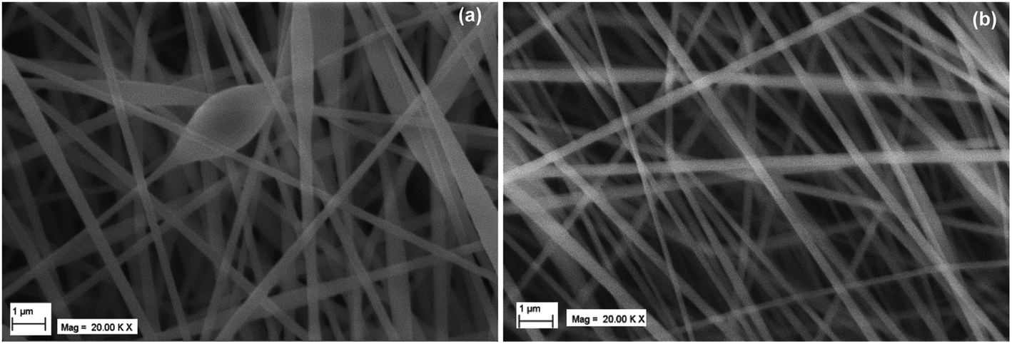 | ||
| Fig. 3 SEM micrograph of nanofibers from 10% of PAN at flow rate (a) 0.5 mL h−1 (b) 1 mL h−1. | ||
Comparison of mobile phase transport
Fig. 4 illustrates the variation in square root of time with solvent migration distance on E-PAN UTLC, commercial available Si-Gel TLC and Si-Gel HPTLC plates. The distance of mobile phase ascent was kept to 30 mm for all three types of plates. The significant reduction in analysis time was observed for 180–200 nm electrospun PAN devices to reach 30 mm compared to commercially available phases (Fig. 4). The observed linear dependence (R2 > 0.99) is predicted using the least squares linear regression method on the basis of the equation y = ax + b (Fig. 4). This dependence is similar to the observed with the other electrospun UTLC and commercially available Si-Gel devices.8 | ||
| Fig. 4 Development time as a function of travel distance of mobile phase (M8) using different TLC devices. | ||
Efficiency
The plate number (N) and plate height (H) in TLC decide the efficiency of separations. The chromatographic efficiency is directly proportional to the plate number (N) and inversely proportional to the plate height (H). The efficiency of PAN derived UTLC plates prepared from the varied concentration of PAN copolymer (8, 10 and 12%) and flow rate (0.2, 0.5 and 1.0 mL h−1) were determined from the obtained RF values and width of analyte spot (Tables S1 and S2 in ESI†). The results presented in Tables S1 and S2† demonstrate that N and H values are closely dependent on the magnitude of hRF and the width of analyte spots. The efficiency of PAN derived UTLC plates was influenced by the applied concentration of amino acids. The better separation of amino acids was achieved on UTLC plates when 0.5% amino acids (∼50 nL) solution was applied onto the plates (Fig. 5). The magnitude of the retardation factor and the nature of color of amino acids achieved on the electrospun PAN based UTLC plates were compared with those obtained on commercially available Si-Gel HPTLC plates (Table 4). The ∼50 nL of 0.5% amino acids solution was loaded onto the both PAN derived nanofiber plates and commercial Si-Gel HPTLC plates. M8 was used as a mobile for the chromatographic behavior of the amino acids on the plates. The distance travelled by the mobile phase on the PAN UTLC plate was fixed as for 20 mm, while for Si-Gel plates the mobile phase was allowed to migrate to 20 mm as well as 50 mm. The hRF values and the spot color of the amino acids on the silica phase and the electrospun UTLC plate were considerably different. Though, both the commercial silica stationary phase and the electrospun PAN stationary phase are polar, but the different retention behavior for identical analytes may be attributed to the difference between their chemical specificity. These results show that electrospun PAN derived nanofibers as a stationary phase provided an alternative for analysis of the amino acids. The superior efficiency of PAN nanofiber E-UTLC plates over silica HPTLC plates is evident by higher values of N and lower values of H (Fig. 6 and 7). This is attributed to the decreased in spot widths of amino acids due to the formation of highly compact spots on PAN derived electrospun UTLC plates. | ||
| Fig. 5 Plate height of different amino acids concentration. | ||
| Amino acids | hRF values | Color formation | |||
|---|---|---|---|---|---|
| E-PAN UTLC plates (20 mm)** | Si-Gel HPTLC plates (20 mm)** | Si-Gel HPTLC plates (50 mm)** | E-PAN UTLC plates | Si-Gel HPTLC plates | |
| a Each value is an average of four replicates. *T = tailed spot; bl (blue), pu (purple), bn (brown), gy (grey), vt (violet), or (orange), yl (yellow), lt pu (light purple), lt gy (light grey); ** indicates plate development distance. | |||||
| Ala | 74 | 38 | 47 | bl | bl/vt |
| Gly | 55T* | 22 | 30 | bl | gy/green |
| Ile | 90 | 65 | 67 | bl | bl/vt |
| Leu | 90 | 68 | 71 | bl | bl/vt |
| Val | 56 | 60 | 66 | bl | bl/vt |
| Phe | 88 | 70 | 76 | pu/bn | vt/gy |
| Trp | 60 | 70 | 74 | gy | lt pu/gy |
| Tyr | 48 | 58 | 60 | vt | pu/gy |
| Asp | 83 | 35 | 38 | pu | vt/gy |
| Glu | 95 | 35 | 41 | bl | bl/vt |
| Arg | 83 | 00 | 04 | bl | vt/gy |
| His | 85 | 35 | 44 | bn/yl | vt/gy |
| Lys | 80 | 00 | 06 | bl | gy |
| Ser | 78 | 33 | 36 | bl | vt/gy |
| Thr | 67 | 45 | 47 | bl/vt | vt/gy |
| Met | 73 | 65 | 71 | bl | vt/gy |
| Cyst | 95 | 28 | 14 | pu | lt gy |
| (Cyst)2 | 95 | 28 | 17 | pu | lt gy |
| Asn | 78 | 35 | 39 | or/yl | vt/gy |
| Gln | 74 | 45 | 48 | bl/vt | vt/gy |
 | ||
Fig. 6 Plate number (N) on E-PAN UTLC plates developed to 20 mm  and Si-Gel HPTLC plates developed to 20 mm and Si-Gel HPTLC plates developed to 20 mm  and 50 mm and 50 mm  . The plate number of gly for E-PAN UTLC plate is not shown due to significant tailing. However, the plate number of arg, lys, cyst and (cyst)2 for Si-Gel HPTLC are not shown due to no or very little mobility. The error bars represent the standard deviation of four replicates. . The plate number of gly for E-PAN UTLC plate is not shown due to significant tailing. However, the plate number of arg, lys, cyst and (cyst)2 for Si-Gel HPTLC are not shown due to no or very little mobility. The error bars represent the standard deviation of four replicates. | ||
 | ||
Fig. 7 Plate heights (H) on E-PAN UTLC plates developed to 20 mm ( ), Si-Gel HPTLC plates developed to 20 mm ( ), Si-Gel HPTLC plates developed to 20 mm ( ) and ) and  (50 mm). The plate height of gly is not shown due to significant tailing on E-PAN UTLC plate. However, the plate height of arg, lys, cyst and (cyst)2 are not shown due to no or very little mobility on Si-Gel HPTLC plate. (50 mm). The plate height of gly is not shown due to significant tailing on E-PAN UTLC plate. However, the plate height of arg, lys, cyst and (cyst)2 are not shown due to no or very little mobility on Si-Gel HPTLC plate. | ||
Limit of detection
The lowest possible detectable amounts per spot for tyr, try, ala, met, ser and lys are presented in Table 5 and these results are clearly showing high sensitivity of the proposed method than the existing TLC and ion exchange resin methods.26,27Application
To explore the efficiency and analytical applicability of proposed method, it was applied for the identification of met and lys in commercial available drugs (Bethadoxin-12M and Beplex forte). The drug sample Bethadoxin-12M syrup have lys (each 5 mL contains 30 mg) and Beplex forte have met (each 1 mL contains 10 mg). Met and lys were detected in drug samples by the use of developed UTLC system as demonstrated by densitograms (Fig. 8 and 9). | ||
| Fig. 8 Densitographic presentation of detection of lys in Bethadoxin 12M syrup. | ||
 | ||
| Fig. 9 Densitographic presentation of detection of met in Beplex forte drug sample. | ||
Conclusion
For the first time, electrospun UTLC-scanning densitometry was applied to record the chromatogram of separated amino acids from drug samples. PAN copolymer nanofibers UTLC plates were fabricated by electrospinning method at different experimental conditions. The diameter, morphology, thickness, and the adherent to aluminium sheet of fabricated nanofibers played a major role in chromatographic separation of amino acids.The increase in diameter of the nanofibers resulted in larger spots and beaded nanofibers were responsible for spreading of spots and their non-reproducible mobility. PAN derived nanofibers sheets obtained from longer electrospinning run was not suitable for chromatographic separation of amino acids due to detachment of nanofibers from aluminium sheet. However, electrospun PAN derived UTLC plates fabricated through 1 h electrospinning run of 10% PAN solution were found appropriate for chromatographic separation. The large difference in hRF values (higher separation possibilities) and brightness of spots colour was attained when the freshly prepared mobile phase containing n-butanol, ethylene glycol and ethyl acetate (5:3:2 v/v) was used. These electrospun UTLC plates shorten amino acids analysis time and lower limits of detection in comparison with Si-Gel plates and ion exchange resin (Na+-form). In future, the electrospun PAN nanofiber derived UTLC plates can be an attractive alternate of commercial available particle based TLC plates and ion exchange resin for separation of amino acid from marketed drug samples.
Acknowledgements
The authors are grateful to Vice Chancellor Dr G. N. Qazi, Jamia Hamdard, New Delhi for providing the required research facilities to the Department of Chemistry, Jamia Hamdard. Dr A. Moheman and Dr A. Kumar are thankful to the University Grant Commission (UGC), New Delhi, India for the award of Dr D. S. Kothari Post-Doctoral Fellowship. Professor A. Mohammad is also obliged to the UGC, New Delhi, India for the award of Emeritus Fellowship.References
- A.-M. Siouffi, Sep. Purif. Rev., 2005, 34, 155–180 CrossRef CAS.
- High-performance thin-layer chromatography (HPTLC), ed. M. M. Srivastva, Springer, Berlin, 2011, p. 360 Search PubMed.
- H. E. Hauck, O. Bund, W. Fischer and M. Schulz, J. Planar Chromatogr.--Mod. TLC, 2001, 14, 234–236 CrossRef CAS.
- H. E. Hauck and M. Schulz, J. Chromatogr. Sci., 2002, 40, 550–552 CAS.
- H. E. Hauck and M. Schulz, Chromatographia, 2003, 57, S313–S315 Search PubMed.
- R. Bakry, G. K. Bonn, D. Mair and F. Svec, Anal. Chem., 2007, 79, 486–493 CrossRef CAS PubMed.
- L. W. Bezuidenhout and M. J. Brett, J. Chromatogr. A, 2008, 1183, 179–185 CrossRef CAS PubMed.
- A. Moheman, M. S. Alam and A. Mohammad, Adv. Colloid Interface Sci., 2016, 229, 1–24 CrossRef CAS PubMed.
- S. R. Jim, A. J. Oka, M. T. Taschuk and M. J. Brett, J. Chromatogr. A, 2011, 1218, 7203–7210 CrossRef CAS PubMed.
- A. J. Oka, S. R. Jim, M. T. Taschuk and M. J. Brett, J. Chromatogr. A, 2011, 1218, 2661–2667 CrossRef PubMed.
- S. R. Jim, A. Foroughi-Abari, K. M. Krause, P. Lib, M. Kupsta, M. T. Taschuk, K. C. Cadienc and M. J. Brett, J. Chromatogr. A, 2013, 1299, 118–125 CrossRef CAS PubMed.
- J. Wannenmacher, S. R. Jim, M. T. Taschuk, M. J. Brett and G. E. Morlock, J. Chromatogr. A, 2013, 1318, 234–243 CrossRef CAS PubMed.
- J. Song, D. S. Jensen, D. N. Hutchison, B. Turner, T. Wood, A. Dadson, M. A. Vail, M. R. Linford, R. R. Vanfleet and R. C. Davis, Adv. Funct. Mater., 2011, 21, 1132–1139 CrossRef CAS.
- S. R. Dhakate, A. Gupta, A. Chaudhari, J. Tawale and R. B. Mathur, Synth. Met., 2011, 161, 411–419 CrossRef CAS.
- P. Rujitanaroj, N. Pimpha and P. Supaphol, J. Appl. Polym. Sci., 2010, 116, 1967–1976 CAS.
- J. He, K. Qi, Y. Zhou and S. Cui, J. Appl. Polym. Sci., 2014, 131, 40137, DOI:10.1002/APP.40137.
- L. Zhang, A. Aboagye, A. Kelkar, C. Lai and H. Fong, J. Mater. Sci., 2014, 49, 463–480 CrossRef CAS.
- H. Kaspar, K. Dettmer, W. Gronwald and P. J. Oefner, Anal. Bioanal. Chem., 2009, 393, 445–452 CrossRef CAS PubMed.
- A. Mohammad, A. Siddiq, A. Moheman and G. E. El-Desoky, J. Liq. Chromatogr. Relat. Technol., 2014, 37, 829–840 CrossRef CAS.
- M. Jaworska, Z. Szulinska, M. Wilk and E. Anuszewska, Talanta, 2010, 83, 513–520 CrossRef CAS PubMed.
- J. D. Vasta, M. Cicchii, J. Sherma and B. Fried, Acta Chromatogr., 2009, 21, 29–38 CrossRef CAS.
- A. S. Khan and F. Faiz, J. Nat. Sci. Math., 2008, 48, 1–17 CAS.
- S. Laskar, A. Sinhababu and K. M. Hazra, Amino Acids, 2001, 21, 201–204 CrossRef CAS PubMed.
- A. Mohammad, A. Moheman and G. E. El-Desoky, Cent. Eur. J. Chem., 2012, 10, 731–750 CAS.
- S. Laskar, A. Sinhababu and K. M. Hazra, J. Appl. Nat. Sci., 2013, 5, 125–127 Search PubMed.
- A. Mohammad, N. Haq and A. Siddiq, J. Sep. Sci., 2010, 33, 3619–3626 CrossRef CAS PubMed.
- S. Z. Qureshi and S. Anwar, Talanta, 1979, 26, 883–884 CrossRef CAS PubMed.
Footnote |
| † Electronic supplementary information (ESI) available. See DOI: 10.1039/c6ra15465d |
| This journal is © The Royal Society of Chemistry 2016 |