The synthesis and comparison of poly(methacrylic acid)–poly(ε-caprolactone) block copolymers with and without symmetrical disulfide linkages in the center for enhanced cellular uptake

Yu-Sheng Liua, Shih-Jer Huanga, Xiao-Shan Huangb, Yi-Ting Wua, Hsuan-Ying Chena, Yu-Lun Loa and Li-Fang Wang*ab
aDepartment of Medicinal & Applied Chemistry, College of Life Science, Kaohsiung Medical University, 100, Shih-Chuan 1st Rd, Kaohsiung 80708, Taiwan. E-mail: lfwang@kmu.edu.tw; Fax: +886-7-3125339; Tel: +886-7-3121101 ext. 2217
bInstitute of Medical Science and Technology, National Sun Yat-Sen University, Kaohsiung 804, Taiwan

Received 13th June 2016 , Accepted 4th August 2016

First published on 4th August 2016


Abstract

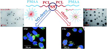
A self-assembled poly(methacrylic acid)–poly(ε-caprolactone) block copolymer with a disulfide linkage, PMAA-b-PCL-SS-PCL-b-PMAA (S-PCL-PMAA)2, was synthesized for enhanced cellular uptake due to a reduction response to glutathione (GSH) and pH-sensitive characteristics. For comparison, a reduction-insensitive PMAA-b-PCL-CC-PCL-b-PMAA (C-PCL-PMAA)2, using a carbon–carbon linkage as a symmetrical center was also synthesized. These block copolymers were synthesized via the combination of ring opening polymerization (ROP) and atom transfer radical polymerization (ATRP) followed by hydrolysis. The similar number of MAA repeating units was controlled in the copolymers containing either disulfide or carbon–carbon linkages. Copolymers could self-assemble into core–shell micelles in an aqueous solution owing to amphiphilicity. The molecular weight of (S-PCL-PMAA)2 increases linearly with reaction time at both reaction temperatures of 40 and 80 °C. Critical micelle concentrations range within 3.09 × 10⁻³ to 6.31 × 10⁻³ mg mL−1 at pH 5 and 3.16 × 10⁻² to 3.98 × 10⁻² mg mL−1 at pH 8. The average hydrodynamic diameters of micelles are ∼200 nm. The cellular uptake of (S-PCL-PMAA)2 increases with incubation time and is higher in the medium with GSH than without in CRL-5802 cells. In contrast, the cellular uptake of (C-PCL-PMAA)2 is insensitive to the presence of GSH. The higher internalization of the micelle containing disulfides in the presence of GSH is attributable to all three pinocytosis pathways involved, including macropinocytosis-, caveolae-, clathrin-mediated endocytosis, but in the absence of GSH clathrin-mediated endocytosis is only involved. The nascent (S-PCL-PMAA)2 is non-cytotoxic to four cell lines. However, the paclitaxel-encapsulated (S-PCL-PMAA)2 shows slightly higher cell-killing ability than free paclitaxel against CRL-5802 cells. Thus, the GSH-responsive (S-PCL-PMAA)2 is a potential drug delivery system.


Introduction

Self-assembly of amphiphilic copolymers in aqueous solutions generally results in aggregates of core–shell structures. Great control of micellar shapes and sizes has an important effect on functions and properties in drug delivery systems (DDS), such as relatively high stability, improved drug solubility, prolonged circulation time, and enhanced elimination half-life in the blood, thus affording a high drug efficacy and low side effects.1,2 Although amphiphilic block copolymer micelles with a distinct hydrophobic core to accommodate hydrophobic drugs and an intrinsically stealthy shell emerge as one of the most promising platforms for DDS, however, one practical challenge for micellar DDS is their low in vivo stability because large volume dilution and interactions with cells and/or proteins present in the bloodstream often lead to micellar dissociation or aggregation, premature drug release, and low drug target ability.3 The crosslinking of micelle core4 or shell5 could greatly improve micellar stability, leading to prolonged circulation time and enhanced accumulation at disease sites. Nevertheless, crosslinked micelles would also significantly retard drug release at the site of action, giving compromised therapeutic effect. This dilemma of the extracellular stability and the intracellular drug release may be solved using stimuli-sensitive cleavable micelles.

Typical cleavable linkages of micelles include acid-labile bonds,6–8 photo-cleavable groups,9 enzymatic-sensitive groups,10 and reduction-responsive disulfide linkages.11,12 The amphiphilic block copolymer micelles containing disulfides are of particular interest because they can be cleaved to the corresponding thiols in the presence of a thiol reducing agent.13 Glutathione (GSH) is found at different concentrations in intracellular (3–10 mM) and extracellular compartments (∼2.8 μM) in living cells,14,15 being an effective cellular trigger that cleaves disulfide linkages in micelles through disulfide–thiol exchange reactions.16 Thus, encapsulated therapeutic agents in the micelles containing disulfides can be effectively released and perform their therapeutic effect at the site of action.

Several amphiphilic block copolymer micelles containing disulfides have been successfully developed for chemo12,17 and/or gene18,19 drugs delivery carriers. Disulfide linkages can be positioned at main chains within hydrophilic–hydrophobic block junctions,18,20,21 or within the center of hydrophobic block segments,13,17,22 and in pendent chains.11,23 The system containing disulfide linkages in the center of symmetrical block copolymer (i.e. A-b-(B-SS-B)-b-A, A and B are block segments) is particularly interesting,13,22 because the cleaved block copolymer with a shorter hydrophobic block length still remain amphiphilic and can reform micelles. This reduction-responsive process leads to a change in size and morphology in micelles. For example, a disulfide-based initiator (bis[2-(2-bromoisobutyryl-oxy)ethyl]disulfide or (BiBOE)2S2) was used to polymerize 2-(methacryloyloxy)ethyl phosphorylcholine (MPC) followed by N-isopropylacryl amide (NIPAM) to yield a PNIPAM-b-(PMPC-SS-PMPC)-b-PNIPAM triblock copolymer via ATRP.13 The reduction-responsive triblock copolymer comprising biocompatible polylactide (PLA) and poly(ethylene glycol) (PEG) blocks, PEG-b-(PLA-SS-PLA)-b-PEG, was synthesized via a combination of ring-opening polymerization and a facile coupling reaction.22 The testing results of these two materials demonstrate GSH could trigger micellar destabilization and change their size distribution.

Poly(ε-caprolactone), PCL, is a biodegradable US Food and Drug Administration (FDA)-approved hydrophobic polymer. It is widely used in various biomedical applications because of its excellent biocompatibility and degradability.24,25 The alkyl segments of PCL are advantageous for DDS because they efficiently encapsulate hydrophobic compounds, slow degradation and provide a sustained release of drugs.26 Poly(methacrylic acid) (PMAA) and its methacrylic ester copolymers (i.e. Eudragit®) have been developed for cutaneous, oral, and parenteral drug delivery systems because of their promising properties like light weight, glass-clear structure, pH-sensitive characteristic, and relatively good hardness.27 It is worthwhile to use PCL as a core and PMAA as a shell because PMAA is a hydrophilic and pH-sensitive polymer and PCL is a highly hydrophobic polymer. The critical micelle concentration (CMC) and drug release rate can be manipulated by adjusting pH conditions, leading to a lower CMC value that benefits the micellar stabilization in the bloodstream circulation.

The difference in endocytic vesicles and subsequent processing may significantly impact the intracellular destination of drug-encapsulated nanoparticles.28–32 It seems no reports on the internalization pathways have been studied in the presence or absence of GSH. Three major chemical inhibitors are commonly utilized to study the internalization pathways of nanoparticles: chlorpromazine for clathrin-mediated endocytosis, genistein for caveolae-mediated endocytosis, and wortmannin for macropinocytosis. Chlorpromazine is a cationic amphiphilic drug that inhibits clathrin-mediated endocytosis by obstructing the assembly of the clathrin adaptor protein at the cell surface.33 Genistein is a specific tyrosine kinase inhibitor that locally disrupts the actin cytoskeleton and prevents recruitment of dynamin II, indispensable for both caveolae-mediated endocytosis and Rho-mediated endocytosis.34,35 Wortmannin is a phosphatidylinositol-3-phosphate inhibitor that inhibits the macropinocytosis pathway.36

In this contribution, a novel monocleavable micelle comprising PMAA and PCL with a reduction-responsive disulfide linkage and a pH-sensitive group, was synthesized via the combination of ring opening polymerization (ROP) and atom transfer radical polymerization (ATRP) followed by hydrolysis. The cleavage of disulfide linkages in the hydrophobic PCL block of the micellar core could cause the destabilization of self-assembled micelles due to change in hydrophobic/hydrophilic balance. Such GSH-triggered micellar destabilization changes the size distribution with an appearance of large aggregates and leads to enhanced release of encapsulated anticancer drugs loaded inside the hydrophobic PCL core. Different internalization pathways responsive to GSH was first found using flow cytometry. The higher cellular uptake of (S-PCL-PMAA)2 with disulfide linkages than (C-PCL-PMAA)2 with carbon linkages was analyzed using confocal laser scanning microscopy. The cell viability with or without an encapsulated paclitaxel was conducted using the MTT assay.

Experimental section

Materials

Bis(2-hydroxyethyl)disulfide, 1,1,4,7,10,10-hexamethyltriethylenetetramine (HMTETA), copper(I) bromide (CuBr), 1,6-hexamethylenediol, dimethyl sulfoxide-d (DMSO-d6), and deuterium chloroform (CDCl3) were purchased from Sigma (St. Louis, MO). Amberlite® IR120, 2-bromo-2-methylpropionyl bromide, ε-caprolactone (CL), rhodamine 123, glutathione (GSH) and pyrene were purchased from Acros (Morris Plains, NJ). Aluminum oxide neutral (Al2O3) was purchased from Seedchem Company PTY. LTD (Melbourne, Australia). Tin(II)-2-ethylhexanoate (Sn(Oct)2) was purchased from Alfa Aesar (Ward Hill, MA). Dithiothreitol (DTT) was purchased from J. T. Baker (Center Valley, PA). Paclitaxel (PTX) was purchased from TCI (Tokyo, Japan). Fetal bovine serum (FBS) was purchased from Biological Industries (Beit Haemek, Israel). Dulbecco's Modified Eagle's Medium (DMEM) was purchased from Invitrogen (Carlsbad, CA).

Preparing PCL-RR-PCL and Br-PCL-RR-PCL-Br (R = S or C)

Poly(ε-caprolactone)-RR-poly(ε-caprolactone) (PCL-RR-PCL) was prepared in neat through the ring-opening polymerization (ROP) with bis(2-hydroxyethyl) disulfide/1,6-hexamethylenediol as an initiator and Sn(Oct)2 as a catalyst.37 In brief, bis(2-hydroxyethyl)disulfide/1,6-hexamethylenediol (6.48 mmol), CL (350.88 mmol) and Sn(Oct)2 (1.08 mmol) were placed into a two-neck round-bottom flask equipped with Graham condenser under an argon atmosphere. The polymerization was conducted at 130 °C under an argon atmosphere. Following 8 hours, the reaction solution was cooled down to room temperature and precipitated into excess cold n-hexane. The precipitate was collected and dried under vacuum.

To prepare a bromo-terminated macroinitiator (Br-PCL-RR-PCL-Br), the PCL-RR-PCL powder (1 mmol) was weighed and put into a two-neck round-bottom flask containing 20 mL of dichloromethane and a magnetic stirring bar at room temperature, followed by adding triethylamine (3 mmol) with stirring for 30 minutes in an ice bath. Subsequently, 2-bromoisobutyl bromide (3 mmol) was slowly added using a syringe. The reaction was conducted under an argon atmosphere. Following 24 hours of reaction, the solution was precipitated into excess cold n-hexane to yield the product. The yields of PCL-RR-PCL and Br-PCL-RR-PCL-Br were ∼90%.

Preparing PtBMA-PCL-RR-PCL-PtBMA (R-PCL-PtBMA)2 and PMAA-PCL-RR-PCL-PMAA (R-PCL-PMAA)2 block copolymers (R = S or C)

Symmetrical block copolymers (R-PCL-PtBMA)2 were synthesized using a molar feed ratio [tBMA][thin space (1/6-em)]:[thin space (1/6-em)][macroinitiator][thin space (1/6-em)]:[thin space (1/6-em)][CuBr][thin space (1/6-em)]:[thin space (1/6-em)][HMTETA] of 400[thin space (1/6-em)]:[thin space (1/6-em)]1[thin space (1/6-em)]:[thin space (1/6-em)]2[thin space (1/6-em)]:[thin space (1/6-em)]2. Macroinitiator (1 mmol), tBMA (400 mmol), HMTETA (2 mmol) were added into a two-neck round-bottom flask containing toluene (2 mL). The reaction was degassed by five consecutive standard freeze–pump–thaw cycles. Next, CuBr (2 mmol) was quickly added to the mixture under an argon atmosphere. The polymerization was allowed to proceed under continuous stirring at 40 or 80 °C for 4–48 hours. The reaction was stopped by diluting with toluene and the solution was passed through an alumina column and Amberlite® IR120 to remove the residual copper catalyst. Finally, the product was obtained from precipitation into excess n-hexane and dried under vacuum.

Symmetrical block copolymers (R-PCL-PMAA)2 were prepared using trifluoroacetic acid (TFA) to hydrolyze the tert-butyl esters of (R-PCL-PtBMA)2.38 The typical procedure was as follows. The (R-PCL-PtBMA)2 copolymers (1 mmol) were dissolved in dichloromethane (10 mL) at room temperature. The solution was cooled to 0 °C and TFA (5 mmol excess to tBMA units) was added with stirring. Following 24 hours of reaction at room temperature, the solution was concentrated and precipitated into cold n-hexane. The product was dried under vacuum.

Reduction of the disulfide linkage of PtBMA-PCL-SS-PCL-PtBMA

The reduction of the disulfide linkage of (S-PCL-PtBMA)2 was carried out in the presence of DTT. The molar ratio of [copolymer][thin space (1/6-em)]:[thin space (1/6-em)][DTT] at 1[thin space (1/6-em)]:[thin space (1/6-em)]10 and the three feeding moles (0.44, 3.52 and 7.04) of CH3ONa were used as the catalyst.37 The CH3ONa powder was dissolved in methanol at a concentration of 1 mg mL−1 as a stock solution. The copolymer (1 mmol) and DTT (10 mmol) were dissolved in 10 mL of tetrahydrofuran (THF) containing different molar amounts of CH3ONa and stirred at room temperature for 24 hours. The product was precipitated and washed twice with cold diethyl ether and dried under vacuum.

Characterization

A Varian Mercury plus-200 spectrometer (Varian; Palo Alto, CA) was used for proton nuclear magnetic resonance (1H-NMR) spectrum analyses to determine the chemical structures of polymers using CDCl3 or DMSO-d6 as a solvent. FTIR spectra were acquired using a Perkin-Elmer System 2000 spectrometer (Norwalk, CT). The molecular weights of copolymers were measured by gel permeation chromatography (GPC) using an Agilent 1100 series (Santa Clara, CA) equipped with a Shodex-KF804 column. Samples were dissolved in THF at a concentration of 5 mg mL−1 and filtered through a 0.45 μm filter prior to injection. THF was used as an eluent at a flow rate of 1 mL min−1. Polystyrene standards were used to generate a calibration curve.

Preparation and characterization of micelles

Micelles were prepared using a dialysis technique. A block copolymer (10 mg) was dissolved in 1 mL of DMSO, put into an MW cut-off 1000 membrane (Spectrum Labs, Rancho Dominguez, CA), and dialyzed against double-deionized (DD) water. The dialyzed solution was freeze-dried to obtain a powdery product. The morphologies of micelles were observed using a transmission electron microscope (TEM, JEM-2000 EXII, JEOL; Tokyo, Japan). A carbon coated 200 mesh copper specimen grid (Agar Scientific Ltd. Essex, UK) was glow-discharged for 1.5 minutes. The micelle powder (2 mg) was dissolved in a vial containing 4 mL of PBS buffer solution (pH 7.4) with or without 10 mM of DTT. At predetermined time intervals, the solution was taken out from the vial and dropped on the copper grid and allowed to dry at room temperature for 5 days. Fluorescence spectra were recorded using a fluorescence spectrophotometer (Cary Eclipse; Varian, CA) with pyrene as a fluorescence probe. A stock solution of pyrene was prepared at a concentration of 6.0 × 10⁻⁷ M in acetone.39 Pyrene excitation spectra were recorded using an emission wavelength at 390 nm. The emission and excitation slit widths were set at 2.5 and 2.5 nm, respectively. Critical micelle concentrations (CMC) were determined from the ratios of pyrene intensities at 339 and 334 nm and calculated from the intersection of two tangent plots of I339/I334 versus the concentrations (5.0 × 10⁻⁵ to 1.0 mg mL−1) of copolymers in 0.1 M PBS of pH 5 or pH 8.

PTX-encapsulated micelles (micelle-PTX)

The method for preparing PTX-encapsulated micelles was slightly modified from that for preparing the micelle in order to improve encapsulation efficiency. In brief, the PTX powder was dissolved in DMSO at 10 mg mL−1 to yield a stock solution. The (S-PCL-PtBMA)2 (10 mg) powder was dissolved in 1 mL of DMSO, followed by adding 0.1 mL of the PTX stock solution. The solution was slowly poured into 20 mL DD water with sonication, which were put into an MW cut-off 1000 membrane and dialyzed against (DD) water for 3 h to remove DMSO and then filtered to remove the unencapsulated PTX. Finally, the micelle-PTX was obtained by freeze-drying. To evaluate the PTX loading efficiency, a dried sample was dissolved in DMSO at a concentration of 1 mg mL−1, followed by sonication for 30 min and centrifugation at 15[thin space (1/6-em)]000 rpm for 5 min. The supernatant solution was filtered with a 0.23 μm disc filter and the PTX concentration was analyzed by high performance liquid chromatography (Agilent 1100 series) with a Hypersil BDS C18 column. The mobile phase consisted of a volume ratio of acetonitrile/DD water as 9/1 and its flow rate was 1 mL min−1. The loading efficiency (LE, %) was calculated using the following equation.
 
LE (%) = amount of drug in micelle/(amount of polymer + amount of drug in micelle) × 100 (1)

Preparing rhodamine 123-conjugated (R-PCL-PMAA)2, (R-PCL-PMAA)2–Rh123

(R-PCL-PMAA)2 (10 mg) and N-(3-dimethylaminopropyl)-N′-ethylcarbodiimide hydrochloride (EDAC) (1 mg) were dissolved in a vial containing 10 mL DD water. The solution was stirred at room temperature for 1 hour. One mg of rhodamine 123 (Rh123) in 1 mL DD water was added to the above solution and stirred at room temperature for 24 hours. The solution was dialyzed against DD water for 3 days using an MW cut-off 1000 membrane. All steps were performed in darkness and the product was collected by freeze-drying.

Cell experiments

CRL-5802, H1299, A549 non-small cell lung carcinoma cell lines and 293T human embryonic kidney cell line were obtained from Dr Cheng at the Biomedical Science and Environmental Biology Department of Kaohsiung Medical University in Taiwan and cultured in DMEM supplemented with 10% FBS and 1% penicillin–streptomycin at 37 °C under humidified 5% CO2.

In vitro cellular uptake studies

A flow cytometer was acquired to study the cellular uptake of micelles in DMEM (without 10% FBS) with or without 10 mM GSH. CRL-5802 cells were seeded at 1 × 10⁵ cells per well in 6-well plates in DMEM supplemented with 10% FBS and incubated at 37 °C for 24 hours. The culture medium was removed and replaced with 2 mL of DMEM (without 10% FBS) containing (R-PCL-PMAA)2–Rh123 (100 μg mL−1) with or without 10 mM GSH. The cells were further incubated at 37 °C for 2, 4 and 6 hours, washed three times with 0.1 M PBS, collected and analyzed using an EPICS XL flow cytometer (Beckman Coulter, Fullerton, CA).

Endocytosis inhibition

CRL-5802 cells were plated at a density of 1 × 10⁵ cells per well in 6-well plates in DMEM supplemented with 10% FBS and incubated for 24 hours. Three inhibitors were selected to pretreat the cells at 37 °C for 30 minutes. The concentrations of inhibitors were used as following: chlorpromazine (10 μg mL−1), wortmannin (50 nM), and genistein (200 μM), respectively.40 After 30 minutes of incubation, the cells were washed and replaced with 2 mL DMEM (without 10% FBS) containing (S-PCL-PMAA)2–Rh123 (100 μg mL−1) with or without 10 mM GSH. The cells were further incubated at 37 °C for 4 hours, washed three times with 0.1 M PBS, collected and analyzed using flow cytometry.

Confocal laser scanning microscopy (CLSM)

CRL-5802 cells were cultured on a glass coverslip in a 12-well plate (1 × 10⁵ cells per well) using DMEM supplemented with 10% FBS for 24 hours. The medium was replaced with 1 mL of DMEM without 10% FBS and (S-PCL-PMAA)2–Rh123 (100 μg mL−1) was added into each well and incubated with or without 10 mM GSH at 37 °C for 2, 4 and 6 hours. The coverslips containing the cells were removed and rinsed three times with 0.1 M PBS. Next, the cells were treated with Hoechst 33342 (5 μg mL−1) to stain the cell nuclei for 15 minutes, washed three times with 0.1 M PBS, and treated with 100 nM Lysotracker Green (DND-26, Invitrogen; Carlsbad, CA) for 30 minutes. The cells were rinsed three times with 0.1 M PBS and fixed with 3.7% paraformaldehyde for 30 minutes. Finally, the cells on the coverslip were washed three times with 0.1 M PBS and mounted with the fluorescent mounting medium on a glass slide for CLSM (Fv 1000; Olympus, Tokyo, Japan) observation.

Cytotoxicity assay of micelles

Cells were seeded in 96-well tissue culture plates at a density of 5 × 10³ per well in medium containing 10% FBS. The cell viabilities of (S-PCL-PMAA)2 micelles were determined after 24 h of incubation with various concentrations of micelles (1–200 μg mL−1) using the tetrazolium-based colorimetric method. 50 μL per well of PBS containing MTT (2 mg mL−1) was added into each well and incubated for 3 h. The solution was removed and 100 μL per well DMSO was added. The number of active cells was estimated by measuring the absorbance at 595 nm.

Cytotoxicity assay of PTX-loaded micelles

The cytotoxicities of PTX and micelle-PTX were measured using the MTT assay. The PTX powder was dissolved in DMSO to yield a stock solution (0.5 mg mL−1), followed by dilution to a concentration of 0.625–160 ng mL−1 in DMEM containing 10% FBS for the cytotoxic test of PTX. The equivalent PTX concentration of the micelle-PTX was controlled within 0.625–160 ng mL−1 as well. CRL-5802 cells were seeded in 96-well culture plates at a density of 5 × 10³ cells per well in DMEM containing 10% FBS for 24 hours. The culture medium was replaced with 100 μL medium containing various concentrations of PTX. Following 48 hours of incubation, the cells were washed three times with 0.1 M PBS and the number of viable cells was measured by estimating their mitochondrial reductase activity using the tetrazolium-based colorimetric method.

Statistical analysis

Means and standard deviations (SD) of data were calculated. Comparison between groups was tested using Student's t-test and *P < 0.05 was considered to be significant.

Results and discussion

Preparation and characterization of copolymers

Scheme 1 illustrates the synthesis route of (S-PCL-PMAA)2 and (C-PCL-PMAA)2 symmetrical copolymers. First, PCL-SS-PCL or PCL-CC-PCL was prepared by ring-opening CL using bis(2-hydroxyethyl)disulfide or 1,6-hexanediol as an initiator, and the degree of polymerization (DP) of PCL was calculated from NMR peak intensities. For PCL-SS-PCL, the peak intensity ratio of (a, –CH2O) at 4.3 ppm and (f, –OCH2C) at 4.07 ppm was utilized to calculate the n value (DP) of PCL (image file: c6ra15307k-t1.tif, Fig. 1a).37 A similar method was repeated to calculate the DP of PCL-CC-PCL using the peak intensity ratio of (e, –CH2OH) at 3.65 ppm and (d, –OCH2C) at 4.07 ppm (image file: c6ra15307k-t2.tif, Fig. S1a).41 The DP of PCL-SS-PCL and PCL-CC-PCL was ∼60. Secondly, Br-PCL-SS-PCL-Br or Br-PCL-CC-PCL-Br was synthesized through the bromination of the hydroxyl groups of PCL with 2-bromo-2-methylpropionyl bromide at 0 °C. The degree of bromination of PCL was ∼95%, estimated from the peak intensity ratio between 1.92 ppm [peak (h), C(CH3)2–Br of 2-bromo-2-methylpropionyl groups] and 4.07 ppm [peak (f), –OCH2 of the PCL] in Fig. 1a. Compared with the upper figure of PCL-SS-PCL, the disappearance of peak (g) in the bottom figure of Fig. 1a, attributable to the end –CH2OH groups of PCL, is a good evidence for successful bromination as well.
image file: c6ra15307k-s1.tif
Scheme 1 The synthesis flow chart of PMAA-PCL-RR-PCL-PMAA.

image file: c6ra15307k-f1.tif
Fig. 1 NMR spectra of (a) PCL-SS-PCL (top) and Br-PCL-SS-PCL-Br (bottom), (b) PtBMA-PCL-SS-PCL-PtBMA with high (H), medium (M), and low (L) lengths of tBMA, and (c) PMAA-PCL-SS-PCL-PMAA with high (H), medium (M), and low (L) lengths of MAA.

Thirdly, the chain extension of Br-PCL-SS-PCL-Br or Br-PCL-CC-PCL-Br with tBMA was proceeded to obtain (S-PCL-PtBMA)2 or (C-PCL-PtBMA)2 symmetrical block copolymers using an ATRP technique. The 1H-NMR spectra of (S-PCL-PtBMA)2 and (C-PCL-PtBMA)2 are shown in Fig. 1b and S1b, respectively. The intensity peak ratio of peak (a) at 4.07 ppm for two methylene protons next to oxygen of PCL and peak (f) at 1.04 ppm for three methyl and two methylene protons of tBMA was calculated to estimate the m value (DP) of PtBMA image file: c6ra15307k-t3.tif,42 and list in Table 1. The molecular weights of block copolymers synthesized at different conditions were analyzed by GPC. The number average molecular weight (Mn) and polydispersity index (PDI) were summarized in Table 1, being in the range of 11[thin space (1/6-em)]700–52[thin space (1/6-em)]300 g mol−1 and 1.29–1.48, respectively. Three block copolymers with DP of tBMA of 320, 150 and 40 measured by 1H-NMR were denoted as high (H), medium (M), and low (L). The GPC profiles of (S-PCL-PtBMA)2 shifted to the high molecular weight region after running ATRP with time (Fig. S2a). The same condition to yield the high tBMA length of (S-PCL-PtBMA)2-H was applied to synthesize (C-PCL-PtBMA)2. Indeed, the Mn of (C-PCL-PtBMA)2 calculated from Fig. S2b was 55[thin space (1/6-em)]300 Da and polydispersity index (PDI) was 1.39, close to 52[thin space (1/6-em)]300 Da of (S-PCL-PtBMA)2-H. When the Mn of (S-PCL-PtBMA)2 and the DP of tBMA were plotted against reaction time (Fig. S3), the Mn and DP increased linearly with an increase in reaction time at both reaction temperatures of 40 or 80 °C. The higher the temperature, the faster the reaction rate because the increase in propagation rate constant kp is much higher than termination rate constant kt due to the lower activation energy of termination.43 The relatively narrow molecular weight distributions (PDI < 1.5) of the synthesized block copolymers imply a well-controlled living radical polymerization.

Table 1 ATRP conditions and GPC data of PtBMA-PCL-SS-PCL-PtBMA
Sample (no.) Molar ratio of PCL/CuBr/HMETTA/tBMA Time (h) Temperature (°C) Mn Mw/Mn DPa of tBMA
a DP was determined by NMR.
1 1/2/2/400 4 40 11[thin space (1/6-em)]700 1.48 34
2 1/2/2/400 8 40 15[thin space (1/6-em)]400 1.35 60
3 1/2/2/400 12 40 22[thin space (1/6-em)]700 1.34 112
4 1/2/2/400 24 40 33[thin space (1/6-em)]000 1.36 184
5 1/2/2/400 48 40 39[thin space (1/6-em)]000 1.39 230
6-L 1/2/2/400 4 80 12[thin space (1/6-em)]500 1.34 40
7 1/2/2/400 8 80 19[thin space (1/6-em)]100 1.35 86
8-M 1/2/2/400 12 80 28[thin space (1/6-em)]100 1.38 150
9 1/2/2/400 24 80 37[thin space (1/6-em)]400 1.39 215
10-H 1/2/2/400 48 80 52[thin space (1/6-em)]300 1.29 320


Finally, the tert-butyl groups of (S-PCL-PtBMA)2 or (C-PCL-PtBMA)2 were hydrolyzed to yield carboxylic groups using TFA as an acid in dichloromethane. The 1H-NMR spectra of (S-PCL-PMAA)2 and (C-PCL-PMAA)2 in d6-DMSO are shown in Fig. 1c and S1b, respectively. Compared with Fig. 1b, the peak intensity at ∼1.42 ppm of Fig. 1c magnificently decreased due to the disappearance of tert-butyl groups.44 Fig. 2 shows the FTIR spectra of (S-PCL-PMAA)2-H and its precursors, where the characteristic hydroxyl (–OH) groups stretched at 3300–3500 cm−1,45 indicating the successful hydrolysis of tBMA. The similar FTIR results of (S-PCL-PMAA)2-L and (S-PCL-PMAA)2-M can be found in Fig. S4.


image file: c6ra15307k-f2.tif
Fig. 2 FTIR spectra of PCL-SS-PCL, PtBMA-PCL-SS-PCL-PtBMA with a high length of tBMA, and its corresponding PMAA-PCL-SS-PCL-PMAA.

Reduction of the disulfide linkage in PtBMA-PCL-SS-PCL-PtBMA and micellar properties

To test whether the central disulfide linkage was cleavable or not, aliquots of (S-PCL-PtBMA)2 were mixed with 10 mmol of DTT (a strong reducing agent) and various mmol amounts of CH3ONa (a catalyst) in THF and stirred at room temperature for 24 hours. Fig. 3a shows the GPC profiles of (S-PCL-PtBMA)2 shifted accordingly to the lower molecular weight region with the higher CH3ONa concentration.46 The Mn of (S-PCL-PtBMA)2 was 70[thin space (1/6-em)]400 Da and changed to 50[thin space (1/6-em)]400, 36[thin space (1/6-em)]100, and 35[thin space (1/6-em)]700 Da in the presence of CH3ONa at 0.44, 3.52, and 7.04 mmol, respectively. The similar result has been reported on polyion complex nano-micelles based on PEG-conjugated polymers containing disulfide linkages,46 where the GPC profiles proved the cleavage of disulfide bonds in the polymer and the resultant decrease in molecular weight.
image file: c6ra15307k-f3.tif
Fig. 3 (a) GPC profiles of PtBMA-PCL-SS-PCL-PtBMA with 10 mmol of DTT and various mmol of CH3ONa as a catalyst in THF; (b) TEM micrographs of PMAA-PCL-SS-PCL-PMAA cast from PBS (pH 7.4) without DTT and with 10 mM DTT for 1 and 7 days.

Consequently, the amphiphilic copolymer of (S-PCL-PMAA)2-H consisting of the dual hydrophilic PMAA segments and the hydrophobic PCL segments containing disulfides, could self-assemble into a micelle in aqueous solutions. Fig. 3b shows the TEM image of the micelle in PBS (pH 7.4). Without DTT, the micelle appeared to be a dense spheroid with a narrow size distribution (∼200 nm). Nevertheless, the spherical micelle became bigger (∼600 nm) with 10 mM DTT for 1 day. At this time point, the loosely hydrophobic domain and clearly hydrophilic corona domain was observed. Following 7 days in the presence of DTT, the region of the core and corona became invisible and the particle size was still ∼600 nm. After that, a similar TEM morphology was observed even following the trace of 14 days (data not shown). This result indicates that the PCL core in (S-PCL-PMAA)2 was responsible to 10 mM DTT, which could either swell or cleave the core compartment. It has been reported self-assembled micelles of the dual disulfide located polylactide (PLA)-based block copolymers composed of disulfide linkages in the dual locations positioned in the PLA cores and PLA/POEOMA (polyoligoethylene oxide methacrylate) interfaces.12 The disulfide linkages were cleaved in response to reductive reactions, causing disassembly of micelles in the presence of 10 mM GSH after 20 hours. The occurrence of aggregation was attributed to both hydrophobicity of cleaved HS-PLA-SH chains and amphiphilicity of cleaved POEOMA-SH chains generated upon the cleavage of the disulfide linkages in the dual locations. Another example of the reduction-responsive triblock copolymers comprising biocompatible PLA and poly(ethylene glycol) (PEG) blocks, PEG-b-(PLA-SS-PLA)-b-PEG,22 was also cleaved to shorter amphiphilic HS-PLA-b-PEG chains, which still formed micelles. Such reduction-responsive process led to a change in size and morphology in micelles. The same explanation can be applied to (S-PCL-PMAA)2. As the disulfide linkages of (S-PCL-PMAA)2 were cleaved by DTT, the PMAA-PCL-SH residue remained amphiphilic and re-assembled into large particles as shown in the TEM images (Fig. 3b).

Using a fluorescence probe technique, the critical micelle concentrations (CMC) of (S-PCL-PMAA)2 were determined at pH 5 or pH 8 with the high and medium length of PMAA (Fig. 4a) but not with the low length of PMAA because it was not successfully fabricated into micelles. The CMC values were calculated from the intersection of two tangent plots of pyrene intensity ratios at I339/I334 versus concentrations. The CMC values of (S-PCL-PMAA)2-H and (S-PCL-PMAA)2-M were 3.16 × 10⁻² mg mL−1 and 3.98 × 10⁻² mg mL−1 at pH 8, and 3.09 × 10⁻³ mg mL−1 and 6.31 × 10⁻³ mg mL−1 at pH 5, respectively. The longer PMAA lengths resulted in the smaller CMC values. For example, the CMC value was half for (S-PCL-PMAA)2 with the high length of PMAA as compared with that of the medium length at pH 5. The micelles had lower CMC values in the lower pH condition because the PMAA segments were protonated to reduce the hydrophilicity. The more hydrophobicity of copolymers led to the sharper rise of aggregate size.47 The solubility of the PMAA segments in water strongly depended on the pH of media as well.48 The more protonation of carboxylate groups in the PMAA segments resulted in a decrease in solubility. To examine the pH-sensitivity of (S-PCL-PMAA)2-H, FTIR was acquired to directly monitor the protonation of the carboxylate groups in the PMAA segments. The carboxylate groups of the PMAA segments were traced at pH 5 or pH 8 (Fig. 4b). The characteristic peaks of protonated and deprotonated carboxylate groups appeared at 1707 cm−1 (C[double bond, length as m-dash]O stretching of COOH) and 1554 cm−1 (asymmetric stretching band of COO), respectively.48 Fig. 4b clearly shows the characteristic COO stretching at pH 8 and C[double bond, length as m-dash]O stretching of COOH at pH 5, suggesting the PMAA segments of the micelle favoring deprotonation and becoming more hydrophilic at pH 8 than pH 5. This fact also explains why the CMC value of the micelle at pH 5 was lower than that at pH 8.


image file: c6ra15307k-f4.tif
Fig. 4 (a) The critical micelle concentration measurements of PMAA-PCL-SS-PCL-PMAA with medium (M) and high lengths (H) of MAA in the plot of I3/I1 vs. concentrations at pH 5 or pH 8; (b) the FTIR spectra of PMAA-PCL-SS-PCL-PMAA with the high length of MAA prepared at pH 5 or pH 8.

Cellular uptake

Flow cytometry was used as a first measure to study cellular uptake of micelles with or without disulfide linkages in the presence and absence of 10 mM GSH. A fluorescent dye, rhodamine 123 (Rh123), was conjugated to (S-PCL-PMAA)2-H micelles. The x-axis represents the fluorescence intensity of the Rh123-labeled micelles (FL3 in logarithmic scale) and the y-axis corresponds to the number of cells of that intensity. In CRL-5802 cells, the internalization of (S-PCL-PMAA)2 was similar with or without 10 mM GSH at 2 hours of incubation but profoundly higher with than without GSH at 4 hours of incubation (Fig. 5a). The effect of GSH on internalization rate was more visible as the incubation time was extended to 6 hours. In contrast, the internalization of (C-PCL-PMAA)2 with carbon–carbon linkages was independent of GSH at 4 hours of incubation (Fig. 5b).
image file: c6ra15307k-f5.tif
Fig. 5 Flow cytometric histograms of (a) PMAA-PCL-SS-PCL-PMAA-H relative to CRL-5802 control cells at 2, 4 and 6 h of incubation and (b) PMAA-PCL-CC-PCL-PMAA relative to CRL-5802 control cells at 4 h of incubation with or without 10 mM GSH.

To directly visualize the internalization of (S-PCL-PMAA)2–Rh123 into CRL-5802 cells at these time points, the cell nuclei were stained with Hoechst 33342 in blue, the endolysosome in green, and the Rh123-labeled micelles in red. The cellular uptake of the micelle in red fluorescence was clearly seen higher with than without GSH at all three incubation time points (Fig. 6). At a long incubation time of 6 hours, most of the internalized (S-PCL-PMAA)2–Rh123 micelles were still associated with endolysosomes, showing an overlapping color in yellow.


image file: c6ra15307k-f6.tif
Fig. 6 Confocal microscopic photographs of PMAA-PCL-SS-PCL-PMAA-H relative to CRL-5802 control cells at 2, 4 and 6 h of incubation with or without 10 mM GSH.

To understand whether the internalization routes of the micelle changed in the presence of GSH, three major internalization pathways were tested using their corresponding chemical inhibitors: chlorpromazine for clathrin-mediated endocytosis, genistein for caveolae-mediated endocytosis, and wortmannin for macropinocytosis. Compared with the control, the cellular uptake of (S-PCL-PMAA)2–Rh123 into CRL-5802 cells in the presence of 10 mM GSH manifestly decreased when the cells were pretreated with chlorpromazine, and slightly decreased when the cells were pretreated with genistein and wortmannin (Fig. 7a). The decrease in fluorescence intensity showed significance (*P < 0.05) in using three chemical inhibitors, suggesting clathrin-, caveolae-, and macropinocytosis-mediated endocytosis as three possible pathways to be involved in the cellular uptake of the micelle. However, in the absence of GSH, the flow cytometric histogram shifted left and mean fluorescence intensities significantly decreased at 4 hours of incubation in the cells pretreated with chlorpromazine alone (Fig. 7b). The left shift of the flow cytometric diagram, suggests clathrin-mediated endocytosis as the only pathway to be involved in the absence of GSH. Thus, the present work confirms the better cellular uptake of the micelle containing disulfide linkages in the presence of GSH was owing to many internalization pathways involved. As aforementioned, the disulfide linkages of (S-PCL-PMAA)2 were hydrolyzed in the presence of GSH and the cleaved HS-PCL-PMAA could self-reassemble into different sizes or morphologies, which might trigger other internalization pathways besides clathrin-mediated endocytosis.


image file: c6ra15307k-f7.tif
Fig. 7 Flow cytometric histograms and mean fluorescence intensities of CRL-5802 cells exposed to PMAA-PCL-SS-PCL-PMAA-H after the cells pretreated with different chemical inhibitors in the presence (a) and absence (b) of GSH at 4 h of incubation.

In vitro cytotoxicity

The cytotoxicity of (S-PCL-PMAA)2 was evaluated using an MTT assay in CRL-5802, 293T, H1299, and A549 cells (Fig. 8a). The micelle did not show obvious cytotoxicity in test concentrations of 1–200 μg mL−1 and cell line dependence. Even when the cells were exposed to the highest concentration of 200 μg mL−1, the cell viability of (S-PCL-PMAA)2 was still >80% in the four cell lines.
image file: c6ra15307k-f8.tif
Fig. 8 Relative cell viabilities of (a) four cell lines exposed to PMAA-PCL-SS-PCL-PMAA-H for 24 h of incubation and (b) CRL-5802 cells exposed to free PTX and PTX-loaded PMAA-PCL-SS-PCL-PMAA-H (n = 8).

The PTX loading efficiency in micelle was ∼2.85%. The cytotoxicity of micelle-PTX was found to be higher than that of free PTX against CRL-5802 cells (Fig. 8b). The IC50 value (the concentration of PTX required to inhibit 50% of cell proliferation) of the micelle-PTX was 99.2 ng mL−1 (0.116 μM), slightly lower than that of free PTX (IC50 = 119.6 ng mL−1, 0.140 μM). Although the similar values of IC50 were found, however, the micelle-PTX is superior to its parent PTX because of an increase in water solubility of PTX, protection of PTX against deactivation during incubation, and responsive PTX release to GSH.

Conclusion

Novel amphiphilic (R-PCL-PMAA)2 symmetrical block copolymers having R = S or C in the center of the hydrophobic PCL block were first synthesized by a combination of well-defined organic and polymeric syntheses via ROP, ATRP, and hydrolysis. The copolymers (R-PCL-PMAA)2 and their precursors synthesized in each step were well characterized by 1H-NMR and FTIR, and molecular weights by GPC. The similar repeating numbers of PCL at ∼60 and PMAA at ∼130 were controlled in both (S-PCL-PMAA)2 and (C-PCL-PMAA)2. The molecular weight of (S-PCL-PtBMA)2 and the DP of tBMA increased linearly with an increase in reaction time at the reaction temperature of 40 or 80 °C. As the results obtained from GPC, the disulfide linkage in the center of (S-PCL-PtBMA)2 could be cleaved with 10 mM DTT in the presence of CH3ONa as the catalyst. From TEM image results, the PCL core containing disulfide linkages in (S-PCL-PMAA)2 was responsive to 10 mM DTT, which could either swell or cleave the core. The cleaved PMAA-PCL-SH still remained amphiphilic and re-assembled into a looser and larger particle. The longer PMAA length in the symmetrical block copolymers resulted in the smaller CMC value. The micelles were pH-sensitive and had lower CMC values in the environment of pH 5 than pH 8. The high internalization of CRL-5802 cells exposed to (S-PCL-PMAA)2 in response to GSH was confirmed by flow cytometry as well as CLSM. The high cellular uptake of (S-PCL-PMAA)2 in the presence of GSH was because clathrin, caveolae, and macropinocytosis, tri-mediated endocytosis pathways were all involved. However, in the absence of GSH, only clathrin-mediated pathway was involved. The (S-PCL-PMAA)2 micelle was non-cytotoxic to CRL-5802, 293T, H1299, and A549 four cell lines. The micelle-PTX showed similar cell-killing ability to free PTX against CRL-5802 cells. To the best of our knowledge, this is the first example to demonstrate the internalization pathway in response to the GSH cellular trigger. The new type of amphiphilic (S-PCL-PMAA)2 copolymers containing disulfide linkages was successfully prepared and its potential application as a hydrophobic drug carrier was illustrated.

Conflict of interest

The authors declare no conflict of interest.

Acknowledgements

We are grateful for the financial support from the Ministry of Science and Technology of Taiwan (MOST104-2314-B-037-006-MY3 and MOST103-2320-B-037-012-MY3). This study is also supported by “Aim for the Top Journals Grant (KMU-DT105009)” and by "NSYSU-KMU Joint Research Project, (NSYSUKMU 105-P018)" from Kaohsiung Medical University. We appreciate the experiment support of a confocal laser scanning microscope and a transmission electron microscope from Center for Research Resources and Development of KMU.

References

  1. N. Rapoport, W. G. Pitt, H. Sun and J. L. Nelson, J. Controlled Release, 2003, 91, 85–95 CrossRef CAS PubMed.
  2. T. Kissel, Y. X. Li and F. Unger, Adv. Drug Delivery Rev., 2002, 54, 99–134 CrossRef CAS PubMed.
  3. K. Kataoka, A. Harada and Y. Nagasaki, Adv. Drug Delivery Rev., 2001, 47, 113–131 CrossRef CAS PubMed.
  4. Y. Wu, W. Chen, F. Meng, Z. Wang, R. Cheng, C. Deng, H. Liu and Z. Zhong, J. Controlled Release, 2012, 164, 338–345 CrossRef CAS PubMed.
  5. A. M. Nystrom and K. L. Wooley, Tetrahedron, 2008, 64, 8543–8552 CrossRef CAS PubMed.
  6. B. Yang, Y. Lv, J.-y. Zhu, Y.-t. Han, H.-z. Jia, W.-h. Chen, J. Feng, X.-z. Zhang and R.-x. Zhuo, Acta Biomater., 2014, 10, 3686–3695 CrossRef CAS PubMed.
  7. M. Oishi, S. Sasaki, Y. Nagasaki and K. Kataoka, Biomacromolecules, 2003, 4, 1426–1432 CrossRef CAS PubMed.
  8. S. Binauld and M. H. Stenzel, Chem. Commun., 2013, 49, 2082–2102 RSC.
  9. H. Zhao, E. S. Sterner, E. B. Coughlin and P. Theato, Macromolecules, 2012, 45, 1723–1736 CrossRef CAS.
  10. J.-Z. Du, D.-P. Chen, Y.-C. Wang, C.-S. Xiao, Y.-J. Lu, J. Wang and G.-Z. Zhang, Biomacromolecules, 2006, 7, 1898–1903 CrossRef CAS PubMed.
  11. B. Khorsand, G. Lapointe, C. Brett and J. K. Oh, Biomacromolecules, 2013, 14, 2103–2111 CrossRef CAS PubMed.
  12. N. R. Ko and J. K. Oh, Biomacromolecules, 2014, 15, 3180–3189 CrossRef CAS PubMed.
  13. C. Li, J. Madsen, S. P. Armes and A. L. Lewis, Angew. Chem., Int. Ed., 2006, 45, 3510–3513 CrossRef CAS PubMed.
  14. S. Son, R. Namgung, J. Kim, K. Singha and W. J. Kim, Acc. Chem. Res., 2011, 45, 1100–1112 CrossRef PubMed.
  15. Q. Zhang, N. Re Ko and J. Kwon Oh, Chem. Commun., 2012, 48, 7542–7552 RSC.
  16. Y. Miura, T. Takenaka, K. Toh, S. Wu, H. Nishihara, M. R. Kano, Y. Ino, T. Nomoto, Y. Matsumoto, H. Koyama, H. Cabral, N. Nishiyama and K. Kataoka, ACS Nano, 2013, 7, 8583–8592 CrossRef CAS PubMed.
  17. M. Ding, J. Li, X. He, N. Song, H. Tan, Y. Zhang, L. Zhou, Q. Gu, H. Deng and Q. Fu, Adv. Mater., 2012, 24, 3639–3645 CrossRef CAS PubMed.
  18. H. Wei, L. R. Volpatti, D. L. Sellers, D. O. Maris, I. W. Andrews, A. S. Hemphill, L. W. Chan, D. S. Chu, P. J. Horner and S. H. Pun, Angew. Chem., Int. Ed., 2013, 52, 5377–5381 CrossRef CAS PubMed.
  19. X. Jiang, J. Liu, L. Xu and R. Zhuo, Macromol. Chem. Phys., 2011, 212, 64–71 CrossRef CAS.
  20. N. Song, W. Liu, Q. Tu, R. Liu, Y. Zhang and J. Wang, Colloids Surf., B, 2011, 87, 454–463 CrossRef CAS PubMed.
  21. B. Khorsand Sourkohi, A. Cunningham, Q. Zhang and J. K. Oh, Biomacromolecules, 2011, 12, 3819–3825 CrossRef CAS PubMed.
  22. A. Cunningham, N. R. Ko and J. K. Oh, Colloids Surf., B, 2014, 122, 693–700 CrossRef CAS PubMed.
  23. Q. Zhang, S. Aleksanian, S. M. Noh and J. K. Oh, Polym. Chem., 2013, 4, 351–359 RSC.
  24. C. Allen, J. N. Han, Y. S. Yu, D. Maysinger and A. Eisenberg, J. Controlled Release, 2000, 63, 275–286 CrossRef CAS PubMed.
  25. C. Allen, Y. S. Yu, D. Maysinger and A. Eisenberg, Bioconjugate Chem., 1998, 9, 564–572 CrossRef CAS PubMed.
  26. K. E. Uhrich, S. M. Cannizzaro, R. S. Langer and K. M. Shakesheff, Chem. Rev., 1999, 99, 3181–3198 CrossRef CAS PubMed.
  27. V. Carelli, G. Di Colo, E. Nannipieri, B. Poli and M. F. Serafini, Int. J. Pharm., 2000, 202, 103–112 CrossRef CAS PubMed.
  28. O. Harush-Frenkel, N. Debotton, S. Benita and Y. Altschuler, Biochem. Biophys. Res. Commun., 2007, 353, 26–32 CrossRef CAS PubMed.
  29. W. Li, C. Chen, C. Ye, T. Wei, Y. Zhao, F. Lao, Z. Chen, H. Meng, Y. Gao, H. Yuan, G. Xing, F. Zhao, Z. Chai, X. Zhang, F. Yang, D. Han, X. Tang and Y. Zhang, Nanotechnology, 2008, 19, 145102 CrossRef PubMed.
  30. J. Z. Rappoport, Biochem. J., 2008, 412, 415–423 CrossRef CAS PubMed.
  31. P. J. Smith, M. Giroud, H. L. Wiggins, F. Gower, J. A. Thorley, B. Stolpe, J. Mazzolini, R. J. Dyson and J. Z. Rappoport, Int. J. Nanomed., 2012, 7, 2045–2055 CAS.
  32. G.-J. Chen, C. Hsu, J.-H. Ke and L.-F. Wang, J. Biomed. Nanotechnol., 2015, 11, 951–963 CrossRef CAS PubMed.
  33. I. A. Khalil, K. Kogure, H. Akita and H. Harashima, Pharmacol. Rev., 2006, 58, 32–45 CrossRef CAS PubMed.
  34. H. Hufnagel, P. Hakim, A. Lima and F. Hollfelder, Mol. Ther., 2009, 17, 1411–1417 CrossRef CAS PubMed.
  35. R. G. Parton, B. Joggerst and K. Simons, J. Cell Biol., 1994, 127, 1199–1215 CrossRef CAS PubMed.
  36. N. Araki, M. T. Johnson and J. A. Swanson, J. Cell Biol., 1996, 135, 1249–1260 CrossRef CAS PubMed.
  37. H. Sun, B. Guo, R. Cheng, F. Meng, H. Liu and Z. Zhong, Biomaterials, 2009, 30, 6358–6366 CrossRef CAS PubMed.
  38. Y. Q. Yang, W. J. Lin, B. Zhao, X. F. Wen, X. D. Guo and L. J. Zhang, Langmuir, 2012, 28, 8251–8259 CrossRef CAS PubMed.
  39. Q. Gao, Q. Liang, F. Yu, J. Xu, Q. Zhao and B. Sun, Colloids Surf., B, 2011, 88, 741–748 CrossRef CAS PubMed.
  40. S.-F. Peng, C.-J. Su, M.-C. Wei, C.-Y. Chen, Z.-X. Liao, P.-W. Lee, H.-L. Chen and H.-W. Sung, Biomaterials, 2010, 31, 5660–5670 CrossRef CAS PubMed.
  41. R. G. J. C. Heijkants, R. V. v. Calck, T. G. van Tienen, J. H. de Groot, P. Buma, A. J. Pennings, R. P. H. Veth and A. J. Schouten, Biomaterials, 2005, 26, 4219–4228 CrossRef CAS PubMed.
  42. Y.-L. Luo, W. Yu and F. Xu, Polym. Bull., 2012, 69, 597–620 CrossRef CAS.
  43. K. Matyjaszewski, Macromolecules, 2012, 45, 4015–4039 CrossRef CAS.
  44. L. Chang, J. Liu, J. Zhang, L. Deng and A. Dong, Polym. Chem., 2013, 4, 1430 RSC.
  45. X. Wei, L. Qi, G. Yang and F. Wang, Talanta, 2009, 79, 739–745 CrossRef CAS PubMed.
  46. H. Huang, X. Zhang, J. Yu, J. Zeng, P. R. Chang, H. Xu and J. Huang, Colloids Surf., B, 2013, 110, 59–65 CrossRef CAS PubMed.
  47. X. M. Song, W. Q. Yao, G. L. Lu, Y. J. Li and X. Y. Huang, Polym. Chem., 2013, 4, 2864–2875 RSC.
  48. M. G. Santonicola, G. W. de Groot, M. Memesa, A. Meszyńska and G. J. Vancso, Langmuir, 2010, 26, 17513–17519 CrossRef CAS PubMed.

Footnote

Electronic supplementary information (ESI) available. See DOI: 10.1039/c6ra15307k

This journal is © The Royal Society of Chemistry 2016
Click here to see how this site uses Cookies. View our privacy policy here.