DOI:
10.1039/C6RA15225B
(Paper)
RSC Adv., 2016,
6, 65960-65975
Synthesis of potential biosorbent from used stevia leaves and its application for malachite green removal from aqueous solution: kinetics, isotherm and regeneration studies
Received
12th June 2016
, Accepted 24th June 2016
First published on 27th June 2016
Abstract
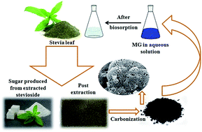
To develop a highly efficient, low-cost adsorbent from waste materials, powdered activated carbon (AC) was synthesized from used stevia leaves post the extraction of the glycosides used for producing a natural sweetener. The modification of the chemical characteristics of the biosorbent surface was achieved by impregnating it with sodium hydroxide (NaOH) at two different weight ratios. The AC biosorbent was applied for the removal of hazardous dye malachite green from an aqueous solution and the effects of the key operating parameters, such as initial dye concentration, agitation time, adsorbent dosage, solution pH, and the presence of electrolyte on the dye removal efficiency, were analyzed. The adsorption data were fitted to different isotherm and kinetic models to predict the adsorption mechanism. The Freundlich isotherm model showed the best fit to the adsorption equilibrium data and the adsorption kinetics followed a pseudo-second order model. A thermodynamic study was carried out and the data showed the endothermic and non-spontaneous nature of the process in the studied temperature range. The high adsorption efficiency shown after the regeneration studies indicated the high potential for the adsorbent to be used as a highly efficient, low-cost biosorbent.
1. Introduction
Water quality maintenance is a major concern in this period of rapid urbanization and industrialization. Most pollutants are released into the environment along with different industrial effluent streams, which can adversely affect the aquatic life as well as other living beings. Dyes are one of the most common categories of industrial materials and are widely used in the textile, paper and pulp, leather, food, cosmetic, and printing industries, as well as distilleries.1 Most dyes are highly toxic and cause several hazardous effects, besides their undesirable visibility in water, recalcitrant nature and organic content. Malachite green (MG) is a basic dye that has a brilliant and high intensity color and is highly visible, even when present at a very low concentration. In addition to being extensively used as a coloring agent in the textile and leather industries, it is also used in aquaculture as a popular antibacterial and antifungal agent as well as a therapeutic agent.2 It adversely works against the protozoan Saprolegnia, a water mold that destroys fish eggs and young fish.2 Moreover, due to its toxic effects, due to its carcinogenic, genotoxic, mutagenic and teratogenic properties on animal and human beings, the continuous removal of excess MG from water sources is highly essential.3
Conventional water purification techniques include physical, chemical, biological and advanced methods such as coagulation, flocculation, electro-coagulation, precipitation, ion exchange, membrane filtration, electrochemical destruction, irradiation, ozonation and adsorption.2 Among all these techniques, adsorption is one of the most efficient and cost-effective treatment methods because it has least possibilities of producing byproducts and hazardous secondary pollutants. Good adsorbents are mostly regenerable and can be reused with high efficiency, which can make the treatment process far more effective and inexpensive. Usually, the novelty of the adsorption process relies on the nature of the adsorbents and the target pollutants. Naturally occurring materials and agricultural wastes are rich sources of raw materials for preparing adsorbents with a high specific surface area and are abundantly available, easily biodegradable, and are renewable materials. Several investigations have been carried out with different agricultural and biological waste materials to test their potential for dye adsorption such as bamboo,4 orange peel,5 hen feathers,6 modified rice straw,7 sugarcane dust,8 algae,9 maize cob,10 banana pith,11 and oil palm trunk fiber.1 The utilization of such natural biological materials as an adsorbent could not only make the process cost effective, but could also influence human efforts toward less waste disposal to the environment.
Stevia (Stevia rebaudiana Bertoni) is an herbaceous plant, belonging to the Asteraceae family, from which natural sweetener is produced. The presence of several glycosides, including stevioside, rebaudioside A, B, D, E, dulcoside A and B, makes the stevia leaves a biological source of natural sweetener.12 Stevioside is 300 times sweeter than sucrose and can be used as a supplementary sweetener for diabetic and phenylketonuria patients, as well as for obese persons because of its heat stability and non-calorific nature.13 After the extraction of natural sweetener from the dry leaves, the waste leaves are discarded, with no after use of these used stevia leaves reported to date.
The present study reports for the first time the synthesis of a highly efficient, chemically treated activated carbon adsorbent from used stevia leaves and then its application to MG removal from an aqueous solution. Accordingly, the adsorption isotherms, kinetics, and thermodynamic phenomenon of the adsorption process were analyzed and the theoretical isotherm models were compared with the experimental results. The renewable capacity of the adsorbent was also expressed following an adsorption–desorption study.
2. Materials and methods
2.1. Materials
The adsorbent was prepared from spent stevia leaves post the extraction of natural sweetener. The powdered dry stevia leaves were purchased from RAS Agro Associates, Maharashtra, India. Malachite green oxalate, C.I. Basic Green 4, Classification Number 42000 (chemical formula: C52H54N4O12, Mol. wt 927) was supplied by Merck Ltd., Mumbai, India and was used as the target pollutant. The chemical structure of the malachite green (MG) and the steviol are shown in Fig. 1(a) and (b), respectively. Sodium hydroxide (NaOH) and hydrochloric acid (HCl) were purchased from Merck Ltd., Mumbai, India. The double distilled water for preparing all the reagents and solutions was obtained from the distillation unit of the departmental research facility of IIT Kharagpur.
 |
| Fig. 1 Chemical structure of (a) malachite green and (b) steviol. | |
2.2. Preparation of the biosorbent
Powdered activated carbon was prepared as the adsorbent by using spent stevia leaves as the precursor. Initially, the dry powdered stevia leaves were mixed with hot distilled water at a ratio of 1
:
14 (g
:
mL) and placed in a water bath at 78 ± 1 °C for 56 min. This specific ratio of the process parameters was selected following the results obtained from an optimized response surface methodology to extract steviosides from dry stevia leaves.14 Post-extraction, the leaves were dried overnight in a hot air oven at 70 °C to remove moisture and other volatile impurities. It was then ground, sieved and carbonized at 400 °C under nitrogen flow (250 mL min−1) for 1 h. The carbonized sample was then chemically activated by separately impregnating with NaOH solution in 1
:
1 and 1
:
2 weight ratios and was then dehydrated at 100 °C in a hot air oven for 12 h. In the next stage, the dehydrated samples were carbonized at 600 °C under nitrogen flow (250 mL min−1) for 1 h at a heating rate of 10 °C min−1.15,16 After carbonization, the activated carbon was thoroughly washed with 0.1 M HCl solution and double distilled water until the pH reached 6–7. Finally, the samples were dried under vacuum at 100 °C for 12 h and stored in a desiccator for future use. The activated carbon obtained from 1
:
1 and 1
:
2 weight ratios with NaOH were labeled as AC1-1 and AC1-2, respectively. The average particle sizes obtained for AC1-1 and AC1-2 were 0.046 and 0.039 mm, respectively, as obtained using a particle size analyzer (Malvern Instruments).
2.3. Characterization of the biosorbent
Both the carbonized samples AC1-1 and AC1-2 were characterized by scanning electron microscopy (SEM), Fourier transform infrared spectroscopy (FTIR), X-ray diffraction (XRD) and N2 adsorption analysis. The morphology and pores created on the surface were investigated using a MERLIN ZEISS EVO 60 SEM system (Germany). The unloaded and loaded images of both the samples were compared to identify the changes in the surface morphology. The amorphous nature of the prepared activated carbon samples was depicted through high resolution XRD analysis using a Panalytical high-resolution XRD-I PW 3040/60 within the spectrum from 20° to 80° (2θ) with a Co Kα radiation at a wavelength (λ) of 1.789 Å. The functional groups and the chemical bonds present over the surface were assessed using a Perkin Elmer spectrum Fourier transform infrared spectroscopy (FTIR) by plotting the percentage transmittance against wavenumber in the range from 4000 to 400 cm−1. The samples were converted into pellets using potassium bromide (KBr) as the binding agent, procured from Sisco Research Laboratories Pvt. Ltd., Mumbai and a hydraulic pellet press, manufactured by Kimaya Engineers, Mumbai. The specific surface area and average pore diameter and pore volume of the samples were measured using the Brunauer–Emmett–Teller method by the N2 gas adsorption–desorption isotherms in the Quantachrome Autosorb automated gas sorption system. The nitrogen bath temperature was 77.3 K and the outgas temperature was fixed at 250 °C for an out-gassing duration of 7 h.
2.4. Preparation of the dye solution and analysis
The dye solution was prepared by dissolving a measured amount of MG in double distilled water to prepare a stock solution of 500 mg L−1. The required concentrations of the solutions were prepared by proper dilution of the stock solution. Initially, a model calibration curve of the absorbance (at a wavelength (λ) of 615 nm
17) against dye concentration was drawn using a UV-Vis spectrophotometer (Spectroquant® UV-VIS Spectrophotometer Pharo 300-Merck) from ten solutions of known concentrations varying from 1 to 10 mg L−1, which were then fitted in a straight line.
2.5. Adsorption experiments
All the experiments were conducted in batch mode and at 30 ± 1 °C (303 K) by varying the initial dye concentration from 50 to 300 mg L−1. In each study, a precisely weighed amount of adsorbent was added to 150 mL of dye solution of known initial concentration taken in a 250 mL conical flask and stirred in a magnetic stirrer at a speed of 300 rpm, allowing equilibrium to be reached. The pH was maintained at 8 during the experiment in each case by using 0.1 M HCl/NaOH solution. The collected product samples were centrifuged at 10
000 rpm for 10 min to separate the solid adsorbent particles18,19 and the absorbance of the supernatant liquid was measured in the spectrophotometer at a wavelength (λ) of 615 nm to determine the final concentration of the dye. The amount of dye adsorbed at equilibrium can be expressed by eqn (1) as follows: |
 | (1) |
where C0 and Ce (mg L−1) are the liquid-phase concentrations of dye at the initial and equilibrium conditions, respectively, V is the volume of the solution (L), and W is the mass of adsorbent used (g). The effects of variations in the initial dye concentration, agitation time, amount of adsorbent dosage, electrolyte effect and initial pH of the solution were studied.
3. Results and discussion
3.1. Characterization results
3.1.1. Scanning electron microscopic (SEM) analysis. Fig. 2(a) and (b) show the SEM images of AC1-1 in unloaded and loaded conditions, respectively, whereas Fig. 2(c) and (d) show the same for adsorbent AC1-2. From Fig. 2(a) and (c), infinite numbers of pores of variable sizes can be observed on the adsorbent surface, which indicates that it has a better adsorption capacity. Fig. 2(b) and (d) show the SEM images of the loaded adsorbents after one cycle of treatment, wherein vacant pores have been fully or partially filled up with the dye molecules.
 |
| Fig. 2 SEM images of (a) unloaded AC1-1, (b) loaded AC1-1, (c) unloaded AC1-2, and (d) loaded AC1-2. | |
3.1.2. XRD analysis. Fig. 3 shows the XRD patterns of the adsorbents AC1-1 and AC1-2 in the range of 20–80°. In both cases for the adsorbents, the broad peaks appeared at about 24° and define the amorphous and carbonaceous nature of the adsorbents.
 |
| Fig. 3 XRD pattern of the biosorbents. | |
3.1.3. FTIR analysis. Fig. 4(a) shows the comparison of the FTIR results of fresh and treated adsorbent AC1-1 and Fig. 4(b) compares the same for adsorbent AC1-2. The broad bands found in the range of 3300–3400 cm−1 can be attributed to the presence of stretching vibrations of the OH and NH bonds,20 whereas the bands present in between 2920 and 2850 cm−1 indicate the presence of symmetric and asymmetric vibrations of the CH2 functional groups.21 The bonds located at wavelengths in between 1823 and 1833 cm−1 are assigned to anhydride C
O stretching,22 whereas the bonds present in between 1615 and 1630 cm−1 refer to stretching of the C
C bonds in aromatic rings.23 In between 1190 and 1135 cm−1, the C
O stretching of esters can be observed. The region in between 800 and 500 cm−1 contains various alkyl halide groups.22 The shift in the positions of the assigned bonds indicate the possible interaction of the biosorbents with MG dye molecules via these functional groups and the loading of MG dye onto the biosorbents.
 |
| Fig. 4 FTIR spectra of (a) AC1-1 (i) before and (ii) after treatment and (b) AC1-2 (i) before and (ii) after treatment and the nitrogen adsorption–desorption isotherm curve obtained from BET experiments for (c) AC1-1 and (d) AC1-2. | |
3.1.4. N2 adsorption analysis. To study the porous structure of the adsorbent particle, adsorption–desorption isotherms were constructed, as shown in Fig. 4(c) and (d) for AC1-1 and AC1-2, respectively.As per the international union of pure and applied chemistry standards (IUPAC), the nature of the curves show a similarity with the Type IV curve classification. According to this, the pores are slit-like in shape and mesoporous in nature, which is associated with capillary condensation occurring inside the pores.24 The specific surface area and average pore diameter, as well as total pore volume of the two adsorbents are mentioned in Table 1.
Table 1 BET analysis of prepared biosorbents obtained from the adsorption isotherm data
Sample |
AC1-1 |
AC1-2 |
Average pore diameter (Å) |
63.7 |
41.5 |
Specific surface area (m2 g−1) |
58.3 |
281.8 |
Total pore volume (cm3 g−1) |
0.09 |
0.29 |
3.2. Effect of agitation time
Fig. 5(a) and (b) show the effect of agitation time on the amount of dye uptake upon varying the initial dye concentration for adsorbent AC1-1 and AC1-2, respectively. All the experiments were carried out at pH 8 with an adsorbent dosage of 0.1 g in 150 mL of solution at a temperature of 303 K. The entire phenomenon of dye uptake by the adsorbent can be subdivided into three parts. Initially, the dye molecules encounter the boundary layer effect near the adsorbent surface and then diffuse through the boundary layer film to reach the surface before finally diffusing through the pores on the adsorbent surface.17 In the initial 10 min, the dye uptake increased rapidly due to a large concentration gradient and the availability of a large number of empty pores. After 10 min, it became gradual, before finally reaching a state of dynamic equilibrium. With increasing the initial dye concentration from 50 to 300 mg L−1, the amount of dye adsorbed increased from 74.87 to 284.45 mg g−1 and from 73.08 to 288.67 mg g−1 for AC1-1 and AC1-2, respectively. Upon increasing the dye concentration, more dye molecules collide with each other, thus producing more resistance for diffusion and therefore more time is required to attain equilibrium.25
 |
| Fig. 5 Effect of agitation time on the amount of dye uptake at various initial concentrations for (a) AC1-1 and (b) AC1-2. | |
3.3. Effect of initial concentration and adsorbent dosage
Fig. 6(a) shows the effect of initial dye concentration on the percentage removal of dye for both the adsorbents. With increasing the dye concentration from 50 to 300 mg L−1, the percentage removal decreased gradually from 99.83 to 63.21 for AC1-1 and from 97.45 to 64.15 for AC1-2. Though increasing the dye concentration results in enhanced dye uptake by the adsorbent, the percentage removal decreases gradually due to saturation of the active pores on the surface. On the other hand, by increasing the adsorbent dosage, the percent removal increases. More available active pores will result in more adsorption of the dye on the surface (Fig. 6(b)). Simultaneously, it is important to note that as the adsorbent dosage was increased, the amount of dye uptake decreased. This is because of the greater number of pores at the fixed initial dye concentration, the fractional uptake of dye per unit weight of adsorbent is less.3
 |
| Fig. 6 (a) Effect of initial dye concentration and (b) adsorbent dosage on percent dye removal. | |
3.4. Effect of solution pH
The pH of the solution is a major parameter influencing the performance of the operation as it influences the surface charge of the adsorbents and the degree of ionization. To understand the mechanism and the favourability of the adsorption process, it is essential to determine the point of zero charge (pHPZC) of the adsorbent. According to this concept, the adsorption of cations is favoured at pH > pHPZC, whereas the adsorption of anions is favoured at pH < pHPZC. In this experiment, the pHPZC of both the adsorbents were determined by the solid addition method. A series of 45 mL of 0.1 M KNO3 solutions were prepared in 100 mL conical flasks, with varying the initial pH value (pHinitial) from 2 to 12 by adding either 0.1 N HCl or NaOH solutions. After that the total volume of solution in each flask was made to 50 mL by adding more KNO3 solution of same strength. After accurately measuring the pH0, 0.05 g adsorbent was added to each flask and the solution was kept securely. The solutions were manually shaken and left for 48 h for the equilibrium to occur. After 48 h, the final pH (pHfinal) of the suspensions were measured and the difference between the initial and final pH values (ΔpH = pHinitial − pHfinal) was plotted against the initial pH, pHinitial. The point of intersection of the resulting curve at which ΔpH is 0 is known as the pHPZC. Fig. 7(a) shows the plot of (pHinitial − pHfinal) against pHinitial and the pHPZC for AC1-1 and AC1-2 came out as 6.78 and 6.63, respectively. As all the experiments were carried out at pH 8 (i.e., >pHPZC), this favoured the adsorption process of cationic MG dye.
 |
| Fig. 7 (a) Point of zero charge and (b) effect of solution pH on percent dye removal. | |
The percentage removal with the pH variation is shown in Fig. 7(b). With increasing the pH from 2 to 8, the percentage removal of dye gradually increased from 80.71 to 95.12 and from 78.62 to 95.48 for AC1-1 and AC1-2, respectively. On further increasing the pH up to 12, the percentage removal remained almost similar, with only a very small variation. The pH of the solution influences the surface charge and functional groups present at the pores as well as the degree of ionization of the material. At lower pH levels, the presence of more H+ ions in the solution competes and prevents the adsorption of other cations on the pores. MG dye molecules (pKa = 10.3) get induced by positive charges at lower pH levels and get prevented from being adsorbed due to electrostatic repulsion with H+ ions. With increasing the pH, the negative charges on the adsorbent surface increases, thus favouring the uptake of dye molecules.3,6
3.5. Effect of electrolyte
Dyes are one of the most common pollutants that come out with textile industrial effluents. Textile industrial effluents usually contain high concentrations of electrolytes and surfactants. Therefore, the effect of electrolytes on the adsorption process was investigated by dissolving NaCl at different concentrations to the dye solutions ranging from 50 to 100 g L−1. Fig. 8 shows the variation in percentage of MG adsorption with increasing NaCl concentration. It can be observed that with increasing the NaCl concentration from 50 to 80 mg L−1, the percentage adsorption of MG decreased and then on further increasing the NaCl concentration up to 100 g L−1, the percent adsorption further increased. In this regard, there are two possible factors that can influence the process performance. Initially, on increasing the NaCl concentration, partial neutralization of the adsorbent surface charge occurs, which competes with the dye molecules adsorption. This reduces the number of active pores available for the dye molecules, which reduces the percentage adsorption of MG. Second, at higher concentration, the electrolytes can decrease the degree of dissociation of the dye molecules and enhance the amount of adsorbed dye. Consequently, at higher NaCl concentration, the Cl− anions compress the electrical double layer and sometimes pair their charges to the dye molecules. Therefore, the repulsion between the MG molecules adsorbed on the surface decreases. This leads to a greater amount of dye adsorption onto the adsorption surface.
 |
| Fig. 8 Effect of electrolyte on dye adsorption. | |
3.6. Adsorption isotherm
To design the adsorption system and to know the adsorption mechanism and properties of the adsorbent surface, the experimental data were fitted to Langmuir, Freundlich and Temkin isotherm models.
The Langmuir isotherm is one of the most widely known models and assumes a monolayer coverage of the adsorbent surface by the target pollutant and homogeneous distribution of the active pores. The adsorption energy is assumed to be constant and independent of the surface coverage.26 The linear form of the Langmuir isotherm model can be expressed by eqn (2):
|
 | (2) |
where
Ce (mg L
−1) is the equilibrium concentration of the dye,
qe (mg g
−1) is the amount of dye adsorbed per unit mass of adsorbent added, and
Q0 (mg g
−1) and
KL (L mg
−1) are the Langmuir constants related to the maximum adsorption capacity and the rate of adsorption, respectively.
The Freundlich isotherm assumes a heterogeneous adsorbent surface having a non-uniform distribution with varying heat of adsorption on the surface.27 The linear form of the Freundlich isotherm can be expressed by eqn (3):
|
 | (3) |
where
KF and
n are the Freundlich constants.
KF (mg g
−1 (L mg
−1)
−1/n) denotes the adsorption capacity and 1/
n is the heterogeneity factor, where
n > 1 denotes a favourable adsorption. The higher the values of
n, the stronger will be the adsorption intensity.
The Temkin isotherm assumes a uniform distribution of binding and that the heat of adsorption of all the molecules in the layer will decrease linearly with the coverage of active sites on the adsorbent surface due to adsorbent and adsorbate interactions.28 The linear form of the Temkin isotherm model can be expressed as (eqn (4)):
|
 | (4) |
where

(J mol
−1) and
A (L g
−1) are the Temkin constants related to the heat of sorption and the equilibrium binding constant corresponding to the maximum binding energy,
R (8.314 J mol
−1 K
−1) is the universal gas constant and
T (K) is the absolute solution temperature.
The parameters of all three isotherm models and the R2 values are reported in Table 2 for both adsorbents at three different temperatures (303, 318 and 333 K), and the linear plots of the three isotherm models at three temperatures are shown in Fig. 9. The Freundlich isotherm model better fits the adsorption equilibrium data as observed from the R2 values, thus indicating the heterogeneous nature of the adsorbent surface. The values of n are greater than unity, which indicate a favourable adsorption of MG onto the surface of the biosorbent. Fig. 10(a) and (b) show the comparison of the theoretical adsorption isotherm equilibrium data with the experimental values, depicting the greater match of the Freundlich isotherm model with the experimental results. Table 3 shows the comparison of the maximum absorption capacity of MG dye by different low-cost biosorbents with the present adsorbent used in this study. The adsorbent obtained from used stevia leaves in this present study shows a higher adsorption capacity than many other biosorbents used to date.
Table 2 Adsorption isotherm parameters for the adsorption of MG on AC1-1 and AC1-2
|
Adsorbent |
AC1-1 |
AC1-2 |
303 K |
318 K |
333 K |
303 K |
318 K |
333 K |
Langmuir isotherm |
Q0 (mg g−1) |
284.1 |
328.9 |
341.3 |
301.2 |
336.7 |
347.2 |
KL (L mg−1) |
0.15 |
0.17 |
0.24 |
0.11 |
0.14 |
0.21 |
R2 |
0.97 |
0.97 |
0.97 |
0.97 |
0.98 |
0.98 |
![[thin space (1/6-em)]](https://www.rsc.org/images/entities/char_2009.gif) |
Freundlich isotherm |
KF (mg g−1 (L mg−1)−1/n) |
110.3 |
118.9 |
129.3 |
75.8 |
87.1 |
108.9 |
n |
5.64 |
5.06 |
4.95 |
3.48 |
3.33 |
3.84 |
R2 |
0.98 |
0.97 |
0.98 |
0.98 |
0.98 |
0.98 |
![[thin space (1/6-em)]](https://www.rsc.org/images/entities/char_2009.gif) |
Temkin isotherm |
A (L g−1) |
83.7 |
60.5 |
68.8 |
3.6 |
4.2 |
12.3 |
B (J mol−1) |
26.5 |
31.7 |
33.7 |
44.8 |
50.8 |
44.8 |
R2 |
0.85 |
0.83 |
0.86 |
0.96 |
0.96 |
0.94 |
 |
| Fig. 9 Langmuir isotherm for (a) AC1-1 and (b) AC1-2; Freundlich isotherm for (c) AC1-1 and (d) AC1-2; Temkin isotherm for (e) AC1-1 and (f) AC1-2. | |
 |
| Fig. 10 Comparison of the experimental equilibrium data with isotherm models for (a) AC1-1 and (b) AC1-2. | |
Table 3 Comparison of the MG adsorption capacity of the present study adsorbent with other reported low-cost adsorbents
Biosorbent |
Capacity (mg g−1) |
References |
AC from used stevia leaf (AC1-1) |
284.45 |
This work |
AC from used stevia leaf (AC1-2) |
288.67 |
This work |
Bentonite |
178.6 |
18 |
Oil palm trunk fiber |
149.4 |
29 |
Modified sphagnum peat moss |
121.9 |
30 |
Pithophora sp., a freshwater algae |
117.6 |
31 |
Conch shell powder |
92.3 |
32 |
Walnut shell |
90.8 |
33 |
Treated ginger waste |
84 |
34 |
Wood apple shell |
80.6 |
35 |
Carbon prepared from borassus bark |
20.7 |
36 |
Palm flower based AC |
20.5 |
37 |
Carbon from dried cashew nut bark |
20.1 |
38 |
Chlorella-based biomass |
18.4 |
39 |
Timber waste |
13.9 |
40 |
Neem sawdust |
4.35 |
41 |
The favourability of the adsorption process can be obtained by one essential characteristic of the Langmuir isotherm expressed by a dimensionless separation factor RL (eqn (5)):42
|
 | (5) |
where
C0 (mg L
−1) is the initial dye concentration and the favourability of the process is denoted as,
RL > 1; unfavourable,
RL = 1; linear, 0 <
RL < 1; favourable,
RL = 0; irreversible.
Fig. 11 shows for both AC1-1 and AC1-2, the RL values are between 0 and 1, indicating favourable adsorption processes. With increasing the initial dye concentration, the RL values decrease gradually due to the greater favourability at higher concentration.
 |
| Fig. 11 Effect of initial dye concentration on the separation factor (RL) at 303 K. | |
3.7. Adsorption kinetics
The adsorption kinetics of MG adsorption on the biosorbent was analyzed through pseudo-first-order, pseudo-second-order and intra-particle-diffusion models. Simultaneously, the adsorption mechanism was also investigated.
3.7.1. Pseudo-first-order kinetics model. This is the first kinetics model developed by Lagergren for adsorption in a liquid/solid system. It assumes the rate of change of solute uptake with time is directly proportional to the difference in the equilibrium amount of solid adsorbed and the amount of solid adsorbed with time.43,44 The linear form of this model can be expressed as (eqn (6)): |
 | (6) |
where qt and qe are the amount adsorbed at time t and at equilibrium (mg g−1) and k1 is the pseudo-first-order rate constant for the adsorption process (min−1).Fig. 12(a) and (b) show the linear plots of log(qe − qt) against t for AC1-1 and AC1-2, respectively. The pseudo-first-order kinetics follows the diffusion through the boundary layer, and most adsorption processes that are diffusion controlled do indeed follow the first-order-kinetics model. From the fitting of the experimental data in this study and the magnitude of the correlation coefficient, it is evident that the adsorption process does not follow the first-order-kinetics model.
 |
| Fig. 12 Pseudo-first-order model for (a) AC1-1 and (b) AC1-2; pseudo-second-order model for (c) AC1-1 and (d) AC1-2; intra-particle diffusion model for (e) AC1-1 and (f) AC1-2. | |
3.7.2. Pseudo-second-order kinetics model. The pseudo-second-order kinetics model was developed by Ho and Mckay45 and is based on the adsorption capacity and studies the behavior over the whole range of adsorption. The linear form of this model can be expressed as (eqn (7)): |
 | (7) |
where qe is the amount of dye adsorbed at equilibrium (mg g−1), qt is the amount of dye adsorbed at time t (mg g−1), and k2 is the equilibrium rate constant of pseudo-second-order sorption (g mg−1 min−1). Fig. 12(c) and (d) are the linear plots of
vs. t for both the adsorbents.From the figure it can be observed that the experimental data match very well with the model, with a correlation coefficient (R2) of 0.99. From the slope and intercept of the plot we can obtain the values of k2 and qe. This adsorption kinetics was found to closely follow the second-order-kinetics model.
3.7.3. Intra-particle-diffusion model. The intra-particle-diffusion model is another kinetics model proposed by Weber and Morris and is used to understand the controlling mechanism of the adsorption process and can be expressed as (eqn (8)):46where qt is the amount of solute adsorbed at time t (mg g−1), kint is the intra-particle-diffusion rate constant (mg g−1 min−1/2) obtained from the slopes of qt vs. t1/2 plots, t is the time (min), and C is the intercept (mg g−1).The entire process of adsorption can be divided into four major steps: (i) dye molecules travel from the bulk solution to the adsorbent surface by bulk diffusion; (ii) film diffusion of the dye through the boundary layer to reach the surface of the adsorbent; (iii) intra-particle or pore diffusion of the dye from the adsorbent surface to the interior pores; (iv) chemical reaction or complex formation with the surface functional groups of the dye at the active sites on the adsorbent surface.47 To understand the effect of pore diffusion, the plots of qt vs. t1/2 are observed. If the plot is linear passing through the origin, then it indicates that the entire process is intra-particle-diffusion controlled or if it provides a linear relationship but does not pass through the origin, it means that the intra-particle diffusion is not the only rate-controlling step and other processes are also involved.25 Fig. 12(e) and (f) are the plots of qt vs. t1/2 for AC1-1 and AC1-2, respectively, at different initial concentrations. It can be observed that there is multilinearity, indicating the involvement of other processes. For higher initial concentrations, there are three segments in the plot. The first segment of the plot indicates rapid external boundary layer diffusion due to the strong electrostatic force of attraction, whereas the second segment indicates gradual adsorption in which the intra-particle diffusion is the rate-controlling step. The last segment is the equilibrium stage, wherein the intra-particle diffusion slows down due to the low solute concentration in solution.17 For the lower initial concentrations, only two segments are observed for the boundary layer diffusion and pore diffusion.
The values of all parameters of the pseudo-first-order kinetics, pseudo-second-order kinetics, and intra-particle diffusion models discussed above are gathered and presented in Table 4.
Table 4 The kinetic parameters for the adsorption of MG onto the biosorbent at 303 K
Pseudo-first-order kinetic model |
C0 (mg L−1) |
AC1-1 |
AC1-2 |
(qe)exp (mg g−1) |
qe1 (mg g−1) |
k1 (min−1) |
R2 |
(qe)exp (mg g−1) |
qe1 (mg g−1) |
k1 (min−1) |
R2 |
50 |
74.87 |
2.19 |
0.038 |
0.57 |
73.08 |
12.9 |
0.03 |
0.79 |
75 |
110.8 |
9.78 |
0.059 |
0.81 |
107.97 |
17.8 |
0.031 |
0.79 |
100 |
141.6 |
8.43 |
0.036 |
0.64 |
142.17 |
26.83 |
0.026 |
0.76 |
125 |
164.8 |
25.64 |
0.03 |
0.63 |
163.62 |
30.13 |
0.024 |
0.72 |
150 |
192.66 |
43.1 |
0.021 |
0.72 |
188.62 |
30.7 |
0.025 |
0.72 |
200 |
218.4 |
21.68 |
0.028 |
0.5 |
221.01 |
25.26 |
0.034 |
0.75 |
300 |
284.45 |
24.14 |
0.034 |
0.67 |
288.67 |
24.8 |
0.032 |
0.66 |
Pseudo-second-order kinetic model |
C0 (mg L−1) |
AC1-1 |
AC1-2 |
(qe)exp (mg g−1) |
qe2 (mg g−1) |
k2 (g mg−1 min−1) |
R2 |
(qe)exp (mg g−1) |
qe2 (mg g−1) |
k2 (g mg−1 min−1) |
R2 |
50 |
74.87 |
74.9 |
0.134 |
1 |
73.1 |
73.47 |
0.012 |
0.99 |
75 |
110.8 |
111.1 |
0.035 |
0.99 |
107.97 |
108.34 |
0.009 |
0.99 |
100 |
141.6 |
141.84 |
0.025 |
0.99 |
142.17 |
142.65 |
0.005 |
0.99 |
125 |
164.8 |
165.56 |
0.005 |
0.99 |
163.62 |
164.2 |
0.004 |
0.99 |
150 |
192.66 |
193.05 |
0.002 |
0.99 |
188.62 |
189.04 |
0.004 |
0.99 |
200 |
218.4 |
218.8 |
0.007 |
0.99 |
221.01 |
221.73 |
0.007 |
0.99 |
300 |
284.45 |
284.9 |
0.008 |
0.99 |
288.67 |
289.86 |
0.007 |
0.99 |
Intra-particle diffusion model |
C0 (mg L−1) |
AC1-1 |
AC1-2 |
(qe)exp (mg g−1) |
kint (mg g−1 min−1/2) |
R2 |
(qe)exp (mg g−1) |
kint (mg g−1 min−1/2) |
R2 |
50 |
74.87 |
0.073 |
0.67 |
73.08 |
0.65 |
0.74 |
75 |
110.8 |
0.176 |
0.71 |
107.97 |
0.81 |
0.83 |
100 |
141.6 |
0.38 |
0.60 |
142.17 |
1.26 |
0.87 |
125 |
164.8 |
0.86 |
0.95 |
163.62 |
1.54 |
0.86 |
150 |
192.66 |
2.24 |
0.93 |
188.62 |
1.37 |
0.8 |
200 |
218.4 |
0.646 |
0.98 |
221.01 |
0.87 |
0.83 |
300 |
284.45 |
0.52 |
0.91 |
288.67 |
0.58 |
0.9 |
3.8. Adsorption mechanism
To design and control an adsorption process, kinetic studies are not enough and investigation of the proper adsorption mechanism is also important. The overall adsorption process involves: (i) the transport of adsorbate molecules from the bulk solution to the outer surface of the adsorbent by boundary layer diffusion, (ii) the adsorbate molecules undergo pore diffusion from the external surface to the pores of the adsorbent and (iii) the adsorption of the adsorbate molecules at the active sites of the pores.
The last step being a very rapid one, the first two steps are generally considered as the rate-controlling steps, either individually or jointly. Weber's pore-diffusion model46 and Boyd's film diffusion model48 are the two most extensively used models to predict the adsorption mechanism.
Boyd's equation helps us to understand whether the film/boundary layer diffusion or the intra-particle diffusion controls the adsorption process. The general equation of this model can be expressed as (eqn (9)):
|
 | (9) |
where
F is the fractional attainment of equilibrium at different times,
t, and
Bt is a function of
F, where
F can be expressed as (
eqn (10)):
|
 | (10) |
where
qt and
qe are the dye uptake (mg g
−1) at time
t and at equilibrium, respectively.
Because it is not possible to directly estimate the values of B for each fraction adsorbed from eqn (9), by applying Fourier transform and integration, Reichenberg obtained the following approximation:49
For F values > 0.85,
|
Bt = −0.4977 − ln(1 − F)
| (11) |
whereas for
F values < 0.85,
|
 | (12) |
The values of B further can be used to predict the values of the effective diffusion coefficient, Di (cm2 min−1) by eqn (13):
|
 | (13) |
where
r is the radius of the adsorbent particle assuming a spherical shape. The calculated
Di values at different initial dye concentrations are given in
Table 5. To understand the adsorption mechanism involved, a plot of
Bt against time was investigated, as shown in
Fig. 13.
Table 5 Effective diffusivity at different initial dye concentration
C0 (mg L−1) |
Effective diffusivity Di × 107 (cm2 min−1) |
AC1-1 |
AC1-2 |
50 |
1.65 |
0.72 |
75 |
1.52 |
0.75 |
100 |
1.39 |
0.62 |
125 |
0.88 |
0.60 |
150 |
0.68 |
0.58 |
200 |
0.94 |
0.91 |
300 |
0.98 |
0.86 |
 |
| Fig. 13 Boyd's plot for MG adsorption onto the adsorbent (a) AC1-1 and (b) AC1-2 at different initial dye concentrations at 30 °C. | |
It is assumed that if Boyd's plot is linear and passes through the origin, then the adsorption process is pore diffusion controlled. Otherwise, if the plot is linear or non-linear but does not pass through the origin, then the process is controlled by boundary or film diffusion and chemical reaction. From Boyd's plot, as shown in Fig. 13, it can be observed that the initial portion of the curves are non-linear in nature and do not pass through the origin. This indicates that for the initial period, the pore diffusion does not control the process and the film diffusion is the major controlling step. This assumption has been validated with Webber's pore-diffusion model, which is another single-resistance model derived from Fick's second law of diffusion, as expressed by eqn (8).
As per the previous discussion, we know that if the plot is linear passing through the origin, this indicates that the entire process is intra-particle-diffusion controlled or if it provides a linear relationship but does not pass through the origin, then it means the intra-particle diffusion is not the only rate-controlling step and other processes are also involved. From Fig. 12(e) and (f), it was previously observed that the plots are multilinear in nature containing two or three linear segments with a significant intercept at the beginning. This suggests that the pore diffusion is not the rate-controlling step for the overall rate of mass transfer at the beginning of the batch adsorption process. Therefore, film diffusion is the rate-controlling step at the initial period, followed by pore diffusion at the later stage.
3.9. Adsorption thermodynamics
To characterize the adsorption process, the thermodynamic parameters are required to be determined. The major thermodynamic parameters are standard enthalpy (ΔH°), standard free energy (ΔG°) and standard entropy (ΔS°).
To understand the spontaneity of the adsorption process, the standard free energy changes (ΔG°) at different temperatures were calculated using eqn (14):
|
ΔG° = −RT ln KL
| (14) |
The values of ΔH° and ΔS° were determined from eqn (15):
|
 | (15) |
where
T (
K) is the temperature,
R (8.314 J mol
−1 K
−1) is the gas constant and
KL (L mg
−1) is the Langmuir constant. The values of Δ
H° and Δ
S° can be calculated, respectively, from the slope and intercept of the Van't Hoff plot of ln
KL vs. 1/
T.
Table 6 shows the calculated values of Δ
H°, Δ
S° and Δ
G° at three different temperatures. The positive values of Δ
H° indicate the endothermic nature of the adsorption process, wherein MG dye uptake increases with the increase in solution temperature.
17,50 The Δ
G° values are positive, which indicates the non-spontaneous nature of the adsorption processes at the range of the temperature studied. It also indicates that the reaction rate is decreasing with the increase in temperature.
17 The positive values of Δ
S° indicate an increasing randomness at the solid/solution interface during the adsorption process.
Table 6 Thermodynamic parameters for the adsorption of MG dye on AC1-1 and AC1-2
Adsorbent |
ΔH° (kJ mol−1) |
ΔS° (J mol−1 K−1) |
ΔG° (kJ mol−1) |
303 K |
318 K |
333 K |
AC1-1 |
12.6 |
25.7 |
4.7 |
4.6 |
3.9 |
AC1-2 |
17.9 |
40.5 |
5.6 |
5.2 |
4.3 |
3.10. Regeneration studies
The effectiveness of any adsorption process not only depends on the efficiency of the adsorbent, but also on the reusability of the same. Moreover, the disposal of spent adsorbent, besides creating a secondary source of pollutants, may also spread toxicity and hazards. An adsorbent with a regenerable capacity always provides advantages of reduced secondary pollutant generation as well as the recovery of valuable adsorbate.50 In this study, the spent biosorbents after one cycle of use were air dried and divided into three equal groups. The three groups of adsorbents were thoroughly and individually washed with distilled water, 0.1 M HNO3 and 0.1 M NaOH solutions to carry out the desorption of the dyes. After drying, the regenerated adsorbents were applied for the adsorption study under similar conditions to predict the adsorption capacity. This adsorption–desorption study was carried out for five consecutive cycles under similar experimental conditions. Fig. 14 shows the percentage dye removal for all three groups for both AC1-1 and AC1-2 for the five consecutive cycles.
 |
| Fig. 14 Percent removal efficiency of the biosorbent after regeneration on (a) AC1-1 and (b) AC1-2. | |
The adsorbents washed with water and 0.1 M HNO3 lost their removal efficiency gradually. Interestingly, the adsorbent washed with 0.1 M NaOH was found to have enhanced removal efficiency even after three cycle of adsorption–desorption and could maintain almost similar removal efficiency as the first time even after five consecutive cycles. NaOH unveils the functional groups (hydroxyl groups) on the adsorbent surface by the removal of natural fats, waxes and low molecular weight lignin compounds, which may improve the removal efficiency after washing with NaOH solution.51
4. Conclusions
In the present study, used stevia leaves post extraction of the glycosides were used to synthesize powdered activated carbon after chemical activation by impregnating with NaOH. The efficiency of the biosorbent was investigated for the adsorption of the basic dye malachite green from an aqueous solution. The effects of initial dye concentration, adsorbent dosage, and solution pH, as well as the electrolyte effect on the percent dye adsorption, were investigated. By increasing the initial dye concentration from 50 to 300 mg L−1, the adsorption capacity increased from 74.87 to 284.45 mg g−1 and 73.08 to 288.67 mg L−1 for AC1-1 and AC1-2, respectively. The adsorption isotherm was found to follow the Freundlich isotherm, whereas the adsorption kinetics followed the pseudo-second-order model with a high correlation coefficient (R2 = 0.99). The adsorption mechanism was found to be film diffusion controlled at the initial stage followed by pore diffusion controlled. The process was thermodynamically endothermic and non-spontaneous in the studied temperature range. From the regeneration studies, it was observed to have good adsorption efficiency even after five adsorption–desorption cycles. These results demonstrate its potential to be an excellent biosorbent for wide application in wastewater remediation.
Acknowledgements
The technical and the financial support provided in the form of research grants from the Chemical Engineering Department, Indian Institute of Technology Kharagpur is greatly acknowledged by the authors.
References
- B. H. Hameed and M. I. El-Khaiary, Batch removal of malachite green from aqueous solutions by adsorption on oil palm trunk fibre: Equilibrium isotherms and kinetic studies, J. Hazard. Mater., 2008, 154, 237–244 CrossRef CAS PubMed.
- E. Bulut, M. Özacar and Ì. A. Şengil, Adsorption of malachite green onto bentonite: Equilibrium and kinetic studies and process design, Microporous Mesoporous Mater., 2008, 115, 234–246 CrossRef CAS.
- G. Crini, H. N. Peindy, F. Gimbert and C. Robert, Removal of C.I. Basic Green 4 (Malachite Green) from aqueous solutions by adsorption using cyclodextrin-based adsorbent: Kinetic and equilibrium studies, Sep. Purif. Technol., 2007, 53, 97–110 CrossRef CAS.
- B. H. Hameed and M. I. El-Khaiary, Equilibrium, kinetics and mechanism of malachite green adsorption activated carbon prepared from bamboo by K2CO3 activation and subsequent gasification with CO2, J. Hazard. Mater., 2008, 157, 344–351 CrossRef CAS PubMed.
- A. Khaled, A. El Nemr, A. El-Sikaily and O. Abdelwahab, Removal of Direct N Blue-106 from artificial textile dye effluent using activated carbon from orange peel: adsorption isotherm and kinetic studies, J. Hazard. Mater., 2009, 165, 100–110 CrossRef CAS PubMed.
- A. Mittal, Adsorption kinetics of removal of a toxic dye, Malachite Green, from wastewater by using hen feathers, J. Hazard. Mater., 2006, B133, 196–202 CrossRef PubMed.
- R. Gong, Y. Jin, F. Chen, J. Chen and Z. Liu, Enhanced malachite green removal from aqueous solution by citric acid modified rice straw, J. Hazard. Mater., 2006, 137, 865–870 CrossRef CAS PubMed.
- Y. S. Ho, W. T. Chiu and C. C. Wang, Regression analysis for the sorption isotherms of basic dyes on sugarcane dust, Bioresour. Technol., 2005, 96, 1285–1291 CrossRef CAS PubMed.
- K. Vasanth Kumar, V. Ramamurthi and S. Sivanesan, Biosorption of malachite green, a cationic dye onto Pithophora sp., a fresh water algae, Dyes Pigm., 2006, 69, 102–107 CrossRef CAS.
- V. B. Jose, R. W. Sugumar and R. Gopalan, Removal of dyestuffs from aqueous solutions using maize cob, Asian J. Chem., 2000, 12, 668–674 CAS.
- C. Namasivayam, D. Prabha and M. Kumutha, Removal of direct red and acid brilliant blue by adsorption onto banana pith, Bioresour. Technol., 1998, 64, 77–79 CrossRef CAS.
- A. Y. Leung and S. Foster, Encyclopedia of Common Natural Ingredients—Used in Food, Drugs and Cosmetics, John Wiley and Sons, Inc., New York, 2nd edn, 1996, p. 478 Search PubMed.
- Chhaya, C. Sharma, S. Mondal, G. C. Majumdar and S. De, Clarification of Stevia extract by ultrafiltration: Selection criteria of the membrane and effects of operating conditions, Food Bioprod. Process., 2012, 90, 525–532 CrossRef.
- C. Rai, G. C. Majumdar and S. De, Optimization of Process Parameters for Water Extraction of Stevioside using Response Surface Methodology, Sep. Sci. Technol., 2012, 47, 1014–1022 CrossRef CAS.
- Y. Sudaryanto, S. B. Hartono, W. Irawaty, H. Hindarso and S. Ismadji, High surface area activated carbon prepared from cassava peel by chemical activation, Bioresour. Technol., 2006, 97, 734–739 CrossRef CAS PubMed.
- K. Mohanty, M. Jha, B. C. Meikap and M. N. Biswas, Removal of chromium(VI) from dilute aqueous solutions by activated carbon developed from Terminalia arjuna nuts activated with zinc chloride, Chem. Eng. Sci., 2005, 60, 3049–3059 CrossRef CAS.
- M. A. Ahmad and R. Alrozi, Removal of malachite green dye from aqueous solution using rambutan peel-based activated carbon: Equilibrium, kinetic and thermodynamic studies, Chem. Eng. J., 2011, 171, 510–516 CrossRef CAS.
- E. Bulut, M. Özacar and I. A. Sengil, Adsorption of malachite green onto bentonite: equilibrium and kinetic studies and process design, Microporous Mesoporous Mater., 2008, 115(3), 234–246 CrossRef CAS.
- V. K. Garg, R. Kumar and R. Gupta, Removal of malachite green dye from aqueous solution by adsorption using agro-industry waste: a case study of Prosopis cineraria, Dyes Pigm., 2004, 62(1), 1–10 CrossRef CAS.
- D. Cuhadaroglu and O. A. Uygun, Production and
characterization of activated carbon from a bituminous coal by chemical activation, Afr. J. Biotechnol., 2008, 7(20), 3703–3710 CAS.
- S. H. Park, S. McClain, Z. R. Tian, O. Giraldo, S. L. Suib and C. Karwacki, Surface and bulk measurements of metals deposited on activated carbon, Chem. Mater., 1997, 9, 176–183 CrossRef CAS.
- R. M. Silverstein, G. C. Bassler and T. C. Morrill, Spectrometric Identification of Organic Compounds, John Wiley and Sons, New York, 4th edn, 1981, p. QD272.S6 S55 Search PubMed.
- G. Socrates, Infrared Characteristic Group Frequencies, Wiley, Chichester, 2nd edn, 1994 Search PubMed.
- K. S. W. Sing, Reporting Physisorption Data for Gas/solid Systems, Pure Appl. Chem., 1982, 54, 2201–2218 CrossRef.
- I. A. W. Tan, A. L. Ahmad and B. H. Hameed, Adsorption isotherms, kinetics, thermodynamics and desorption studies of 2,4,6-trichlorophenol on oil palm empty fruit bunch-based activated carbon, J. Hazard. Mater., 2009, 164, 473–482 CrossRef CAS PubMed.
- I. Langmuir, The adsorption of gases on plane surfaces of glass, mica and platinum, J. Am. Chem. Soc., 1918, 40, 1361–1403 CrossRef CAS.
- H. M. F. Freundlich, Over the adsorption in solution, J. Phys. Chem., 1906, 57, 385–470 CAS.
- M. I. Temkin and V. Pyzhev, Kinetics of ammonia synthesis on promoted iron catalyst, Acta Phys. Chim., 1940, 12, 327–356 CAS.
- B. H. Hameed and M. I. El-Khaiary, Batch removal of malachite green from aqueous solutions by adsorption on oil palm trunk fibre: Equilibrium isotherms and kinetic studies, J. Hazard. Mater., 2008, 154, 237–244 CrossRef CAS PubMed.
- F. Hemmati, R. Norouzbeigi, F. Sarbisheh and H. Shayesteh, Malachite green removal using modified sphagnum peat moss as a low-cost biosorbent: Kinetic, equilibrium and thermodynamic studies, J. Taiwan Inst. Chem. Eng., 2016, 58, 482–489 CrossRef CAS.
- K. Vasanth Kumar, S. Sivanesan and V. Ramamurthi, Adsorption of malachite green onto Pithophora sp., a fresh water algae: Equilibrium and kinetic modeling, Process Biochem., 2005, 40, 2865–2872 CrossRef.
- S. Chowdhury and P. Das Saha, Mechanistic, kinetic, and thermodynamic evaluation of adsorption of hazardous malachite green onto conch shell powder, Sep. Sci. Technol., 2011, 46, 1966–1976 CrossRef CAS.
- M. K. Dahri, M. R. R. Kooh and L. B. L. Lim, Water remediation using low cost adsorbent walnut shell for removal of malachite green: equilibrium, kinetics, thermodynamic and regeneration studies, J. Environ. Chem. Eng., 2014, 2, 1434–1444 CrossRef CAS.
- R. Ahmad and R. Kumar, Adsorption studies of hazardous malachite green onto treated ginger waste, J. Environ. Manage., 2010, 91, 1032–1038 CrossRef CAS PubMed.
- A. S. Sartape, A. M. Mandhare, V. V. Jadhav, P. D. Raut, M. A. Anuse and S. S. Kolekar, Removal of malachite green dye from aqueous solution with adsorption technique using Limonia acidissima (wood apple) shell as low cost adsorbent, Arabian J. Chem., 2013 DOI:10.1016/j.arabjc.2013.12.019.
- S. Arivoli, M. Hema, P. Martin and D. Prasath, Adsorption of malachite green onto carbon prepared from borassus bark, Arabian J. Sci. Eng., Sect. A, 2009, 34(2), 31–42 CAS.
- S. Nethajia, A. Sivasamya, G. Thennarasua and S. Saravananb, Adsorption of Malachite Green dye onto activated carbon derived from Borassus aethiopum flower biomass, J. Hazard. Mater., 2010, 181, 271–280 CrossRef PubMed.
- S. Parthasarathy, N. Manju, M. Hema and S. Arivoli, Removal of malachite green from industrial waste-water by activated carbon prepared from cashew nut bark, Int. J. Chem., 2011, 2(2), 41 Search PubMed.
- W. T. Tsai and H. R. Chen, Removal of malachite green from aqueous solution using low-cost chlorella-based biomass, J. Hazard. Mater., 2010, 175, 844–849 CrossRef CAS PubMed.
- Uma and Y. C. Sharma, Removal of Malachite Green from Aqueous Solutions by Adsorption on to Timber Waste, Int. J. Environ. Eng. Manag., 2013, 4(6), 631–638 Search PubMed.
- S. D. Khattri and M. K. Singh, Removal of malachite green from dye wastewater using neem sawdust by adsorption, J. Hazard. Mater., 2009, 167, 1089–1094 CrossRef CAS PubMed.
- T. W. Weber and R. K. Chakkravorti, Pore and solid diffusion models for fixed-bed adsorbers, AIChE J., 1974, 20, 228–238 CrossRef CAS.
- S. Langergren and B. K. Svenska, Zur theorie der sogenannten adsorption geloester stoffe, K. Sven. Vetenskapsakad. Handl., 1898, 24(4), 1–39 Search PubMed.
- K. Periasamy and C. Namasvayam, Process Development for Removal and Recovery of Cadmium from Wastewater by a Low-Cost Adsorbent: Adsorption Rates and Equilibrium Studies, Ind. Eng. Chem. Res., 1994, 33, 317–320 CrossRef CAS.
- Y. S. Ho and G. Mckay, Sorption of dye from aqueous solution by peat, Chem. Eng. J., 1998, 70, 115–124 CrossRef CAS.
- W. J. Weber Jr and J. C. Morris, Kinetics of adsorption on carbon from solution, J. Sanit. Eng. Div., Am. Soc. Civ. Eng., 1963, 89, 31–59 Search PubMed.
- V. Sivakumar, M. Asaithambi, P. Sivakumar and N. Gopal, Removal of Congo Red Dye Using an Adsorbent Prepared from Martynia annua, L. Seeds, Am. Chem. Sci. J., 2014, 4(4), 424–442 CrossRef CAS.
- G. E. Boyd, A. W. Adamson and L. S. Myers Jr, The exchange adsorption of ions from aqueous solutions by organic zeolites, II: kinetics, J. Am. Chem. Soc., 1947, 69, 2836–2848 CrossRef CAS PubMed.
- D. Reichenberg, Properties of ion exchange resins in relation to their structure. III. Kinetics of exchange, J. Am. Chem. Soc., 1953, 75, 589–597 CrossRef CAS.
- X. K. OuYang, R. N. Jin, L. P. Yang, Y. G. Wang and L. Y. Yang, Bamboo-derived porous bioadsorbents and their adsorption of Cd(II) from mixed aqueous solutions, RSC Adv., 2014, 4, 28699–28706 RSC.
- M. K. Dahri, M. R. R. Kooh and L. B. L. Lim, Water remediation using low cost adsorbent walnut shell for removal of malachite green: Equilibrium, kinetics, thermodynamic and regeneration studies, J. Environ. Chem. Eng., 2014, 2, 1434–1444 CrossRef CAS.
|
This journal is © The Royal Society of Chemistry 2016 |
Click here to see how this site uses Cookies. View our privacy policy here.