Molecular-structure control of electron transfer dynamics of push–pull porphyrins as sensitizers for NiO based dye sensitized solar cells

Lei Zhanga, Ludovic Favereaub, Yoann Farreb, Antoine Maufroyb, Yann Pellegrinb, Errol Blartb, Muriel Hisslerc, Denis Jacqueminbd, Fabrice Odobel*b and Leif Hammarström*a
aDepartment of Chemistry – Ångström Laboratory, Uppsala University, Box 523, Uppsala SE75120, Sweden. E-mail: leif.hammarstrom@kemi.uu.se
bUniversité LUNAM, Université de Nantes, CNRS, Chimie et Interdisciplinarité: Synthèse, Analyse, Modélisation (CEISAM), UMR 6230, 2 rue de la Houssinière, 44322 Nantes cedex 3, France. E-mail: Fabrice.Odobel@univ-nantes.fr
cInstitut des Sciences Chimiques de Rennes, UMR 6226 263 Avenue du General Leclerc CS 74205 35042, Rennes cedex, France
dInstitut Universitaire de France, 1, rue Descartes, 75005 Paris Cedex 5, France

Received 11th June 2016 , Accepted 8th August 2016

First published on 8th August 2016


Abstract

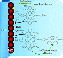
Porphyrin dyes were synthesized for use in p-type (NiO) dye sensitized solar cells based on different design principles. One porphyrin was designed with a significant charge transfer character in the excited state because of push–pull effects of the substituents. Another porphyrin had instead an appended NDI acceptor group (NDI = naphthalene diimide). The dyes were characterized by spectroscopic, electrochemical and DFT methods. Solar cells based on sensitized, meso-porous NiO showed rather poor performance compared to other organic dyes, but with a clear improvement for the dye with the NDI acceptor. Ultrafast transient absorption spectroscopy and nanosecond laser photolysis showed that hole injection into NiO was followed by unusually rapid charge recombination, predominantly on a 50–100 ps time scale, which is likely the main reason for the poor photovoltaic performance. Again the porphyrin with the NDI group showed a more long-lived charge separation that should lead to better dye regeneration in a solar cell, which can explain its better photovoltaic performance.


Introduction

Porphyrin derivatives constitute one of the most important classes of dyes, and these macrocycles have been widely used as components in molecular arrays for photoinduced energy and electron transfer schemes,1 for solar hydrogen production as catalysts2–5 or as sensitizers6 and as dyes in organic solar cells7–9 and in dye-sensitized solar cells (DSSCs).10–13 In particular, porphyrins stand among the most efficient sensitizers ever reported in conventional n-type Grätzel cells with record power conversion efficiencies (PCEs) around 13%.13 p-Type DSSC (p-DSSC) are photoelectrochemical devices that are related to classical Grätzel cells except that the photoexcited dye injects a hole in the SC instead of an electron.14–16 In p-DSSC the sensitizer injects a hole in the valence band of the semi-conductor, typically NiO, while in classical Grätzel cells an electron is injected into the conduction band of the semi-conductor, e.g., TiO2.17 The combination of sensitized p-type and n-type materials allows for construction of a tandem DSSC, with high theoretical energy conversion efficiency; however, p-type DSSCs are much less investigated than their n-type counter parts and they currently display much lower efficiency (2.5%).18 Owing to the broad applicability of porphyrins for solar energy conversion, including DSSCs, we have been interested in designing porphyrin derivatives for p-DSSCs, with the anticipation that they could deliver large PCEs as well. Indeed, zinc porphyrins fulfill all the required properties to act as efficient sensitizers for NiO: large hole injection driving force, significant light harvesting efficiency and an excited state that feature large charge transfer character in push–pull systems. Another attractive feature of zinc porphyrins is the strong reducing ability of these dyes after hole injection (ca. −1.3 V vs. SCE), which could be a valuable feature for application in dye sensitized solar fuel devices.19–24 However, surprisingly porphyrin dyes have been rarely investigated in p-DSSCs and our preliminary studies revealed that they exhibit very modest photovoltaic performances in NiO based DSSCs.25–27 To unveil the main reasons of the low performances of porphyrin sensitizers in p-DSSC and to advance towards efficient and rational design strategies of better performing dyes, we have decided to investigate in details the photophysical deactivation processes of a series of porphyrin dyes by transient absorption spectroscopy (Chart 1). Indeed, understanding the electron transfer mechanisms and dynamics is important to guide the synthesis to more effective p-type dye molecules.28–33
image file: c6ra15195g-c1.tif
Chart 1 Structures of the porphyrin sensitizers investigated in this study.

The ZnPref and ZnP-TPA-NO2 dyes were chosen in this study to investigate the impact of the push–pull character on the PCE by comparing a dye with a negligible charge transfer character of the excited-state (ZnPref) to a dye exhibiting a significant one (ZnP-TPA-NO2). The dyad ZnP-NDI has an appended acceptor group and is a better performing dye in a p-type solar cell than both ZnPref and ZnP-TPA-NO2, which prompted us to elucidate the reasons behind this property. In this work, we found that all dyes performed poorly in NiO-based solar cells compared to most other dyes. By transient absorption spectroscopy we could prove that the dye–NiO+ charge recombination reaction was faster than for most other dyes and dyads, which is the main reason for the comparatively low energy conversion efficiency.

Experimental section

General

1H and 13C NMR were recorded on a Bruker ARX 300 MHz, AVANCE 300 and 500 Bruker. Chemical shifts for 1H and 13C NMR spectra are referenced to residual protium in the deuterated solvent of CDCl3 δ = 7.26 ppm for 1H and δ = 77.16 ppm for 13C. Spectra were recorded at room temperature, chemical shifts are given in ppm and coupling constants in Hz. High-resolution mass spectra (HR-MS) were obtained by MALDI-TOF-TOF (Autoflex III, Bruker), both working in ion positive mode. Electrochemical measurements were made under an argon atmosphere at room temperature in DMF and in presence of 0.1 M Bu4NPF6 as supporting electrolyte. Cyclic voltammetry experiments were performed by using an Autolab PGSTAT 302N potentiostat/galvanostat. A standard three-electrode electrochemical cell was used. All potentials were measured and are quoted relative to saturated calomel electrode (SCE). The working electrode was platinum (3 mm in diameter). The auxiliary electrode was a stainless steel wire. In all the experiments the scan rate was 100 mV s−1. UV visible absorption spectra were recorded on a UV 2501PC Shimadzu spectrophotometer, using 1 cm path length cells.

Chemicals were purchased from Sigma-Aldrich or Alfa Aesar and used as received. Thin-layer chromatography (TLC) was performed on aluminum sheets precoated with Merck 5735 Kieselgel 60F254. Column chromatography was carried out with Merck 5735 Kieselgel 60F (0.040–0.063 mm mesh). The compounds ZnP-NDI,26 bromoporphyrin 1,26 monoiodo trisisopropyl-ethynyl porphyrin 3,34 and N,N-di(4-benzoic acid tert-butyl ester)-4-trimethylsilanylethynyl-phenylboronate ester 4[thin space (1/6-em)]35 were prepared from previously published procedures. Identity and purity were confirmed by means of NMR spectroscopy. The synthesis and characterization of ZnPref and ZnP-TPA-NO2 is described in the ESI.

Solvents employed for this study [dichloromethane (Aldrich, UV spectroscopy grade), ethanol (Aldrich, 99.5% spectroscopy grade) and propylene carbonate (Aldrich, 99% ACS reagent grade)] were used without further purification unless otherwise noted.

Theoretical calculations

All calculations have been achieved with the Gaussian09 program,36 using Time-Dependent Density Functional Theory (TD-DFT) to characterize the excited-states.37 During these calculations, the long alkyl chains were replaced by methyl moieties. The computational protocol proceeds through a four step strategy that allows quantifying the charge transfer features of organic dyes: (i) the ground-state geometrical parameters have been determined at the B3LYP/6-311G(d,p) level38 and the LanL08 pseudopotential and basis set for the zinc atom, through a force-minimization process using a SCF convergence threshold of 10−10 a.u.; (ii) the vibrational spectra of each derivative have been determined analytically at the same level of theory to ascertain that all structures correspond to true minima of the potential energy surface; (iii) the first 15 low-lying excited-states have been determined within the vertical TD-DFT approximation using the CAM-B3LYP/6-311++G(2d,p)39 level of theory with the LanL08(f) pseudopotential and basis set for the metallic center; (iv) the charge-transfer parameters have been estimated with the so-called dCT metric40,41 using the same level of theory as in the third step. All calculations systematically include a modeling of bulk solvent effects (here CH2Cl2) with the polarizable continuum model (PCM),42 in its linear-response non-equilibrium flavour for the TD-DFT calculations.
Solar cells fabrication and photovoltaic measurements. Conductive glass substrates (F-doped SnO2) were purchased from Solaronix (TEC15, sheet resistance 15 Ω per square). Conductive glass substrates were successively cleaned by sonication in soapy water, then acidified ethanol for 10 min before being fired at 450 °C for 30 min. Once cooled down to room temperature, FTO plates were screen printed with NiO using a home-made paste. The NiO screen-printing paste was produced by preparing a slurry of 3 g of NiO nanopowder (Inframat) suspended in 10 mL of distilled ethanol and ball-milled (500 rpm) for 24 h. The resulting slurry was mixed in a round-bottom flask with 10 mL of 10 wt% ethanolic ethyl cellulose (Sigma Aldrich) solution and 20 mL terpineol, followed by slow ethanol removal by rotary evaporation. The dried film was calcined in air at 400 °C for 0.5 h. The prepared NiO electrodes were soaked while still hot (80 °C) in a 0.16 mM solution of each dye during 16 h. A mixture of distilled acetonitrile and tert-butanol was used (1/1, v/v) for each bath.

The composition of the electrolyte is 0.5 M 1,2-dimethyl-3-butylimidazolium iodide, 0.5 M LiI, 0.1 M I2, 0.5 M 4-tert-butylpyridine and 0.1 M guanidinium thiocyanate in acetonitrile electrolyte. Platinum counter electrodes were prepared by depositing a few drops of an isopropanol solution of hexachloroplatinic acid in distilled isopropanol (2 mg per mL) on FTO plates (TEC7, Solaronix). Substrates were then fired at 375 °C for 30 min. The photocathode and the counterelectrode were placed on top of each other and sealed using a thin transparent film of Surlyn polymer (DuPont, 25 μm) as spacer. A drop of electrolyte was introduced through a predrilled hole in the counter electrode by vacuum backfilling, the hole was then sealed by a glass stopper with Surlyn. The cell had an active area of 0.25 cm2.

The current–voltage characteristics were determined by applying an external potential bias to the cell and measuring the photocurrent using a Keithley model 2400 digital source meter. The solar simulator is an Oriel Lamp calibrated to 100 mW cm−2.

Sensitized NiO films for spectroscopic characterization. The sensitized NiO films were prepared as above. A drop of 0.1 M LiClO4 in propylene carbonate (PC) was placed onto the film and a clean piece of glass slide placed onto to create a sandwich cell configuration. Capillary forces enable the covering glass to adhere to the glass substrate possessing the sensitized film. Typical optical densities of the sensitizer were 0.3–0.6 at the excitation wavelength.
Time-correlated single photon counting (TCSPC). The detailed description of the experimental setup has been described recently.43 Briefly, the sample was excited with a picosecond diode laser (Edinburgh Instruments, EPL470) at 470 nm (77.1 ps pulses). The laser pulse energy was ca. 15 pJ and was attenuated by neutral density filters before the sample (often by more than 1 order of magnitude) to the desired count rate of 1% or less of the excitation frequency. Photoemission detection was made with a multi-channel plate, with a cut-off filter (λ < 530 nm) between the sample and detector. Decay curves obtained by single photon counting were analyzed by iterative reconvolution using an exponential decay model with 1 or 2 components in the SpectraSolve program. The instrument response function (IRF: 60 ps) was free to move relative to the decay during analysis.
Steady state measurements. Steady state absorption spectra for freshly prepared solutions in deionized ethanol were measured on Cary 50 UV-visible spectrometer. Fluorescence measurements were recorded on a Fluorolog III emission spectrometer after excitation at 450 nm, using a P928 PMT detector and a single photon counting mode. The spectra were corrected for the wavelength dependent response of the detection system. Solution phase samples were measured in quartz cuvettes with the desired solvent (1 cm for steady-state measurements and 1 mm for femtosecond measurements). Optical densities of solution samples for emission spectroscopy were on the order of 0.1 at the excitation wavelength, while those for transient absorption in solution and NiO films were between 0.3 and 0.6.
Transient absorption measurements. Nanosecond and femtosecond transient spectroscopy methods have been described previously.44 The femtosecond transient absorption spectrometer consists of a 1 kHz Ti:sapphire amplifier (Legend-HE-Cryo, Coherent) pumped by a frequency doubled Q-switched Nd:YLF laser and seeded by a mode locked Ti:sapphire oscillator (Vitesse-800, Coherent). The output is 800 nm pulses with a temporal width of about 100 fs. The output is split to form pump and probe beams. Desired pump wavelength was obtained with a TOPAS-white, and with neutral density filters the energy of each pulse was kept between 200 and 400 nJ. The white light continuum probe was obtained by focusing part of the 800 nm light on a moving CaF2 plate. Polarization of the pump was set at magic angle, 54.7°, relative to the probe. Instrumental response time depends on pump and probe wavelengths, but are typically about 150 fs. The NiO samples were translated to avoid photodegradation.

Data analysis were done in MATLAB (The MathWorks, Inc.), a robust trust-region reflective Newton nonlinear-least-squares method was used for the fits of time traces. Traces (ΔA vs. t) were fitted to a sum of exponentials convolved with a Gaussian shaped response. An artifact signal was also included in the fit that is due to cross phase modulation during pump and probe overlap. All spectra were corrected for chirp in the white light probe, time zero was set at maximum pump-probe temporal overlap. The region around pump wavelength was removed due to scatter of pump light.

Transient absorption spectra in the microsecond time domain were recorded on a Nanosecond Flash Photolysis Spectrometer. Nanosecond transient absorption and emission were measured with a Q-switched YAG laser (Quanta Ray, Spectra Physics) that delivered ca. 10 ns pulse at 10 pulses per second. Pulse energies were 3–5 mJ per pulse at the sample. Kinetic traces were measured with a PMT and a digital oscilloscope while transient spectra were collected using a CCD (ANDOR).

Results and discussion

Synthesis of the sensitizers

The synthesis of the two new sensitizers ZnPref and ZnP-TPA-NO2 is rather straightforward as it is based on established Sonogashira or Suzuki cross-coupling reactions performed with already published reagents such as 1,45 3 (ref. 34) and 4 (ref. 35) (see Scheme 1). Sonogashira cross-coupling of the bromoporphyrin 1 with triisopropysilylethynyl affords with a 78% yield the zinc porphyrin 2. Deprotection of TIPS group by tetrabutylammonium fluoride (TBAF) followed by Sonogashira cross-coupling reaction with 4-iodo benzene gave ZnPref with 99% yield over the two steps (Scheme 1). Suzuki cross-coupling of the monoiodo trisisopropyl-ethynyl porphyrin 3 with N,N-di(4-benzoic acid tert-butyl ester)-4-trimethylsilanylethynyl-phenylboronate ester 4 gives porphyrin 5 with 95% yield. The TIPS protecting group of the alkyne of porphyrin 5 was cleaved with TBAF and the resulting porphyrin was metallated with zinc acetate before being involved in the final Sonogashira cross-coupling reaction with para iodo-nitrobenzene to afford porphyrin 6 with 67% yield over these three steps. Finally, the hydrolysis of the tert-butyl ester of the latter in trifluoroacetic acid followed by metalation of the free base porphyrin with zinc acetate furnished the sensitizer ZnP-TPA-NO2 with a quantitative yield.
image file: c6ra15195g-s1.tif
Scheme 1 Synthesis of ZnPref and ZnP-TPA-NO2. Conditions and reagents: (a) Pd(PPh3)4, CuI, Et3N, THF, reflux, 78% (b) TBAF, THF, RT and then Pd(dba)2, AsPh3, Et3N, THF, 50 °C, 99% over two steps; (c) Pd(PPh3)4, Ba(OH)2, H2O, THF/H2O (v/v: 3/1), reflux; 95%, (d) TBAF, THF, RT then Zn(OAc)2, CH2Cl2/MeOH (v/v: 1/1), reflux and finally 1-iodo-4-nitrobenzene, Pd2(dba)3, AsPh3, Et3N, THF, reflux; 67%, (e) NaOH (1 M), THF/MeOH (v/v: 8/1), reflux; 92%.

Electronic absorption and emission spectra

The UV-vis absorption spectra of the dyes ZnPref, ZnP-TPA-NO2 and ZnP-NDI are shown in Fig. 1 and the absorption and emission characteristics are collected in Table 1. All three dyes exhibit the intense Soret band at ∼440 nm together with the Q-bands over the range of 500–700 nm. The direct attachment of the ethynyl group on the porphyrin ring causes a broadening of the Soret band and an intensification of the second Q band as already reported.46,47 The spectra of ZnPref and ZnP-NDI are almost superimposable, except for the UV 350–400 nm region that exhibits the typical vibronic π–π* transitions associated with NDI unit.48 This indicates that the NDI unit has a little impact on the electronic structure of the porphyrin macrocycle certainly because the HOMO and LUMO orbital of the NDI have nodes on the nitrogen of the bisimide units.49 In contrast, the ethynyl nitrobenzene unit in ZnP-TPA-NO2 increases the electron delocalization in the molecule, induces a perceptible charge transfer band and also breakes the symmetry which cause a strong widening of the Soret band and the red shift of the Soret and Q-bands (cf. Fig. 1 and theoretical calculations below).
image file: c6ra15195g-f1.tif
Fig. 1 Overlay of the absorptions spectra of dyes ZnPref (blue), ZnP-TPA-NO2 (black) and ZnP-NDI (red) recorded in dichloromethane solution.
Table 1 Absorption and emission data of the porphyrins recorded in CH2Cl2 at room temperature
Dyes λabs/nm (ε/M−1 cm−1) λem/nm E00a (eV)
a Calculated from the average of the absorption maxima for the lowest energy Q-band in the absorption spectrum and the highest energy Q-band in the fluorescence spectrum: image file: c6ra15195g-t1.tif.
ZnPref 440 (350[thin space (1/6-em)]000), 567 (14[thin space (1/6-em)]500), 613 (14[thin space (1/6-em)]000) 624, 672 2.01
ZnP-NDI 379.5 (35[thin space (1/6-em)]000); 441 (364[thin space (1/6-em)]000); 567 (14[thin space (1/6-em)]000); 614 (16[thin space (1/6-em)]000) 625.5; 670 2.00
ZnP-TPA-NO2 450 (105[thin space (1/6-em)]000); 570 (10[thin space (1/6-em)]300); 630 (16[thin space (1/6-em)]800) 637, 682 1.96


As far as the fluorescence properties are concerned, all these three porphyrins dyes are emissive, but the dyad ZnP-NDI is strongly quenched by electron transfer to the NDI unit (see below).

Electrochemistry and spectroelectrochemistry

First, the redox potentials of the porphyrins were recorded by electrochemistry to calculate the driving force of the photoinduced hole transfer into NiO valence band (ΔG0inj) and that of the dye regeneration reaction (ΔG0reg) (Table 2 and Fig. 2). The potential of the zinc porphyrin oxidation is not much affected by the phenyl ethynyl substituent, while the zinc porphyrin centred reduction process becomes much easier in ZnP-TPA-NO2, which carries the electron withdrawing nitro-phenyl substituent. In the dyad ZnP-NDI, the first reduction process occurs at a more positive potential because it is localized on the NDI unit. Overall, the values of the Gibbs free energies of the photoinduced hole transfer and dye regeneration reactions indicate that both processes are significantly exergonic with these three dyes, although the regeneration of NDI by triiodide in the dyad ZnP-NDI has a much smaller driving force than in the other systems (Table 2 and Fig. 2).
Table 2 Reduction potentials, and reaction free energies for photoinduced hole transfer in NiO (ΔG0inj) and dye regeneration by I3/IG0reg), of the three porphyrin dyes. The reduction potentials are taken as the average of the cathodic and anodic peak potentials, as recorded in CH2Cl2, and are referenced versus saturated calomel electrode (SCE)
Dyes EOx(P+/P) ERed(P/P) ERed(NDI/NDI) ERed(P*/P)a ΔG0injb (eV) ΔG0regc (eV)
a Calculated according to the equation: ERed(P*/P) = ERed(P/P) + E00.b Calculated according to the equation: ΔG0inj = e(EVB(NiO) − ERed(P*/P)) with EVB(NiO) denoting the potential of the valence band of NiO and taken as 0.3 V vs. SCE.c Calculated according to the equation: ΔG0reg = e(ERed(A/A) − E(I3/I2˙)), with E(I3/I2˙) taken as −0.32 V vs. SCE and A = porphyrin for the two first derivatives and NDI for ZnP-NDI, respectively.
ZnPref 0.72 −1.41 0.60 −0.30 −1.09
ZnP-NDI 0.75 −1.46 −0.59 0.54 −0.24 −0.27
ZnP-TPA-NO2 0.73 −1.02   0.94 −0.64 −0.70


image file: c6ra15195g-f2.tif
Fig. 2 Energy diagram of the relevant levels for the hole photoinjection and dye regeneration reactions in NiO based p-DSSC with iodide and cobalt based redox mediator.

Spectroelectrochemical experiments were undertaken to determine the absorption spectrum of the radical anion of the zinc porphyrin for the transient absorption spectroscopy study (see below). The spectra of the reduced zinc porphyrins ZnPref and ZnP-TPA-NO2 were recorded in DMF with Bu4NPF6 as supporting electrolyte with an optically transparent thin layer electrode (OTTLE). We assumed that the spectrum of ZnP in ZnP-NDI must be very close to that of ZnPref owing to the similarity of their absorption spectra and it was consequently not recorded. Upon reduction, the spectrum of both dyes (ZnPref and ZnP-TPA-NO2) evolves in the same way (Fig. 3). The Soret band intensity decreases and widens while the Q-bands red shift and form a broad band extending to 800 nm. These features are in good agreement with a radical anion centered on the π-aromatic system of the porphyrin ring and consistent with the spectrum of the radical anion of already reported zinc porphyrins50,51 (see also below).


image file: c6ra15195g-f3.tif
Fig. 3 Evolution of the absorption spectrum of ZnPref (left) and ZnP-TPA-NO2 (right) upon electrochemical reduction at −1.8 V and −1.5 V vs. SCE, respectively, in OTTLE in DMF. The initial spectrum is shown in black while the final is in red.

Theoretical modeling

The results of first-principle are given in Table 3 (see also Fig. S5 for representation of the MOs). The determined vertical transition energies are in good agreement with the experimental spectra (see above), with weak Q-bands at ca. 600 nm and very intense Soret bands at ca. 410 nm. As in the measured data, both ZnPref and ZnP-NDI present similar signatures but for an additional band centred on the NDI core (see Fig. 4), confirming the above interpretation. The behaviour of the dyad ZnP-NDI can be readily explained by the fact the NDI moiety is almost perfectly perpendicular to the phenyl ring (DFT yields a 87° dihedral angle) and that the NDI unit does not electronically communicate with the porphyrin owing to the nodes on the HOMO and LUMO (see Fig. 4 and S5). As can be seen in Table 3, the CT distance associated to all transitions in ZnPref and ZnP-NDI are extremely small (<1.1 Å) and are thus well-localized π–π* excited-states, as expected. The bands of ZnP-TPA-NO2 are slightly bathochromically shifted due to the increase of delocalization, and a notable CT character can be seen (CT distance of 4.73 Å) for the most intense state in the Soret region. As can be seen in Fig. 4, these CT corresponds to a transfer from the porphyrin core to the nitro group, whereas the donor moiety has no significant role. In this dye, the Q-bands have a much smaller CT character (see Table 3 and bottom of Fig. 4). The spin density for the radical anion of ZnP-TPA-NO2 is shown in Fig. 5 and confirms that it is indeed mainly centered on the ZnP core though a small contribution is found on the nitro group.
Table 3 Results of TD-DFT calculations performed on the three dyes: selected vertical transition wavelength and associated oscillator strengths, as well as determined CT distance (see Experimental section for details)
Dyes λabs/nm (f) δXT
ZnPref 598 (0.20); 580 (0.00); 410 (2.22); 405 (1.53) 0.82; 0.77; 1.08; 1.08
ZnP-NDI 596 (0.24); 579 (0.00); 411 (2.56); 404 (1.53); 350 (0.43) 0.71; 0.50; 0.14; 0.86; 0.04
ZnP-TPA-NO2 604 (0.41); 581 (0.01); 433 (2.33); 408 (1.33) 0.55; 1.04; 4.73; 1.82


image file: c6ra15195g-f4.tif
Fig. 4 Density difference plots obtained for the 350 nm absorption in ZnP-NDI (top) and the 433 nm (center) and 604 nm (bottom) absorption of ZnP-TPA-NO2. The blue (red) regions indicate increase (decrease) of electron density upon absorption and are drawn with a contour threshold of 0.0004 au.

image file: c6ra15195g-f5.tif
Fig. 5 Spin density difference for the radical anion ZnP-TPA-NO2. A contour threshold of 0.002 au is used.

Photovoltaic performance of porphyrin-sensitized NiO films

The three porphyrin systems were used as sensitizers in nanocrystalline NiO-based photocathodes using the iodide/triiodide couple as redox shuttle (see Experimental section). The short circuit photocurrent density (JSC), open circuit voltage (VOC), fill factor (ff) and energy conversion efficiency (η) recorded under stimulated AM1.5G solar light (100 mW cm−2) are listed in Table 4.
Table 4 Photovoltaic characteristics of the solar cells made with mesoporous NiO electrodes sensitized with the porphyrin dyes using I/I3 electrolyte and recorded under stimulated AM1.5G solar light (100 mW cm−2)
Dyes JSC (mA cm−2) VOC (mV) ff (%) η (%)
ZnPref 0.19 98 35 0.006
ZnP-NDI 1.38 127 32 0.056
ZnP-TPA-NO2 0.29 107 38 0.012


As previously reported with other zinc porphyrin sensitizers, except for the dyad ZnP-NDI, the dyes ZnPref and ZnP-TPA-NO2 give poor photovoltaic performance (Table 4). Examination of the driving forces of both hole injection and dye regeneration processes indicate that the thermodynamics must certainly not be the main reason of the low efficiencies (Table 2). Accordingly, transient absorptions spectroscopy measurements were carried out to shed some light on the origin of the unexpectedly low performances of these dyes with NiO based p-DSSCs.

Time-resolved spectroscopy measurements

Transient absorption and time-resolved emission measurements were made to investigate the excited state charge transfer dynamics of all porphyrin dyes both in ethanol solution and on NiO film. Excitation at 470 nm in the TCSPC experiments show fluorescence lifetimes of 2–3 ns for ZnPref and ZnP-TPA-NO2 in ethanol solution (Table S1, Fig. S1), which is quite typical for the lowest singlet excited state (S1) of Zn(II)-porphyrins.52 The majority of the S1 decay is due to intersystem crossing to the lowest triplet state (T1).52 In contrast, the ZnP-NDI fluorescence is very short-lived, ca. 15 ps, suggesting rapid electron transfer towards the NDI unit. These results are supported by the transient absorption experiments in EtOH solution following ∼120 fs laser excitation at 560 nm. The transient absorption spectra and summarized kinetic data are shown in the ESI (Table S2, Fig. S2 and S3). The transient absorption spectra recorded for ZnPref and ZnP-TPA-NO2 are typical for a Zn-porphyrin S1 excited state: an intense Soret band bleach in the 400–440 nm region, a broad positive band culminating at around 465 nm and extending to about 700 nm, with an overlapping negative feature around 625 nm due the Q-band bleach. The transients show very small spectral changes during the time window of the experiment, i.e., 0–2 ns, because the transient spectra of the S1 and T1 states are very similar. Nevertheless they agree with the 2–3 ns lifetime of the S1 state determined by TCSPC, see ESI for details. To determine the spectra and lifetime of the T1 state, nano-second laser flash photolysis with 560 nm, 10 ns excitation was used (Fig. S3). These experiments show triplet state spectra that are similar to the final spectra of the fs experiments, and decays with τ = 1.9 μs (ZnPref) and τ = 4.6 μs (ZnP-TPA-NO2) in argon purged solution. Note that the ethynyl substituent shortens the triplet lifetime significantly compared to the case of ZnTPP.46,53

For ZnP-NDI instead the fs-experiments show rapid transient absorption changes with strong Soret and Q-band recovery described by τ = 11.6 ps and a concomitant shift and decay of the 480 nm band. A slower decay component with τ = 149 ps shows a narrow band at 460 nm and a bleach at 440 nm. This is attributed to the charge separation to form ZnP+–NDI with τ = 11.6 ps followed by charge recombination with τ = 149 ps. Unfortunately, the transient spectra of ZnP+ and ZnP look almost the same, and have large bands around 460 nm just like the S1 and T1 state (this is a well-known difficulty with porphyrins).51,54 In addition, the main NDI band is also seen around 475 nm. Therefore, the ZnP+–NDI state is not clearly distinguishable. Nevertheless, the short emission lifetime and rapid transient absorption decay is most readily attributed to electron transfer to NDI, followed by a rapid recombination.

Femtosecond transient absorption experiments were also performed on the sensitized NiO films in order to understand their electron transfer dynamics. Fig. 6 shows the transient absorption spectra of Zn porphyrin dyes on the NiO surface. Generally, for all three dyes, decay of the transient bands and ground state bleach were much faster than observed for the dyes in homogeneous solution. The transient absorption spectra of NiO/ZnPref and NiO/ZnP-NDI present similar features with a fast recovery of the ground state bleach and the small stimulated emission signal observed at ∼660 nm (cf. the 1–0 fluorescence in Table 1) during the initial few picoseconds. The positive absorption bands at <410 nm and >460 nm also decay rapidly. However, more transient signals remain after 1.9 ns for NiO/ZnP-NDI than for NiO/ZnPref. The transient spectrum for NiO/ZnP-TPA-NO2 is similar and decays almost completely within 2 ns, like for NiO/ZnPref, but the Q-band bleach is broader.


image file: c6ra15195g-f6.tif
Fig. 6 Femtosecond transient absorption spectra of porphyrin dyes on the NiO film with 0.1 M LiClO4/PC electrolyte after excitation at 560 nm: (a) NiO/ZnPref, the inset kinetic trace is probed at 600 nm, (b) NiO/ZnP-TPA-NO2, the inset kinetic trace is probed at 600 nm, and (c) NiO/ZnP-NDI, the inset kinetic trace is probed at 700 nm. The Soret band region of a and c is not reliable as the high optical density consumed most of the probe light.

To exclude that the rapid excited state quenching is due to dye aggregation, experiments were performed with samples in which chenodeoxycholic acid (CDCA) was used as co-adsorbant, which is known to break up aggregates. As the transient absorption decayed even faster in these samples (Fig. 7), we can exclude aggregation as an explanation for the rapid excited state decay on NiO.


image file: c6ra15195g-f7.tif
Fig. 7 Kinetic comparison of the transient absorption at 490 nm for NiO/ZnP-TPA-NO2 samples without CDCA (green circles) and with 0.5 mM (light blue triangles) or 5 mM CDCA (yellow diamonds). The solid lines are multi-exponential fits to the data.

Global analysis of the time evolution of the TA spectra measured upon 560 nm excitation was performed using a sum of exponential functions to determine the number of time constants required to represent the data. The data for NiO/ZnPref could be well processed using five exponential functions with results 0.21 ps, 2.1 ps, 8.9 ps, 51.7 ps and ∞ time constants. These time constants should not be interpreted as unique processes, but they represent an approximate description of the multi-exponential kinetics typically observed for DSCs. Decay associated spectra (DAS, Fig. S4a) show that the 0.21 ps and 2.1 ps components are similar to decay of the S1 excited state in solution. The species associated spectra (SAS; Fig. 8a) show that the bleach/stimulated emission at 620 nm for the 0.21 ps component shifts to 640 nm for the 8.9 ps component, which suggests that the three first τ1τ3 components mainly reflect hole injection. The constant τ4 = 51.7 ps instead represents rapid recombination of the reduced ZnP and the injected hole, and there is very little (<1%) signal left after 2 ns. The results for ZnP-TPA-NO2 are very similar, with very little signal remaining after 2 ns (Fig. 6b, 8b and S4b). To summarize, although the transient spectra are similar to those in homogeneous solution, the rapid quenching and the details of the results from global analysis of the data (DAS and SAS) allows us to conclude that hole injection into NiO occurs from excited ZnPref and ZnP-TPA-NO2with time constants around 0.2–10 ps, and is followed by rapid recombination, mainly on the 50–100 ps time scale.


image file: c6ra15195g-f8.tif
Fig. 8 Species associated TA spectra of porphyrins dyes on NiO from global analysis, (a) NiO/ZnPref, (b) NiO/ZnP-TPA-NO2 and (c) NiO/ZnP-NDI.

For NiO/ZnP-NDI, the global analysis gave similar results, although the presence of the secondary electron acceptor NDI made the transient signals more long-lived (Fig. 6c); the DAS and SAS are presented in Fig. 8c and S4c. The τ1 = 0.42 ps and τ2 = 2.3 ps components show clearly vibrational relaxation and decay of stimulated emission at 620 nm and 680 nm. The SAS of the τ3 = 16.3 ps, τ4 = 219 ps and τ5 > 2 ns components do not show the stimulated emission, and has a more prominent band around 700, which can be attributed to ZnP (Fig. 3). The corresponding band shape is hard to see for the other two dyes as the remaining signal magnitude is so much smaller. There is no clear evidence of NDI reduction, yet the charge separated state is clearly more long-lived with the ZnP-NDI dyad. These reactions were further investigated on a ns to μs time scale.

Nano-second laser flash photolysis was performed to further investigate the charge recombination processes, as well as dye regeneration. The transient absorption after a 10 ns laser flash in the Soret band at 450 nm are shown in Fig. 9. Note that the ns-pulse gives more photons per sample area than the fs-pulses, and also excited in the strong Soret band. Thus, we could obtain detectable transient signals although most of the excited species had decayed on the <2 ns time scale, before the time of observation. First, we see that on the ns to μs time scale all three dyes give a signal that is different from the S1 and T1 states in homogeneous solution (cf. ESI). The transient spectra of all three dyes on the “dry” film exhibit similar shape, with a narrow and positive band at 450–480 nm, which is agreement with the spectroelectrochemistry for the porphyrin radical anion (Fig. 3). With added PC solvent on the NiO film, the signal for ZnP-NDI broadens a little compared to without solvent, but there are still clear bleaches of the Soret and Q-bands from porphyrin, so if the electron goes out to NDI acceptor, it is only in some of the molecules, resulting in a broadened spectra at 480 nm by the absorption of the NDI. The driving force is large for electron shift from ZnP to NDI, −ΔG0 ≈ 0.85 eV (Table 2). Moreover, we have successfully used NDI as secondary acceptor in other dyes, and obtained direct spectroscopic evidence that the electron transferred rapidly (1–20 ps) and completely onto this unit, which resulted in orders of magnitude more long-lived charge separation.55–58 It is surprising that ZnP does not reduce NDI even on the ns time scale. Nevertheless, the NDI group apparently stabilizes the charge separated state so that more of it survives onto the ns time scale.


image file: c6ra15195g-f9.tif
Fig. 9 Nanosecond TA spectra at t = 50 ns of porphyrin dyes on NiO films with (right) and without PC solvent (left), excitation at 450 nm: ZnPref (black), ZnP-TPA-NO2 (blue), ZnP-NDI (red).

Conclusion

The conclusions that can be drawn from this study is that the dye–NiO+ charge recombination is unusually fast in these dyes; at 1 ns after excitation there is very little charge separated state left. This would lead to very inefficient regeneration by the I3/I electrolyte, and is certainly the main reason for the poor photovoltaic performance of these dyes.

The creation of the push–pull dye ZnP-TPA-NO2, with a TPA spacer between the porphyrin group and the NiO, led to no significant increase in charge separation lifetime or photovoltaic performance compared to ZnPref. This is surprising, but was also shown for a series of dipyrrolopyrolidine dyes.55 It is conceivable that the dyes lie flat on the NiO surface, so that molecular control of the location of the excess electron in the reduced state has no or only little effect. However, charge recombination is slower for ZnP-NDI, the dye with an appended acceptor unit and this also shows a significantly better photovoltaic performance. This points to the possibility to further improve porphyrin dyes for sensitized NiO devices by suitable molecular design.

Acknowledgements

This project is financial support from the Knut & Alice Wallenberg Foundation and the Swedish Energy Agency. ANR is gratefully acknowledged for the financial support of these researches through the program POSITIF (ANR-12-PRGE-0016-01), CNRS, Région des Pays de la Loire for the projects NiOPhotoCat and LUMOMAT, and COST CM1202 program (PERSPECT H2O). L.Z. acknowledges the China Scholarship Council (CSC) for a doctoral scholarship support. D.J. thanks Dr F. Anne (Nantes) who performed preliminary calculations. D.J. acknowledges the European Research Council (ERC) for financial support in the framework of a Starting Grant (Marches-278845). This research has used resources of the GENCI-CINES/IDRIS and the CCIPL (Centre de Calcul Intensif des Pays de Loire).

References

  1. M. R. Wasielewski, Chem. Rev., 1992, 92, 435–461 CrossRef CAS.
  2. I. Bhugun, D. Lexa and J.-M. Saveant, J. Am. Chem. Soc., 1996, 118, 3982–3983 CrossRef CAS.
  3. B. B. Beyene, S. B. Mane and C.-H. Hung, Chem. Commun., 2015, 51, 15067–15070 RSC.
  4. D. K. Bediako, B. H. Solis, D. K. Dogutan, M. M. Roubelakis, A. G. Maher, C. H. Lee, M. B. Chambers, S. Hammes-Schiffer and D. G. Nocera, Proc. Natl. Acad. Sci. U. S. A., 2014, 111, 15001–15006 CrossRef CAS PubMed.
  5. Y. Han, H. Fang, H. Jing, H. Sun, H. Lei, W. Lai and R. Cao, Angew. Chem., Int. Ed., 2016, 55, 5457–5462 CrossRef CAS PubMed.
  6. K. Ladomenou, M. Natali, E. Iengo, G. Charalampidis, F. Scandola and A. G. Coutsolelos, Coord. Chem. Rev., 2015, 304–305, 38–54 CrossRef CAS.
  7. H. Qin, L. Li, F. Guo, S. Su, J. Peng, Y. Cao and X. Peng, Energy Environ. Sci., 2014, 7, 1397–1401 CAS.
  8. Y. I. H. Chao, J.-F. Jheng, J.-S. Wu, K.-Y. Wu, H.-H. Peng, M.-C. Tsai, C.-L. Wang, Y.-N. Hsiao, C.-L. Wang, C.-Y. Lin and C.-S. Hsu, Adv. Mater., 2014, 26, 5205–5210 CrossRef CAS PubMed.
  9. K. Gao, L. Li, T. Lai, L. Xiao, Y. Huang, F. Huang, J. Peng, Y. Cao, F. Liu, T. P. Russell, R. A. J. Janssen and X. Peng, J. Am. Chem. Soc., 2015, 137, 7282–7285 CrossRef CAS PubMed.
  10. A. Yella, H.-W. Lee, H. N. Tsao, C. Yi, A. K. Chandiran, M. K. Nazeeruddin, E. W.-G. Diau, C.-Y. Yeh, S. M. Zakeeruddin and M. Grätzel, Science, 2011, 334, 629–634 CrossRef CAS PubMed.
  11. L.-L. Li and E. W.-G. Diau, Chem. Soc. Rev., 2013, 42, 291–304 RSC.
  12. H. Imahori, T. Umeyama and S. Ito, Acc. Chem. Res., 2009, 42, 1809–1818 CrossRef CAS PubMed.
  13. S. Mathew, A. Yella, P. Gao, R. Humphry-Baker, F. E. CurchodBasile, N. Ashari-Astani, I. Tavernelli, U. Rothlisberger, K. NazeeruddinMd and M. Grätzel, Nat. Chem., 2014, 6, 242–247 CrossRef CAS PubMed.
  14. F. Odobel, L. Le Pleux, Y. Pellegrin and E. Blart, Acc. Chem. Res., 2010, 43, 1063–1071 CrossRef CAS PubMed.
  15. F. Odobel, Y. Pellegrin, E. A. Gibson, A. Hagfeldt, A. L. Smeigh and L. Hammarström, Coord. Chem. Rev., 2012, 256, 2414–2423 CrossRef CAS.
  16. F. Odobel and Y. Pellegrin, J. Phys. Chem. Lett., 2013, 4, 2551–2564 CrossRef CAS.
  17. A. Hagfeldt, G. Boschloo, L. Sun, L. Kloo and H. Pettersson, Chem. Rev., 2010, 110, 6595–6663 CrossRef CAS PubMed.
  18. I. R. Perera, T. Daeneke, S. Makuta, Z. Yu, Y. Tachibana, A. Mishra, P. Bäuerle, C. A. Ohlin, U. Bach and L. Spiccia, Angew. Chem., Int. Ed., 2015, 54, 3758–3762 CrossRef CAS PubMed.
  19. Y. Kou, S. Nakatani, G. Sunagawa, Y. Tachikawa, D. Masui, T. Shimada, S. Takagi, D. A. Tryk, Y. Nabetani, H. Tachibana and H. Inoue, J. Catal., 2014, 310, 57–66 CrossRef CAS.
  20. Z. Yu, F. Li and L. Sun, Energy Environ. Sci., 2015, 8, 760–775 CAS.
  21. H. Tian, ChemSusChem, 2015, 8, 3746–3759 CrossRef CAS PubMed.
  22. L. Hammarstrom, Acc. Chem. Res., 2015, 48, 840–850 CrossRef PubMed.
  23. D. L. Ashford, M. K. Gish, A. K. Vannucci, M. K. Brennaman, J. L. Templeton, J. M. Papanikolas and T. J. Meyer, Chem. Rev., 2015, 115, 13006–13049 CrossRef CAS PubMed.
  24. J. R. Swierk and T. E. Mallouk, Chem. Soc. Rev., 2013, 42, 2357–2387 RSC.
  25. M. Borgström, E. Blart, G. Boschloo, E. Mukhtar, A. Hagfeldt, L. Hammarström and F. Odobel, J. Phys. Chem. B, 2005, 109, 22928–22934 CrossRef PubMed.
  26. A. Maufroy, L. Favereau, F. B. Anne, Y. Pellegrin, E. Blart, M. Hissler, D. Jacquemin and F. Odobel, J. Mater. Chem. A, 2015, 3, 3908–3917 CAS.
  27. H. Tian, J. Oscarsson, E. Gabrielsson, S. K. Eriksson, R. Lindblad, B. Xu, Y. Hao, G. Boschloo, E. M. J. Johansson, J. M. Gardner, A. Hagfeldt, H. Rensmo and L. Sun, Sci. Rep., 2014, 4, 4282 Search PubMed.
  28. P. Qin, J. Wiberg, E. A. Gibson, M. Linder, L. Li, T. Brinck, A. Hagfeldt, B. Albinsson and L. Sun, J. Phys. Chem. C, 2010, 114, 4738–4748 CAS.
  29. L. J. Antila, P. Myllyperkio, S. Mustalahti, H. Lehtivuori and J. Korppi-Tommola, J. Phys. Chem. C, 2014, 118, 7772–7780 CAS.
  30. A. Morandeira, G. Boschloo, A. Hagfeldt and L. Hammarström, J. Phys. Chem. B, 2005, 109, 19403–19410 CrossRef CAS PubMed.
  31. A. Morandeira, G. Boschloo, A. Hagfeldt and L. Hammarström, J. Phys. Chem. C, 2008, 112, 9530–9537 CAS.
  32. Z. Bian, T. Tachikawa, S.-C. Cui, M. Fujitsuka and T. Majima, Chem. Sci., 2012, 3, 370 RSC.
  33. J. Warnan, Y. Pellegrin, E. Blart, L. Zhang, A. Brown, L. Hammarström, D. Jacquemin and F. Odobel, Dyes Pigm., 2014, 105, 174–179 CrossRef CAS.
  34. L. Favereau, J. Warnan, F. B. Anne, Y. Pellegrin, E. Blart, D. Jacquemin and F. Odobel, J. Mater. Chem. A, 2013, 1, 7572–7575 CAS.
  35. M. Weidelener, A. Mishra, A. Nattestad, S. Powar, A. J. Mozer, E. Mena-Osteritz, Y.-B. Cheng, U. Bach and P. Bäuerle, J. Mater. Chem., 2012, 22, 7366–7379 RSC.
  36. M. J. Frisch, G. W. Trucks, H. B. Schlegel, G. E. Scuseria, M. A. Robb, J. R. Cheeseman, G. Scalmani, V. Barone, B. Mennucci, G. A. Petersson, H. Nakatsuji, M. Caricato, X. Li, H. P. Hratchian, A. F. Izmaylov, J. Bloino, G. Zheng, J. L. Sonnenberg, M. Hada, M. Ehara, K. Toyota, R. Fukuda, J. Hasegawa, M. Ishida, T. Nakajima, Y. Honda, O. Kitao, H. Nakai, T. Vreven Jr, J. E. Peralta, F. Ogliaro, M. Bearpark, J. J. Heyd, E. Brothers, K. N. Kudin, V. N. Staroverov, R. Kobayashi, J. Normand, K. Raghavachari, A. Rendell, J. C. Burant, S. S. Iyengar, J. Tomasi, M. Cossi, N. Rega, J. M. Millam, M. Klene, J. E. Knox, J. B. Cross, V. Bakken, C. Adamo, J. Jaramillo, R. Gomperts, R. E. Stratmann, O. Yazyev, A. J. Austin, R. Cammi, C. Pomelli, J. W. Ochterski, R. L. Martin, K. Morokuma, V. G. Zakrzewski, G. A. Voth, P. Salvador, J. J. Dannenberg, S. Dapprich, A. D. Daniels, O. Farkas, J. B. Foresman, J. V. Ortiz, J. Cioslowski and D. J. Fox, Gaussian 09 Revision A.02, Gaussian Inc, Wallingford CT, 2009, p. 2009 Search PubMed.
  37. D. Jacquemin and C. Adamo, in Top. Curr. Chem., ed. N. Ferré, M. Filatov and M. Huix-Rotllant, Springer International Publishing, Cham, 2016, pp. 347–375 Search PubMed.
  38. A. D. Becke, J. Chem. Phys., 1993, 98, 5648–5652 CrossRef CAS.
  39. T. Yanai, D. P. Tew and N. C. Handy, Chem. Phys. Lett., 2004, 393, 51–57 CrossRef CAS.
  40. T. Le Bahers, C. Adamo and I. Ciofini, J. Chem. Theory Comput., 2011, 7, 2498–2506 CrossRef CAS PubMed.
  41. D. Jacquemin, T. L. Bahers, C. Adamo and I. Ciofini, Phys. Chem. Chem. Phys., 2012, 14, 5383–5388 RSC.
  42. J. Tomasi, B. Mennucci and R. Cammi, Chem. Rev., 2005, 105, 2999–3094 CrossRef CAS PubMed.
  43. A. El-Zohry, A. Orthaber and B. Zietz, J. Phys. Chem. C, 2012, 116, 26144–26153 CAS.
  44. J. Petersson, M. Eklund, J. Davidsson and L. Hammarstrom, J. Phys. Chem. B, 2010, 114, 14329–14338 CrossRef CAS PubMed.
  45. A. Maufroy, L. Favereau, F. B. Anne, Y. Pellegrin, E. Blart, M. Hissler, D. Jacquemin and F. Odobel, J. Mater. Chem. A, 2015, 3, 3908–3917 CAS.
  46. J. Fortage, J. Boixel, E. Blart, L. Hammarström, H. C. Becker and F. Odobel, Chem.-Eur. J., 2008, 14, 3467–3480 CrossRef CAS PubMed.
  47. F. Odobel, S. Suresh, E. Blart, Y. Nicolas, J.-P. Quintard, P. Janvier, J.-Y. Le Questel, B. Illien, D. Rondeau, P. Richomme, T. Haupl, S. Wallin and L. Hammarström, Chem.–Eur. J., 2002, 8, 3027–3046 CrossRef CAS.
  48. S. V. Bhosale, C. H. Jani and S. J. Langford, Chem. Soc. Rev., 2008, 37, 331–342 RSC.
  49. F. Chaignon, M. Falkenstroem, S. Karlsson, E. Blart, F. Odobel and L. Hammarström, Chem. Commun., 2007, 64–66 RSC.
  50. G. Balducci, G. Chottard, C. Gueutin, D. Lexa and J.-M. Saveant, Inorg. Chem., 1994, 33, 1972–1978 CrossRef CAS.
  51. P. Neta, J. Phys. Chem., 1981, 85, 3678–3684 CrossRef CAS.
  52. K. Kalyanasundaram, Photochemistry of Polypyridine and Porphyrin Complexes, Academic Press, London, 1992 Search PubMed.
  53. S. M. LeCours, S. G. DiMagno and M. J. Therien, J. Am. Chem. Soc., 1996, 118, 11854–11864 CrossRef CAS.
  54. M. Andersson, M. Linke, J.-C. Chambron, J. Davidsson, V. Heitz, L. Hammarström and J.-P. Sauvage, J. Am. Chem. Soc., 2002, 124, 4347–4362 CrossRef CAS PubMed.
  55. Y. Farré, L. Zhang, Y. Pellegrin, A. Planchat, E. Blart, M. Boujtita, L. Hammarström, D. Jacquemin and F. Odobel, J. Phys. Chem. C, 2016, 120, 7923–7940 Search PubMed.
  56. E. A. Gibson, A. L. Smeigh, L. L. Pleux, J. Fortage, G. Boschloo, E. Blart, Y. Pellegrin, F. Odobel, A. Hagfeldt and L. Hammarström, Angew. Chem., Int. Ed., 2009, 48, 4402–4405 CrossRef CAS PubMed.
  57. A. Morandeira, J. Fortage, T. Edvinsson, L. Le Pleux, E. Blart, G. Boschloo, A. Hagfeldt, L. Hammarström and F. Odobel, J. Phys. Chem. C, 2008, 112, 1721–1728 CAS.
  58. Z. Huang, G. Natu, Z. Ji, M. He, M. Yu and Y. Wu, J. Phys. Chem. C, 2012, 116, 26239–26246 CAS.

Footnote

Electronic supplementary information (ESI) available. See DOI: 10.1039/c6ra15195g

This journal is © The Royal Society of Chemistry 2016