DOI:
10.1039/C6RA15181G
(Paper)
RSC Adv., 2016,
6, 90371-90383
Thienopyrimidine sulphonamide hybrids: design, synthesis, antiprotozoal activity and molecular docking studies†
Received
11th June 2016
, Accepted 1st September 2016
First published on 1st September 2016
Abstract
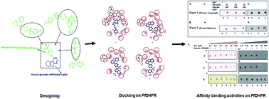
A series of hybrid compounds containing the thienopyrimidine scaffold as a DHFR inhibitor fused with a bioactive sulphonamide piperazine skeleton were synthesized and evaluated against the chloroquine and pyrimethamine resistant K1 strain of Plasmodium falciparum and the HM1:1MSS strain of Entamoeba histolytica, respectively. A few of the compounds showed better results than the standard drugs. The toxicity of the hybrids was measured on the Chinese hamster cell line. The majority of the compounds had low toxicity. The binding modes of the most potent antimalarial compounds (5, 6 and 8) were also investigated against PfDHFR using molecular docking and enzyme binding studies, whereby 5 and 6 were found to the most promising against PfDHFR. The present studies suggest that these hybrids might be possible antiprotozoal lead compounds worth further investigation.
1. Introduction
The world's population is plagued by numerous parasitic protozoal diseases that are a huge burden to society. Amoebiasis, caused by Entamoeba histolytica (E. histolytica), a protozoan, leads to a loss of about 100
000 lives annually.1 Effective chemotherapy with the 4-nitroimidazole class of drugs exists but is marred by the fact that it is genotoxic to humans; in addition, there are reports of the development of resistance to the present chemotherapy by the parasite, which can cause major health problems.2,3 Malaria, another protozoal disease, caused by Plasmodium falciparum (P. falciparum), results in a mortality rate of approximately half a million people globally.4 Fear of the disease is now even more alarming due to the emergence of chloroquine-resistant and multiple drug resistance strains of the malaria parasites.5 This has accelerated the search for hybrid anti-malarials to overcome the problem of drug resistance.
Designing hybrid molecules in which two or more pharmacophores are covalently linked to form one molecule is an upcoming field in this direction.6,7 Such designs allow for synergic action, resulting in an active moiety and a reduction in unwanted side effects.8 Thienopyrimidines are a highly promising form of heterocyclic scaffolds possessing a diverse range of biological activities, such as antituberculosis, anti-inflammatory, anticancer, antiviral and antimalarial.9–13 The enzyme dihydrofolate reductase (DHFR), a key enzyme in the folate biosynthetic pathway, is critical to the biosynthesis of DNA, RNA and some amino acids and is an important target for the development of novel chemotherapeutic agents for malaria, cancer and various microbial diseases. Thieno[2,3-d]pyrimidines have been reported to be DHFR inhibitors.14–16 A literature survey revealed that compounds bearing the piperazine sulphonamide scaffold possess a broad range of pharmacological activities, such as anti-HIV, anti-microbial, anti-proliferative, anti-fungal, anti-diabetic, anti-convulsant, anti-TB and antiprotozoal.17–24
In view of the above properties of the thienopyrimidine and piperazine sulphonamide skeletons, the two pharmacophores were covalently linked to generate a single hybrid molecule (Fig. 1), which was evaluated against the HM1:IMSS strain of E. histolytica and the K1 strain of P. falciparum in anticipation that the hybrid molecules would show potent antiprotozoal activity due to the synergistic effect of the two scaffolds. The in vitro results of the hybrids were further validated by molecular docking and enzyme binding studies on PfDHFR.
 |
| Fig. 1 Design of the hybrid compounds. | |
2. Results and discussion
2.1. Chemistry
The 4-piperazine-1-yl-5,6,7,8-tetrahydrobenzothiopheno[2,3-d]pyrimidine-based sulphonamides were synthesized by a reported method.24 Cyclohexanone was condensed with ethyl cyanoacetate under basic conditions and then cyclized in the presence of sulfur to construct the thiophene core (1) through the Gewald reaction, as shown in Scheme 1. Cyclization with formamide gave the basic thienopyrimidine core (2), which on reaction with POCl3 gave the 4-chloro-5,6,7,8-tetrahydrobenzothiopheno[2,3-d]pyrimidine (3). Piperazine was reacted with 4-chloro-5,6,7,8-tetrahydrobenzothiopheno[2,3-d]pyrimidine in ethanol, resulting in the formation of the intermediate 4-piperazine-1-yl-5,6,7,8-tetrahydrobenzothiopheno[2,3-d]pyrimidine (4).25 The reaction proceeded via aromatic nucleophilic substitution. The thienopyrimidine sulphonamide hybrids (5–14) were synthesized by the reaction of 4-piperazine-1-yl-5,6,7,8-tetrahydrobenzothiopheno[2,3-d]pyrimidine (4) with different substituted sulphonyl chlorides in dichloromethane. Triethylamine was used as a base at 0 °C to room temperature (Scheme 1). The compounds were recrystallized from dichloromethane and methanol (1
:
9).
 |
| Scheme 1 Synthesis of 4-piperazin-1-yl-5,6,7,8-tetrahydrobenzo[4,5]theino[2,3-d]pyrimidine-based sulphonamides. Reagents and conditions: (a) S8, Et3N, EtOH (b) formamide, reflux (c) POCl3, reflux (d) piperazine, EtOH (e) R-sulphonyl chloride, Et3N, DCM. | |
2.2. Biology
2.2.1. Antiamoebic activity. The antiamoebic potential of the hybrid compounds was tested on the HM1:IMSS strain of E. histolytica by a microdilution method. The antiamoebic activity was compared with the standard amoebicidal drug metronidazole (MNZ) with a 50% inhibitory concentration (IC50) of 1.86 ± 0.02 μM. The results manifested that the intermediate 4 had an IC50 value of 3.29 ± 0.02 μM, which suggested that the intermediate was less potent than the standard drug MNZ. Further reaction of the intermediate with different substituted sulphonyl chlorides yielded compounds exhibiting a range of IC50 values (0.35 ± 0.03 to 5.8 ± 0.02 μM), as shown in Table 1. The structure activity relationship (SAR) of compounds 10 and 12 (IC50 0.78 ± 0.02 μM and 0.35 ± 0.03 μM, respectively) bearing electron donating groups showed a two-fold increase in activity as inhibitors of E. histolytica compared to MNZ. The presence of aliphatic substitution in compounds 9 and 11 (IC50 1.36 ± 0.01 μM and 1.70 ± 0.01 μM, respectively) led to an enhancement of the antiamoebic potency. The enhancement in the activity observed on moving from methyl to propyl substituted sulphonamides may be attributed to the elongation of the alkyl chain. Furthermore, it was observed that the introduction of an electron withdrawing group, as in compounds 7 (IC50 3.0 ± 0.02 μM), 8 (2.82 ± 0.02 μM) and 14 (IC50 2.02 ± 0.02 μM), or bulky groups, as in compounds 5 (5.8 ± 0.02 μM) and 6 (IC50 4.6 ± 0.02 μM), had a negative effect on the activity.
Table 1 In vitro antiamoebic and antimalarial activity of thienopyrimidine sulphonamide hybrids against the HM1:IMSS strain of E. histolytica and the K1 strain of P. falciparum
2.2.2. Antimalarial activity. All the synthesized compounds were evaluated for their inhibitory effects on P. falciparum K1 growth by in vitro assay. The inhibitory effects of the compounds are presented in Table 1. Based on the results, all the compounds showed parasite growth inhibition, with the different IC50 values ranging from <0.1–18.5 ± 0.03 μM. The IC50 value of the intermediate 4 and final compound 13 were 2.07 ± 0.01 and 3.15 ± 0.02 μM, respectively. The most potent compounds against P. falciparum in culture were 5 and 8 (>0.1 μM), with IC50 values less than that of chloroquine. The antimalarial potency of compounds 7 and 14 (IC50 7.85 ± 0.02 μM and 18.5 ± 0.03 μM, respectively) were found to be very low compared to chloroquine. According to the structure–activity relationship, compound 5 containing a t-butyl group at the para position was most active, while compound 7 containing an ortho NO2 group and 14 containing a Cl group at the para position of the benzene ring showed a low inhibition of parasite growth. However, compound 6 containing a naphthalene group as the substitution at this para position was found to be active, with an IC50 value of 0.1 ± 0.001 μM, and its antimalarial activity was similar to that of the reference drug chloroquine. Compound 8 containing two chloro groups at positions 2 and 5 inhibited P. falciparum growth (IC50 < 0.1 μM) at low concentration, while compounds 9 and 11, with aliphatic sulphonamide substitution, showed fairly good activity, with IC50 values of 1.02 ± 0.01 μM and 0.9 ± 0.001 μM, respectively. In addition, compounds 10 and 12, with electron donating groups, also gave nearly the same IC50 values of 1.98 ± 0.01 μM and 1.94 ± 0.02 μM, respectively. The variation in IC50 values obtained for the antimalarial and antiamoebic activities of some of these compounds may be due to the different uptake capability of these parasites or the availability of drug targets in their cells.
2.2.3. Cytotoxic activity. The IC50 values of compounds (5, 6, 8, 9, 10, 11 and 12) could not be determined, even when the concentration was raised up to 50 μM (Table 1). The cytotoxicity of the compounds was assessed by MTT assay on the Chinese hamster ovary (CHO) normal cell line. A confluent population of CHO cells was treated with increasing concentrations of compounds and the number of viable cells was measured after 48 h by MTT-cell viability assay based on mitochondrial reduction of the yellow MTT tetrazolium dye to a highly coloured blue formazan product. This assay usually shows a high correlation with number of live cells and cell proliferation. The concentration range for all the compounds (5, 6, 8, 9, 10, 11, 12, MNZ and CQ) are mentioned in Fig. 2, which illustrates that all the active compounds and standard compounds had low cytotoxicity in the concentration range of 1.56–25 μM for 48 h. At 50 μM for 48 h, only three compounds, 5, 6 and 8, showed the maximum viability and least cytotoxicity. Therefore, it can be concluded that the cytotoxicity of all the compounds (5, 6, 8, 9, 10, 11 and 12) was found to be concentration-dependent and all the screened compounds were least cytotoxic against the Chinese hamster ovary (CHO) normal cell line in the concentration range of 1.56–25 μM.
 |
| Fig. 2 Viability of CHO normal cells in response to different compounds. Cells were plated in triplicate for 48 h and treated with the compounds. Cells treated with DMSO were used as the control. | |
2.3. Pharmacokinetics
Most drugs fail in clinical trials due to weak pharmacokinetic properties and cellular toxicity. Therefore, the in silico pharmacokinetic profile of selected compounds was evaluated to determine the putative bioavailability for PfDHFR inhibitors (Table 2). The physico-chemical properties, especially aqueous solubility (log
S), lipophilicity (clog
P), polar surface area (PSA) and molecular weight (MW), are directly related to the absorption and bioavailability of a drug molecule.26 These properties directly affect the movement of a drug from the site of administration into the blood. The CYPs (cytochrome P450) play a significant role in drug metabolism and are equally important for the disposition of drugs in the body as well as for their pharmacological and toxicological effects.27
Table 2 Pharmacokinetics profile of the known inhibitors and synthesized compoundsa
ADMET |
TOPKAT |
Compound no. |
BBB |
Alog P |
Sol |
HIA |
HTL |
HT_ Prob |
PPB |
CYP2D6 |
PSA |
Ames Mut |
Prob |
Enrichment |
WOE |
BBB: blood brain barrier, level 0–4, having high penetration to no penetration, HIA: human intestinal absorption level, ideal value range from 0–1 as good to moderate, Sol. (solubility level): ideal value of solubility level is 3, HTL: hepatotoxicity level, ideal value range from 0–1 as good to moderate, HT-Prob: hepatotoxicity probability < 0.5 is ideal, CYP2D6 < 0.5 is good and denoted with level 0, PPB: plasma protein binding value 0 is good and compounds are accessible with BBB, Alog P value should not be greater than 5.0 and a polar surface area ≤ 140 is ideal. Ames Mut: Ames mutagen prediction, Prob: Ames probability; enrichment: Ames enrichment; WOE-prediction (weight of evidence); M: (mutagen); NM: (non-mutagen); C: (carcinogen); NC: (non-carcinogen). |
Synthesize molecules |
5 |
1 |
5.348 |
1 |
0 |
0 |
0.34 |
2 |
0 |
63.82 |
NM |
0.27 |
0.48 |
NC |
6 |
1 |
4.856 |
1 |
0 |
0 |
0.17 |
1 |
0 |
63.82 |
NM |
0.62 |
1.12 |
NC |
8 |
1 |
5.277 |
1 |
0 |
0 |
0.41 |
2 |
0 |
63.82 |
NM |
0.41 |
0.74 |
NC |
![[thin space (1/6-em)]](https://www.rsc.org/images/entities/char_2009.gif) |
Known inhibitors |
Chloroquine |
3 |
0.029 |
4 |
0 |
0 |
0.33 |
0 |
0 |
42.55 |
NM |
0.15 |
0.28 |
C |
ADMET (DS3.5) was used to obtain the probable pharmacokinetic profile of the compounds. ADMET utilizes QSAR models to compute the ADMET-related properties for small molecules. The log
P value (lipophilicity) is a significant property for the prediction of the per oral bioavailability of drug molecules.28 The results showed that, of the synthesized compounds, six had ideal Alog
P values ≤ 5, except for synthesized compounds like 5 and 8 (Alog
P = 4.856). However, a log
P value up to 6 is classed as considerable for a drug molecule.29 Similarly, all the selected synthesized compounds showed a moderate to good range of solubility levels (solubility levels = 0 to 4). In regards the best synthesized compounds, six showed better solubility at level 1, Alog
P = 4.856, having a good absorption level 0 and good hepatotoxicity level, i.e. 0. All the other compounds (5, 6 and 8) showed hepatotoxicity probability scores ≤ 0.5. Furthermore, the observed human intestinal absorption (HIA) values were excellent for all the compounds. The blood-brain-barrier (BBB) penetration ability of the compounds is high when the prediction value is zero and is least for a prediction value of 4. All the synthesized molecules showed better capability for the blood brain barrier (BBB = 0–4).
The antiprotozoal drug chloroquine, with a good ADMET profile (log
P = 0.029, BBB = 3, solubility = 4 and HIA = 0), showed less hypotoxicity effect and was also not reactive to CYP2D6. The CYP2D6 probability of all the synthesized compounds was <0.5, demonstrating that all the compounds were non-inhibitors to the CYP2D6 enzyme. For good druggability, the ideal plasma protein binding (PPB) level is 0. All the synthesized compounds with chloroquine showed better PPB activity. PSA is dependent on the conformation and hydrogen bonding, which implies a single low-energy conformer of the molecule. For the activity of a drug, the optimum value of PSA is ≤90 Å.30 The hydrogen bonding and log
P value are the two key descriptors to define the PSA of drug molecules. Here, all the predicted compounds showed significant PSA values.
The computer-aided toxicity predictor TOPKAT was used to examine the cellular toxicity of the synthesized compounds. Assessing the carcinogenic and mutagenic effects of the compounds with WOE prediction (weight of evidence) and Ames prediction was our primary goal. Here, we utilized various models and toxicity end points (irritation, teratogenicity, sensitization, neuro-toxicity and immunotoxicology) that are often employed in drug development. All the selected compounds with known inhibitors showed Ames probability scores ≤ 7 and were thus non-mutagen. The other toxicity predictor, WOE (weight of evidence), was employed to examine the relative level of certainty of compounds that may cause cancer in humans. All the compounds were found to be noncarcinogenic, except where a known inhibitor is predicted. The ADMET score and TOPKAT property data of the virtual synthesized compounds with a standard drug suggested that the selected compounds could be exploited as bioactive compounds.
2.4. Molecular docking
The binding mode of the active thienopyrimidine-based sulphonamide antimalarial compounds (5, 6, 8) was determined to understand the protein–ligand contacts and their interaction strength, so these compounds were docked in the active site of the crystal structure of PfDHFR using AutoDock. Initially, WR99210, the co-crystallised compound in the active site of wild PfDHFR, was docked and it was observed that the root-mean-square deviation (RMSD) between the original and re-scored docked ligand was 1.19 Å, thus validating the docking protocol. Compounds 5, 6 and 8 were then docked and it was observed that they fitted into the active pocket of PfDHFR, with binding energies of −9.84 kcal mol−1, −9.95 kcal mol−1 and −9.59 kcal mol−1, respectively. The thienopyrimidine-based sulphonamide compounds exhibited a similar pose as that of the co-crystallised ligand WR99210 in the binding region (Fig. 3). The 2D interaction plots indicated that the three ligands (5, 6 and 8) also exhibited H-bonding with Ile164, as also observed in WR99210. Also, these compounds showed conserved hydrophobic interactions with the active site residues Ile14, Cys15, Ala16, Val45, Leu46, Lys49, Asp54, Met55, Phe58, Ser111, Ile112 and Tyr170. The molecular docking studies thereby supported the data that such sulphonamide analogues could act as antiprotozoal agents by inhibiting P. falciparum DHFR.
 |
| Fig. 3 Ligplot images showing the hydrogen bonding interaction and hydrophobic contacts (with green dashed lines and red arcs with radiating lines, respectively) for the ligand (A) WR99210, (B) compound 5, (C) compound 6 and (D) compound 8. | |
2.5. PfDHFR marker interaction with ligands
It was revealed that the interaction of the PfDHFR protein with the standard compound (CQ) was to bind to the active site region and change the conformation of the protein, which creates a dilemma between the protein and compound in experiment-1 (Fig. 4A), while the other set in experiment-2 showed that in the interactions of the PfDHFR protein with the DNA substrate (DNA oligo labelled with Y32ATP hot), the conformation of the protein did not change from its native form. Therefore, the substrate binds to a few regions of the protein, and due to this, a low signal was visualized in the autoradiogram (Fig. 4B). In all the experiments, BSA was used as a negative control (C) and chloroquine (CQ) as a positive control (Fig. 4A and B). In the test experiments, the results clearly showed the binding affinity of the PfDHFR marker protein at various concentrations (2.5–100 μM) of the compounds. The results revealed that at lower concentrations of compounds, the conformation of the protein does not change. Hence, the substrate binded to its own region and more signals were visualised. When the concentration of the compounds was increased from 50 μM to 100 μM, the binding region of the DNA oligo was covered, resulting in a low signal, which means the interaction of the compounds and proteins was significant (Fig. 4C, compound 5, a, b and compound 6, c, d). In the case of compound 8, there was no change in protein conformation, even on increasing the concentration of the compound. The substrate showed a constant signal as the protein did not bind to the compound significantly (Fig. 4C, compound 8, e and f). Hence, compounds 5 and 6 may have better binding affinity towards PfDHFR than compound 8.
 |
| Fig. 4 Affinity binding activities on PfDHFR. (A) (protein + compounds), (B) (protein + substrate), (C) lanes 1–6, different concentrations of compounds (a, c and e) and scan result of PfDHFR with the phosphor imager (b, d and f). | |
3. Experimental
All the required chemicals were purchased from Merck and Aldrich Chemical Company (USA). Precoated aluminium sheets (silicagel 60 F254, Merck Germany) were used for the thin-layer chromatography (TLC) and the spots were visualized under UV light. Elemental analysis was carried out on a CHNS Elementar instrument (Vario EL-III, Germany) and the results were within ±0.3% of the theoretical values. IR spectra were recorded on a Bruker FT-IR spectrophotometer. 1H NMR and 13C NMR spectra were recorded on a Bruker Spectrospin DPX 300 MHz instrument using CDCl3 or DMSO as a solvent and trimethylsilane as an internal standard. The splitting patterns were designated as follows: s, singlet; d, doublet; m, multiplet. Herein, chemical shift values are given in ppm. The FAB mass spectra of the compounds were recorded on a JEOL SX 102/DA-6000 mass spectrometer using argon/xenon (6 kV, 10 Ma) as the FAB gas and m-nitrobenzyl alcohol (NBA) in the matrix.
3.1. Synthesis of intermediate (4)
To a stirred solution of 4-chloro-5,6,7,8-tetrahydrobenzothiopheno[2,3-d]pyrimidine (44 mmol) in 150 mL ethanol was added piperazine (156 mmol). The resulting solution was refluxed for 12 h. The completion of reaction was monitored by TLC, and concentration under vacuo gave a crude solid mixture, which was then taken in 250 mL of dichloromethane and washed with saturated sodium bicarbonate solution until no piperazine was seen in the organic layer (TLC). The combined organic extracts were dried over anhydrous sodium sulphate, concentrated and then recrystallized from dichloromethane.
3.1.1. 4-Piperazin-1-yl-5,6,7,8-tetrahydrobenzo[4,5]theino[2,3-d]pyrimidine (4). Yield 70%; m.p. 280 °C; molecular formula: C14H18N4S; elemental analysis: calculated: C 61.28%, H 6.61%, N 20.42%, S 11.69%; found C 61.24%, H 6.60%, N 20.44%, S 11.67%; FT-IR νmax (cm−1): 3240 (NH), 1531 (C
N), 972 (C–N); 1HNMR (300 MHz, CDCl3) δ (ppm): 8.54 (s, 1H, pyrimidine), 4.36 (s, 1H, NH), 3.66–3.59 (m, 2H), 3.43–3.30 (m, 2H), 2.89 (s, 4H) 2.72 (s, 2H), 1.94–1.82 (m, 4H); 13CNMR (CDCl3) δ (ppm): 168.27, 162.46, 151.49, 135.12, 127.23, 121.44, 51.90, 45.66, 26.76, 25.81, 23.00, 22.83; ESI-MS: m/z = 275 [M + 1].
3.2. General procedure for the synthesis of (4-[4-aryl/alkyl-sulphonyl)-piperazine-1-yl]-5,6,7,8-tetrahydrobennzo[4,5]theino[2,3-d]pyrimidine (5–14)
To a stirred solution of 4-piperazin-1-yl-5,6,7,8-tetrahydrobenzo[4,5]theino[2,3-d]pyrimidine (1.81 mmol) in 10 mL dichloromethane at 0 °C was added triethylamine (1.81 mmol) and different substituted sulfonyl chlorides (1 mmol) for 5–8 h. The reaction was monitored by TLC. The reaction mixture was poured on water and extracted from dichloromethane. The crude solid was recrystallized from dichloromethane and methanol.
3.2.1. 4-[4-(4-tert-Butylbenzenesulphonyl)-piperazine-1-yl]-5,6,7,8-tetrahydrobenzo[4,5]theino[2,3-d]pyrimidine (5). Yield 88%; m.p. 161 °C; molecular formula C21H24N4O2S; elemental analysis: calculated: C 58.85%, H 5.64%, N 13.07%, S 14.96% found: 58.89%, H 5.671%, N 13.11%, S 14.89%; FT-IR νmax (cm−1): 1533 (C
N), 1340, 1164 (SO2), 960 (CN); 1HNMR (300 MHz, CDCl3) δ (ppm): 8.47 (s, 1H, pyrimidine), 7.734 (d, 2H, J = 8.4 Hz, ArH), 7.58 (d, 2H, J = 8.4 Hz, ArH), 3.55 (t, 4H, J = 4.5 Hz, CH2N), 3.22 (t, 4H, J = 4.5 Hz, CH2N), 2.88 (t, 4H, J = 6.0 Hz, CH2), 1.93–1.91 (m, 2H, CH2), 1.77–1.63 (m, 2H, CH2); 1.36 (s, 9H, C(CH3)3); 13CNMR (CDCl3) δ (ppm): 161.40, 156.81, 151.30, 135.76, 133.11, 127.63, 127.70, 126.14, 121.21, 49.94, 45.37, 35.19, 31.06, 26.61, 25.77, 22.91, 22.75; ESI-MS: m/z = 471 [M + 1].
3.2.2. 4-[4-(Naphthalene-1-sulphonyl)piperazine-1-yl]-5,6,7,8-tetrahydrobenzo[4,5]theino[2,3-d]pyrimidine (6). Yield 85%; m.p. 185 °C; molecular formula C24H24N4O2S2; elemental analysis: calculated: C 62.04%, H 5.21%, N 12.06%, S 13.80% found: C 62.10%, N 12.07%, H 5.03%, S 13.56%; FT-IR νmax (cm−1): 3045 (C
C), 1528 (C
N), 1129, 1075 (SO2), 960 (CN); 1HNMR (300 MHz, CDCl3) δ (ppm): 8.459 (s, 1H, pyrimidine), 8.39 (s, 1H, ArH), 8.037–7.953 (m, 3H, ArH), 7.80–7.70 (m, 1H), 7.68–7.28 (m, 2H, ArH), 3.55 (t, 4H, J = 5.6 Hz, CH2N), 3.29 (t, 4H, J = 6.9 Hz, CH2), 2.86 (m, 4H, CH2), 1.91–1.88 (m, 2H), 1.79–1.72 (m, 2H); 13C NMR (CDCl3) δ (ppm): 161.41, 151.32, 135.82, 134.99, 133.28, 132.27, 129.45, 129.27, 129.15, 128.97, 127.99, 127.67, 126.65, 122.86, 50.01, 45.49, 26.56, 25.77, 22.87, 22.72; ESI-MS: m/z = 465 [M + 1].
3.2.3. 4-[4-(2-Nitro-1-sulphonyl)-piperazine-1-yl]-5,6,7,8-tetrahydrobenzo[4,5]theino[2,3-d]pyrimidine (7). Yield 75%; m.p. 155 °C; molecular formula C20H21N5O4S2; elemental analysis: calculated: C 52.27%, H 4.61%, N 15.24%, S 13.95% found: C 52.53%, H 4.665%, N 15.37%, S 13.76%; FT-IR νmax (cm−1): 1538 (C
N), 1435, 1317 (NO2), 1360, 1072 (SO2); 1HNMR (300 MHz, CDCl3) δ (ppm): 8.52 (s, 1H, pyrimidine), 8.04–7.99 (m, 1H, ArH), 7.74–7.68 (m, 3H, ArH), 3.52 (s, 8H, CH2N), 2.89 (s, 4H), 1.95–1.80 (m, 4H); 13CNMR (CDCl3) δ (ppm): 168.66, 161.55, 151.33, 148.35, 135.99, 133.93, 131.74, 131.46, 130.99, 126.71, 124.25, 121.44, 50.24, 45.38, 26.59, 25.79, 22.90, 22.72; ESI-MS: m/z = 460 [M + 1].
3.2.4. 4-[4-(2,5-Dichloro-benzenesulphonyl)-piperazine-1-yl]-5,6,7,8-tetrahydrobenzo[4,5]theino[2,3-d]pyrimidine (8). Yield 84%; m.p. 160 °C; molecular formula C20H20Cl2N4O2S2; elemental analysis: calculated: C 52.27%, H 4.61%, N 15.24%, S 13.95%, found: C 52.53%, H 4.66%, N 15.37%, S 13.88%; FT-IR νmax (cm−1): 1533 (C
N), 1366, 1128 (SO2), 962, (CN), 707 (C–Cl); 1HNMR (300 MHz, CDCl3) δ (ppm): 8.534 (s, 1H, pyrimidine), 8.10 (s, 1H, ArH), 7.49 (s, 2H, ArH), 3.55–3.41 (t, 8H, J = 5.7 Hz, CH2N), 2.90–2.88 (d, 4H, J = 5.1, CH2), 1.95–1.83 (m, 4H, CH2); 13C NMR (CDCl3) δ (ppm): 168.61, 161.33, 151.26, 139.65, 135.995, 134.61, 129.56, 129.07, 126.60, 121.27, 49.89, 45.37, 26.53, 25.75, 22.88, 22.69; ESI-MS: m/z = 484 [M + 1].
3.2.5. 4-[4-(Propane-1-sulphonyl)-piperazine-1-yl]-5,6,7,8-tetrahydrobenzo[4,5]theino[2,3-d]pyrimidine (9). Yield 86%; m.p. 155 °C; molecular formula C17H24N4O2S2; elemental analysis: calculated: C 53.38%, N 14.65%, H 6.85%, S 16.76% found: C 54.17%, N 14.70%, H 6.88%, S 16.66%; FT-IR νmax (cm−1): 1533 (C
N), 1318, 1138 (SO2), 945 (CN); 1HNMR (300 MHz, CDCl3) δ (ppm): 8.55 (s, 1H, pyrimidine), 3.69–3.50 (m, 8H, CH2N), 2.97–2.84 (m, 6H), 1.97–1.84 (m, 6H), 1.12 (t, 3H, J = 7.5 Hz, CH3); 13CNMR (CDCl3) δ (ppm): 168.59, 161.64, 150.70, 136.00, 126.73, 121.48, 52.24, 49.35, 45.23, 26.64, 25.82, 22.91, 16.86, 13.83; ESI-MS: m/z = 381 [M + 1].
3.2.6. N-{4-[4-(5,6,7,8-Tetrahydrobenzo[4,5]theino[2,3-d]pyrimidin-4-yl)-piperazine-1-sulphonyl]-phenyl}-acetamide (10). Yield 88%; m.p. 380 °C; molecular formula C22H25N5O3S2; elemental analysis: calculated: C 56.03%, H 5.34%, N 14.85%, S 13.60% found: C 56.50%, H 5.21%, N 14.80%, S 13.70%; FT-IR νmax (cm−1): 3353 (NH), 1661 (C
O), 1529 (C
N), 1131, 1158 (SO2), 936 (CN); 1HNMR (300 MHz, CDCl3) δ (ppm): 8.46 (s, 1H, pyrimidine), 7.73 (d, J = 9.0 Hz, 2H, ArH), 7.02 (d, J = 9.0 Hz, 2H, ArH), 3.88 (s, 3H), 3.54–3.50 (m, 4H), 3.48–3.45 (m, 4H), 2.88–2.77 (m, 4H), 1.96–1.67 (m, 4H); 13CNMR (CDCl3) δ (ppm): 163.34, 157.95, 156.15, 146.04, 130.49, 124.60, 122.24, 121.46, 115.96, 109.12, 50.38, 44.64, 40.17, 21.32, 20.52, 17.66, 17.47; ESI-MS: m/z = 472 [M + 1].
3.2.7. 4-(4-Methane sulphonyl piperazin-1-yl)-5,6,7,8-tetrahydro-benzo[4,5]theino[2,3-d]pyrimidine (11). Yield 88%; m.p. 150 °C; molecular formula C15H20N4O2S2; elemental analysis: calculated: C 51.11%, H 5.77%, N 15.90%, S 18.19%, found: C 51.03%, H 5.77%, N 15.88%, S 18.18%; FT-IR νmax (cm−1): 1526 (C
N), 1355, 1150 (SO2), 938 (CN); 1HNMR (300 MHz, CDCl3) δ (ppm): 8.55 (s, 1H, pyrimidine), 3.57–3.54 (m, 4H, CH2N), 3.42–3.14 (m, 4H, CH2N), 2.91–2.88 (m, 4H, CH2N), 2.86 (s, 3H, CH3), 1.96–1.84 (m, 4H, CH2); 13CNMR (CDCl3) δ (ppm): 168.71, 161.54, 151.39, 136.04, 126.67, 121.46, 50.16, 45.28, 34.91, 26.63, 25.81, 22.93, 22.75; ESI-MS: m/z = 353 [M + 1].
3.2.8. 4-[4-(4-Methoxy-benzenesulphonyl)-piperazine-1-yl]-5,6,7,8-tetrahydro-benzo[4,5]theino[2,3-d]pyrimidine (12). Yield 90%; m.p. 180 °C; molecular formula C21H24N4O2S2; elemental analysis: calculated: C 56.74%, H 5.44%, N 12.60%, S 14.42%, found: C 56.76%, H 5.77%, N 12.74%, S 14.27%; FT-IR νmax (cm−1): 1529 (C
N), 1345, 1145 (SO2), 936 (CN); 1HNMR (400 MHz, CDCl3) δ (ppm): 8.48 (s, 1H, pyrimidine), 7.74 (d, 2H, J = 6.6 Hz, ArH), 7.04 (d, 2H, J = 6.9 Hz, ArH), 3.90 (s, 3H, OCH3), 3.60 (t, 4H, J = 4.4 Hz, CH2N), 3.20 (t, 4H, J = 4.4 Hz, CH2N), 2.91 (t, 2H, J = 6.0 Hz, CH2), 2.82 (t, 2H, J = 5.4 Hz, CH2), 1.97–1.81 (m, 2H, CH2), 1.81–1.76 (m, 2H, CH2); 13CNMR (CDCl3) δ (ppm): 169.64, 165.76, 161.13, 150.44, 144.15, 140.05, 135.29, 129.22, 127.73, 120.43, 55.38, 49.76, 45.87, 26.49, 25.60, 24.48, 22.86, 22.56; ESI-MS: m/z = 445 [M + 1].
3.2.9. 4-[4-Benzenesulphonyl-piperazine-1-yl]-5,6,7,8-tetrahydro-benzo[4,5]theino[2,3-d]pyrimidine (13). Yield 83%; m.p. 200 °C; molecular formula C21H26N4O2S2; elemental analysis: calculated: C 57.95%, H 5.35%, N 13.52%, S 15.47%, found: C 57.87%, H 5.40%, N 13.45%, S 15.32%; FT-IR νmax (cm−1): 3340 (C
C), 1532 (C
N), 1351, 1165 (SO2), 940 (CN); 1HNMR (300 MHz, CDCl3) δ (ppm): 8.46 (s, 1H, pyrimidine), 7.80 (d, 2H, J = 7.2 Hz, ArH), 7.66–7.54 (m, 3H, ArH), 3.52–3.51 (m, 4H, CH2N), 3.21–3.20 (m, 4H, CH2N), 2.86–2.78 (m, 4H, CH2), 1.92–1.90 (m, 2H, CH2), 1.76–1.75 (m, 2H, CH2); 13CNMR (CDCl3) δ (ppm): 168.36, 161.34, 151.14, 135.91, 135.84, 133.12, 129.24, 127.71, 126.68, 121.21, 49.90, 45.45, 26.58, 25.78, 22.89, 22.71; ESI-MS: m/z = 415 [M + 1].
3.2.10. 4-[4-(4-Chlorobenzenesulphonyl-piperazine-1-yl)]-5,6,7,8-tetrahydro-benzo[4,5]theino[2,3-d]pyrimidine (14). Yield 81%; m.p. 206 °C; molecular formula C21H25N4O2S2Cl; elemental analysis: calculated: C 53.50%, N 12.48%, H 4.71%, S 4.28%; found: C 53.05%, N 12.42%, H 4.75%, S 4.40%; FT-IR νmax (cm−1): 1529 (C
N), 1340, 1164 (SO2), 960 (CN), 822 (CCl); 1HNMR (300 MHz, CDCl3) δ (ppm): 8.49 (s, 1H, pyrimidine), 7.75 (d, 2H, J = 8.4 Hz, ArH), 7.56 (d, 2H, J = 8.4 Hz, ArH), 3.55–3.52 (m, 4H, CH2N), 3.22–3.21 (m, 4H, CH2), 2.88–2.80 (m, 4H, CH2), 1.94–1.92 (m, 2H, CH2), 1.79–1.78 (m, 2H, CH2); 13CNMR (CDCl3) δ (ppm): 168.70, 161.63, 151.41, 137.79, 136.05, 133.71, 133.32, 133.26, 131.79, 130.51, 126.66, 121.50, 50.41, 45.22, 26.62, 25.81, 22.91, 22.75; ESI-MS: m/z = 450 [M + 1].
4. X-ray crystal structure determination
Three-dimensional X-ray data were collected on a Bruker Kappa Apex CCD diffractometer at low temperature for compounds 9 and 13 by the ϕ-ω-scan method (Fig. 5–7). Reflections were measured from a hemisphere of data collected from frames, each of them covering 0.3° in ω. 18
298 of the measured reflections for compound 9 and 90
862 for compound 13 were corrected for Lorentz and polarization effects and for absorption by multi-scan methods based on symmetry-equivalent and repeated reflections. Of these, 3348 and 4187 independent reflections, respectively, exceeded the significance level (∣F∣/σ∣F∣) > 4.0. After data collection, in each case, multi-scan absorption correction (SADABS)31 was applied, and the structures were solved by direct methods and refined by full matrix least-squares on F2 data using the SHELX suite of programs.32 For compound 9, the hydrogen atoms were left to refine freely, except for C(15A), C(16A), C(17A), C(15B), C(16B) and C(17B), which were included in the calculated positions and refined in the riding mode. For compound 13, the hydrogen atoms were left to refine freely. The refinements were done with an allowance for the thermal anisotropy of all the non-hydrogen atoms. A final difference Fourier map showed no residual density in the crystals: 0.427 and −0.329 e Å−3 for compound 9 and 0.349 and −0.474 e Å−3 for compound 13. A weighting scheme w = 1/[σ2(Fo2) + (0.056500P)2 + 0.805900P] for compound 9 and w = 1/[σ2(Fo2) + (0.044100P)2 + 4.008800P] for compound 13, where P = (|Fo|2 + 2|Fc|2)/3, were used in the latter stages of refinement. Crystals of compound 9 presented a disorder on the propane-1-sulphonyl group. The disorder was resolved and the atomic sites observed and refined with anisotropic atomic displacement parameters. The site occupancy factor was 0.72116 for C(15A), C(16A) and C(17A). Further details of the crystal structure determination are given in SA1.†
 |
| Fig. 5 ORTEP plot of the compound 4-[4-(propane-1-sulphonyl)-piperazine-1-yl]-5,6,7,8-tetrahydrobenzo[4,5]theino[2,3-d]pyrimidine (9). All the non-hydrogen atoms are presented by their 50% probability ellipsoids. Hydrogen atoms are omitted for clarity. | |
 |
| Fig. 6 ORTEP plot of the compound 4-[4-benzenesulphonyl-piperazine-1-yl]-5,6,7,8-tetrahydro-benzo [4,5]theino[2,3-d]pyrimidine (13). All the non-hydrogen atoms are presented by their 50% probability ellipsoids. Hydrogen atoms are omitted for clarity. | |
 |
| Fig. 7 Atropoisomers aR- and aS- present in the crystal packing of compound 13. | |
CCDC 1410920 for compound 9 and 1410921 for compound 13 contains the supplementary crystallographic data for the structures reported in this paper.†
5. Pharmacological evaluation
5.1. Antiamoebic activity
Compounds 4–14 were screened in vitro for antiamoebic activity against the HM1:IMSS strain of E. histolytica by a microdilution method.33 E. histolytica trophozoites were cultured in culture tubes by using the Diamond TYIS-33 growth medium.34 The test compounds (1 mg) were dissolved in DMSO (40 μL) to the level at which level no inhibition of amoeba occurs.35,36 The stock solutions of the compounds were prepared freshly before use at a concentration of 1 mg mL−1. Two-fold serial dilutions were made in the wells of a 96-well microtiter plate (costar). Each test included metronidazole as a standard amoebicidal drug, control wells (culture medium plus amoebae) and a blank (culture medium only). All the experiments were carried out in triplicate at each concentration level and repeated three times. The amoeba suspension was prepared from a confluent culture by pouring off the medium at 37 °C and adding 5 mL of fresh medium, then chilling the culture tube on ice to detach the organisms from the side of flask. The number of amoeba per mL was estimated with the help of a haemocytometer, using trypan blue exclusion to confirm the viability. The suspension was diluted to 105 organism per mL by adding fresh medium, and then 170 μL of this suspension was added to the test and control wells in the plate so that the wells were completely filled (total volume, 340 μL). An inoculum of 1.7 × 10⁴ organisms per well was chosen so that confluent, but not excessive growth, took place in the control wells. The plate was sealed and gassed for 10 min with nitrogen before incubation at 37 °C for 72 h. After incubation, the growth of amoeba in the plate was checked with a low power microscope. The culture medium was removed by inverting the plate and shaking gently. The plate was then immediately washed with sodium chloride solution (0.9%) at 37 °C. This procedure was completed quickly and the plate was not allowed to cool, in order to prevent the detachment of amoeba. The plate was allowed to dry at room temperature and the amoebae were fixed with methanol and when dried, stained with (0.5%) aqueous eosin for 15 min. The stained plate was washed once with tap water, then twice with distilled water and then allowed to dry. A 200 μL portion of 0.1 N sodium hydroxide solution was added to each well to dissolve the protein and release the dye. The optical density of the resulting solution in each well was determined at 490 nm with a micro plate reader. The % inhibition of amoebal growth was calculated from the optical densities of the control and test wells and plotted against the logarithm of the dose of the drug tested. Linear regression analysis was used to determine the best fitting line, from which the IC50 value was found.
5.2. In vitro antimalarial activity
The synthetic compounds 4–14 were tested for their inhibitory effects on parasite growth by in vitro assay.37 Various concentrations of each compound were obtained by 10-fold serial dilution in a 96-well tissue culture plate. The control reaction was contained in a 0.1% (v/v) final concentration of DMSO as a solvent without any compounds. Each concentration of compounds was tested in triplicate. The synchronized ring stage P. falciparum K1 strain was prepared from mixed stage parasite culture by 5% D-sorbitol treatment, and the ring stage parasite was then mixed with culture medium containing RPMI 1640 supplemented with 10% human serum. The prepared parasite was distributed on each well containing 1% parasitemia and 2% haematocrit in a total volume of 100 μL. The reaction was incubated at 37 °C in conditions with 5% CO2 for 72 h, then frozen and kept at −20 °C until measuring the reaction.
The fluorescence intensity measurement based on the incorporation of SYBR Green fluorescence dye into dsDNA replicated during parasite growth was performed. Each plate was thawed at room temperature for 3 h and culture samples in each well were gently mixed by pipetting. Then, 100 μL of mixed SYBR Green I (SYBR® Green I Nucleic Acid Gel Stain 10
000× concentrate in DMSO, Invitrogen) with lysis buffer at pH 7.5 containing 20 mM Tris, 5 mM EDTA, 0.008% (w/v) saponin and 0.08% (v/v) Triton X-100 was added into each well and incubate at room temperature for 1 h. The fluorescence intensity was measured with a Varioskan Flash Multimode Reader (Thermo Scientific, Vantaa, Finland) with excitation and emission wavelengths of 485 and 535 nm, respectively. The percentage of fluorescence intensity of each compound concentration compared with the control reaction without any compound was representative of the parasite amount. A standard curve of % fluorescence intensity and compound concentrations was generated and IC50 was analysed using SigmaPlot 12.0 software.
5.3. Cell viability (MTT) assay
The compounds (5, 6, 8, 9, 10, 11, 12, MNZ and CQ) with lower IC50 values than metronidazole and chloroquine were subjected to the MTT assay. The compounds were dissolved in DMSO to 10 mM mL−1 and stored in a freezer until further use. The compounds were diluted to 100 μM mL−1 with culture medium, followed by 2-fold serial dilution with 1% DMSO in medium. The Chinese hamster ovary (CHO) cells were obtained from NCCS (Pune, India). The cells were cultured in RPMI (Sigma) with 10% foetal bovine serum, 1% penicillin-streptomycin-neomycin and incubated at 37 °C in a humidified incubator with 5% CO2. The effect of compounds 5, 6, 8, 9, 10, 11, 12, the standard drug metronidazole (MNZ) and chloroquine (CQ) on cell proliferation was measured using an MTT-based assay.38 Briefly, the cells (5000/well) were incubated in triplicate in a 96-well plate in the presence of various concentrations of compounds 5, 6, 8, 9, 10, 11, 12, MNZ, CQ and the vehicle (DMSO) alone in a final volume of 100 μL at 37 °C and 5% CO2 in a humidified atmosphere chamber for 48 h. At the end of this time period, 20 μL of MTT solution (5 mg mL−1 in PBS) was added to each well, and the cells were incubated at 37 °C in a humidified atmosphere chamber for 4 h. After 4 h, the supernatant was removed from each well. The coloured formazan crystal produced from MTT was dissolved in 100 μL of DMSO, and then the absorbance (A) value was measured at 570 nm by a multi-scanner auto reader. The following formula was used for the calculation of the percentage cell viability (CV): CV (%) = (A of the experimental samples/A of the control) × 100.
5.4. ADMET predictions
Discovery Studio 3.5 (Accelrys San Diego, USA) was used to generate the ADMET values. Typically, the Absorption, Distribution, Metabolism, Excretion and Toxicity (commonly abbreviated as ADMET) properties are considered before designing a drug as these properties play an important role in the clinical phases. Also, the administration of these properties before drug designing leads to cost savings in drug design.39 These studies resulted in the identification of a number of antiprotozoal compounds. The after-effects of drug intake are assessed by TOPKAT, which assesses the toxicological end points by Quantitative Structure Toxicity Relationship (QSTR). Ames mutagen predication, Ames probability, Ames enrichment and weight of evidence were tested through the toxicity profile of the compounds.
5.5. Molecular docking
Molecular docking in recent years has been recognised as a well-established computational technique for predicting the pose and interaction energy between receptor and ligand molecules.40–43 The crystal structure of wild-type Plasmodium falciparum dihydrofolate reductase (PDB id: 1J3I) co-crystallized with the ligand (WR99210) was utilized for the docking studies.44 To identify the key residues of PfDHFR that interact with the thienopyrimidine sulphonamide hybrids (5, 6 and 8), we used Autodock 4.2. Docking simulations were performed using the Lamarckian Genetic algorithm (LGA). The grid maps representing the ligand were calculated with Autogrid. The dimensions of the grid were 40 × 40 × 40 grid points with a spacing of 0.375 Å between the grid points and centred on the ligand (27.71, 6.508 and 58.3 coordinates).45 Docking was performed by creating an initial population of 150 individuals, a maximum number of evaluation of 2
500
000 and the maximum number of generations default. Ten docking conformations (poses) were generated and the best docked conformation was selected based on the Autodock binding energy. For generating the 2D interaction plots depicting the hydrogen and hydrophobic interactions between the ligand–receptor complexes, Ligplot+ was used.46
5.6. PfDHFR marker interaction with ligands
Plasmodium falciparum dihydrofolate reductase (PfDHFR) interactions with three selected synthesized compounds (5, 6 and 8) were done by using a dot blot method with slight modification.47 The PfDHFR protein was purchased from Sigma. To check the purity on 10% SDS-PAGE (sodium dodecyl sulphate-polyacrylamide gel electrophoresis) and examine the effect of compounds on PfDHFR protein for binding affinity. BSA (1.0 μg), PfDHFR enzyme and various concentrations of compounds with the standard drug chloroquinine were dot-blotted on a pre-charged nitrocellulose membrane. This was then incubated in a blocking buffer, which was prepared using 25 mM NaCl, 10 mM MgCl2, 10 mM HEPES, 0.1 mM EDTA, 1 mM DTT, and 3% BSA. The substrate (DNA stretch) was labelled at the 5′-end by using T4 polynucleotide kinase (NEB, England) with 1.85 MBq of YP32 ATP and purified using a sepharose 4B bead column (Pharmacia, Sweden). The membrane was first blocked and then incubated for 1 h in a binding buffer at 37 °C. An additional recharged membrane was dot-blotted with rising concentrations of the compounds (2.5–100 μM) to check for the loading of proteins (100 nM). This membrane was probed with alkaline phosphatase conjugated anti-his HRP antibody (Sigma Chemical Co.) for 1 h at room temperature. After blocking in blocking buffer (1% BSA in Tris-buffered saline), the blot was washed three times (5 min × 3) with binding buffer and developed by DAB (3,3′-diaminobenzidine)hydrogen peroxide solution. In parallel experiment, the other blot was already probed with DNA substrate and then exposed for autoradiography.
6. Conclusion
Thienopyrimidine bearing the piperazine sulphonamide skeleton were designed and synthesized as a hybrid agent possessing enhanced antiprotozoal activity. Compounds 9, 10, 11 and 12 showed the most promising results against the HM1:IMSS strain of E. histolytica; whereas the most potent compounds against P. falciparum in cultures were 5 and 8 with IC50 values less than 0.1 μM. Docking studies with PfDHFR showed that the inhibitors place themselves nicely into the active site of the enzyme and revealed the interacting residues in the binding pocket. These results reveal that the compounds (5 and 6) bind to the enzyme properly and showed significant signal as compared to 8. Compounds 5 and 6 were found to be the best compounds against PfDHFR compared to the others. All these observations from the in vitro and in silico studies suggest that compounds 5 and 6 are potent, and could be further studied for the development of a novel pharmacophore against PfDHFR.
Notes and references
- K. S. Ralston and W. A. Petri, Essays Biochem., 2011, 51, 193 CrossRef CAS PubMed.
- A. Bendesky, D. Menendez and P. Ostrosky-Wegman, Mutat. Res., 2002, 11, 133 Search PubMed.
- S. Becker, P. Hoffman and E. R. Houpt, Am. J. Trop. Med. Hyg., 2011, 84, 581 CrossRef CAS PubMed.
- World Health Organization, status, 2014, http://www.who.int/malaria/media/world%20malaria%20report%202013/en/.
- I. Petersen, R. Eastman and M. Lanzer, FEBS Lett., 2011, 585, 1551 CrossRef CAS PubMed.
- C. Viegas-Junior, A. Danuello, V. D. S. Bolzani, E. J. Barreiro and C. A. M. Fraga, Curr. Med. Chem., 2007, 14, 1829 CrossRef CAS PubMed.
- L. K. Gediya and V. C. Nijar, Expert Opin. Drug Discovery, 2009, 4, 1099 CrossRef CAS PubMed.
- N. Sharma, D. Mohanakrishnan, A. Shard, A. Sharma, A. K. Sinha and D. Sahal, J. Med. Chem., 2011, 55, 297 CrossRef PubMed.
- S. Ananthan, E. R. Faaleolea, R. C. Goldman, J. V. Hobrath, C. D. Kwong, B. E. Laughon, J. A. Maddry, A. Mehta, L. Rasmussen, R. C. Reynolds, J. A. Secrist III, N. Shindo, D. N. Showe, M. I. Sosa, W. J. Suling and E. L. White, Tuberculosis, 2009, 89, 334 CrossRef CAS PubMed.
- V. Alagarsamy, S. Vijayakumar and V. R. Solomon, Biomed. Pharmacother., 2007, 61, 285 CrossRef CAS PubMed.
- Y. D. Wang, S. Johnson and D. Powell, Bioorg. Med. Chem. Lett., 2005, 15, 3763 CrossRef CAS PubMed.
- T. Horiuchi, J. Chiba, K. Uoto and T. Soga, Bioorg. Med. Chem. Lett., 2009, 19, 305 CrossRef CAS PubMed.
- A. Cohena, P. Suzanneb, J. C. Lancelot, P. Verhaeghe, A. Lesnard, L. Basmaciyana, S. Huttera, M. Lageta, A. Dumetrea, L. Paloqued, E. Deharod, M. D. Crozete, P. Rathelote, P. Dallemagne, A. Lorthiois, C. H. Sibleyg, P. Vanellee, A. Valentin, D. Mazier, S. Rault and N. Azas, Eur. J. Med. Chem., 2015, 95, 16 CrossRef PubMed.
- O. Donkor, H. Li and S. F. Queener, Eur. J. Med. Chem., 2003, 38, 605 CrossRef PubMed.
- A. Gangjee, W. Li, R. L. Kisliuk, V. Cody, J. Pace, J. Piriano and J. Makin, J. Med. Chem., 2009, 52, 4892 CrossRef CAS PubMed.
- A. Gangjee, Y. Qiu, W. Li and R. L. Kisliuk, J. Med. Chem., 2008, 51, 5789 CrossRef CAS PubMed.
- L. Rong-Jian, A. T. John, Z. Tatiana, K. Olga, K. Vitalii, K. Svetlana, S. Sergey, P. Jason, T. Sagun, B. Enugurthi, Y. Yang, W. Jian, F. Stephanie, F. Shelly, S. Alana, Z. Jiying, S. O. Sherry, G. Michael, B. Dani, B. Brian, K. Barney, J. Peter, K. Alisher, M. You-An, J. Cynthia, L. Changhui, P. Tatiana, Z. Tong, C. Alexander, L. Rongshi and S. Connie, J. Med. Chem., 2007, 50, 6535 CrossRef PubMed.
- J. N. N. S. Chandra, C. T. Sadashiva, C. V. Kavitha and K. S. Rangappa, Bioorg. Med. Chem., 2006, 14, 6621 CrossRef CAS PubMed.
- C. S. A. Kumar, S. B. B. Prasad, K. Vinaya, S. Chandrappa, N. R. Thimmegowda, Y. C. S. Kumar, S. Sanjay and K. S. Rangappa, Eur. J. Med. Chem., 2009, 44, 1223 CrossRef PubMed.
- Z. Gang, C. T. Pauline, A. Robert, C. Jianhua, W. K. David, K. Rongze, F. L. Joe, S. John, W. Heping, R. J. Herr, J. Z. Andrew, Y. Jinhai, L. Sang, W. Samuel, A. B. Todd, M. M. Paul, X. Yiming and S. W. Scott, Bioorg. Med. Chem. Lett., 2011, 21, 2890 CrossRef PubMed.
- X. Jason, W. Zhao-Kui, L. Huan-Qiu, I. Manus, B. Eva, N. Jill, C. Lihren, C. John, S. M. Tarek, X. Xin, S. Vipin, T. May, X. Yuzhe, L. Xiangping, H. Seung, T. James and S. Eddine, J. Med. Chem., 2008, 51, 4068 CrossRef PubMed.
- K. P. Harish, K. N. Mohana, L. Mallesha and B. N. P. Kumar, Eur. J. Med. Chem., 2013, 65, 276 CrossRef CAS PubMed.
- S. Talath and A. K. Gadad, Eur. J. Med. Chem., 2006, 41, 918 CrossRef CAS PubMed.
- A. Salahuddin, A. Inam, R. L. van Zyl, D. C. Heslop, C. T. Chen, F. Avecilla, S. M. Agarwal and A. Azam, Bioorg. Med. Chem., 2013, 21, 3080 CrossRef CAS PubMed.
- C. Wu, M. S. Coumar, C. Chu, W. Lin, Y. Chen, C. Chen, H. Shiao, S. Rafi, S. Wang, H. Hsu, C. Chen, C. Chang, T. Chang, T. Lien, M. Fang, K. Yeh, C. Chen, T. Yeh, S. Hsieh, J. T. A. Hsu, C. Liao, Y. Chao and H. Hsieh, J. Med. Chem., 2010, 53, 7316 CrossRef CAS PubMed.
- H. Van De Waterbeemd and E. Gifford, Drug Discovery, 2003, 2, 192 CrossRef CAS PubMed.
- C. de Graaf, N. P. Vermeulen and K. A. Feenstra, J. Med. Chem., 2005, 48, 2725 CrossRef CAS PubMed.
- V. Srimai, M. Ramesh, K. S. Parameshwar and T. Parthasarathy, Med. Chem. Res., 2013, 22, 5314 CrossRef CAS.
- P. Singh and F. Bast, Med. Chem. Res., 2014, 23, 1690 CrossRef CAS.
- H. Pajouhesh and G. R. Lenz, NeuroRx, 2005, 2, 541 CrossRef PubMed.
- G. M. Sheldrick, SHELX, Acta Crystallogr., Sect. A: Found. Crystallogr., 2008, 64, 112–122 CrossRef CAS PubMed.
- G. M. Sheldrick, SADABS, version 2.10, University of Göttingen, Germany, 2004 Search PubMed.
- C. W. Wright, M. J. O'Neill, J. D. Phillipson and D. C. Warhurst, Antimicrob. Agents Chemother., 1988, 32, 1725 CrossRef CAS PubMed.
- L. S. Diamond, D. R. Harlow and C. C. R. Cunnick, Trans. R. Soc. Trop. Med. Hyg., 1978, 72, 431 CrossRef CAS PubMed.
- F. D. Gillin, D. S. Reiner and M. Suffness, Antimicrob. Agents Chemother., 1982, 22, 342 CrossRef CAS PubMed.
- A. T. Keene, A. Harris, J. D. Phillipson and D. C. Warhurst, Planta Med., 1986, 52, 278 CrossRef.
- M. A. Rason, T. Randriantsoa, H. Andrianantenaina, A. Ratsimbasoa and D. Menard, Trans. R. Soc. Trop. Med. Hyg., 2008, 102, 346 CrossRef PubMed.
- T. Mosmann, J. Immunol. Methods, 1983, 65, 55 CrossRef CAS PubMed.
- H. E. Selick, A. P. Bereford and M. H. Tarbit, Drug Discovery Today, 2002, 7, 109 CrossRef PubMed.
- M. F. Ansari, S. M. Siddiqui, S. M. Agarwal, K. S. Vikramdeo, N. Mondal and A. Azam, Bioorg. Med. Chem. Lett., 2015, 25, 3545 CrossRef CAS PubMed.
- A. Salahuddin, S. M. Agarwal, F. Avecilla and A. Azam, Bioorg. Med. Chem. Lett., 2012, 22, 5694 CrossRef CAS PubMed.
- M. Y. Wani, F. Athar, A. Salauddin, S. M. Agarwal, A. Azam, I. Choi and A. R. Bhat, Eur. J. Med. Chem., 2011, 46, 4742 CrossRef CAS PubMed.
- I. S. Yadav, P. P. Nandekar, S. Shrivastava, A. Sangamwar, A. Chaudhury and S. M. Agarwal, Gene, 2014, 539, 82 CrossRef CAS PubMed.
- J. Yuvaniyama, P. Chitnumsub, S. Kamchonwongpaisan, J. Vanichtanankul, W. Sirawaraporn, P. Taylor, M. D. Walkinshaw and Y. Yuthavong, Nat. Struct. Biol., 2003, 10, 357 CrossRef CAS PubMed.
- A. Inam, R. L. Van Zyl, N. J. van Vuuren, C. Chen, F. Avecilla, S. M. Agarwal and A. Azam, RSC Adv., 2015, 5, 48368 RSC.
- R. A. Laskowski and M. B. Swindells, J. Chem. Inf. Model., 2011, 51, 2778 CrossRef CAS PubMed.
- E. C. B. Nadala Jr and P. C. Loh, J. Virol. Methods, 2000, 175 CrossRef CAS.
Footnote |
† Electronic supplementary information (ESI) available. CCDC 1410920 and 1410921. For ESI and crystallographic data in CIF or other electronic format see DOI: 10.1039/c6ra15181g |
|
This journal is © The Royal Society of Chemistry 2016 |
Click here to see how this site uses Cookies. View our privacy policy here.