
Design, synthesis and biological evaluation of a hybrid compound of berberine and magnolol for improvement of glucose and lipid metabolism†
Yan Lia,
Xiao Yuanb,
Xianglu Rongc,
Ying Gaoa,
Zhibin Qiua,
Zhipeng Zhangc,
Dongbin Zhoub and
Weimin Li*a
aSchool of Chinese Materia Medica, Guangzhou University of Chinese Medicine, Guangzhou 510006, People's Republic of China. E-mail: 13925023915@139.com; Fax: +86 20 39358290
bGuangzhou Pi & Pi Technology Inc, Guangzhou 510006, People's Republic of China
cCenter Laboratory, Guangdong Pharmaceutical University, Guangzhou 510006, People's Republic of China
First published on 15th August 2016
Abstract
The discovery and structural optimization of lead compounds is the main task in the research and development of new drugs. In order to search for new compounds with greater activities and lower toxicity, the synthesis of two or more basic structures of drugs bonded together has been designed on the basis of combination principles. Here, a hybrid compound consisting of berberine (Ber) and magnolol (Mag) was synthesized and its biological activities were evaluated. We named the hybrid compound Huanghousu (HHS), and its size and structure were confirmed by spectroscopy (1H NMR, 13C NMR and HRMS spectra). The LD50 of HHS was determined in NIH mice by intraperitoneal injection according to the modified Karber's method. Treatment of transgenic aP2-SREBP-1c mice with HHS markedly reduced blood triglycerides (TG) and improved sugar tolerance. In addition, we also evaluated the effects of berberine, magnolol and HHS on the proliferation and differentiation of 3T3-L1 preadipocytes and investigated the underlying mechanism. The adipocyte differentiation-related genes PPARγ and C/EBPα were tested using a real-time quantitative polymerase chain reaction (real-time PCR). In addition, FAS, UCP2 and adiponectin mRNA, which are related to adipocyte adipogenesis, were also measured in mature 3T3-L1 adipocytes induced by differentiation medium. The efficacy of HHS in preventing obesity was somewhat greater than that of magnolol or berberine. Taken together, these results indicated that HHS was effective in improving disorders of glucose and lipid metabolism in vivo and regulating lipid metabolism-related gene expression in vitro.
1. Introduction
Diabetes mellitus (DM) is a complex endocrine and metabolic disorder characterized by high levels of glucose and lipids in the blood.1,2 A recent study found that the estimated prevalence of diabetes in Chinese adults is 11.6% and the prevalence of prediabetes is 50.1%.3 It is estimated that approximately 4% of the population worldwide suffer from diabetes, which is expected to increase by 5.4% in 2025.4 Obesity is a key risk factor for type 2 diabetes as it desensitizes glucose recipient organs to the action of insulin.2 Growing evidence shows that obesity is characterized by the intra-abdominal accumulation of visceral fat, which depends on the proliferation of adipocytes and differentiation of preadipocytes.5–7 The expression of lipogenic genes in adipocytes is known to be mainly regulated by CCAAT-enhancer-binding proteins (C/EBPs) and peroxisome proliferator-activated receptor-γ (PPARγ).8–10 It has long been known that transient expression of the C/EBPβ and C/EBPδ genes is manifested in the early stage of cell differentiation, and C/EBPα is expressed in the late stage. Once C/EBPα is activated, it can trigger the expression of several genes that influence the intracellular deposition of lipid droplets, insulin resistance, the metabolism of adipose and the balance of energy. It has been reported that the activation of PPARγ upregulates the expression of FAS and UCP2, which results in increases in glucose uptake and lipogenesis.Traditional Chinese medicine (TCM) is based on herbal extracts containing natural active components. In TCM, herb couples, as the basic compositional units of Chinese herbal formulae, have specific clinical significance.11,12 Coptis Magnolia Decoction is a representative formula composed of Rhizoma coptidis and Magnolia officinalis in mass prescriptions, which is derived from The Complete Record of Holy Benevolence, a traditional Chinese medical classic compiled during the Song dynasty. Berberine and magnolol are its major active components, which can be used as lead compounds for chemical drugs with the corresponding structural modifications. Berberine is an isoquinoline alkaloid isolated from the Chinese herbal extract Rhizoma coptidis, which mainly exists in Ranunculaceae, Rutaceae and Berberidaceae. The high medicinal value of berberine and the physiological activities of berberine derivatives have recently attracted significant attention owing to their extensive pharmacological effects and usefulness in biomedical applications.13,14 Studies have shown that berberine possesses anti-inflammatory,15 hypotensive, anti-platelet aggregation,16–18 hypoglycemic19,20 and hypolipidemic21,22 activities. Also, derivatization of the berberine core structure has resulted in the development of molecules with enhanced biological effects.23,24 Magnolol is a polyphenolic biphenyl compound from the Chinese medicinal herb Magnolia officinalis, which has been reported to have multiple biological effects, including anti-inflammatory,25 antioxidant,26,27 antiangiogenic28 and antidiabetic29 activities. Moreover, Rhizoma coptidis and Magnolia officinalis are widely used for the treatment of diabetes and hyperlipidemia in Chinese traditional herbal medicines and prescriptions.
Although research into the synthesis and structural modification of berberine and magnolol has been extensively carried out recently, little information has been available on the synthesis of hybrid compounds consisting of berberine and magnolol. Therefore, this study aims to investigate the process of the structural combination of berberine and magnolol and to synthesize a new compound with pharmacodynamic activity. In this study, the structure of the new compound was confirmed by spectroscopy (1H NMR, 13C NMR and HRMS spectra). We investigated the hypoglycemic and hypolipidemic activities of the new compound using transgenic aP2-SREBP-1c mice in vivo. Furthermore, we determined its antiobesity effects using 3T3-L1 cells in vitro, which measured mRNA expression of adipogenic transcription factors using real-time PCR.
2. Results and discussion
2.1 Design of berberine–magnolol hybrid compound (HHS)
The synthesis of HHS was conveniently undertaken, as outlined in Scheme 1. With berberine hydrochloride as a starting material, berberrubine 1 was synthesized by demethylation of the 9-position under microwave irradiation in excellent yield (92%). Afterwards, berberrubine 1 on reaction with 1,2-dibromoethane in acetonitrile as the solvent, followed by heating under reflux, gave 9-(2-bromoethyl)berberine hydrochloride 2 in good yield (56%). Finally, the hybrid compound 3 (HHS) was obtained by the reaction of 9-(2-bromoethyl)berberine hydrochloride 2 with magnolol and Na2CO3 (1![[thin space (1/6-em)]](https://www.rsc.org/images/entities/char_2009.gif) :1.5:6) in acetonitrile under reflux in moderate yield (24%). After crystallization and purification, the structural characterization of these products was performed by 1H NMR, 13C NMR and HRMS spectroscopy, and the profile of HHS was established by HPLC (ESI†).
:1.5:6) in acetonitrile under reflux in moderate yield (24%). After crystallization and purification, the structural characterization of these products was performed by 1H NMR, 13C NMR and HRMS spectroscopy, and the profile of HHS was established by HPLC (ESI†).
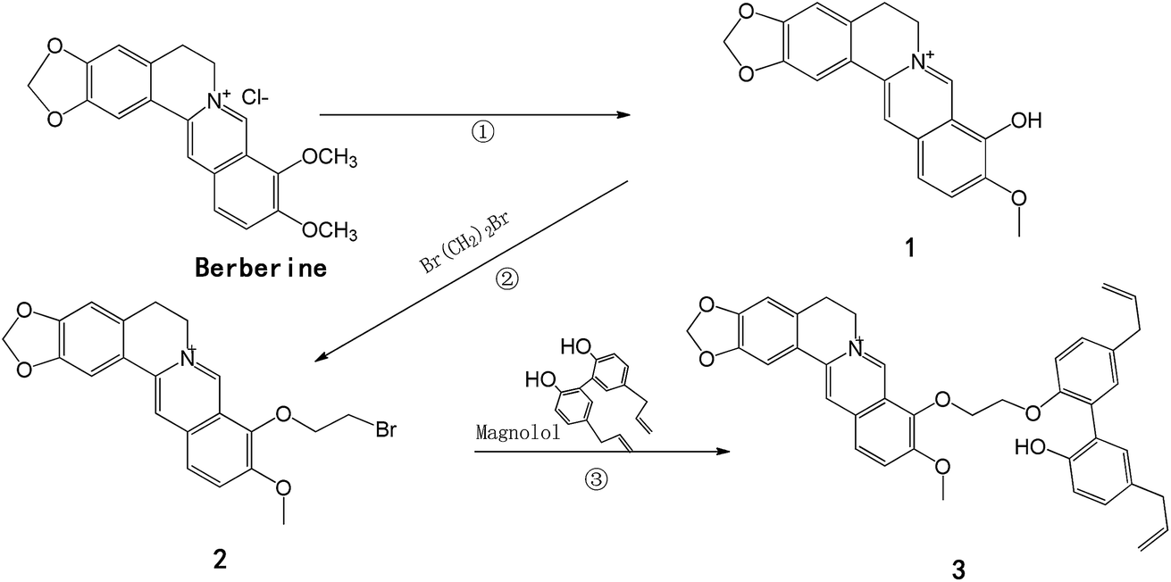 | ||
| Scheme 1 Synthesis of HHS. Reagents and conditions: (1) DMF, microwave power (400 W), reflux, 15 min, 92%; (2) acetonitrile, 1,2-dibromoethane, reflux, 3 h, 56%; (3) acetonitrile, anhydrous sodium carbonate, reflux, 8 h, 24%. | ||
Previous studies have suggested that berberrubine was usually synthesized by vacuum pyrolysis, with low yield and purity owing to uneven heating.30 In a preliminary experiment, we adopted a microwave synthesis method as described in the literature,31,32 but no chemical transformation occurred. After that, we used DMF, which has a high boiling point and high stability, as the reaction medium under microwave irradiation to afford berberrubine in 92% yield. 9-(2-Bromoethyl)berberine hydrochloride was synthesized using acetonitrile as the solvent. There have been few studies on the use of two natural active molecules bonded together through a chemical bond to synthesize a hybrid compound.
2.2 Acute toxicity study
After intraperitoneal injection of HHS, the various behaviors of mice were observed closely for the first 4 h. Mice administered with HHS at a dose of 132.1 mg kg−1 displayed no significant changes in behavior or breathing or postural abnormalities. However, significant changes occurred in mice in other groups, including unsteady walking, difficult breathing, and death, even the existence of moderate organ damage. Moreover, the mortality of mice treated with doses of 188.7 and 385.0 mg kg−1 was 0.2 and 0.8, respectively, in a dose-dependent manner. The LD50 value (lethal dose 50) was 279 mg kg−1, and the 95% confidence interval was 234.6–332.5 mg kg−1 (Table 1). The results obtained from this study demonstrate that after a single intraperitoneal injection the acute toxicity of HHS was lower than that of berberine, whereas the toxicity of berberine in previous reports was shown by LD50 values ranging between 38.8 and 85.5 mg kg−1 (ref. 33) and an LD50 value of 50 mg kg−1.34| Group | Dose (mg kg−1) | Mortality | LD50 (mg kg−1) | 95% confidence interval (mg kg−1) |
|---|---|---|---|---|
| a These data were calculated according to the modified Karber's method. | ||||
| 1 | 550.0 | 10/10 | 279.3 | 234.6–332.5 |
| 2 | 385.0 | 8/10 | ||
| 3 | 269.5 | 4/10 | ||
| 4 | 188.7 | 2/10 | ||
| 5 | 132.1 | 0/10 | ||
| Control | 0 | 0 | ||
| ∑p = 2.4 | ||||
2.3 Effect of HHS on glucose and lipid metabolism in aP2-SREBP-1c mice
It has been known that an excessive amount of white tissue (obesity) is a crucial factor in insulin resistance.35–37 In contrast to all other mouse models of insulin resistance,38,39 aP2-SREBP-1c mice are not obese, but exhibit a distinct syndrome that includes lipodystrophy, insulin resistance, diabetes mellitus and fatty liver.40,41 In the present study, we examined the effect of HHS on glucose and lipid metabolism disorders. An OGTT was performed at the end of 9 weeks. As shown in Fig. 1A, the blood glucose level in the model group was much higher than that in the control group. However, the mice treated with HHS displayed a significant decrease in blood glucose level compared with the model group at 20 and 60 min. Moreover, the AUC was significantly lower in the HHS group than that in the model group (Fig. 1B). As shown in Fig. 1C, at the end of 8 and 13 weeks, the plasma TG level in the model group was significantly higher than that in the control group, whereas the HHS group exhibited a significantly decreased TG level compared with the model group. According to an analysis of the TG level, compared with 0 weeks (0.85 ± 0.43) the TG levels at 8 and 13 weeks in the control group displayed no significant change, and the level in the model group increased significantly. Conversely, after treatment with HHS for 8 and 13 weeks, the plasma TG level decreased significantly compared with 0 weeks. These data indicated that HHS improved glucose and lipid metabolism in aP2-SREBP-1c mice. | ||
| Fig. 1 In vivo effects of HHS on glucose and lipid metabolism in aP2-SREBP-1c mice. Mice at 12 weeks of age were divided into 3 groups. HHS was suspended in a 5% aqueous solution of CMC-Na and administered orally for 13 weeks. (A) 9 weeks after final dosing, following an overnight fast, an oral gavage of 50% glucose (2 g kg−1 body weight) was performed on each animal. Blood glucose levels were estimated from blood samples just prior to glucose administration (0 min) and 20, 60 and 120 min after glucose administration. (B) By an oral glucose tolerance test (OGTT), the area under a blood glucose concentration curve (AUC) was calculated from the formula: AUC = [1/4 × 0 min (mmol L−1) + 1/2 × 20 min (mmol L−1) + 3/4 × 60 min (mmol L−1) + 120 min (mmol L−1)]. (C) At the end of 8 and 13 weeks, mice were anesthetized with diethyl ether after fasting for 12 h and plasma triglyceride levels were measured. Each group was composed of 8 mice. Data are presented as means ± SD. *p < 0.05, **p < 0.01 (one-way ANOVA) compared with the model group. | ||
2.4 Cytotoxicity of HHS to 3T3-L1 preadipocytes
In the present study, the cytotoxicity of berberine, magnolol and HHS to 3T3-L1 preadipocytes was investigated using CCK-8. The results are shown in Fig. 2. Within the range of 0–80 μM, the cell viability of the magnolol group decreased with an increase in concentration in a dose-dependent manner (Fig. 2A). When the concentration was greater than 20 μM, berberine had an obvious inhibitory effect on 3T3-L1 preadipocytes (Fig. 2B). However, the result showed that HHS could significantly reduce cell viability when the concentration reached 10 μM (Fig. 2C). Moreover, the IC50 values of berberine, magnolol and HHS were 19.43 ± 0.56, 27.93 ± 0.63 and 11.54 ± 0.52 μM, respectively. These observations suggested that HHS exerted a stronger effect than berberine or magnolol on the viability of 3T3-L1 preadipocytes. | ||
| Fig. 2 Cytotoxic activity of magnolol (Mag) (A), berberine (Ber) (B) and HHS (C). Cells were seeded in a 96-well plate at a density of 5000–8000 cells per well and incubated for 24 h. Then, various concentrations of berberine, magnolol and HHS (1.25, 2.5, 5, 10, 20, 40 and 80 μM) were added to the medium, which was incubated for 48 h. Cells treated with 0.1% DMSO served as controls (con). Values are means ± SD (n = 3 in each group). | ||
2.5 Differentiation of 3T3-L1 preadipocytes and Oil Red O staining
The cultured 3T3-L1 preadipocytes were in a fibroblast-like form with a long fusiform or polygonal shape (Fig. 3A). The cells could successfully be trans-differentiated into adipocytes with an appreciable amount of accumulated lipid droplets after induction for 10 days, which was confirmed by staining with Oil Red O (Fig. 3B and C).42 Lipids and Oil Red O were extracted using isopropanol, and the absorbance was measured at 520 nm. The result of staining with Oil Red O is shown in Fig. 3D. The OD520 value was significantly increased in the model group compared with the control group. | ||
| Fig. 3 3T3-L1 preadipocytes were successfully differentiated into adipocytes. Differentiated 3T3-L1 cells were identified using staining with Oil Red O. (A) Controls (preadipocytes), (B) differentiated adipocytes, (C) model (differentiated adipocytes, using staining with Oil Red O), (D) Oil Red O was extracted from the cells using isopropyl alcohol, and the absorbance was measured at 520 nm. Three independent experiments were performed and data are shown as the mean ± SD. **p < 0.01 compared with the model group. | ||
2.6 Effects of HHS on expression of differentiation-related genes in 3T3-L1 adipocytes
Research shows that PPARγ and C/EBPα play major roles in the process of adipocyte differentiation, and are also critical in the regulation of lipid metabolism and glucose homeostasis.43–46 To quantitatively describe the effects of berberine, magnolol and HHS at different concentrations on adipocyte differentiation, we examined the expression of PPARγ2 and C/EBPα using real-time PCR. As shown in Fig. 4A and B, the levels of PPARγ2 and C/EBPα mRNA were significantly increased in the MD group compared with the NC group, and HHS was effective in reducing their levels. Ten days after initiating treatment, the groups treated with Mag and Ber at 5 μM experienced a significant effect on PPARγ2 and C/EBPα mRNA levels compared with the MD group, which was consistent with previous studies.47–49 The group treated with Mag at 2.5 μM did not undergo substantial change in PPARγ2 and C/EBPα mRNA levels. Treatment with HHS significantly decreased the PPARγ2 mRNA level in a dose-dependent manner. However, surprisingly, the group treated with HHS at 5 μM underwent a less effective decrease in the C/EBPα mRNA level than the group treated with Ber at 5 μM. Although the group treated with HHS experienced a significant decrease in the C/EBPα mRNA level, this was not in a dose-dependent manner. By a comparison of the effects of both doses of HHS on PPARγ2 and C/EBPα, we found that HHS at 2.5 μM led to a greater reduction in mRNA expression than berberine or magnolol. Thus, we concluded that HHS to some extent displayed a stronger inhibitory effect on PPARγ2 and C/EBPα mRNA levels than berberine or magnolol. | ||
| Fig. 4 Effects of HHS on mRNA expression levels of PPARγ2 and C/EBPα in 3T3-L1 cells. Three independent experiments were performed and data are shown as means ± SD. *p < 0.05, **p < 0.01 compared with the DM group. NC = control group (cells treated with 0.1% DMSO). MD = model group (mature 3T3-L1 adipocytes). | ||
2.7 Effects of HHS on expression of adipogenesis-related genes in 3T3-L1 adipocytes
Adipocyte differentiation is a complex process that is controlled and regulated by various adipocyte-specific genes such as FAS, UCP2, adiponectin and others, which participate in creating the adipocyte phenotype.50 Here, we focused more on downstream genes of PPARγ2 and C/EBPα and studied how their mRNA expression is influenced by HHS. When 3T3-L1 preadipocytes differentiated into adipocytes, we tested the mRNA levels of FAS, UCP2 and adiponectin in all groups. As shown in Fig. 5A–C, significant increases in the mRNA expression of FAS and UCP2 were observed in the MD group compared with the NC group, which confirmed the presence of lipid accumulation. After treatment with berberine, magnolol and HHS, it was found that cells treated with 5 μM HHS experienced greater reductions in the mRNA expression of FAS and UCP2 than those treated with berberine and magnolol, whereas the mRNA expression of adiponectin increased more obviously, which demonstrated a good antiobesity effect.51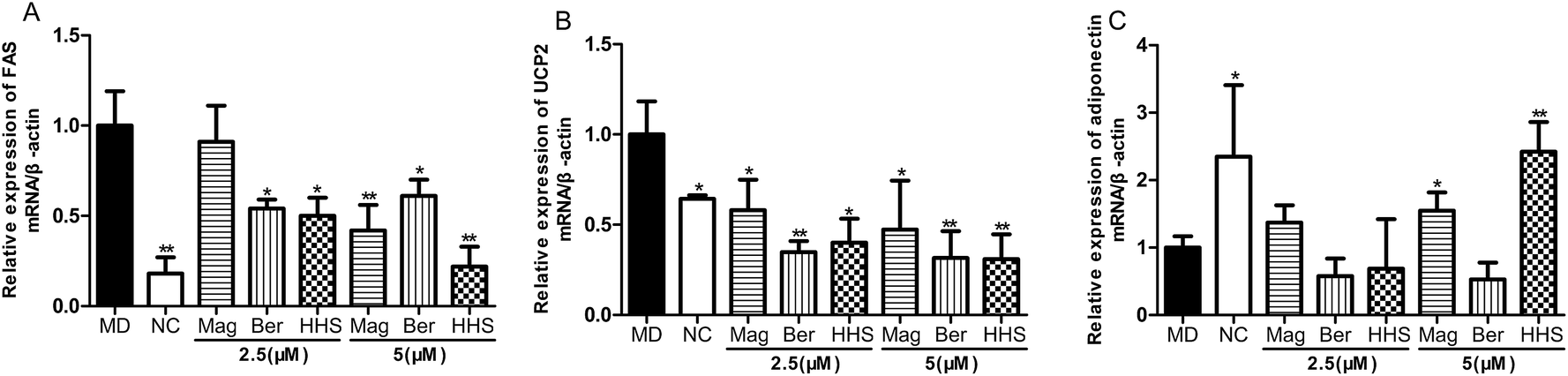 | ||
| Fig. 5 Effects of HHS on mRNA expression levels of (A) FAS, (B) UCP2 and (C) adiponectin in 3T3-L1 cells. Three independent experiments were performed and data are shown as means ± SD. *p < 0.05, **p < 0.01 compared with the MD group. NC = control group (cells treated with 0.1% DMSO). MD = model group (mature 3T3-L1 adipocytes). | ||
3. Experimental
3.1 Reagents and chemicals
Berberine and magnolol (purity ≥ 98%) were purchased from the China Drugs and Biological Products Inspection Institute (Beijing, China). Dulbecco's modified Eagle's medium (DMEM) and fetal bovine serum (FBS) were purchased from Gibco BRL (Grand Island, NY). Insulin (INS), 3-isobutyl-1-methylxanthine (IBMX), dexamethasone (DEX) and Oil Red O were purchased from Sigma Aldrich (St Louis, MO). RNAiso Plus, PrimeScript™ RT Reagent Kit with gDNA Eraser (Perfect Real Time), and SYBR® Premix Ex Taq™ II (Tli RNaseH Plus) were purchased from Takara (Tokyo, Japan). All other chemicals and reagents were of analytical grade and were purchased from Aldrich Chemical Co. (Beijing, China). 1H and 13C NMR spectra were recorded using a Bruker Avance 400 at room temperature with tetramethylsilane (TMS) as an internal standard and chloroform-d (CDCl3) as solvent. Mass spectra were recorded using a DSQ mass spectrometer (Thermo, USA).3.2 Synthesis of HHS
Berberrubine 1. Red needle crystals; mp 281.6–282.4 °C. 1H NMR (CDCl3, 300 MHz) δ: 7.35 (1H, s, H-1), 6.76 (1H, s, H-4), 3.04 (1H, t, J = 5.8, H-5), 4.53 (1H, t, J = 5.8, H-6), 9.20 (1H, s, H-8), 6.83 (1H, d, J = 9.1, H-11), 7.46 (1H, d, J = 9.1, H-12), 7.97 (1H, s, H-13), 5.95 (1H, s, H-15), 3.81 (1H, s, H-16); 13C NMR (CDCl3, 300 MHz) δ: 106.27 (C-1), 121.81 (C-1a), 151.42 (C-2), 149.97 (C-3), 109.14 (C-4), 131.16 (C-4a), 57.04 (C-6), 143.72 (C-8), 134.21 (C-8a), 147.84 (C-9), 147.84 (C-10), 120.21 (C-11), 123.21 (C-12), 131.16 (C-12a), 124.22 (C-13), 136.23 (C-14), 103.68 (C-15), 55.94 (C-16); LC-MS (ESI+, m/z) [C19H16NO4]+: 322.2 [M − Cl]+.
9-(2-Bromoethyl)berberine hydrochloride 2. Yellow crystals; mp 273.6–274.4 °C. 1H NMR (CDCl3, 300 MHz) δ: 7.68 (1H, s, H-1), 6.97 (1H, s, H-4), 3.87 (2H, t, J = 5.8, H-5), 4.74 (2H, t, J = 5.8, H-6), 9.87 (1H, s, H-8), 8.04 (1H, d, J = 9.1, H-11), 8.15 (1H, d, J = 9.1, H-12), 8.74 (1H, s, H-13), 6.11 (2H, s, H-15), 3.30 (3H, s, H-16), 4.65 (2H, t, J = 5.6, H-17), 4.12 (2H, t, J = 5.6, H-18); 13C NMR (CDCl3, 300 MHz) δ: 106.43 (C-1), 151.64 (C-2), 149.39 (C-3), 109.29 (C-4), 131.77 (C-4a), 27.59 (C-5), 57.53 (C-6), 143.27 (C-8), 134.68 (C-8a), 146.24 (C-9), 146.24 (C-10), 121.57 (C-11), 124.94 (C-12), 131.77 (C-12a), 127.53 (C-13), 139.26 (C-14), 121.34 (C-14a), 103.31 (C-15), 56.88 (C-16), 57.53 (C-17), 74.49 (C-18); LC-MS (ESI+, m/z) [C21H19BrNO4]+: 429.1 [M − Cl]+.
:1). The crude product obtained was evaporated in a vacuum to obtain pure compound 3 (HHS) in 24% yield.
HHS (3). {9-(2′-((5′,5′-Diallyl-2′-hydroxy-[1′,1′′-biphenyl]-2′-yl)oxy)ethoxy)-10-methoxy-5,6-dihydro-[l,3]dioxolo[4,5-g]isoquinolino[3,2-a]isoquinolin-7-ium}.
Yellow solid; mp 145.2–146.1 °C. 1H NMR (CDCl3, 500 MHz) δ: 7.64 (1H, s, H-1), 6.91 (1H, s, H-4), 3.87 (2H, t, J = 5.8, H-5), 4.74 (2H, t, J = 5.8, H-6), 9.38 (1H, s, H-8), 7.90 (1H, d, J = 9.1, H-11), 8.04 (1H, d, J = 9.1, H-12), 8.56 (1H, s, H-13), 6.11 (2H, s, H-15), 3.30 (3H, s, H-16), 4.65 (2H, t, J = 5.6, H-17), 4.12 (2H, t, J = 5.6, H-18), 7.00 (1H, d, H-21), 7.11 (1H, d, J = 8.45, H-23), 6.69 (1H, d, J = 8.45, H-24), 3.00 (1H, d, H-25), 6.39 (1H, d, H-30), 5.99–5.86 (1H, m, J = 9.6, 16.9, H-26), 5.81–5.67 (1H, m, J = 9.6, 16.9, H-27); 13C NMR (CDCl3, 300 MHz) δ: 106.81 (C-1), 152.38 (C-2), 150.21 (C-3), 109.65 (C-4), 131.76 (C-4a), 28.39 (C-5), 57.83 (C-6), 143.27 (C-8), 134.13 (C-8a), 146.70 (C-9), 152.06 (C-10), 123.88 (C-12), 145.30 (C-12a), 127.65 (C-13), 121.34 (C-14a), 121.75 (C-11), 103.97 (C-15), 56.88 (C-16), 69.31 (C-17), 75.19 (C-18), 155.69 (C-19), 125.04 (C-20), 132.70 (C-21), 129.66 (C-22), 129.25 (C-23), 113.69 (C-24), 40.49 (C-25), 131.76 (C-26), 116.09 (C-27), 153.83 (C-28), 126.51 (C-29), 129.92 (C-30), 133.57 (C-31), 129.25 (C-32), 115.86 (C-33), 40.65 (C-34), 131.93 (C-35), 116.59 (C-36); LC-MS (ESI+, m/z) [C39H36NO6]+: 615.3 [M − Cl]+.
3.3 Acute toxicity study
The strain of mice used in this study was NIH, which was originally bred by the National Institutes of Health (NIH). Both sexes of SPF (specific pathogen-free) NIH mice were investigated by regular methods for toxicological evaluation. NIH mice weighing 18–22 g [no. SCXK (Yue) 2008-0002], half male and half female, were purchased from the Medical Laboratory Animal Center of Guangdong (Guangdong, China). Mice were housed in a temperature-controlled animal facility with a 12 hour light and dark cycle and were free to access water and food. Procedures for animal care and handling were in strict accordance with the Chinese legislation on the care and use of laboratory animals. The Animal Experiment Center of Guangdong Pharmaceutical University approved the experimental procedures and protocols conducted during this study. After acclimating for 1 week, ten mice were treated with each dose level of HHS (132.1, 188.7, 269.5, 385.0 and 550.0 mg kg−1). After intraperitoneal injection of HHS according to the modified Karber's method, mice were observed for 14 days and signs of toxicity and mortality were recorded.3.4 In vivo pharmacologic test
Transgenic aP2-SREBP-1c mice were introduced from Jackson Lab (USA) at 6 weeks of age and the Model Animal Research Center of Nanjing University was commissioned for purification [no. J003393 SCXK (Su) 2010-0001]. Mice were housed in a temperature-controlled animal facility with a 12 hour light and dark cycle and were free to access water and food. Procedures for animal care and handling were in strict accordance with the Chinese legislation on the care and use of laboratory animals. The Animal Experiment Center of Guangdong Pharmaceutical University approved the experimental procedures and protocols conducted during this study. After the processes of animal reproduction and genotype identification, transgenic mice at 12 weeks of age were divided into a model group and an HHS group, and the wild-type littermates served as a control group (8 mice in each group). Then, the animals were treated with 40 mg kg−1 HHS or a 0.5% aqueous solution of CMC-Na orally once daily by gavage for 13 weeks. At the end of 8 and 13 weeks, the animals were anesthetized with diethyl ether after fasting for 12 h, and blood samples were collected by retro-orbital venous plexus puncture. Serum triglyceride levels were determined using enzymatic kits. At the end of 9 weeks, following an overnight fast, an oral gavage of 50% glucose (2 g kg−1 body weight) was performed on each animal. Blood glucose levels were estimated from blood samples just prior to glucose administration (0 min) and 20, 60 and 120 min after glucose administration. By an oral glucose tolerance test (OGTT), the area under a blood glucose concentration curve (AUC) was calculated from the formula: AUC = [1/4 × 0 min (mmol L−1) + 1/2 × 20 min (mmol L−1) + 3/4 × 60 min (mmol L−1) + 120 min (mmol L−1)]. All samples were stored at −80 °C until further analysis.3.5 In vitro cell culture and biological studies
3.6 Statistical analysis
Data are presented as means ± SD. Statistical analysis was performed using SPSS 20.0 statistical software by one-way analysis of variance (ANOVA) followed by Fisher's LSD tests. Graphical representations were performed using GraphPad Prism 5. Differences were considered to be significant when p < 0.05.4. Conclusion
In the present study, based on the association principle, we designed and synthesized a hybrid compound of berberine and magnolol as a combined drug, which possessed potential biological activities, as well as safety and low toxicity. The method of linking the two molecules together was carried out to obtain the target product based on chemical bonding. The results of an in vivo study demonstrated that the administration of 40 mg kg−1 HHS orally to aP2-SREBP-1c mice reduced plasma glucose and lipid levels. In addition, HHS could significantly downregulate the expression of PPARγ2, C/EBPα, FAS and UCP2 mRNA and upregulate the expression of adiponectin mRNA in 3T3-L1 adipocytes. Although the underlying mechanism of action of HHS is not currently well understood, the reduction in TG levels induced by HHS in a mouse model is remarkable. For further studies, as the 1, 9, 10, and 13-positions of berberine are open to structural modification, the binding of these positions to magnolol can be considered.References
- E. A. Nyenwe, T. W. Jerkins, G. E. Umpierrez and A. E. Kitabchi, Metabolism, 2011, 60, 1–23 CrossRef CAS PubMed.
- M. E. Cerf, Front. Endocrinol., 2013, 4, 1–12 Search PubMed.
- Y. Xu, L. Wang, J. He, Y. Bi, M. Li, T. Wang, L. Wang, Y. Jiang, M. Dai, J. Lu, M. Xu, Y. Li, N. Hu, J. Li, S. Mi, C. S. Chen, G. Li, Y. Mu, J. Zhao, L. Kong, J. Chen, S. Lai, W. Wang, W. Zhao and G. Ning, J. Am. Med. Assoc., 2013, 310, 948–959 CrossRef CAS PubMed.
- S. H. Kim, S. H. Hyun and S. Y. Choung, J. Ethnopharmacol., 2006, 104, 119–123 CrossRef PubMed.
- S. Fujioka, Y. Matsuzawa, K. Tokunaga and S. Tarui, Metabolism, 1987, 36, 54–59 CrossRef CAS PubMed.
- J. R. Acosta, I. Douagi, D. P. Andersson, J. Bäckdahl, M. Rydén, P. Arner and J. Laurencikiene, Diabetologia, 2016, 59, 560–570 CrossRef CAS PubMed.
- P. Trayhurn and J. H. Beattie, Proc. Nutr. Soc., 2001, 60, 329–339 CrossRef CAS PubMed.
- K. Iizuka, R. K. Bruick, G. Liang, J. D. Horton and K. Uyeda, Proc. Natl. Acad. Sci. U. S. A., 2004, 101, 7281–7286 CrossRef CAS PubMed.
- S. Ishii, K. Iizuka, B. C. Miller and K. Uyeda, Proc. Natl. Acad. Sci. U. S. A., 2004, 101, 15597–15602 CrossRef CAS PubMed.
- T. M. Loftus and M. D. Lane, Curr. Opin. Genet. Dev., 1997, 7, 603–608 CrossRef CAS PubMed.
- L. Wang, G. B. Zhou, P. Liu, J. H. Song, Y. Liang, X. J. Yan, F. Xu, B. S. Wang, J. H. Mao, Z. X. Shen, S. J. Chen and Z. Chen, Proc. Natl. Acad. Sci. U. S. A., 2008, 105, 4826–4831 CrossRef CAS PubMed.
- D. A. Guo, A. Lu and L. Liu, J. Ethnopharmacol., 2012, 141, 547–548 CrossRef PubMed.
- J. Tang, Y. B. Feng, S. W. Tsao, N. Wang, R. Curtain and Y. W. Wang, J. Ethnopharmacol., 2009, 126, 5–17 CrossRef CAS PubMed.
- M. Tillhon, L. M. Guamán Ortiz, P. Lombardi and A. I. Scovassi, Biochem. Pharmacol., 2012, 84, 1260–1267 CrossRef CAS PubMed.
- C. L. Kuo, C. W. Chi and T. Y. Liu, Cancer Lett., 2004, 203, 127–137 CrossRef CAS PubMed.
- R. Verpoorte, J. Nat. Prod., 1985, 48, 397–433 Search PubMed.
- R. Rajesh, N. Sahu, C. Shah and R. Karpoormath, Curr. Top. Med. Chem., 2014, 14, 253–273 Search PubMed.
- P. J. Facchini, Annu. Rev. Plant Physiol. Plant Mol. Biol., 2001, 52, 29–66 CrossRef CAS PubMed.
- H. S. Bodiwala, S. Sabde, D. Mitra, K. K. Bhutani and I. P. Singh, Eur. J. Med. Chem., 2011, 46, 1045–1049 CrossRef CAS PubMed.
- S. L. Zhang, J. J. Chang, G. L. Damu, B. Fang, X. D. Zhou, R. X. Geng and C. H. Zhou, Bioorg. Med. Chem. Lett., 2013, 23, 1008–1012 CrossRef CAS PubMed.
- S. H. Leng, F. E. Lu and L. J. Xu, Acta Pharmacol. Sin., 2004, 25, 496–502 CAS.
- W. H. Peng, K. L. Lo, Y. H. Lee, T. H. Hung and Y. C. Lin, Life Sci., 2007, 81, 933–938 CrossRef CAS PubMed.
- L. Zhang, J. Li, F. Ma, S. Yao, N. Li, J. Wang, Y. Wang, X. Wang and Q. Yao, Molecules, 2012, 17, 11294–11302 CrossRef CAS PubMed.
- X. Deng, X. X. Zhao, J. Han, J. J. Wang, W. L. Huang, H. Qian and L. Ge, Lett. Drug Des. Discovery, 2015, 9, 489–493 CrossRef.
- J. Park, J. Lee, E. Jung, Y. Park, K. Kim, B. Park, K. Jung, E. Park, J. Kim and D. Park, Eur. J. Pharmacol., 2004, 496, 189–195 CrossRef CAS PubMed.
- D. Y. Chuang, M. H. Chan, Y. Zong, W. Sheng, Y. He, J. H. Jiang, A. Simonyi, Z. Gu, K. L. Fritsche, J. Cui, J. C. Lee, W. R. Folk and D. Lubahn, J. Neuroinflammation, 2013, 10, 1–14 CrossRef PubMed.
- M. Akiko, F. Aya, L. Cheng, K. Shota, F. Yoshiyasu and M. Yasuhide, Parkinson's Dis., 2012, 1–9 Search PubMed.
- G. D. Kim, J. Oh, H. J. Park, K. Bae and S. K. Lee, Int. J. Oncol., 2013, 43, 600–610 CAS.
- E. J. Sohn, C. S. Kim, Y. S. Kim, D. H. Jung, D. S. Jang, Y. M. Lee and J. S. Kim, Life Sci., 2007, 80, 468–475 CrossRef CAS PubMed.
- H. S. Bodiwala, S. Sabde and D. Mitra, Eur. J. Med. Chem., 2011, 46, 1045–1049 CrossRef CAS PubMed.
- B. Das and K. Srinivas, Synth. Commun., 2002, 32, 3027–3029 CrossRef CAS.
- D. C. Arantzazu, J. Carmen, L. C. Vicente, L. Olatz, D. C. Abel, P. C. Fernando, V. Yosu, D. Andrés, M. Luis, G. Javier and G. Rosa, Tetrahedron, 2015, 71, 6148–6154 CrossRef.
- M. M. Kheir, Y. Wang, L. Hua, J. Hu, L. Li, F. Lei and L. Du, Food Chem. Toxicol., 2010, 48, 1105–1110 CrossRef CAS PubMed.
- K. V. Anis, G. Kuttan and R. Kuttan, Pharm. Pharmacol. Commun., 1999, 5, 697–700 CrossRef CAS.
- J. A. Martyn, M. Kaneki and S. Yasuhara, Anesthesiology, 2008, 109, 137–148 CrossRef PubMed.
- S. Wueest, F. Item, F. C. Lucchini, T. Challa, W. Müller, M. Blüher and D. Konrad, Diabetes, 2016, 65, 140–148 CAS.
- G. H. Antonio, M. V. Vicente, R. R. Jose, D. F. Ana, G. M. Manuel and G. O. Luis, Prev. Med., 2016, 82, 59–64 CrossRef PubMed.
- B. M. Spiegelman, L. Choy, G. S. Hotamisligil, R. A. Graves and P. Tontonoz, J. Biol. Chem., 1993, 268, 6823–6826 CAS.
- B. M. Spiegelman and J. S. Flier, Cell, 1996, 87, 377–389 CrossRef CAS PubMed.
- I. Shimomura, R. E. Hammer, J. A. Richardson, S. Ikemoto, Y. Bashmakov, J. L. Goldstein and M. S. Brown, Genes Dev., 1998, 12, 3182–3194 CrossRef CAS PubMed.
- I. Shimomura, Y. Bashmakov and J. D. Horton, J. Biol. Chem., 1999, 274, 30028–30032 CrossRef CAS PubMed.
- Z. Wu, E. D. Rosen, R. Brun, S. Hauser, G. Adelmant, A. E. Troy, C. Mckeon, G. J. Darlington and B. M. Spiegelman, Mol. Cell, 1999, 3, 151–158 CrossRef CAS PubMed.
- E. D. Rosiglitazone, C. H. Hus, X. Wang, S. Sakai, M. W. Freeman, F. J. Gonzalez and B. M. Spiegelman, Genes Dev., 2002, 16, 22–26 CrossRef PubMed.
- R. K. Petersen, L. Madsen, L. M. Pedersen, P. Hallenborg, H. Hagland, K. Viste, S. O. Døskeland and K. Kristiansen, Mol. Cell. Biol., 2008, 28, 3804–3816 CrossRef CAS PubMed.
- C. F. Vogel, E. Sciullo, S. Park, C. Liedtke, C. Trautwein and F. Matsumura, J. Biol. Chem., 2004, 279, 88886–88894 CrossRef PubMed.
- J. E. Reusch, L. A. Colton and D. J. Klemm, Mol. Cell. Biol., 2000, 2, 1008–1020 CrossRef.
- C. Huang, Y. B. Zhang, Z. W. Gong, X. Sheng, Z. Li, W. Zhang and Y. Qin, Biochem. Biophys. Res. Commun., 2006, 348, 571–578 CrossRef CAS PubMed.
- B. H. Choi, I. S. Ahn, Y. H. Kim, J. W. Park, S. Y. Lee, C. K. Hyun and M. S. Do, Exp. Mol. Med., 2006, 38, 599–605 CrossRef CAS PubMed.
- J. H. Chen, H. C. Kuo, K. F. Lee and T. H. Tsai, Toxicol. Appl. Pharmacol., 2014, 279, 294–302 CrossRef CAS PubMed.
- E. D. Rosen, C. J. Walkey, P. Puigserver and B. M. Spiegelman, Genes Dev., 2000, 14, 1293–1307 CAS.
- L. M. Belalcazar, W. Lang, S. M. Haffner, D. C. Schwenke, A. Kriska, A. Balasubramanyam, R. C. Hoogeveen, F. X. Pi-Sunyer, R. P. Tracy and C. M. Ballantyne, Diabetes Care, 2015, 38, 1544–1550 CrossRef CAS PubMed.
- S. P. Poulos, M. V. Dodson and G. J. Hausman, Exp. Biol. Med., 2010, 235, 1185–1193 CrossRef CAS PubMed.
Footnote |
| † Electronic supplementary information (ESI) available: The 1H NMR, 13C NMR and HRMS spectra of the synthesized compounds; HPLC profile of HHS; the oligonucleotide primer sequence used for real-time PCR. See DOI: 10.1039/c6ra15100k |
| This journal is © The Royal Society of Chemistry 2016 |