DOI:
10.1039/C6RA15081K
(Paper)
RSC Adv., 2016,
6, 94959-94966
A highly sensitive naphthaoxazole-based cell-permeable ratiometric chemodosimeter for hydrazine†
Received
10th June 2016
, Accepted 20th September 2016
First published on 20th September 2016
Abstract
The environmental toxicity, detonable characteristics and widespread usage of hydrazine in industrial activities, coupled with the fact that it is a leading candidate as a hydrogen reservoir, mean that selective methods for the detection of trace levels of hydrazine are much needed. We report herein a ratiometric chemodosimeter (P1) for the highly efficient detection of hydrazine at a lowest level of 1.79 × 10⁻⁹ M. The P1 probe was designed by the judicious anchoring of a naphthaoxazole skeleton to malononitrile. The same chemodosimeter also enables the bioimaging of hydrazine in live cells. The performance of P1 was tested in the form of test paper strips, as well as in the solid state for the identification of hydrazine vapours. The sensing mechanism was established through spectroscopic techniques and was further fortified through theoretical calculations using density functional theory.
Introduction
Hydrazine (N2H4) is extensively used in various chemical industries, such as those that manufacture pharmaceuticals, pesticides, photography chemicals, emulsifiers and dyes etc.1a–d In addition, hydrazine also plays an important role in propulsion systems and hydrazine fuel cells, as a highly explosive fuel.2 In contrast to its usefulness, hydrazine is a model toxin. Because it is highly water soluble, it gets easily absorbed through inhalation and oral and dermal uptake, leading to the damage of various organs and systems,3 especially the nervous system.1c It has also been reported as a neurotoxin with severe mutagenic effects, causing infections of the respiratory tract and damage to the lungs, liver, kidneys and central nervous system, etc.4 It has been identified as a potential carcinogen by the U.S. Environmental Protection Agency (EPA) with a threshold limit value (TLV) of 10 ppb.5
Apart from the above mentioned Janus-faced properties of hydrazine, recently it has been identified as one of the leading candidates to be used as a hydrogen reservoir, owing to its high content of hydrogen.6 Therefore, the development of highly selective, sensitive, efficient and economic methods for the rapid detection of hydrazine is urgently required. Although hydrazine can be analysed through various already available analytical instrumental techniques,7 these are time-consuming, require complicated sample processing and are also destructive for tissues and cells. These analytical methods are also not compatible with in vivo measurements.8 Nevertheless, fluorescent techniques are extremely attractive due to their high sensitivity, low cost, easy implementation, and real-time (on the spot) detection of analytes.9
Until now, only a few fluorescent sensors for hydrazine have been reported, and almost all of them detect hydrazine through a fluorescence off–on mechanism.10 The main bottlenecks of this approach are poor selectivity, strong background signals and narrow measurement range. A number of literature reports and our own experiences in the same field prompted us to fabricate a new ratiometric fluorescent probe for hydrazine (P1). Ratiometric sensors have advantages over on–off type sensors as they allow simultaneous measurement of fluorescence intensities at two different wavelengths. At the same time, ratiometric sensors provide an inbuilt correction for environmental effects11 and are able to perceive colour changes for rapid visual sensing.12
For constructing ratiometric sensors, a number of approaches, such as fluorescence resonance energy transfer (FRET), intramolecular charge transfer (ICT) and excited state intramolecular proton transfer (ESIPT), are normally employed.13 Although the construction design of P1 is based on an ICT approach (Scheme 1), the mechanistic aspect of the functioning of P1 involves a chemodosimetric approach. This involves hydrazine mediating the removal of the electron deficient malononitrile component of P1 and becoming bound to itself, resulting in the formation of an adduct with clear optical and spectral changes. This P1 sensor presents a number of advantages over previously reported sensors for hydrazine, in terms of its lowest detection limit and appropriate solvent system. The P1 sensor also exhibits a lowest detection limit of ∼1.79 × 10⁻⁹ M for hydrazine in aqueous medium. Yet another positive point of P1 worth mentioning here is its large Stoke's shift (∼110 nm) upon its interaction with hydrazine.
 |
| | Scheme 1 Chemical structure of probe P1. | |
Experimental section
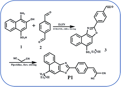
Synthesis of 2-(4-formylphenyl)naphtho[1,2-d]oxazole-5-sulfonic acid (3)
2-(4-Formylphenyl)naphtho[1,2-d]oxazole-5-sulfonic acid served as precursor for P1 and was synthesized as per Scheme 2. 1.0 mmol absolute ethanolic solution of terephthaldehyde was added to an equimolar absolute ethanolic solution of 4-amino-3-hydroxy-1-naphthalene sulphonic acid also containing 2 mmol of triethylamine. The reaction mixture was stirred overnight at room temperature, resulting in a clear yellow solution. Then the volume of the reaction mixture was reduced to its minimum on a rotary evaporator, followed by washing it with diethyl ether and finally recrystallizing it from ethanol. The obtained aldehyde (off-white colour) was characterized using FT-IR, 1H NMR & 13C NMR (Fig. S1–S3, ESI†).
 |
| | Scheme 2 Synthetic route for the synthesis of the probe P1. | |
Important spectroscopic data for 3
Yield: 90%; IR/cm−1: 3470, 2936, 2738, 2677, 1698, 1612, 1585, 1475, 1397, 1352, 1333, 1185, 1051, 1011, 927, 834, 769, 645, 600, 535, 491; 1H NMR (300 MHz, DMSO, TMS): δ ppm = 10.11 (s, –CHO, 1H), 9.04–9.03 (d, -ArH, 1H), 8.52–8.45 (m, ArH, 3H), 8.36 (s, ArH, 1H), 8.14–8.12 (d, ArH, 2H), 7.74–7.64 (m, ArH, 2H), 3.09–3.02 (q, –CH2, 6H), 1.16–1.11 (t, –CH3, 9H); 13C NMR (75 MHz, DMSO, 298 K, TMS): δ ppm = 192.62, 161.70, 161.41, 146.49, 146.37, 143.93, 143.67, 137.72, 137.58, 137.51, 131.27, 130.24, 128.92, 128.82, 127.90, 127.63, 127.35, 127.15, 126.02, 125.43, 121.43, 109.75, 45.78, 8.62.
Synthesis of 2-(4-(2,2-dicyanovinyl)phenyl)naphtho[1,2-d]oxazole-5-sulfonic acid (P1)
1 mmol of 3, 1 mmol of malononitrile and 3 drops of piperidine were taken in 15 ml of absolute ethanol in a round bottom flask. The reaction mixture was stirred for ∼2 h at room temperature. The yellow precipitate obtained was filtered, washed with diethyl ether and dried. The resulting crude product was recrystallized from methanol to obtain a bright yellow powder. Single crystals of P1 were obtained by slow evaporation of its saturated solution in DMF![[thin space (1/6-em)]](https://www.rsc.org/images/entities/char_2009.gif) :methanol (1:1). The probe P1 was characterized using FT-IR, 1H NMR and 13C NMR, ESI-MS, and single crystal XRD (Fig. S4–S7 and Table 1, ESI†).
:methanol (1:1). The probe P1 was characterized using FT-IR, 1H NMR and 13C NMR, ESI-MS, and single crystal XRD (Fig. S4–S7 and Table 1, ESI†).
Important spectroscopic data for P1
Yield: 85%; IR/cm−1: 3102, 3044, 2937, 2231 (–C![[double bond, length as m-dash]](https://www.rsc.org/images/entities/char_e001.gif) N), 1640, 1584, 1420, 1373, 1307, 1219, 1192, 1050, 1012, 936, 844, 824, 767, 645, 617, 598, 540, 418; 1H NMR (300 MHz, DMSO, TMS): δ ppm = 9.03–9.00 (d, -ArH, 1H), 8.62 (s, 1H), 8.47–8.45 (m, ArH, 3H), 8.31 (s, ArH, 1H), 8.16–8.13 (d, ArH, 2H), 7.75–7.63 (m, ArH, 2H), 3.03–3.10 (q, –CH2, 6H), 1.17–1.12 (t, –CH3, 9H); 13C NMR (75 MHz, DMSO, 298 K, TMS): δ ppm = 161.13, 160.10, 146.53, 144.19, 137.55, 133.49, 131.26, 130.77, 128.97, 127.72, 127.37, 127.19, 126.00, 125.46, 121.42, 114.08, 113.04, 109.66, 82.94, 45.76, 8.60; ESI-MS (m/z): calculated for C21H11N3O4S = 401.0, found = 401.8.
N), 1640, 1584, 1420, 1373, 1307, 1219, 1192, 1050, 1012, 936, 844, 824, 767, 645, 617, 598, 540, 418; 1H NMR (300 MHz, DMSO, TMS): δ ppm = 9.03–9.00 (d, -ArH, 1H), 8.62 (s, 1H), 8.47–8.45 (m, ArH, 3H), 8.31 (s, ArH, 1H), 8.16–8.13 (d, ArH, 2H), 7.75–7.63 (m, ArH, 2H), 3.03–3.10 (q, –CH2, 6H), 1.17–1.12 (t, –CH3, 9H); 13C NMR (75 MHz, DMSO, 298 K, TMS): δ ppm = 161.13, 160.10, 146.53, 144.19, 137.55, 133.49, 131.26, 130.77, 128.97, 127.72, 127.37, 127.19, 126.00, 125.46, 121.42, 114.08, 113.04, 109.66, 82.94, 45.76, 8.60; ESI-MS (m/z): calculated for C21H11N3O4S = 401.0, found = 401.8.
Synthesis of P1–N2H4
The receptor P1 (0.5 mmol) was mixed with 0.5 mmol of hydrazine in methanol at room temperature and was stirred for 24 hours. The yellow precipitate thus formed was filtered and dried. This was characterized by FTIR, 1H NMR & 13C NMR along with ESI-MS (Fig. S8–S11, ESI†).
Important spectroscopic data for P1–N2H4
Yield: 75%; IR/cm−1: 3385, 2938, 2937, 1621, 1411, 1323, 1298, 1211, 1179, 1106, 1048, 958, 830, 645, 600, 522; 1H NMR (300 MHz, DMSO, TMS): δ ppm = 9.03 (d, -Ar, 1H), 8.83 (s, 1H), 8.56–8.32 (m, ArH, 3H), 8.12–8.03 (m, ArH, 3H), 7.73–7.63 (m, ArH, 2H), 4.41 (s, –NH2, 2H), 3.15–3.04 (q, –CH2, 6H), 1.15–1.11 (t, –CH3, 9H); 13C NMR (75 MHz, DMSO, 298 K, TMS): δ ppm = 162.75, 146.04, 142.90, 139.80, 137.71, 136.43, 128.90, 127.49, 127.31, 127.04, 125.99, 125.72, 125.35, 124.83, 121.57, 112.17, 109.84, 45.84, 8.75, 8.50; ESI-MS: (m/z) calculated for C18H13N3O4S = 367.0, found = 367.7.
Results and discussion
X-ray crystallographic studies
The structure of P1 was confirmed through single crystal X-ray analysis. The P1 probe crystallizes in a triclinic lattice with space group P![[1 with combining macron]](https://www.rsc.org/images/entities/char_0031_0304.gif) . An ORTEP view of the asymmetric unit of P1 is shown in Fig. 1, while crystal data and structural refinement details are listed in Table 1 in the ESI.†
. An ORTEP view of the asymmetric unit of P1 is shown in Fig. 1, while crystal data and structural refinement details are listed in Table 1 in the ESI.†
 |
| | Fig. 1 Single crystal structure of P1. | |
The asymmetric unit contains two molecules of P1 with coplanar antiparallel orientation. These two molecules are held together through weak interactions like C⋯C (C18⋯C32; 3.279 Å), O⋯H–C (O8⋯H–C18; 2.469 Å), O⋯H–C (O2⋯H–C45; 2.569 Å) and O⋯C (O2⋯C45; 3.107 Å), as shown in Fig. 2(a). A triethylamine molecule is also seen, trapped with P1 through weak interactions of hydrogen bonding via O⋯H–N (O3⋯H–N4), as shown in Fig. 2(b). In addition to these, a few more similar weak interactions like C⋯C (C1⋯C39; 3.658 Å and C12⋯28; 3.363 Å), N⋯C (N3⋯C3; 3.227 Å and N7⋯C7; 2.717 Å) and N⋯HC (N3⋯H–C3; 2.742 Å) are also observed, which are responsible for knitting a beautiful supramolecular network, as seen in Fig. 2(c) and (d).
 |
| | Fig. 2 (a) Antiparallel coplanar orientation of P1; (b) triethylamine molecule interaction through H-bonding; (c) and (d) weak supramolecular packing of P1. | |
Absorption spectral response
The sensing properties of P1 towards hydrazine were primarily examined through observing the corresponding absorption spectral changes. The 1.0 × 10⁻⁵ M pure PBS buffer solution (pH 7.4, 10 mM) of P1 showed two distinct absorption bands at 308 nm and 378 nm. The former is attributed to a π–π* transition and the latter to an ICT (intramolecular charge transfer) transition.14 Upon concomitant additions of hydrazine (0–45 equiv.) to the above solution of P1, there was a gradual decrease in terms of the intensity of the absorption band at ∼378 nm, accompanied by the appearance of a new band at 352 nm. Finally, a colour change from yellow to colourless was observed (Fig. 3). The presence of a clear isosbestic point at ∼359 nm confirms the formation of a new species, as a result of the interaction between P1 and hydrazine.
 |
| | Fig. 3 UV-Visible titration spectra of P1 (1 × 10⁻⁵ M) in pure PBS buffer (pH 7.4, 10 mM), with different concentrations of hydrazine (0–45 equiv.). | |
The selectivity of P1, assessed through naked eye observation of colour change and absorption mode, was checked over a wide range of species; i.e. cations, anions and various amines (Al3+, Cr3+, Mn2+, Fe3+, Co2+, Ni2+, Co2+, Cu2+, Zn2+, Cd2+, Hg2+, Pb2+, S2O32−, HSO3−, HSO4−, HPO42−, H2PO4−, PO43−, BzO−, SO32−, S2−, F−, Cl−, Br−, I−, AcO−, PPi, ClO4−, BF4−, aniline, hydroxylamine, 4-nitrophenylhydrazine, ammonia, phenyl hydrazine, pyridine, ethylamine, triethylamine, methylamine, urea, ethylenediamine, cysteine, benzylamine, thioureas, 2,4-dinitrophenylhydrazine, butylamine, semicarbazide, thiocarbohydrazide and carbohydrazide), as detailed in Fig. S12 and S13 in the ESI.† The absorbance ratio at two different wavelengths in the form of a bar graph revealed that there is no considerable hindrance from any species mentioned above in the selective detection of hydrazine by the probe P1 (Fig. S14 and S15, ESI†). We further recorded the time-dependent optical change in P1 upon addition of hydrazine. This was virtually completed within 40 seconds (Fig. 4). The detection limit from the UV-visible titration was calculated to be as low as 1.03 × 10⁻⁸ M, as shown in Fig. S16, ESI.†
 |
| | Fig. 4 Reaction time profile of P1 in the presence of hydrazine in pure PBS buffer (pH 7.4, 10 mM) from the UV-visible study. The kinetic study was performed at room temperature. | |
Fluorescence spectral response
The fluorescence spectral response of P1 towards hydrazine (Fig. 5) was further investigated in pure PBS buffer (pH 7.4, 10.0 mM). The 1.0 μM solution of P1 emitted light at ∼510 nm with a quantum yield of 0.218 at λex = 350 nm. Interestingly, concomitant additions of hydrazine (0–300 equiv.) to the above solution resulted in the disappearance of a weakly emissive band of P1 at ∼510 nm and the progressive appearance of a new strong emission band at ∼400 nm with a quantum yield of 0.501. Moreover, the ratio of fluorescence intensities at ∼400 nm and ∼510 nm (I400/I510) exhibited a ∼19-fold enhancement in the presence of hydrazine compared to that of the free probe P1. The colour of the fluorescence clearly changed from yellow to blue at this stage.
 |
| | Fig. 5 Fluorescence titration spectra of P1 (1 μM) in pure PBS buffer (pH 7.4, 10 mM), with different concentrations of hydrazine (0–300 equiv.) (λex = 350 nm). | |
Furthermore, other analytes including both cations and anions, such as Al3+, Cr3+, Mn2+, Fe3+, Co2+, Ni2+, Co2+, Cu2+, Zn2+, Cd2+, Hg2+, Pb2+, S2O32−, HSO3−, HSO4−, HPO42−, H2PO4−, PO43−, Bzo−, SO32−, S2−, F−, Cl−, Br−, I−, AcO−, PPi, ClO4− and BF4−, didn't cause any obvious changes to the emission spectrum of P1. At the same time, none of them caused any interference in the ratiometric fluorescence detection of hydrazine (Fig. 6(a)). The visual response under UV light is shown in Fig. S17, ESI.†
 |
| | Fig. 6 Fluorescence response in the form of a bar graph representation of P1 with (a) various cations and anions: R-P1, 1-hydrazine, 2-Al3+, 3-Cr3+, 4-Mn2+, 5-Fe3+, 6-Co2+, 7-Ni2+, 8-Cu2+, 9-Zn2+, 10-Cd2+, 11-Hg2+, 12-Pb2+, 13-S2O32−, 14-HSO3−, 15-HSO4−, 16-HPO42−, 17-H2PO4−, 18-PO43−, 19-BzO−, 20-SO32−, 21-S2−, 22-F−, 23-Cl−, 24-Br−, 25-I−, 26-Aco−, 27-PPi, 28-ClO4−, 29-BF4−; inset: emission spectra of P1 with the same analytes; (b) fluorescence response in the form of a bar graph representation of P1 with various amines/oxidising agents: R-P1, 1-hydrazine, 2-aniline, 3-4-nitrophenylaniline, 4-ammonia, 5-hydroxylamine, 6-phenylhydrazine, 7-pyridine, 8-ethylamine, 9-triethylamine, 10-methylamine, 11-urea, 12-ethylenediamine, 13-cysteine, 14-benzylamine, 15-thiourea, 16-2,4-dinitrophenylhydrazine, 17-butylamine; 18-semicarbazide (SC), 19-thiocarbohydrazide (TCH), 20-carbohydrazide (CH), 21-KMnO4, 22-K2Cr2O7, 23-H2O2; inset: emission spectra of P1 with the same analytes, λex = 350 nm. | |
To establish the complete selectivity of P1 towards hydrazine, we also studied the corresponding fluorescent responses of probe P1 (1.0 μM) to a number of other amines similar to hydrazine; i.e. aniline, hydroxylamine, 4-nitrophenylhydrazine, ammonia, phenylhydrazine, pyridine, ethylamine, triethylamine, methylamine, urea, ethylenediamine, cysteine, benzylamine, thiourea, 2,4-dinitrophenylhydrazine, butylamine, semicarbazide, thiocarbohydrazide and carbohydrazide, etc. Fig. 6(b) reveals that no significant variation was observed with the above analytes also. This proves the high degree of specificity of P1 towards hydrazine. The visual response under UV light is shown in Fig. S18, ESI.† To further check the practical ability of probe P1 as a hydrazine selective fluorescent sensor, we carried out competitive experiments in the presence of the above mentioned amines and a variety of cations and anions separately. The results are shown in the form of the bar graph (Fig. S19 and S20, ESI†). No significant variation in the emissive characteristics of P1 was observed.
A linear relationship was observed between the ratio of fluorescence intensities and the concentration of hydrazine in the range of 1.0 × 10⁻⁵ to 5.0 × 10⁻⁵ M. The detection limit was calculated to be as low as 1.79 × 10⁻⁹ M (Fig. 7). These results suggest that the probe P1 can detect hydrazine quite well at the subpicomolar level in aqueous solution.
 |
| | Fig. 7 Calibration curve for the determination of the detection limit. | |
pH study
pH studies help in determining the optimum pH range for the functioning of any probe in aqueous medium. The recognition behaviour of probe P1 towards hydrazine at different pH was studied (Fig. S21, ESI†). Fluorescence intensity values for probe P1 were found to range from 4.0–12.0. The fluorescence intensity increases beyond pH 12, indicating the detectable hydrolysis of the probe.15 However, upon the addition of hydrazine, the fluorescence intensity increased in the pH range 4.0–11.0. Thus P1 presents a wide pH range compared to a few recently reported probes that are only active at low pH (pH < 5).16 Hence P1 can be applied over a wide pH window in aqueous medium for the specific optical detection of hydrazine.
Kinetic study
We also studied the reaction kinetics of the reaction between P1 and hydrazine. The reaction rate constant of P1 with hydrazine was estimated assuming pseudo-first-order kinetics. The reaction was monitored at 400 nm at room temperature. The rate constant was determined according to equation given in the ESI.† The constant k′ was obtained as shown in the ESI.† The negative slope of the line yielded a pseudo-first-order rate constant of 0.145 min−1.
Binding behaviour of hydrazine with P1
The highly sensitive and selective ratiometric response of P1 towards hydrazine was speculated to be as a consequence of corresponding hydrazone formation, which ultimately leads to modulation in the ICT of P1. In fact, the ICT was hindered, leading to a hypsochromic shift in both the absorption and emission modes (Scheme 3).
 |
| | Scheme 3 Change in the fluorescence emission of probe P1 on hydrazone formation with complete cessation of the ICT process. | |
The above probable mechanistic pathway to detect hydrazine was further confirmed using 1H NMR, 13C NMR, and ESI-MS. The 1H NMR titration study (in DMSO-d6) confirmed hydrazone formation as a result of the reaction between P1 and hydrazine. The corresponding 1H NMR spectrum (Fig. 8) exhibits a slight shift in the peaks, in addition to the appearance of a new peak at δ 4.42 ppm, due to amine protons. The ESI-MS study of P1 showed a characteristic m/z peak at 401.8, while upon addition of hydrazine, the occurrence of a new m/z peak at 367.7 confirmed the formation of corresponding hydrazine, as indicated through 1H NMR titration studies also (Fig. S7 and S11, ESI†).
 |
| | Fig. 8 1H NMR titration spectra of P1 and P1 + hydrazine in DMSO-d6. | |
Furthermore, comparing the 13C NMR spectra of P1 and the resulting product P1–N2H4, the chemodosimetric action of P1 was further strengthened by the disappearance of peaks at 83.94, 113.04 and 114.08 which correspond to the malononitrile group present in P1. The occurrence of peaks at 8.50 and 112.17 clearly indicate the complete release of malononitrile from P1. The peaks at 160.13, 144.16, 131.29 and 130.77 in P1 became shifted to 139.80, 142.90, 128.90 and 127.49 respectively, upon addition of hydrazine (Fig. 9).
 |
| | Fig. 9 Partial 13C NMR spectra of P1 and P1–N2H4 in a DMSO-d6 solution. | |
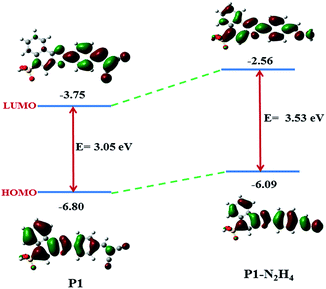
Molecular orbital calculations
To further correlate the structure and optical response of the probe P1 towards hydrazine, theoretical calculations were performed using the density functional theory with B3LYP, employing the 6-311+G** basis set implanted in a Gaussian 03 program.17,18 The optimized geometries and calculated electron distributions in the frontier molecular orbitals of P1 and P1–N2H4 are shown in Fig. 10 and 11 respectively. The molecular orbital diagram shows a relatively high electron density over the naphthalene sulphonic acid moiety in the HOMO, while the malononitrile group is highly populated in the LUMO. This justifies the ICT character of P1. The probe P1 adopts a completely planar conformation, providing an efficient π-conjugation and favouring an effective ICT transition between the donor naphthalene sulphonic acid and the acceptor nitrile group. Being a D–π–A type molecule, P1 is expected to act as a strong dipole upon photoexcitation. Upon reaction with hydrazine, the hydrazone (i.e. P1–N2H4) adduct was formed. This involved the removal of the strong electron withdrawing malononitrile group, leading to a reduction in its polarity.
 |
| | Fig. 10 The optimized structures of (a) P1 and (b) P1–N2H4 (note: for clarity we optimized the structures by taking a –H atom instead of –NH(C2H5)3). | |
 |
| | Fig. 11 The HOMO and LUMO energy levels of P1 and P1–N2H4. | |
Thus, as a whole, charge separation is more distinct in P1 than in P1–N2H4 and the absence of a strong electron withdrawing group in the latter widens the HOMO–LUMO gap, as compared to that of P1 (Fig. 11). This is in good agreement with the blue shifts in the absorption and emission spectra observed upon the treatment of P1 with hydrazine.
Detection of hydrazine using test paper strips
The probe P1 itself fluoresces in the solid state, and its corresponding emission spectrum is shown in Fig. 12(a). A solid state sensor for the detection of hydrazine was fabricated by soaking P1 onto Whatman paper strips. After dipping into a 5 μM solution of P1 in pure PBS, the strips were then dried. These paper strips appeared yellow and emitted a yellow fluorescence under UV light. Upon soaking them in hydrazine solution for a few minutes, the colour became bleached. These strips then emitted blue colour under UV light. The probe P1 again performed well when it was employed for vapour phase sensing of hydrazine (Fig. 12(b)). Solid state sensors are convenient to use and are much easier to carry around. The above results indicate that the probe P1 has remarkable potential for practical usage.
 |
| | Fig. 12 (a) The fluorescence spectra of P1 in the solid state. Inset: the photographs display the solid sample of P1 taken under 365 nm UV light (b); (i) colorimetric and fluorescence response of test strips of P1 and P1–N2H4; (ii) and (iii) colorimetric and fluorescence response of P1 on hydrazine vapour exposure. | |
Cell culture and fluorescence imaging
To explore the biological applicability of P1, an MTT assay and live cell imaging were performed. The MTT assay suggested that the probe has low cytotoxicity to living cells (Fig. S22, ESI†). HeLa cells incubated with P1 (10 μM) for 30 min at 37 °C in phosphate-buffered saline showed intracellular green fluorescence, indicating that probe P1 was permeable to the cell membrane. No blue channel fluorescence was observed at this stage. When hydrazine (50 μM) was added and incubated for about 2 h, obvious fluorescence emerged in the blue channel and the green fluorescence intensity decreased. The excitation wavelength of the laser was 405 nm, and emissions were centred at 405 ± 20 nm and 500 ± 20 nm (Fig. 13).
 |
| | Fig. 13 Confocal fluorescence images of HeLa cells. Cells incubated with probe P1 (10 μM) for 0.5 h (top); image of cells after treatment with probe P1 (10 μM) for 0.5 h and subsequent treatment of the cells with 50 μM hydrazine for 2 h (bottom). (a) and (e) Bright-field images of the HeLa cells in the samples; (b) and (f) blue emission (405 ± 20 nm); (c) and (g) green emission (500 ± 20 nm); (d) and (h) overlap; λex = 405 nm; scale bar: 50 μm. | |
Conclusions
The above results suggest that P1 is an efficient intracellular hydrazine signalling agent that demonstrates a ratiometric change in fluorescence emission from green to blue channel in aqueous medium. Additionally, it shows excellent performance in test paper strips and for vapour phase sensing of hydrazine. Live cell imaging experiments established that the probe is also profitable for tracking hydrazine in live cells. The nano-molar detection limit of P1 makes it even more lucrative.
Acknowledgements
Shweta acknowledges UGC, New Delhi for JRF/SRF [19-12/2010 (i) EU-IV]. AK thanks SERB, New Delhi for the Young Scientist award [YSS/2015/000057]. Neeraj acknowledges the UPE-JRF fellowship [chem./2012-13/154]. SKA thanks the CSIR, New Delhi for JRF/SRF [09/013(0475)/2012-EMR-I].
References
-
(a) U. Ragnarsson, Chem. Soc. Rev., 2001, 30, 205 RSC;
(b) S. S. Narayanan and F. Scholz, Electroanalysis, 1999, 11, 465 CrossRef CAS;
(c) S. Garrod, M. E. Bollard, A. W. Nicholls, S. C. Connor, J. Connelly, J. K. Nicholson and E. Holmes, Chem. Res. Toxicol., 2005, 18, 115 CrossRef CAS PubMed;
(d) K. Yamada, K. Yasuda, N. Fujiwara, Z. Siroma, H. Tanaka, Y. Miyazaki and T. Kobayashi, Electrochem. Commun., 2003, 5, 892 CrossRef CAS.
-
(a) H. W. Schiessl, Hydrazine and its derivatives, Kirk-Othmer Encyclopedia of Chemical Technology, John Wiley & Sons, Inc., 2000 Search PubMed;
(b) J. W. Mo, B. Ogorevc, X. Zhang and B. Pihlar, Electroanalysis, 2000, 12, 48 CrossRef CAS;
(c) S. D. Zelnick, D. R. Mattie and P. C. Stepaniak, Aviat., Space Environ. Med., 2003, 74, 1285 CAS;
(d) A. Serov and C. Kwak, Appl. Catal., B, 2010, 98, 1 CrossRef CAS.
-
(a) E. H. Vernot, J. D. MacEwen, R. H. Bruner, C. C. Haus, E. R. Kinkead, D. E. Prentice, A. Hall III, R. E. Schmidt, R. L. Eason, G. B. Hubbard and J. T. Young, Fundam. Appl. Toxicol., 1985, 5, 1050 CrossRef CAS PubMed;
(b) S. M. Sanins, J. A. Timbrell, C. Elcombe and J. K. Nicholson, Arch. Toxicol., 1992, 66, 489 CrossRef CAS PubMed;
(c) A. Umar, M. M. Rahman, S. H. Kim and Y. B. Hahn, Chem. Commun., 2008, 166 RSC.
-
(a) Y. Y. Liu, I. Schmeltz and D. Hoffman, Anal. Chem., 1974, 46, 885 CrossRef CAS PubMed;
(b) C. A. Reilly and S. D. Aust, Chem. Res. Toxicol., 1997, 10, 328 CrossRef CAS PubMed;
(c) S. Garrod, M. E. Bollard, A. W. Nicholls, S. C. Connor, J. Connelly, J. K. Nicholson and E. Holmes, Chem. Res. Toxicol., 2005, 18, 115 CrossRef CAS PubMed.
- U.S. Environmental Protection Agency (EPA), Integrated Risk Information System (IRIS) on Hydrazine/Hydrazine Sulfate, National Center for Environmental Assessment, Office of Research and Development, Washington, DC, 1999 Search PubMed.
-
(a) S. K. Singh, X. B. Zhang and Q. Xu, J. Am. Chem. Soc., 2009, 131, 9894 CrossRef CAS PubMed;
(b) S. K. Singh and Q. Xu, J. Am. Chem. Soc., 2009, 131, 18032 CrossRef CAS PubMed;
(c) J. Wang, X. B. Zhang, Z. L. Wang, L. M. Wang and Y. Zhang, Energy Environ. Sci., 2012, 5, 6885 RSC;
(d) S. K. Singh and Q. Xu, Chem. Commun., 2010, 46, 6545 RSC;
(e) R. Eloirdi, S. Rossignol, C. Kappenstein, D. Duprez and N. Pillet, J. Propul. Power, 2003, 19, 213 CrossRef CAS.
-
(a) Y. Liu, I. Schmeltz and D. Hoffmann, Anal. Chem., 1974, 46, 885 CrossRef CAS PubMed;
(b) D. Elder, D. Snodin and A. Teasdale, J. Pharm. Biomed. Anal., 2011, 54, 900 CrossRef CAS PubMed;
(c) M. Sun, L. Bai and D. Liu, J. Pharm. Biomed. Anal., 2009, 49, 529 CrossRef CAS PubMed;
(d) H. Bhutani, S. Singh, S. Vir, K. Bhutani, R. Kumar, A. K. Chakraborti and K. Jindal, J. Pharm. Biomed. Anal., 2007, 43, 1213 CrossRef CAS PubMed;
(e) A. Safavi and M. Baezzat, Anal. Chim. Acta, 1998, 358, 121 CrossRef CAS;
(f) G. Collins, S. Latturner and S. Rose-Pehrsson, Talanta, 1995, 42, 543 CrossRef CAS PubMed;
(g) R. Weeks Jr, S. Yasuda and B. Dean, Anal. Chem., 1976, 48, 159 CrossRef;
(h) G. Collins and S. Rose-Pehrsson, Analyst, 1994, 119, 1907 RSC;
(i) C. Batchelor-McAuley, C. Banks, A. Simm, T. Jones and R. Compton, Analyst, 2006, 131, 106 RSC;
(j) X. Gu and J. Camden, Anal. Chem., 2015, 87, 6460 CrossRef CAS PubMed.
- J. Zhang, L. Ning, J. Liu, J. Wang, B. Yu, X. Liu, X. Yao, Z. Zhang and H. Zhang, Anal. Chem., 2015, 87, 9101 CrossRef CAS PubMed.
-
(a) A. P. de Silva, H. Q. N. Gunaratne, T. A. Gunnlaugsson, J. M. Huxley, C. P. McCoy, J. T. Rademacher and T. E. Rice, Chem. Rev., 1997, 97, 1515 CrossRef CAS PubMed;
(b) S. Goswami, A. Manna, S. Paul, A. K. Das, K. Aich and P. K. Nandi, Chem. Commun., 2013, 49, 2912 RSC;
(c) D. T. Quang and J. S. Kim, Chem. Rev., 2010, 110, 6280 CrossRef CAS PubMed;
(d) A. P. Demchenko, Introduction to Fluorescence Sensing, Springer, New York, 2008 Search PubMed.
-
(a) X. Chen, Y. Xiang, Z. Li and A. Tong, Anal. Chim. Acta, 2008, 625, 41 CrossRef CAS PubMed;
(b) M. G. Choi, J. Hwang, J. O. Moon, J. Sung and S.-K. Chang, Org. Lett., 2011, 13, 5260 CrossRef CAS PubMed;
(c) G. E. Collins and S. L. Rose-Pehrsson, Anal. Chim. Acta, 1993, 284, 207 CrossRef CAS;
(d) G. E. Collins and S. L. Rose-Pehrsson, Analyst, 1994, 119, 1907 RSC;
(e) A. B. Brown, T. L. Gibson, J. C. Baum, T. Ren and T. M. Smith, Sens. Actuators, B, 2005, 110, 8 CrossRef CAS;
(f) S. W. Thomas III and T. M. Swager, Adv. Mater., 2006, 18, 1047 CrossRef;
(g) A. K. Mahapatra, R. Maji, K. Maiti, S. K. Manna, S. Mondal, S. S. Ali, S. Manna, P. Sahoo, S. Mandal, Md. R. Uddin and D. Mandal, RSC Adv., 2015, 5, 58228 RSC;
(h) B. Chen, X. Suna, X. Lib, H. Ågren and Y. Xie, Sens. Actuators, B, 2014, 199, 93 CrossRef CAS.
-
(a) J. Wu, W. Liu, J. Ge, H. Zhang and P. Wang, Chem. Soc. Rev., 2011, 40, 3483 RSC;
(b) Y. Kurishita, T. Kohira, A. Ojida and I. Hamachi, J. Am. Chem. Soc., 2010, 132, 13290 CrossRef CAS PubMed;
(c) X. Peng, Y. Xu, S. Sun, Y. Wu and J. Fan, Org. Biomol. Chem., 2007, 5, 226 RSC;
(d) X. Wang, J. Cao and C. Zhao, Org. Biomol. Chem., 2012, 10, 4689 RSC;
(e) L.-K. Zhang, Q.-X. Tong and L.-J. Shi, Dalton Trans., 2013, 42, 8567 RSC.
- J. V. Mello and N. S. Finney, Angew. Chem., 2001, 40, 1536 CrossRef CAS.
- M. H. Lee, J. S. Kim and J. L. Sessler, Chem. Soc. Rev., 2015, 44, 4185 RSC.
- Y.-D. Lin and T. J. Chow, RSC Adv., 2013, 3, 17924 RSC.
- D.-Y. Qu, J.-L. Chen and B. Di, Anal. Methods, 2014, 6, 4705 RSC.
- M. Choi, J. Moon, J. Bae, J. Lee and S. Chang, Org. Biomol. Chem., 2013, 11, 2961 CAS.
-
(a) A. D. Becke, J. Chem. Phys., 1993, 98, 5648 CrossRef CAS;
(b) R. Ditchfield, W. J. Herhe and J. A. Pople, J. Chem. Phys., 1971, 54, 724 CrossRef CAS;
(c) C. Lee, W. Yang and R. G. Parr, Phys. Rev. B: Condens. Matter Mater. Phys., 1988, 37, 785 CrossRef CAS.
- M. J. Frisch, et al., Gaussian 03, Gaussian, Inc., Pittsburgh, PA, 2003 Search PubMed.
Footnote |
| † Electronic supplementary information (ESI) available: 1H and 13C NMR, IR and ESI-MS spectra of P1 and P1–N2H2. UV-visible and fluorescence response with various anions. CCDC 1453378 (P1). For ESI and crystallographic data in CIF or other electronic format see DOI: 10.1039/c6ra15081k |
|
| This journal is © The Royal Society of Chemistry 2016 |
Click here to see how this site uses Cookies. View our privacy policy here. 


