
Removal of mercury(II) and methylene blue from a wastewater environment with magnetic graphene oxide: adsorption kinetics, isotherms and mechanism†
Yongfu Guo*,
Juan Deng,
Junyan Zhu,
Xiaoji Zhou and
Renbi Bai
Center for Separation and Purification Materials & Technologies, Suzhou University of Science and Technology, Suzhou 215009, P. R. China. E-mail: yongfuguo@mail.usts.edu.cn; Tel: +86-512-68092987
First published on 25th August 2016
Abstract
To enhance the adsorption capacity and affinity of graphene oxide (GO) for heavy metals and dyes, a magnetic graphene oxide composite (MGO) was synthesized with magnetic Fe3O4 and graphene oxide and used to study the adsorption performance for the heavy metal Hg(II) and the dye methylene blue (MB). The adsorbents were characterized by XRD, FTIR, zeta potential, BET, SEM-EDS, magnetic properties, Raman and XPS analyses. Batch adsorption experiments were performed to evaluate the adsorption conditions and reusability. The results show that the as-prepared MGO had a much higher Langmuir surface area of 1259.9 m2 g−1. The kinetic data well fitted the pseudo-second order and intra-particle diffusion equations. The adsorption isotherm of MGO for Hg(II) and MB were best described by the Langmuir model with a maximum adsorption capacity of 71.3 and 306.5 mg g−1, respectively, which surpassed the adsorption capacities of many other materials. And this result was also much higher than the adsorption capacity of GO for Hg(II) and MB of only 32.7 and 216.7 mg g−1. Overall the adsorption processes of Hg(II) and MB onto MGO were controlled by intra-particle diffusion and involved some chemisorption. The thermodynamics indicated that the adsorption process of MGO for Hg(II) and MB was endothermic and spontaneous in nature. Moreover, the adsorption capacity of MGO was still over 80% of the initial saturation adsorption capacity after being used five times. Lastly, we found out that the as-synthesized MGO was efficient for Hg(II) removal from real chloralkali wastewater. Results of this work suggest that the magnetic GO nanoparticles may be a promising adsorbent for the adsorption of heavy metals and dyes from wastewater.
1. Introduction
With the rapid development of industry, the amount of wastewater containing heavy metals and dyes resulting from metallurgy, the chemical industry, electroplating, printing and dyeing and other industries has doubled and doubled again. The polluted water enter into rivers, lakes and other water bodies accompanied by various chemical changes and eventually becomes one of the main sources of pollutants in water bodies.Heavy metals, including mercury, chromium, lead and nickel, can cause great harm to human health and the environment. Among these heavy metals, the toxicity of mercury is the most serious due to its typical characteristics of persistence, easy migration, bioaccumulation and biotoxicity.1–4 Minamata disease incident caused by methyl mercury pollution in Japan last century still makes people “turn pale at the mere mention of mercury”.2
The inorganic mercury can directly cause certain damage to human kidneys and livers through food chain. And organic mercury is harmful to human central nervous, reproductive and retinal nerve system.5 Today, mercury pollution has become a global issue.6,7
Dyes are also one of the main water pollutants which can cause serious damage to human beings and aquatic biota due to their carcinogenic and mutagenic characteristics.8 It was reported that the amount of total dyes exceeds the 700![[thin space (1/6-em)]](https://www.rsc.org/images/entities/char_2009.gif) 000 tons per year.9 Therefore, it is very important and highly desirable to effectively remove mercury and dyes from water and wastewater.
000 tons per year.9 Therefore, it is very important and highly desirable to effectively remove mercury and dyes from water and wastewater.
In order to remove the mercury and dyes from water, a great many of technologies have been employed, such as chemical precipitation, membrane separation, ion exchange, adsorption and biological methods, and so on.5
Among these methods, adsorption method has the advantages of low cost, simple operation, not easy to cause secondary pollution and higher removal efficiency, etc. However, conventional adsorbents, including activated carbon, clay, silica, cellulose, zeolite and chitosan, etc., have a relatively weak affinity to mercury and dyes, resulting in low adsorption capacity.5,10–12 Thus, it is imperative to find the materials with high affinity to both mercury and dyes.
Graphene oxide (GO), a kind of novel carbon based material, has been got considerable research attention for environmental applications, due to its enormous specific surface area, high electron mobility, good mechanical stability and the possibility of decoration.5,8,13,14 Nowadays, GO material has been widely used to separate and enrich the aqueous environmental pollutants, and shows a good application prospect as an alternative material for conventional sorbents.
However, owing to the strong interactions of π–π bond between graphitic layers and inert surface chemical properties, GO is easy to get aggregated or agglomerated in aqueous solution, resulting in low effective surface area and the limitation for wide application as adsorption materials.5,15
Nowadays, some of the magnetic nanoparticles, such as Fe3O4 nanoparticles and cobalt ferrite nanoparticles, are more and more widely used in the field of water treatment, due to their high adsorption characteristics, not easy to agglomerate advantage and easy separation characteristics.10,16,17
If magnetic groups can be introduced onto GO, GO will enhance its adsorption capacity and affinity to metals and dyes, at the same time, increase its separation efficiency from aqueous solution.
Mudasir et al. has reported that the adsorption capacity (qe, mg g−1) of dithizone-immobilized natural zeolite for Hg(II) has only 13.1 μmol g−1.12 Some studies with GO-based materials applied for Hg(II) removal have been performed by Bao et al. The adsorption capacities for Hg(II) are 20 and 30.9 mg g−1 with Fe3O4/GO and thiol-functionalized-Fe3O4/GO as adsorbents, respectively. Henriques et al. have also reported that the adsorption capacities of Hg(II) and MB are 35 and 70 mg g−1, when modified 3D GO and Fe3O4/GO are used as adsorbents, respectively.14,18,19
In view of the typical characteristics of mercury (mainly Hg(II)) and dyes in water, this paper will take them as the representatives of pollutants in water bodies to make a joint study. Methylene blue (MB), a kind of typical and widely used cationic dye, will be used as the research representative of dyes.
In this study, a magnetic graphene oxide composite (MGO) was synthesized with magnetic iron and GO by combining the easy separation characteristics of magnetic iron and the enormous advantage of GO. The prepared MGO material was characterized by XRD, FTIR, zeta potential, BET, SEM and magnetic property. In order to further understand the interaction and adsorption mechanism between the MGO and contaminants, batch adsorption tests for the removal of Hg(II) and MB were carried out, and the corresponding kinetics, equilibrium modeling and thermodynamics of adsorption were also discussed in detail.
2. Materials and experiment methods
2.1 Chemicals and materials
Graphite oxide was purchased from Nanjing XFNANO Materials Tech. Co., Ltd. FeCl2·4H2O, FeCl3·6H2O and ammonia water were purchased from Sinopharm Chemical Reagent Co., Ltd. And all other chemicals were of analytical grade. The structures and characteristics of Hg(II) (HgCl2) and MB are displayed in Table S1.†2.2 Preparation of MGO
The co-precipitation method was applied to prepare magnetic MGO nanoparticles. Firstly, 0.20 g graphite oxide was dispersed in 20 mL of ultra-pure water and sonicated for 13 h to form GO suspension. Secondly, the mole ratio of Fe2+ and Fe3+ salts was kept constant at 1:1, namely, 0.11 g FeCl2·4H2O and 0.15 g FeCl3·6H2O were dissolved into 50 mL of ultra-pure water and subsequently, 20 mL of 28 wt% ammonia water was dropped into the above solution containing irons under vigorous stirring at 200 rpm for 30 min. Then the GO suspension was dropped into the above mixed solution containing black precipitate with vigorous stirring at 200 rpm for 12 h. After stirring, the above mixed solution was heated and concentrated to get viscous at 353 K, and then the concentrated solution was placed inside a 50 mL of Teflon lined stainless autoclave. The autoclave was sealed and then heated in an oven at 453 K and air atmosphere for 10 h. Subsequently, the sample was washed several times with ultra-pure water after cooled at room temperature and till the solution became neutral. Finally, the sample MGO was dried overnight at 333 K in a vacuum oven, and then stocked for further use.
2.3 Sample characterizations
The X-ray diffraction (XRD) patterns were recorded on a XRD system (D\max-2550, Japan) with Cu Kα radiation. The diffracted intensities were recorded from 5° to 70° at 2θ angles. The spectra of the functional groups on the surface of the materials were recorded in the range of 500–4000 wave number (cm−1) on a FTIR Spectrum (Nicolet-6700, Thermo, USA). The zeta potential values of the materials in various pH solutions were obtained by electrophoresis method (ZetaPALS, Brookhaven, USA). Specific surface area (Brunauer–Emmett–Teller, BET) along with average pore diameter and total pore volume was determined using a nitrogen adsorption apparatus (ASAP2020, Quantachrome, USA). A scanning electron microscope (SEM, PhenomG3, Phenom-world, USA) was used to observe the surface morphologies of the materials studied at an accelerating voltage of 20 kV.Magnetization measurements were carried out using a vibrating sample magnetometer (VSM, Lakeshore 7404, USA) with an applied magnetic field of about 15 kOe at room temperature. Raman spectra were recorded on a microscope Raman spectrometer (DRX, Thermo Fisher Scientific, USA) with a 532 nm laser. The Raman peak intensity was extracted from the maximum value after baseline subtraction over corresponding spectral range.
The analysis of oxide specimens was performed with an X-ray photoelectron spectroscopy (XPS, Thermo-VG Scientific, ESCALAB 250, USA). The Mg Kα radiation was used in the experiments. The binding energies (BE) were calculated with respect to the C 1s peak set at 284.6 eV.
2.4 Preparation of standard solution
In order to keep the Hg(II) solution for a long time, a certain volumes of 1 vol% HNO3 solution and 0.01 vol% HCl solution were used as diluting agents and preservation solution. 0.169 g of HgCl2 powder was dissolved in a 500 mL of volumetric flask and then diluted to 500 mL with the above diluting agents. The as-obtained solution was 500 mg L−1 of standard solution containing Hg(II). 100 mg L−1 of standard solution containing MB was obtained by dissolving 0.025 g MB into 250 mL of ultra-pure water. The tested concentration of Hg(II) or MB was obtained with stepwise dilution method for the standard solution.2.5 Batch adsorption experiments
The adsorption experiments of Hg(II) and MB by MGO and GO were determined with batch experiments. Batch adsorption experiments were carried out in a 100 mL conical flasks filled with 20 mL solution of Hg(II) or MB with different concentrations. A certain amounts of adsorbents were added into the above flasks. The pH of the aqueous solution was adjusted by adding 0.2 mol L−1 NaOH or HCl solutions.Then, a certain amounts of dried adsorbents were dispersed in the above solution with desired initial pH and temperature under the conditions of 200 rpm, contact time tHg = 4 h (for Hg(II)) and tMB = 6 h (for MB) to achieve adsorption equilibrium. The initial concentrations of Hg(II) and MB are C0(Hg) = 100 mg L−1 and C0(MB) = 45 mg L−1, respectively.
After adsorption equilibration, the adsorbents were separated by external magnetic field for MGO or filtrated through 0.45 μm filter to obtain supernatant liquid for GO.
The concentrations of Hg(II) and MB in solution before and after adsorption were determined according to the absorbance at the wavelength of 253.7 nm by a cold vapor atomic adsorption spectrophotometry (F732-VJ, Jiangfeng, China) and 664 nm by a UV-vis spectrometer (UV3600, Shimadzu, Japan), respectively. Before analyzing the concentrations of Hg(II) and MB, a calibration curve was depicted to ensure precision of measurements.
Blank solutions (without any adsorbents) were treated similarly. All the experiments were performed in triplicate and the average of the data was taken for final value and later calculation.
2.6 Desorption and regeneration experiments
The desorption study is very important since the regeneration process decides the economic cost of adsorbents. In this study, a certain amount of GO or MGO was added into separate 20 mL of 100 mg L−1 Hg(II) solution or 20 mg L−1 MB solution and then shaken for a certain time at room temperature using a shaker water bath at a constant rate of 200 rpm. And the pH of Hg(II) and MB solution was 6 and 9, respectively.After adsorption, the adsorbents were collected by filtration with 0.45 μm filter or external magnetic field and then were dispersed into 20 mL of ethanol solution (8 vol% HCl acid), and stirred for 1 h after sonication for 0.5 h. After treated with ethanol solution, the adsorbents were washed with ultra-pure water for 3–5 times. The regenerated GO and MGO were then used for repeated adsorption/desorption cycles for 5 times to study their recyclability.
3. Results and discussion
3.1 Characterization of GO and MGO
The XRD spectra of GO and MGO are shown in Fig. 1. The diffraction peaks of GO were found at approximately 2θ values of 11.1°, 22.2° and 43.3° which corresponded to the layer-to-layer distance (d-spacing) of 0.79 nm (d001), 0.40 nm (d002) and 0.22 nm (d100).8 It can be seen from Fig. 1 that the six intense peaks of MGO at 2θ values of 30.6°, 35.7°, 43.6°, 53.9°, 57.6° and 63.1° indexed to (111), (220), (311), (400), (422), (511) and (440), respectively, which are consistent with the standard XRD data for the cubic spinel structure of Fe3O4, indicating the existence of Fe3O4 nanoparticles in the as-prepared composite.17,20,21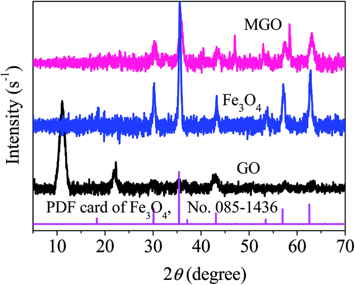 | ||
| Fig. 1 XRD patterns of GO and MGO. | ||
In order to further explore the surface characteristics of GO and MGO materials, FTIR analysis was performed and the revealed spectra can be seen in Fig. 2.
 | ||
| Fig. 2 FTIR characterization of GO and MGO. | ||
The strong peaks of 3151 and 3142 cm−1 were attributed to the –OH stretching vibrations,8,17 The peak at 1728 cm−1 could be assigned to the skeletal vibration of C![[double bond, length as m-dash]](https://www.rsc.org/images/entities/char_e001.gif) O in the carbonyl and carboxyl groups,17 The peak at 1586 cm−1 was associated with the stretching vibration of CC.17 The absorption bands at about 1401 and 1050 cm−1 were corresponding to OC–O stretching in the carboxyl group and C–O–C stretching vibrations.17,22 The strong Fe–O characteristic stretching vibration peak at 580 cm−1 was observed, proving that Fe3O4 was successfully anchored onto GO sheets,20,22 which was consistent with the data of XRD.
O in the carbonyl and carboxyl groups,17 The peak at 1586 cm−1 was associated with the stretching vibration of CC.17 The absorption bands at about 1401 and 1050 cm−1 were corresponding to OC–O stretching in the carboxyl group and C–O–C stretching vibrations.17,22 The strong Fe–O characteristic stretching vibration peak at 580 cm−1 was observed, proving that Fe3O4 was successfully anchored onto GO sheets,20,22 which was consistent with the data of XRD.
In addition, the intensities of absorption bands at 3142 and 1401 cm−1 were enhanced significantly compared with those of GO, indicating that the amount of oxygen-containing functional groups on the surface of MGO significantly increased after GO was modified with magnetic iron. The result was advantageous to the adsorption of Hg(II) and MB with positive charges.
The positive and negative charges on the surface of materials are very important for the removal of pollutants by adsorption, especially by electrostatic attraction.8
As depicted in Fig. 3, the isoelectric points of GO and MGO were less than 3 and the surfaces of GO and MGO were negative charges when pH value was greater than 3.0. Moreover, the zeta potential gradually became more negative with the increasing of pH, which meant that there were more negative charges on the exterior surface of GO and MGO materials. Compared with GO, the values of zeta potential of MGO were lower when pH of solution was greater than 6.5.
 | ||
| Fig. 3 Zeta potentials of GO and MGO under the condition of various pH. | ||
For example, the mean potential values of MGO decreased by at least 20.5% at pH from 8 to 9 after GO was modified, which meant more amount of acidic groups with negative charges were loaded on the exterior surface of MGO. Namely, the amount of oxygen-containing functional groups on the surface of MGO increased after GO was modified with magnetic iron, which offered a more possible affinity for MGO to Hg(II) and MB compared with GO. The results were favorable for the adsorption for cationic Hg(II) and MB with positive charges by electrostatic attraction.
To further investigate the physical characteristics of the as-prepared magnetic nanoparticles, adsorption–desorption isotherm and BJH pore size distribution curves of GO and MGO were carried out, as shown in Fig. S1.† According to the N2 adsorption–desorption analysis, the BET surface area and BJH desorption cumulative pore volume of MGO were 58.6 m2 g−1 and 0.088 cm3 g−1, respectively, much higher than those of GO of 9.2 m2 g−1 and 0.028 cm3 g−1.
According to the data in Table S2,† the BET surface area and BJH pore volume of MGO were at least 6 and 15 times as much as those of GO, respectively, and the mean particle size of MGO decreased by 65.4%. The characteristic change of MGO meant that MGO might have better adsorption capability compared with GO.
From the SEM-EDS spectrums shown in Fig. 4, it can be seen that irons were successfully loaded onto the GO. In addition, according to EDS detection for the mean ratio of C/O at five different positions on the surface of GO and MGO, the mass ratios of C/O of GO and MGO were 2.1 and 0.8, respectively. The mass ratios of C/Fe/O of MGO were 1:1.3:0.8. And the amount of oxygen content on the surface of MGO increased to 40.6 wt% from 31.4 wt%, which was consistent with the data of FTIR. And the detail data of EDS were showed in Table S3.†
 | ||
| Fig. 4 SEM-EDS spectra of GO (a) and MGO (b). | ||
The micrographs in Fig. 4 show the typical SEM images of GO and MGO. The surface of GO (inserted in Fig. 4(a)) exhibited many wrinkles, smooth surface and a more silk-like wave sheet structure which was the unique characteristic of graphene. After combined with magnetic iron (inserted in Fig. 4(b)), MGO material had a much rougher surface, which revealed that many small magnetic iron had been assembled on the surface of MGO layers.
The magnetization property of MGO composite was investigated at room temperature by measuring the magnetization curve using VSM, as shown in Fig. 5. The saturation magnetization of MGO was 31.5 emu g−1, indicating that MGO composite had a high magnetism. The graph inserted in Fig. 5 showed that MGO composite was easily separated from solutions via additional exterior magnets.
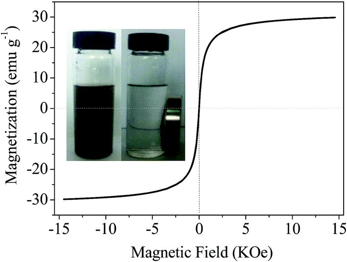 | ||
| Fig. 5 Magnetization curves of MGO at room temperature. Inserts displayed the MGO nanomaterial dispersion in solution and magnetic separation from solution. | ||
The Raman spectra of GO and MGO were characterized by two main features of D and G modes, as shown in Fig. 6. The D-band originated from the vibration of sp3 defective or out-of-order carbon atoms in the graphitic structure, and the G-band was associated with the first order bond stretching of sp2 carbon atoms.8 The Raman spectra of MGO showed a D-band at 1590 cm−1 and a G-band at 1327 cm−1.
 | ||
| Fig. 6 Raman spectra of GO and MGO. | ||
In the presence of Fe3O4, the intensity ratio of D to G bands (ID/IG) of MGO increased from 1.1 of GO to 1.5. The change of the ID/IG value may be related to the increase of disorder and defects of the GO matrix, due to chemical bonds between the carbon matrix and magnetic iron nanoparticles.8
XPS analysis of GO and MGO were displayed in Fig. 7. The C 1s spectra of GO and MGO appeared at 284.6 eV and 284.7 eV, respectively, were assigned to C–C or CC bonding of graphene, shown in Fig. 7(a) and (b). The peaks located at 286.6 eV and 286.7 eV were ascribed to epoxy and alkoxy.19,22
 | ||
| Fig. 7 XPS spectra of GO and MGO. | ||
The peaks at 286.2 eV and 286.5 eV could be assigned to the carbon-linking hydroxyl groups (C–OH) in MGO.19 The peaks at 287.3 eV and 287.4 eV were assigned to carbonyl groups (CO) in MGO.19 The peaks occurred at 288.5 eV and 288.4 eV, were assigned to COOH, illustrating the increase of carboxyl groups in MGO.19,23 The above results show that the oxygen-containing functional groups in MGO significantly increased after the co-precipitation of magnetic particles, which is consistent with the results of FTIR.
The peak of O 1s (529.2 eV, shown in Fig. 7(c)), is attributed to rich oxygen functional groups as well as oxygen atoms in Fe3O4.24 The peaks of O 1s (530.7 eV) and Fe 2p (710.5 eV and 723.2 eV, shown in Fig. 7(d)) confirmed the existence of Fe3O4 in MGO nanosheets.20,25
3.2 Adsorption performance experiments
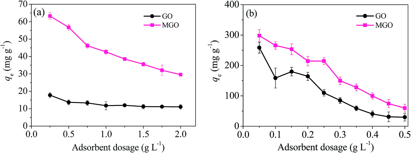 | ||
| Fig. 8 Effects of adsorbent dosage on the adsorption capacity for Hg(II) (a) and MB (b). Condition: temperature T = 298 K, pH = 6 (for Hg(II)), pH = 9 (for MB). | ||
From Fig. 8, it can be seen that the equilibrium adsorption capacities for Hg(II) and MB were decreased with the increasing ratio of solid to liquid. The qe values of MGO for Hg(II) were 2–3 times as much as those of GO for Hg(II). Compared with the qe values of GO for MB, the qe values of MGO increased by averagely 38%.
One of the reason of the decreasing adsorption capacity was due to too more active sites exceeding the demand of the saturated adsorption, which made a large number of effective active sites not used, resulting in the reduction of adsorption capacity.
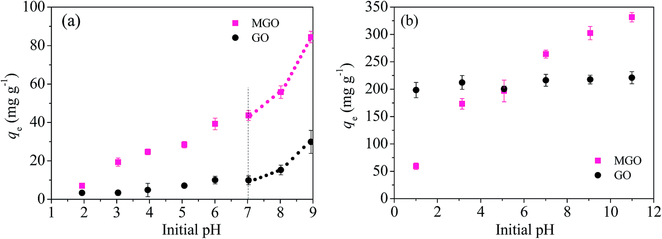 | ||
| Fig. 9 Adsorption capacities under various pH conditions. Condition: T = 298 K, adsorbent dosage WHg = 0.5 g L−1 and WMB = 0.15 g L−1. | ||
The reasons were follows: (1) as the pH increased, more electronegative charges, including –OH and –COOH, were formed on the surface of the adsorbents (see the zeta potential shown in Fig. 3), which was verified by the data of FTIR.24,29 These oxygen-containing functional groups with negative charges can exert a strong attraction to positively charged Hg(II) and MB by electrostatic interactions, which makes the adsorption capacity of MGO increase significantly.
(2) The excessive presence of H+ occupied the surface active sites of MGO, resulting in the competition adsorption between H+ and Hg(II), leading to a lower adsorption percentage at low pH.
By changing pH of the solution, mercury is predicted to exist predominantly as a monomeric species, Hg2+, and multinuclear hydroxide complexes such as [Hg(OH)]+ and Hg(OH)2.30 Part of Hg2+ will transform into Hg(OH)2 in solution and subsequently result in the generation of [HgOH]+ via hydrolysis at high pH. [HgOH]+ has higher mobility than Hg2+, which is favorable for the adsorption of Hg(II).5,30
(3) Slightly soluble Hg(OH)2 can be found when solution pH is higher than 5, especially over pH of 8. The presence of Hg(OH)2 promotes the removal of Hg(II) from aqueous solution, resulting in that it seems a high apparent adsorption capacity for Hg(II). However, it is possible that the adsorption capacity for Hg(II) decreased at high pH, which can be seen at pH from 8 to 9 (see the dotted line shown in Fig. 9(a)).
Thus, to reduce the impact of pH on adsorption capacity, the solution pH of 6 and 9 was chosen as one of the factors of the subsequent experiments for the adsorption of Hg(II) and MB, respectively.
Furthermore, the adsorption capacities of MGO for Hg(II) and MB were 39.2 and 302.4 mg g−1, which were 292% and 39% higher than those of GO of 10.0 and 217.7 mg g−1, respectively. The results show that the adsorption capacity of MGO for Hg(II) and MB significantly increased after GO modified with magnetic iron.
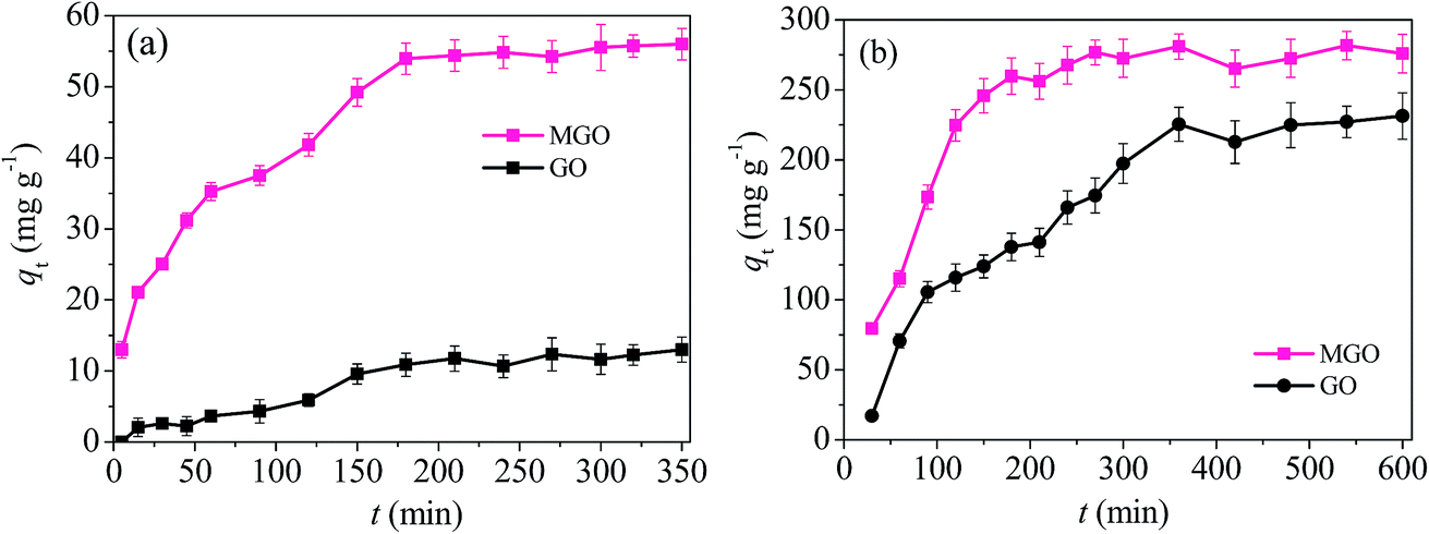 | ||
| Fig. 10 Adsorption capacities of Hg(II) and MB as a function of adsorption time t. Condition: T = 298 K, WHg = 0.5 g L−1, WMB = 0.15 g L−1, pHHg = 6 and pHMB = 9. | ||
The instantaneous adsorption capacities (qt, mg g−1) of Hg(II) and MB onto GO and MGO presented an increasing trend with the prolong of time. The instantaneous adsorption (0–4 h) onto GO and MGO, was governed by fast external diffusion and main surface adsorption,31 following by a milder and gradual ascend (over 4 h) and finally reaching an equilibrium state plateau.
This was due to the existence of the concentration gradient difference of the pollutants between water phase and the adsorbent surface which made the pollutants moved rapidly to the adsorbent surface. Besides, the existence of a large number of free active sites on the surface of the adsorbents also resulted in the rapid increase of adsorption capacity. As the increasing of time, the pollutant concentration gradient decreased and free active sites on the surface of the adsorbents reduced. Finally, the adsorption rate began to slow and gradually tended to balance.
Compared with GO, MGO had a shorter time of about 4 h to achieve adsorption balance for Hg(II) and MB. The adsorption performance of MGO for Hg(II) and MB were significantly enhanced after loaded with magnetic iron. The adsorption capacities of MGO for Hg(II) and MB were 54.8 and 267.6 mg g−1, which were 412.1% and 61.3% higher than the adsorption capacities with GO of 10.7 and 165.9 mg g−1, respectively. The results show again that the modification with magnetic iron can significantly enhance the adsorption performance of GO for heavy metal Hg(II) and dye MB.
 | ||
| Fig. 11 Effects of temperature on adsorption behavior of Hg(II) (a) and MB (b) onto GO and MGO. Condition: WHg = 0.5 g L−1, WMB = 0.15 g L−1, pHHg = 6 and pHMB = 9. | ||
3.3 Adsorption kinetics
Adsorption kinetics describing the Hg(II) and MB adsorption rate is one of the important characteristics which control the residual time of adsorbate adsorption at the solid–liquid interface. Hence, the pseudo-first-order, pseudo-second-order and intra-particle diffusion equations were applied to describe the adsorption kinetics of Hg(II) and MB adsorption onto GO and MGO, and expressed in the ESI.†The adsorption kinetic studies were carried out with C0(Hg) = 100 mg L−1, C0(MB) = 45 mg L−1, WHg = 0.5 g L−1, WMB = 0.15 g L−1, pHHg = 6, pHMB = 9, agitation rate of 200 rpm and absolute temperature of 298 K. Residual pollutant concentration was measured at different time intervals.
The linear fitting results of the three models are shown in Fig. 12. The kinetic parameters calculated from the fitting plots were summarized in Tables 1 and 2.
 | ||
| Fig. 12 Pseudo-first-order kinetics (a) and (c), pseudo-second-order kinetics (b) and (d), intra-particle diffusion kinetics (e) and (f) for adsorption of Hg(II) and MB onto GO and MGO. | ||
| Adsorbents | Pseudo-first-order | Pseudo-second-order | |||||
|---|---|---|---|---|---|---|---|
| qe,exp (mg g−1) | qe,cal (mg g−1) | k1 (min−1) | R2 | qe,cal (mg g−1) | k2 (g mg−1 min−1) | R2 | |
| GO | 16.7 | 14.8 | 0.0093 | 0.904 | 18.6 | 0.0004 | 0.980 |
| MGO | 59.9 | 53.6 | 0.0154 | 0.956 | 64.4 | 0.0002 | 0.991 |
| Adsorbents | Intra-particle diffusion | |||||
|---|---|---|---|---|---|---|
| kd1 (mg g−1 min−0.5) | C1 (mg g−1) | R12 | kd2 (mg g−1 min−0.5) | C2 (mg g−1) | R22 | |
| GO | 1.4 | 2.1 | 0.992 | 0.35 | 16.1 | 0.945 |
| MGO | 3.4 | 18.7 | 0.984 | 0.39 | 60.7 | 0.982 |
| Adsorbents | Pseudo-first-order | Pseudo-second-order | |||||
|---|---|---|---|---|---|---|---|
| qe,exp (mg g−1) | qe,cal (mg g−1) | k1 (min−1) | R2 | qe,cal (mg g−1) | k2 (g mg−1 min−1) | R2 | |
| GO | 231.3 | 313.5 | 0.007 | 0.959 | 256.4 | 0.0002 | 0.983 |
| MGO | 275.9 | 176.5 | 0.009 | 0.804 | 303.1 | 0.0005 | 0.984 |
| Adsorbents | Intra-particle diffusion | |||||
|---|---|---|---|---|---|---|
| kd1 (mg g−1 min−0.5) | C1 (mg g−1) | R12 | kd2 (mg g−1 min−0.5) | C2 (mg g−1) | R22 | |
| GO | 15.7 | 70.8 | 0.996 | 2.2 | 176.2 | 0.952 |
| MGO | 21.5 | 50.8 | 0.989 | 0.81 | 263.6 | 0.873 |
The correlation coefficients R2 indicated a better fitting of pseudo-second-order equation with the experimental data compared with other kinetic models for Hg(II) (R2 = 0.991) and MB (R2 = 0.984), suggesting a chemisorption occurrence (shown in Fig. 12(b) and (d)). The calculated values (qe,cal) resulted from pseudo-second-order equation were 64.4 and 303.1 mg g−1 for Hg(II) and MB, respectively, showing a good agreement with the experimental data (59.9 mg g−1 and 275.9 mg g−1).
The experimental data were fitted well by two straight lines in the intra-particle diffusion model shown in Fig. 12(e) and (f), corresponding to two different adsorption stages and two intra-particle diffusion rate constant kdi. The first steeper section did not pass through the origin which pointed out that intra-particle diffusion was not only the rate-controlling step, but also affected by boundary layer diffusion process.14 The second less pronounced portion was attributed to the adsorption equilibrium stage for which the intra-particle diffusion started to slow down due to low pollutant concentration left in the solution.
3.4 Adsorption isotherms
To obtain an insight on the mechanism of adsorption, the adsorption capacity was further analysed by assuming that the equilibrium did exist between liquid adsorbate phase and the solid adsorbent phase at any time t for the given temperatures.32 Thus, the adsorption capacity was fitted with the Langmuir model which assumes that the adsorbent surface is homogeneous and the magnitude of the adsorption energy is equivalent to each active adsorption sites.12 Each of the active sites of the adsorbent adsorbs one of adsorbate molecules. When the active sites have not been fully occupied yet with adsorbate molecules, the increase of the adsorbate concentration in the solution will always be accompanied by an increase in the adsorption capacity of the adsorbate adsorbed. Conversely, if the active sites have been fully occupied with the adsorbate molecules, the increase of adsorbate concentration will not promote the increase in the adsorption capacity.12However, the employment of an asymmetric or heterogeneous adsorption model is essential for the carbon materials with complicated surface structure. The most common and empirical model is Freundlich model.
The above two models are the most widely isotherm models used for analyzing adsorption isotherm data, and show a correlation between the activity of adsorbents and the amount of adsorbates adsorbed at a constant temperature. In addition, the data from the two models can also provide some physicochemical information on how the adsorption proceeds and how adsorbates interact with adsorbents.8 The linear equations for Langmuir and Freundlich isotherm models are expressed in the ESI.†
The adsorption isotherms of Hg(II) and MB onto the GO and MGO at different C0 concentrations are given in Fig. 13, and the fitted data were listed in Table 3.
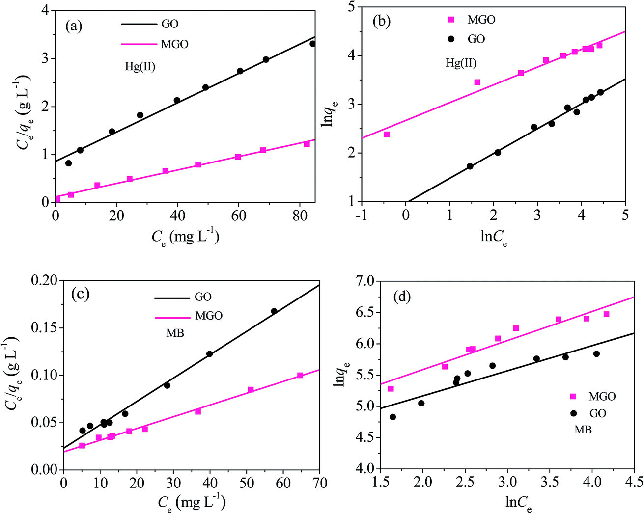 | ||
| Fig. 13 Langmuir (a) and (c), Freundlich (b) and (d) isotherms for the adsorption of Hg(II) and MB onto GO and MGO. Condition: WHg = 0.5 g L−1, WMB = 0.15 g L−1, pHHg = 6, pHMB = 9 and T = 298 K. | ||
| Adsorbents | Langmuir isotherm for Hg(II) adsorption | Freundlich isotherm for Hg(II) adsorption | |||||
|---|---|---|---|---|---|---|---|
| Qm (mg g−1) | KL (L mg−1) | R2 | RL | 1/n | KF (mg1−n Ln g−1) | R2 | |
| GO | 32.7 | 0.04 | 0.987 | 0.22 | 0.51 | 14.4 | 0.974 |
| MGO | 71.3 | 0.12 | 0.988 | 0.08 | 0.37 | 2.6 | 0.971 |
| Adsorbents | Langmuir isotherm for MB adsorption | Freundlich isotherm for MB adsorption | |||||
|---|---|---|---|---|---|---|---|
| Qm (mg g−1) | KL (L mg−1) | R2 | RL | 1/n | KF (mg1−n Ln g−1) | R2 | |
| GO | 216.7 | 0.137 | 0.989 | 0.04 | 0.40 | 78.5 | 0.831 |
| MGO | 306.5 | 0.129 | 0.991 | 0.04 | 0.47 | 105.3 | 0.923 |
From Table 3 it can be seen that under all conditions, the Langmuir isotherm fitted better with the experimental data and the adsorption process follows a Langmuir type (R2 > 0.98) monolayer adsorption.
However, it should be noticed that the R2 for Hg(II) adsorption from Freundlich model was greater than 0.97. And the constant of n calculated from the Freundlich isotherm was found in the range of favorable adsorption (n > 1), suggesting a high adsorption strength and chemical adsorption, which was consistent with the large KL value of the Langmuir model and indicated that Hg(II) and MB were facile to be adsorbed onto MGO.
For Hg(II) and MB adsorption onto MGO, the separation factor RL values are much less than 0.1 in the range of 0.04–0.08, thereby confirming that the adsorption of Hg(II) and MB onto MGO is a favorable process.
The adsorption capacities qe of MGO for Hg(II) and MB from the Langmuir isotherm were 71.3 and 306.5 mg g−1 at 298 K, respectively, and also much higher than those from GO of only 32.7 and 216.7 mg g−1. The result shows that the method of GO modified with magnetic iron is highly effective.
The larger parameters of k1, k2, KL and KF from kinetic and isotherm models of MGO, respectively, suggested that MGO adsorbent had faster adsorption rate and higher adsorption capacity for Hg(II) and MB and the adsorption capacity of MGO surpassed that of many materials as shown in Table 4.
| Adsorbents | BET (m2 g−1) | Pollutants | Qm (mg g−1) | Ref. |
|---|---|---|---|---|
| MGO | 58.6 | Hg(II) | 71.3 | This work |
| MGO | 58.6 | MB | 306.5 | This work |
| Modified 3D GO | 400 | Hg(II) | 35 | 14 |
| Synthesized FeS2 | 20.1 | Hg(II) | 180.5 | 33 |
| Cross-linked polydithiocarbamate | 11.5 | Hg(II) | 22.1 | 34 |
| Chitosan magnetic composite microsphere | Hg(II) | 73.3 | 21 | |
| 3-Mercaptopropyltriethoxysilane CNT/Fe3O4 | 97.2 | Hg(II) | 65.5 | 35 |
| Polyacrylic acid MGO | MB | 291 | 19 | |
| MGO | MB | 70 | 19 | |
| GO aerogel | 42 | MB | 416.7 | 36 |
| Graphene/magnetite composite | MB | 43.8 | 37 | |
| Chitosan/MGO | 412.2 | MB | 243.3 | 12 |
3.5 Adsorption thermodynamics
During any adsorption process, the changes of energy and entropy should be considered so as to determine what process will take place spontaneously and whether the process is spontaneous or not. The values of thermodynamic parameters have a great meaning for practical application for an adsorption process.38 The thermodynamic parameters, including the change of Gibbs free energy (ΔG°, kJ mol−1), enthalpy (ΔH°, kJ mol−1) and entropy (ΔS°, kJ mol−1 K−1) associated with the adsorption process should be used to determine spontaneity and heat change of the adsorption process, and were evaluated by the following equations:|
ΔG° = −RTlnKd
| (1) |
 | (2) |
Kd vs. 1/T) shown in Fig. 14. And the results of ΔG°, ΔS° and ΔH° are shown in Tables 5 and 6.
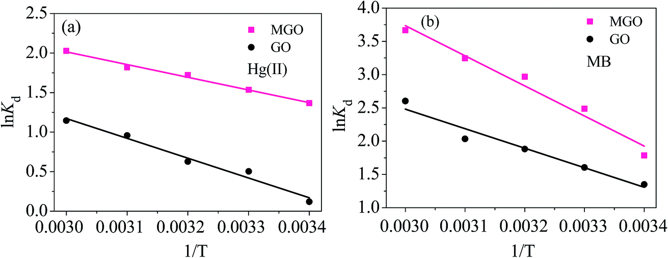 | ||
| Fig. 14 Thermodynamic fittings of Hg(II) and MB onto GO and MGO. | ||
| Adsorbents | ΔH° (kJ mol−1) | ΔS° (J mol−1 K−1) | ΔG° (kJ mol−1) | ||||
|---|---|---|---|---|---|---|---|
| 298 K | 303 K | 313 K | 323 K | 333 K | |||
| GO | 20.8 | 72.3 | −0.29 | −1.26 | −1.63 | −2.57 | −3.17 |
| MGO | 13.4 | 46.8 | −3.38 | −3.87 | −4.48 | −4.88 | −5.61 |
| Adsorbents | ΔH° (kJ mol−1) | ΔS° (J mol−1 K−1) | ΔG° (kJ mol−1) | ||||
|---|---|---|---|---|---|---|---|
| 298 K | 303 K | 313 K | 323 K | 333 K | |||
| GO | 34.4 | 93.8 | −3.34 | −3.64 | −5.13 | −5.29 | −7.09 |
| MGO | 27.6 | 63.9 | −4.42 | −6.37 | −7.85 | −8.85 | −10.15 |
The adsorption process was found endothermic in nature with positive values of ΔH° calculated from the fitting plots in Fig. 14. The result was also supported by the increase in value of Hg(II) and MB adsorption with the ascending temperature, which was accordant with the results of Fig. 11.
Furthermore, the data from Tables 5 and 6 show that the ΔH° values of MGO were less than those of GO, which indicates that the adsorption system of MGO tends to be more unstabilized compared with the system of GO. And the reactive activity of the MGO system should be higher than that of GO, resulting in higher adsorption capacity of MGO.10,15
The positive value of ΔS° suggested the increasing randomness at the solid/liquid interface during the adsorption of Hg(II) and MB ions onto GO and MGO nanoparticles.38
The negative values of ΔG° indicated the feasibility of the process and spontaneous nature of Hg(II) and MB adsorption onto GO and MGO adsorbents. Moreover, the decreasing of ΔG° as temperature rises indicated that the adsorption was more favorable at higher temperatures, which was consistent with the results of Fig. 11 and the small values of ΔG° indicated a strong interaction between the adsorbed pollutants and adsorbents.8
3.6 Desorption and regeneration experiments
The effects of regenerated GO and MGO on the adsorption of Hg(II) and MB in five cycles were shown in Fig. 15. It can be seen that although the adsorption capacities of MGO for Hg(II) and MB declined with the increasing cycles, there were still high adsorption performance of MGO for both two pollutants in the fifth cycle. Compared with the adsorption capabilities in first cycle, the adsorption capabilities of GO for Hg(II) and MB decreased by approximately 59.8% and 51.8% after five cycles, respectively. However, the decrease of adsorption capabilities of MGO was only about 19.7% and 19.0% after five cycles, respectively. This result indicated that MGO could be desorption and regenerated with high efficiency and reused in the wastewater treatment applications. The decrease of adsorption capability can be attributed to the loss of active sites after each desorption step.29,39 | ||
| Fig. 15 Desorption and regeneration cycles of Hg(II) (a) and MB (b) onto GO and MGO. | ||
3.7 Practicability of MGO for purifying real wastewater containing Hg(II)
Chloralkali wastewater is a kind of common industrial wastewater resulted from the production activity of chloralkali or polyvinyl chloride. There are all kinds of pollutants, including metal ions of Hg2+, Mg2+, Fe2+, Fe3+ and Na+, inorganic ions of Cl− and S2−, and also other pollutants in the chloralkali wastewater. The characteristics of raw wastewater are summarized in Table S4.† In order to evaluate the practical efficiency of MGO material in application, a batch adsorption experiment was made with MGO on chloralkali wastewater after pre-treated.Based on the experiment data, the MGO material exhibited a very high removal efficiency (over 99%) for mercury ions from chloralkali wastewater with WHg = 0.5 g L−1 and contact time tHg = 4 h. Although the cationic existence of Mg2+, Fe2+, Fe3+ and Na+ may interfere with removal of Hg(II) ions onto the MGO, the water quality of chloralkali wastewater was greatly improved, and the concentration of total mercury in the effluent met the national discharge standard of pollutants for chloralkali wastewater. The results show that the prepared MGO adsorbents had a satisfactory application effects.
3.8 Adsorption mechanism
Mercury adsorption onto MGO surface may take place through physical or chemical adsorption. The results of FTIR and adsorption test show that the oxygen containing functional groups play a key role during the adsorption process of Hg(II) and MB, and the increase of oxygen containing functional groups in MGO greatly promote the adsorption of Hg(II) and MB onto MGO. Based on the results of pH effect on the adsorption of Hg(II) and MB onto MGO, the reaction mechanisms of oxygen containing functional groups and Hg2+ specie or MB were formulated as the following suggested equations:5,10| R–OH → R–O− + H+ (dissociate and de-protonate) | (3) |
| R–COOH → R–COO− + H+ (dissociate and de-protonate) | (4) |
| 2R–O− + Hg2+ → R–O–Hg–O–R (ionic-pair formation) | (5) |
| 2R–COO− + Hg2+ → R–COO–Hg–OOC–R (ionic-pair formation) | (6) |
| R–O− + MB+ → R–O–MB (ionic-pair formation) | (7) |
| R–COO− + MB+ → R–COO–MB (ionic-pair formation) | (8) |
The above reactions suggest that the more OH− ions are available to react with H+ in the solution under lower zeta potentials (high solution pH), which allows the reactions to proceed properly. And high solution pH is favorable for the adsorption for cationic Hg(II) and MB with positive charges by electrostatic attraction.
The correlation coefficients R2 resulted from pseudo-second-order equation suggests a chemisorption of Hg(II) and MB, and the overall adsorption rates of Hg(II) and MB onto GO and MGO are controlled by chemical adsorption. In this study, Ci (thickness of the boundary layer at stage I, shown in Table 2) is always a non-zero value, revealing that the adsorption process is controlled not only by intra-particle diffusion but involves some main chemisorption process.10,14
In addition, the two kdi values of Hg(II) and MB derived from intra-particle diffusion sharply decreased from kd1 to kd2, which indicated that the adsorption process of Hg(II) and MB onto MGO could be explained with the followings: (i) the first stage (i = 1) represents the boundary layer diffusion of solute molecules and external surface adsorption of pollutants; (ii) the second stage (i = 2) describes the gradual adsorption stage attributing to the intra-particle diffusion and the final equilibrium stage.8,16
The good fittings of Langmuir model shows monolayer process and the Freundlich model showed that the adsorption process of Hg(II) and MB on the surface of MGO had not only single molecular layer occupation, but also asymmetric or multilayer occupation.32
The values of ΔH° of thermodynamics study indicate the adsorption process is endothermic in nature and the values of ΔH° are high enough to ensure strong interaction between the adsorbates and adsorbents, and suggest that the adsorption of Hg(II) and MB involve chemical reactions due to hydrogen bond, chemical bond or protonation and so on.8,40
Moreover, generally, when ΔG° is less than −40 kJ mol−1, the adsorption mainly involves chemisorption.41 Based on the data of adsorption isobar (Fig. 14), the change of ΔH° and ΔG°, and also the results of pseudo-second-order and Langmuir isotherm, the adsorption process of Hg(II) and MB onto GO and MGO can be mainly attributed to chemisorption and controlled by intra-particle diffusion.
4. Conclusions
In summary, MGO coupled characters of graphene oxide and magnetism were prepared with a simple and cost effective method for removing Hg(II) and MB simultaneously from aqueous solution. The XRD, SEM and XPS properties evidenced that iron oxides were successfully loaded on the surface of GO. The analysis of FTIR and zeta potentials presented that more amount of acidic groups with negative charges were loaded on the exterior surface of MGO. Furthermore, MGO nanomaterial was easily separated from water with external magnetic field and had higher desorption and regeneration efficiency, which can hopefully reduce water treatment cost.Kinetic data showed a good correlation to a pseudo-second-order equation and the overall adsorption process of Hg(II) and MB onto MGO were controlled not only by intra-particle diffusion but involved some chemisorption. Intra-particle diffusion may be the rate-controlling step.
The isotherm fitting data from Langmuir and Freundlich models indicated that the adsorption process of Hg(II) and MB on the surface of MGO had not only single molecular layer occupation, but also asymmetric or multilayer occupation. And the calculated equilibrium adsorption capacities of MGO for Hg(II) and MB were 71.3 and 306.5 mg g−1, respectively, which surpassed the adsorption capacities of many materials.
The thermodynamic data indicated that the adsorption process of MGO for Hg(II) and MB was endothermic and spontaneous in nature. The adsorption activity and affinity of MGO for Hg(II) and MB were higher than those of GO.
Results of this work suggest that the magnetic GO nanoparticles may be a promising adsorbent for the adsorption of heavy metals and dyes from wastewater using the technology of magnetic separation.
Acknowledgements
This work was supported by the National Natural Science Foundation of China (No. 51578354), Natural Science Foundation of Jiangsu Province (No. BK20141179), the Practice Innovation Training Program Projects of the Jiangsu College Students (No. 201410332005Z), Suzhou Key Laboratory of Separation and Purification Materials & Technologies (SZS201512), Qing Lan Project and Overseas Training Program of the Outstanding Young Teachers and Principals of Universities of Jiangsu Provincial Department of Education.References
- L. Xu, J. Chen, L. Yang, Z. Niu, L. Tong, L. Yin and Y. Chen, Chemosphere, 2015, 119, 530 CrossRef CAS PubMed.
- M. McNutt, Science, 2013, 341, 1430 CrossRef CAS PubMed.
- D. P. Krabbenhoft and E. M. Sunderland, Science, 2013, 341, 1457 CrossRef CAS PubMed.
- X. Y. Li, X. Y. Gao, L. H. Ai and J. Jiang, Chem. Eng. J., 2015, 274, 238 CrossRef CAS.
- S. Kabiri, D. N. H. Tran, M. A. Cole and D. Losic, Environ. Sci.: Water Res. Technol., 2016, 2, 390 CAS.
- W. J. Lee and G. N. Bae, Environ. Sci. Technol., 2009, 43, 1522 CrossRef CAS PubMed.
- R. Stolle, H. Koeser and H. Gutberlet, Appl. Catal., B, 2014, 144, 486 CrossRef CAS.
- Y. F. Guo, J. Deng, J. Y. Zhu, C. Zhou, C. Y. Zhou, X. J. Zhou and R. B. Bai, RSC Adv., 2016, 6, 39762 RSC.
- J. A. Gonzalez, M. E. Villanueva, L. L. Piehl and G. J. Copello, Chem. Eng. J., 2015, 280, 41 CrossRef CAS.
- L. M. Cui, X. Y. Guo, Q. Wei, Y. G. Wang, L. Gao, L. G. Yan, T. Yan and B. Du, J. Colloid Interface Sci., 2015, 439, 112 CrossRef CAS PubMed.
- A. Mehdinia, M. Akbari, T. B. Kayyal and M. Azad, Environ. Sci. Pollut. Res., 2015, 22, 2155 CrossRef CAS PubMed.
- M. Mudasir, K. Karelius, N. H. Aprilita and E. T. Wahyuni, J. Environ. Chem. Eng., 2016, 4, 1839 CrossRef CAS.
- P. N. Diagboya, B. I. Olu-Owolabi and K. O. Adebowale, RSC Adv., 2015, 5, 2536 RSC.
- B. Henriques, G. Goncalves, N. Emami, E. Pereira, M. Vila and P. Marques, J. Hazard. Mater., 2016, 301, 453 CrossRef CAS PubMed.
- Y. K. Zhang, T. Yan, L. G. Yan, X. Y. Guo, L. M. Cui, Q. Wei and B. Du, J. Mol. Liq., 2014, 198, 381 CrossRef CAS.
- G. D. Jiang, Q. Chang, F. F. Yang, X. Y. Hu and H. Q. Tang, Chin. J. Chem. Eng., 2015, 23, 510 CrossRef CAS.
- Y. H. Wang, L. L. Li, C. N. Luo, X. J. Wang and H. M. Duan, Int. J. Biol. Macromol., 2016, 86, 505 CrossRef CAS PubMed.
- J. Bao, Y. Fu and Z. H. Bao, Nanoscale Res. Lett., 2013, 8, 6 CrossRef PubMed.
- J. W. Zhang, M. S. Azam, C. Shi, J. Huang, B. Bin, Q. X. Liu and H. B. Zeng, RSC Adv., 2015, 5, 32272 RSC.
- L. L. Chen, D. L. Zhao, S. H. Chen, X. B. Wang and C. L. Chen, J. Colloid Interface Sci., 2016, 472, 99 CrossRef CAS PubMed.
- X. Tao, K. Li, H. Yan, H. Yang and A. M. Li, Environ. Pollut., 2016, 209, 21 CrossRef CAS PubMed.
- J. Zhang, J. L. Gong, G. M. Zenga, X. M. Ou, Y. Jiang, Y. N. Chang, M. Guo, C. Zhang and H. Y. Liu, Appl. Surf. Sci., 2016, 370, 335 CrossRef CAS.
- X. Wang, H. Tang, S. Huang and L. Zhu, RSC Adv., 2014, 4, 60102 RSC.
- L. Cui, Y. Wang, L. Hu, L. Gao, B. Du and Q. Wei, RSC Adv., 2015, 5, 9759 RSC.
- X. Y. Li, L. H. Ai and J. Jiang, Chem. Eng. J., 2016, 288, 789 CrossRef CAS.
- X. X. Yang, Y. H. Li, Q. J. Du, J. K. Sun, L. Chen, S. Hu, Z. H. Wang, Y. Z. Xia and L. H. Xia, J. Colloid Interface Sci., 2015, 453, 107 CrossRef CAS PubMed.
- M. Hadavifar, N. Bahramifar, H. Younesi and Q. Li, Chem. Eng. J., 2014, 237, 217 CrossRef CAS.
- C. Zhang, J. H. Sui, J. Li, Y. L. Tang and W. Cai, Chem. Eng. J., 2012, 210, 45 CrossRef CAS.
- Y. B. Song, X. D. Song, C. J. Cheng and Z. G. Zhao, RSC Adv., 2015, 5, 87030 RSC.
- H. Alijani, Z. Shariatinia and A. A. Mashhadi, Chem. Eng. J., 2015, 281, 468 CrossRef CAS.
- N. A. Travlou, G. Z. Kyzas, N. K. Lazaridis and E. A. Deliyanni, Chem. Eng. J., 2013, 217, 256 CrossRef CAS.
- S. T. Song, Y. F. Hau, N. Saman, K. Johari, S. C. Cheu, H. Kong and H. Mat, J. Environ. Chem. Eng., 2016, 4, 1685 CrossRef CAS.
- Y. H. Duan, D. S. Han, B. Batchelor and A. Abdel-Wahab, Colloids Surf., A, 2016, 490, 326 CrossRef CAS.
- O. S. Akintola, T. A. Saleh, M. M. Khaled and O. C. S. Al Hamouz, J. Taiwan Inst. Chem. Eng., 2016, 60, 602 CrossRef CAS.
- C. Zhang, J. Sui, J. Li, Y. Tang and W. Cai, Chem. Eng. J., 2012, 210, 45 CrossRef CAS.
- S. Zamani and N. S. Tabrizi, Res. Chem. Intermed., 2015, 41, 7945 CrossRef CAS.
- L. H. Ai, C. Y. Zhang and Z. L. Chen, J. Hazard. Mater., 2011, 192, 1515 CrossRef CAS PubMed.
- S. Nasirimoghaddam, S. Zeinali and S. Sabbaghi, J. Ind. Eng. Chem., 2015, 27, 79 CrossRef CAS.
- P. Tan, J. Sun, Y. Hu, Z. Fang, Q. Bi, Y. Chen and J. Cheng, J. Hazard. Mater., 2015, 297, 251 CrossRef CAS PubMed.
- M. B. Ahmed, J. L. Zhou, H. H. Ngo and W. S. Guo, Sci. Total Environ., 2015, 532, 112 CrossRef CAS PubMed.
- A. Roy, B. Adhikari and S. B. Majumder, Ind. Eng. Chem. Res., 2013, 52, 6502 CrossRef CAS.
Footnote |
| † Electronic supplementary information (ESI) available. See DOI: 10.1039/c6ra14651a |
| This journal is © The Royal Society of Chemistry 2016 |