Identification of novel pyrazole–rhodanine hybrid scaffolds as potent inhibitors of aldose reductase: design, synthesis, biological evaluation and molecular docking analysis

Hina Andleeba, Yildiz Tehseenb, Syed Jawad Ali Shahb, Imtiaz Khana, Jamshed Iqbal*b and Shahid Hameed*a
aDepartment of Chemistry, Quaid-i-Azam University, Islamabad-45320, Pakistan. E-mail: shameed@qau.edu.pk; Fax: +92-51-9064-2241; Tel: +92-51-9064-2133
bCentre for Advanced Drug Research, COMSATS Institute of Information Technology, Abbottabad-22060, Pakistan. E-mail: drjamshed@ciit.net.pk; Fax: +92-992-383441; Tel: +92-992-383591 Tel: +92-992-383596

Received 4th June 2016 , Accepted 30th July 2016

First published on 8th August 2016


Abstract

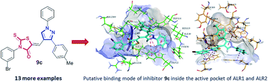
In an effort to develop a new class of potent aldose reductase inhibitors, a series of 1,3-diarylpyrazole assimilated 3-substituted 4-oxo-2-thioxo-1,3-thiazolidines (9a–n) was designed, and synthesized in good to excellent yields by a pharmacophore integration approach. The structures of the newly synthesized pyrazole–rhodanine derivatives were established by readily available spectroscopic methods (FTIR, 1H and 13C NMR) and mass spectrometry. The hybrid compounds were evaluated as aldehyde and aldose reductase inhibitors. The biological screening results identified several compounds as remarkable inhibitors of ALR1 and ALR2. Among them, compounds 9c and 9k showed excellent activity (and complete selectivity) towards the aldose reductase enzyme with IC50 values of 1.22 ± 0.67, and 2.34 ± 0.78 μM, respectively, as compared to the standard drug (sorbinil; IC50 = 3.10 ± 0.20 μM). The molecular docking analysis of the most potent inhibitor 9c was performed in order to identify the putative binding modes inside the active pocket of the enzymes. These newly discovered aldose reductase inhibitors are believed to represent valuable lead structures to further streamline the generation of candidate compounds to target a number of pathological conditions, most strikingly long-term diabetic complications.


Introduction

Aldose reductase (AR) has been implicated in the etiology of diabetic complications that may be attributed to a significant flux of glucose through the polyol pathway, induced in tissues such as nerves, retina, lens and kidney at elevated blood glucose levels. Diabetes, a complex and chronic metabolic disorder, is now recognized as a public health problem. According to epidemiological data, 366 million people were diagnosed with diabetes in 2011, and this number is expected to rise sharply to 552 million people within the next 20 years.1–3 The economic cost attributable to the management of this disease markedly influences countries' health budgets, being the highest of any disease category.4,5 Hence, the inhibition of aldose reductase is emerging as a major therapeutic strategy for the treatment of hyperglycemia-induced cardiovascular pathologies.6 Progression of chronic diabetes results in long-term, debilitating complications such as cataractogenesis and microangiopathy (including nephropathy, retinopathy and neuropathy) thought to be linked to excess free glucose in corresponding tissues.7,8 Numerous observations have provided evidence of aldose reductase (AR) implicated in diabetic complications.9,10 Inhibition of aldose reductase, the first enzyme of the polyol pathway, is therefore a useful strategy for prevention and treatment of complications of chronic diabetes. A considerable effort in this direction has led to the discovery of a large number of AR inhibitors.11–14

Aldose reductase (ALR2; EC 1.1.1.21) is the first rate-determining enzyme in the polyol pathway and catalyses the reduction of glucose to sorbitol with concomitant conversion of NADPH into NADP+ (Fig. 1). Sorbitol is in turn converted into fructose with accompanied reduction of NAD+ by sorbitol dehydrogenase.15 Under normal circumstances, glucose is predominantly converted to glucose-6-phosphate by hexokinase and then enters the glycolytic pathway, whereas only a small amount of glucose is metabolized through the polyol pathway due to a relatively low affinity of ALR2 for this substrate. During hyperglycemia, the polyol metabolic pathway is activated and the increased flux of glucose through the polyol pathway triggers the accumulation of sorbitol, which mainly happens in tissues demonstrating insulin-independent uptake of glucose, such as lens, kidney, retina, and peripheral nerves.16 Because of the excess sugar alcohol (polyol), the lens imbibes water, causing osmotic imbalance. Eventually, increased sodium and decreased potassium levels and decreased glutathione levels lead to cataract formation. Topical administration of aldose reductase inhibitors has been shown to prevent the cataract in rats.17


image file: c6ra14531k-f1.tif
Fig. 1 Polyol pathway for glucose metabolism.

According to recent reports, AR is up-regulated not only under hyperglycemic conditions but also in other pathological states including cardiac disorders (myocardial ischemia and ischemia–reperfusion injury, congestive heart failure, cardiac hypertrophy and cardiomyopathy), inflammation, mood disorders, renal insufficiency, ovarian abnormalities and human cancers such as liver, breast, ovarian, cervical and rectal cancers.5,18–20 These pathological processes have become major threats to human health in the 21st century. Intense efforts have been directed toward the development of effective aldose reductase inhibitors,21 however, only a few compounds have reached clinical trials, mostly thiazolidine-2,4-diones such as glitazones (ciglitazone, troglitazone, pioglitazone and rosiglitazone).22–26 So far, a 2-thioxothiazolidine-4-one, epalrestat (Kinedak),27,28 marketed in Japan and China, is the only commercially available ARI drug (Fig. 2). Apart from epalrestat, AR inhibitors have failed in clinical trials because of poor pharmacokinetic properties and side effects,29 and even epalrestat has been withdrawn from the market in some countries because of its side effects. Thus, it is important to develop novel ARIs with improved efficacy and less toxic profile.


image file: c6ra14531k-f2.tif
Fig. 2 Representative examples of aldose reductase inhibitor (epalrestat; A) and pyrazole-based antidiabetic agents (B and C).

Pyrazoles, key structural fragments in drug discovery, occupy a significant position in medicinal chemistry due to their diverse spectrum of biological activities including anti-viral, antitumor,30–32 anti-inflammatory,33 antihyperglycemic,34,35 and antidiabetic activities.36–41 Inspired by these findings, and in a continuation of our research interests for the development of novel and effective antidiabetic agents,42a,b we focused our attention on the heterocyclic core of epalrestat, (thioxothiazolidine) and pyrazole, exploited previously for numerous pharmacological properties. Idea was to couple the two bioactive nuclei (thioxothiazolidine and pyrazole) using a hybrid pharmacophore approach to develop a novel scaffold with favorable chemical modifications and enhanced aldose reductase inhibitory activities, which to the best of our knowledge, has not been reported previously (Fig. 3). Prompted by this speculation, we embarked on the synthesis of target pyrazole–rhodanine hybrid compounds with structural variations on the aromatic moiety and determined their AR inhibitory potency using an in vitro biological assay. The molecular docking investigations provided the insights on the putative binding mode of the potent inhibitor inside the active site of ALR1 and ALR2.


image file: c6ra14531k-f3.tif
Fig. 3 Structural design of pyrazole–thioxothiazolidine hybrid compounds by pharmacophore integration approach.

Results and discussion

Chemistry

Synthesis of intermediate pharmacophores 4a–b and 8a–g for the formation of target compounds 9a–n was accomplished according to the synthetic routes illustrated in Schemes 1 and 2. Synthesis was performed using modified literature procedure.43a–d,44 3-Bromoaniline/p-toluidine (1) (1.0 equiv.) was reacted with carbon disulfide (2.0 equiv.) in diethyl ether in the presence of triethylamine (2.0 equiv.) as a base to furnish triethylammonium N-aryl dithiocarbamate (2), which on subsequent reaction with sodium chloroacetate in ethanol/water (1[thin space (1/6-em)]:[thin space (1/6-em)]1) afforded sodium 2-(N-4-substituted phenylcarbamothioylthio)acetate (3). The 3-aryl-2-thioxo-1,3-thiazolidin-4-ones (4a and 4b) were synthesized in up to 85% yield by the cyclization of 3 in boiling hydrochloric acid.
image file: c6ra14531k-s1.tif
Scheme 1 Synthetic pathway to 3-aryl-2-thioxo-1,3-thiazolidin-4-ones.

image file: c6ra14531k-s2.tif
Scheme 2 Synthetic pathway to 1,3-diaryl-1H-pyrazole-4-carbaldehyde derivatives (8a–g).

One of the most important feature of this synthetic route is the use of 3-(4-substituted phenyl)-1-phenyl-1H-pyrazole-4-carbaldehyde as a key starting material for further transformation. The Vilsmeier–Haack cyclization of hydrazones provided an efficient route for the preparation of 1H-pyrazole-4-carbaldehydes. Compounds (8a–g) were prepared in two steps. In the first step, substituted acetophenones (6) and phenyl hydrazine (5) were reacted in the presence of glacial acetic acid in ethanol to afford the hydrazone derivative (7). The treatment of hydrazone (7) under Vilsmeier–Haack (DMF–POCl3) conditions in the second step furnished corresponding 4-carboxaldehyde functionalized pyrazole derivatives (8a–g).45

The reaction was carried out at 0–70 °C for 3 h using 3 equivalents of the Vilsmeier–Haack reagent, followed by treatment with base (NaHCO3aq.) affording a series of 3-aryl-1H-pyrazole-4-carbaldehydes (8a–g) in good yields (Scheme 2).

The pyrazole–rhodanine hybrid compounds 3-(4-substituted phenyl)-5-((1-phenyl-3-aryl-1H-pyrazol-4-yl)methylene)-2-thioxothi-azolidin-4-ones (9a–n) were synthesized by the Knoevenagel condensation of 3-(4-substituted phenyl)-2-thioxothiazolidin-4-ones (4a and 4b) with 1-phenyl-3-(aryl)-1H-pyrazole-4-carbaldehydes (8a–g).

The feasibility of the condensation reaction was initially examined using rhodanine (4a) and pyrazole-4-carbaldehyde (8a) as model substrates (Table 1). The reaction was conducted using acetic acid as a solvent and NaOAc (1.0 equiv.) as a base, leading to the desired product 9a in 40% yield (entry 1). Other solvents such as toluene and water in combination with bases (NH4OAc and TBAB) gave inferior results (entries 2 and 3). The change of solvent to methanol with the addition of crystalline EDDA as a base produced compound 9a in 49% yield (entry 4). Notably, EDDA prepared in situ (from EDA and AcOH) remained a very appropriate base for this condensation reaction. It was observed that a maximum yield of 81% was achieved using chloroform/ethanol as a solvent at room temperature (entry 5). However, the change of solvent to 1,4-dioxane gave 9a in 70% yield only (entry 6).

Table 1 Optimization of reaction conditions for the synthesis of 9aa

image file: c6ra14531k-u1.tif

Entry Solvent Base (mmol) T (°C) Time (h) Yieldb (%)
a The reactions were conducted using 4a (1.0 mmol) and 8a (1.2 mmol).b Isolated yield. TBAB = tetrabutylammonium bromide; EDDA = ethylenediammonium diacetate; EDA = ethylenediamine.
1 AcOH NaOAc (1.0 mmol) 120 24 40
2 PhMe NH4OAc (1.0 mmol) 110 48 10
3 H2O TBAB (1.0 mmol) 100 36 13
4 MeOH EDDA (1.0 mmol) 65 3 49
5 CHCl3[thin space (1/6-em)]:[thin space (1/6-em)]MeOH (8[thin space (1/6-em)]:[thin space (1/6-em)]1) EDA (0.2 mmol) in AcOH (2 mmol) 25 1 81
6 1,4-Dioxane EDA (0.2 mmol) in AcOH (2 mmol) 25 3 70


The scope of the Knoevenagel condensation reaction for the preparation of a library of pyrazole–rhodanine hybrid compounds was then explored under the optimized reaction conditions (Table 2). With both the electron-rich and electron-poor substituents on the aryl rings attached to rhodanine and pyrazole cores, the reaction proceeded smoothly and provided the corresponding hybrid compounds (9a–n) in good to excellent yields. Gratifyingly, functional groups such as methoxy, methyl, nitro, fluoro, bromo, and hydroxyl were very compatible in the present reaction conditions. Some of the functional groups (halogen & hydroxyl) offer the opportunity for further functionalization to construct more complex molecules.

Table 2 Scope of Knoevenagel condensation for the synthesis of pyrazole–rhodanine hybrids (9a–n)
image file: c6ra14531k-u2.tif


All the synthesized compounds were characterized by readily available analytical methods including FT-IR, 1H and 13C NMR spectroscopy and mass spectrometry. In the IR spectra, the stretching vibrations observed around 1600–1560 and 1350–1320 cm−1 were attributed to C[double bond, length as m-dash]N (pyrazole) and C[double bond, length as m-dash]S functionalities, respectively. The strong bands for carbonyl stretching frequencies appeared in the region 1710–1698 cm−1.

In 1H NMR spectra, the most characteristic [double bond, length as m-dash]C–H in the pyrazole ring resonated in the range 8.15–8.88 ppm. The olefinic proton ([double bond, length as m-dash]C–H) appeared in rather deshielded region of 7.66–7.88 ppm confirming the presence of Z-configuration for the exocyclic double bond as is evident from literature.45a–e The reason for this deshielding is attributed to the cis position of the carbonyl oxygen of rhodanine ring to the [double bond, length as m-dash]CH hydrogen, and hence Z configuration owing to the high degree of thermodynamic stability credited to intramolecular hydrogen bond that can be formed between the hydrogen atom of [double bond, length as m-dash]CH group and the oxygen atom in rhodanine carbonyl.45a 13C NMR spectra showed diagnostic chemical shifts around 193.97–190.54 ppm due to the presence of C[double bond, length as m-dash]S in the rhodanine ring, whereas, the C[double bond, length as m-dash]O (rhodanine ring) and C[double bond, length as m-dash]N (pyrazole ring) functional groups resonated in the regions 167.12–165.63 and 155.63–153.96 ppm, respectively. Finally, the high resolution mass spectra further confirmed the formation of the target compounds 9a–n.

Enzyme inhibition studies

All of the designed compounds (9a–n) were tested for their potential inhibition against aldehyde and aldose reductase enzymes. The isozymes ALR1 and ALR2 belong to the aldo-keto reductases superfamily of enzymes and thus share almost 65% similarity on structure as well as sequence homology. As these are related closely, unwanted effects can be seen with parallel inhibition as ALR1 is normally involved in detoxification of toxic aldehydes.46 The main objective of the study was to identify most potent and selective inhibitors of ALR1 and ALR2 with an aim to control long-term diabetic complications using the novel synthesized rhodanine derivatives (9a–n). The inhibitory response of the synthesized compounds was tested at various concentrations against aldose reductase and produced significant inhibition. The percentage inhibition of each tested compound was calculated and IC50 ± SEM (μM) values of most potent compounds were determined using graph pad prism. The inhibition data of the tested series (9a–n) is presented in Table 3.
Table 3 Inhibitory efficacy of pyrazole–rhodanine hybrids (9a–n) against aldehyde and aldose reductase enzymes

image file: c6ra14531k-u3.tif

Compound R1 R2 IC50 ± SEM (μM)/% inhibition
ALR1 ALR2
a Reported IC50 of 3.42 μM of sorbinil by Rakowitz et al.47a and Ali et al.47b
9a 3-Br H 2.93 ± 0.77 19.76%
9b 3-Br 4-OMe 12.93% 22.68 ± 3.27
9c 3-Br 4-Me 39.38% 1.22 ± 0.67
9d 3-Br 4-NO2 16.45% 38.52%
9e 3-Br 4-F 22.03 ± 2.11 5.56 ± 1.66
9f 3-Br 4-Br 66.13 ± 5.67 8.08 ± 2.69
9g 3-Br 2-OH 40.92% 42.67%
9h 4-Me H 1.72 ± 0.08 7.54 ± 2.67
9i 4-Me 4-OMe 11.07% 4.24 ± 1.32
9j 4-Me 4-Me 15.93% 32.55%
9k 4-Me 4-NO2 21.23% 2.34 ± 0.78
9l 4-Me 4-F 7.07% 46.12%
9m 4-Me 4-Br 2.28 ± 0.70 6.33 ± 2.11
9n 4-Me 2-OH 5.99 ± 1.23 32.93%
Valproic acid 57.4 ± 10
Sorbinila 3.10 ± 0.20


Among the evaluated compounds, the order showing the highest potency of inhibition was 9h > 9m > 9a > 9n > 9e > 9f against ALR1 while the inhibition potency pattern against ALR2 was 9c > 9k > 9i > 9e > 9m > 9h > 9f > 9b. Table 3 clearly demonstrated that the compounds 9a, 9e, 9h, 9m and 9n possess better activity as compared to the standard inhibitor (valproic acid) against ALR1 while compounds 9c and 9k show potent activity as compared to the standard inhibitor (sorbinil) against ALR2.

The structure–activity relationship analysis of the tested 3-substituted(1,3-diaryl pyrazol-5-yl)methylene rhodanine derivatives revealed that the selectivity and inhibitory activity profile was dependent on the functional groups attached at various positions of the aromatic ring. For example, compound 9h showed the highest potency against ALR1 with an IC50 value of 1.72 ± 0.08 μM, thus approximately 30-fold stronger inhibition as compared to the standard inhibitor (valproic acid; IC50 = 57.4 ± 10 μM), while compound 9c showed highest potency against ALR2 with an IC50 value of 1.22 ± 0.67 μM, more than 2-fold stronger inhibition as compared to the standard inhibitor (sorbinil; IC50 = 3.10 ± 0.20 μM). Compound 9h being the most potent towards ALR1 incorporates methyl group at the para-position of the aryl ring attached to the rhodanine skeleton and an unsubstituted phenyl ring attached to the pyrazole core. Compound 9h also demonstrates more selectivity towards ALR1 as compared to ALR2. Introduction of an electron-withdrawing halo group on the phenyl ring of pyrazole produced compound 9m which is approximately 25-fold stronger inhibitor than valproic acid. The replacement of the methyl group (rhodanine ring) with an electron-withdrawing bromo group at meta-position reduces the inhibitory efficacy but, still this affect is ∼20-fold higher than the standard inhibitor. Thus, it can be concluded from the results that both electron-rich and electron-poor groups at the aryl rings are essential for the recognition of the enzyme pocket to show the inhibition potential. The structure–activity relationship also illustrated that the presence of functional groups like methoxy, nitro, fluoro (in some cases) at the para-position of the aryl ring attached to the pyrazole moiety resulted in reduced inhibition of ALR1, whereas when these groups were replaced with bromo and hydroxyl functionalities, the results demonstrated higher inhibitory potency towards ALR1.

In case of ALR2 inhibition, the lead compound 9c features a combination of electron-withdrawing (bromo) group at the meta-position of the aryl ring (attached to rhodanine) and mild electron-donating (methyl) group at the para-position of the aryl ring linked to the pyrazole core. The replacement of bromo group in 9c with a methyl substituent and methyl group with a more polarized nitro substituent furnished compound 9k which is also a potent inhibitor with an IC50 value of 2.34 ± 0.78 μM. Furthermore, compounds 9c and 9k were also identified as selective inhibitors of ALR2. The aryl rings substituted only with electron-rich (methyl and methoxy) groups resulted in compound 9i which shows significant inhibitory effect with an IC50 value of 4.24 ± 1.32 μM. In general, the present study explored a new class of aldose reductase inhibitors based on pyrazole–rhodanine compounds.

Molecular docking

Molecular docking study of compound 9c (z-isomer) was carried out using FlexX docking utility of LeadIT from BioSolveIT GmbH, Germany.48 As evident from the in vitro results, the compound 9c shows more potent inhibition of aldose reductase (ALR2) as compared to aldehyde reductase (ALR1). To understand the molecular basis of such difference in activity, the molecular docking study of the compound is carried out against both enzyme systems.

To perform molecular docking study, the parameters were optimized after successfully reproducing the co-crystallized reference ligand poses with root mean square deviation of 1.890 and 0.695 Å inside ALR1 and ALR2, respectively.

Inside the active pocket of ALR1, the oxygen moiety of thioxothiazolidin-4-one heterocycle ring of compound 9c forms hydrogen bonding interaction with amino acid Arg309. The compound fails to form any hydrogen bonding interaction with catalytic amino acids His113, previously reported in case of tolrestat and fidarestat49 which may lead to its poor binding nature and lower binding affinity inside ALR1. Pi–alkyl interactions with amino acid Lys23, Phe125, Ala219 and Pro301 were also observed. The Hyde assessment50 of compound 9c inside the ALR1 active site determined maximum binding affinity of −10 kJ mol−1.

Despite the inability of the compound 9c to form any hydrogen bonding interactions with previously reported51 catalytic amino acids such as Tyr48, His110 and Trp111, the compound binds to active pocket of ALR2 with higher binding affinity of −32 kJ mol−1. The reason for higher binding affinity of the compound 9c inside ALR2 was determined by Hyde assessment that reveals that the phenyl group attached to the adjacent pyrazole ring of the compound 9c lies in a perfect alignment inside the specificity pocket formed by amino acids His110, Trp111, Thr113 and Leu300 and thus leading to higher binding affinity.

Large difference in the binding free energy i.e. −32 kJ mol−1 (inside ALR2) against −10 kJ mol−1 (inside ALR1) of compound 9c strongly supports the selectivity determined under in vitro enzyme study. The putative binding mode of the compound 9c inside ALR1 and ALR2 is illustrated in Fig. 4.


image file: c6ra14531k-f4.tif
Fig. 4 Putative binding mode of compound 9c (colored cyan) inside active pocket of ALR1 (colored green) and ALR2 (colored brown). The hydrogen bonding interactions, Pi–Pi interaction and Pi–sulphur interactions are shown in green, pink and yellow colored dashed lines and Pi–charge interaction is shown in solid yellow lines.

OSIRIS drug properties and toxicity profile

The synthesized pyrazole–rhodanine hybrid compounds (9a–n) were screened computationally using online OSIRIS property explorer52 server and were found to possess an average drug score because of their lower water solubility and higher polar surface area. The compounds with –NO2 substituted groups i.e. 9d and 9k were found to show lowest overall drug scores, for compound 9d, the estimated overall drug score was found to be 0.16 because of its lowest drug likeness of −7.42 while overall drug score of compound 9k was found to be 0.19 with drug likeness of −5.58. Highest overall drug score was found to be 0.41 for compound 9n because of the presence of a hydroxyl group with enhanced water solubility. Details of the OSIRIS drug properties are included in Table 4. When all the synthesized compounds were assessed for drug toxicity on tumorigenic, mutagenic, irritability and reproductive toxicity, they were found non-toxic.
Table 4 Drug properties of pyrazole–rhodanine hybrids (9a–n) determined by OSIRIS property explorer
Compounds clog[thin space (1/6-em)]P Solubility Mol. mass TPSA Drug likeness Overall drug score
9a 4.54 −7.33 517 95.52 2.8 0.31
9b 4.47 −7.35 547 104.7 2.78 0.29
9c 4.88 −7.67 531 95.52 1.28 0.26
9d 3.61 −7.79 562 141.3 −7.42 0.16
9e 4.64 −7.64 535 95.52 2.1 0.28
9f 5.26 −8.16 595 95.52 2.74 0.22
9g 4.19 −7.03 533 115.7 2.35 0.32
9h 4.16 −6.84 453 95.52 4.67 0.39
9i 4.09 −6.86 483 104.7 4.63 0.38
9j 4.50 −7.18 467 95.52 4.46 0.35
9k 3.23 −7.30 498 141.3 −5.58 0.19
9l 4.26 −7.15 471 95.52 3.93 0.36
9m 4.88 −7.67 531 95.52 2.79 0.28
9n 3.81 −6.54 469 115.7 4.24 0.41


Conclusions

In summary, the present study reports successful design and synthesis of a new class of aldose reductase inhibitors based on pyrazole–rhodanine hybrid skeleton. A diverse range of structural variations on the aromatic moiety validated the scope of synthesized derivatives. The designed compounds were tested for their ALR1 and ALR2 inhibitory potency determined through an in vitro biological assay using valproic acid and sorbinil as standard inhibitors. Several new compounds demonstrated a strong potential to act as selective and potent inhibitors of both isozymes. Among them, 9a, 9e, 9h, 9m and 9n exhibited better activity as compared to the standard inhibitor (valproic acid) against ALR1, while compounds 9c and 9k displayed strong inhibitory potential as compared to the standard inhibitor (sorbinil) against ALR2. The structure–activity relationship analysis revealed existence of a delicate balance of attached functional groups to the aryl rings (attached to pyrazole and rhodanine) for potent inhibitory activity, whereas molecular modelling investigations identified the putative binding modes of the lead inhibitors in the active pocket of the enzymes. Hence, the identified inhibitors from the tested series of novel hybrid compounds (9a–n) may serve as selective and promising leads for the development of ALR1 and ALR2 inhibitors to treat long term diabetic complications.

Experimental section

Substrates and reagents

The acetophenones and glacial acetic acid were purchased form Sigma-Aldrich (Steinheim, Germany). Anilines and ethylenediamine were the products of Merck (Germany). The reagents used were of analytical grade. Ethanol, chloroform and methanol were supplied by Lab scan (Patuman, Thailand). Dichloromethane and diethyl ether were the products of Riedel de Haen (Seelze, Germany) while ethyl acetate and acetone were obtained from commercial sources. All solvents used were either anhydrous or dried.

Instrumentation

All reactions were carried out using oven-dried glassware. Thin layer chromatography (TLC) was performed on Merck precoated 60F254 (0.2 mm) DF-aluminium sheets. Compounds were visualized by exposure to UV light at 254 nm. Flash column chromatography was carried out using silica gel (Fisher Scientific 60 Å particle size 35–70 micron). Melting points were recorded in open capillaries using Gallenkamp melting point apparatus (MP-D) and are uncorrected. FTIR spectra were recorded on a Thermoscientific Fourier Transform Infra-Red Spectrophotometer Nicolet 6700 (USA) using attenuated total refraction (ATR) technique. NMR spectra were acquired on Bruker DQX400 and AV300 spectrometers at room temperature. All chemical shifts are reported in parts per million (ppm). For CDCl3, the shifts are referenced to 7.27 ppm for 1H NMR spectroscopy and 77.0 ppm for 13C NMR spectroscopy. High-resolution mass spectra were recorded on a VG Autospec spectrometer by chemical ionization or on a Micromass LCT electrospray ionization mass spectrometer operating at a resolution of 5000 full width half height. The elemental analysis was performed on Leco CHNS-932 Elemental Analyzer, Leco Corporation (USA).

General procedure for the synthesis of 3-(4-substituted phenyl)-2-thioxo-1,3-thiazolidin-4-ones (4a,b)

To a suspension of aniline (1) (0.1 mol) and triethylamine (0.2 mol) in a 250 mL round bottom flask at 0 °C was dropwise added carbon disulfide (0.2 mol) dissolved in diethyl ether (50 mL). The reaction mixture was allowed to stir at the same temperature for 5–6 h. After complete precipitation, the triethylammonium dithiocarbamate (2) was filtered in vacuo and washed with diethyl ether. The solid product was used in the next step without further purification.

To a stirred solution of sodium chloroacetate (0.1 mol) in H2O (35 mL) was slowly added a solution of triethylammonium dithiocarbamate (2) (0.1 mol) separately dissolved in water/ethanol (1[thin space (1/6-em)]:[thin space (1/6-em)]1) and the resulting mixture was allowed to stir at 70 °C. After completion of the reaction (TLC), the mixture was cooled to room temperature and cautiously poured into boiling hydrochloric acid (12 N, 40 mL). After 5 minutes, the solution was allowed to cool slowly to room temperature to precipitate the desired 3-(4-substituted phenyl)-2-thioxo-1,3-thiazolidin-4-one derivatives (4a,b). The precipitated solid was filtered and washed with water followed by cold ethanol, dried and recrystallized (ethanol).44,45 The spectro-analytical data was consistent to that reported in literature.53,54

General procedure for the synthesis of phenyl-3-aryl-1H-pyrazole-4-carbaldehydes (8a–g)

To a stirred solution of phenyl hydrazine 5 (12 mmol) in absolute ethanol (20 mL) was added an appropriate acetophenone 6 (10 mmol) and glacial acetic acid (0.3 mL). The reaction mixture was stirred at 70 °C. After completion of the reaction (TLC), the excess solvent was evaporated to yield a crude solid which was filtered, washed with cold ethanol, dried and recrystallized (ethanol) to yield the corresponding hydrazone (7).

The appropriate acetophenone phenylhydrazone (1.0 mmol) was added to a cold solution of dimethylformamide (4 mL) and phosphorus oxychloride (0.5 g, 3.0 mmol), and the resulting mixture was heated at 70 °C for 3 h. After cooling to room temperature, the reaction mixture was poured into crushed ice, neutralized with an aqueous solution of sodium bicarbonate. The precipitated solid was filtered and purified by flash column chromatography (20% EtOAc in petroleum ether) to afford pyrazole-4-carbaldehydes (8a–g).45 The spectro-analytical data were consistent with those reported in literature.55–59

General procedure for the synthesis of 3-aryl-5-((1-phenyl-3-aryl-1H-pyrazol-4-yl)methylene)-2-thioxothiazolidin-4-ones (9a–n)

To a stirred solution of 3-(4-substituted phenyl)-2-thioxothiazolidin-4-one (4a or b) (1.0 mmol) in chloroform/methanol (8[thin space (1/6-em)]:[thin space (1/6-em)]1; 18 mL), glacial acetic acid (2 mmol) was added followed by ethylenediamine (0.2 mmol, 1.1 μL). The resulting mixture was stirred for 15 minutes followed by the addition of an appropriate 1-phenyl-3-(4-substituted phenyl)-1H-pyrazole-4-carbaldehyde (8a–g) (1.2 mmol) at ambient temperature. After completion of the reaction (TLC; 40% acetone in petroleum ether), the excess solvent was removed to afford a bright yellow solid. The crude solid was washed with water, filtered, dried and recrystallized (chloroform/ethanol) to yield pyrazole–rhodanines (9a–n).
(Z)-3-(3-Bromophenyl)-5-((1,3-diphenyl-1H-pyrazol-4-yl)methylene)-2-thioxothiazolidin-4-one (9a). Yellow solid (81%): mp 253–256 °C; Rf: 0.72 (30% acetone/n-hexane); IR (ATR, cm−1): 3011 (Ar-H), 1727 (C[double bond, length as m-dash]O), 1612 (C[double bond, length as m-dash]N), 1568, 1509 (C[double bond, length as m-dash]C), 1215 (C[double bond, length as m-dash]S); 1H NMR (400 MHz, CDCl3) δ 8.16 (1H, s, [double bond, length as m-dash]C–Hpyrazole), 7.77–7.75 (3H, m, ArH and [double bond, length as m-dash]C–Holefinic), 7.74–7.64 (3H, m, ArH), 7.59–7.49 (6H, m, ArH), 7.47–7.41 (2H, m, ArH), 7.30–7.27 (1H, m, ArH); 13C NMR (100.6 MHz, CDCl3) δ 191.56 (C[double bond, length as m-dash]S), 166.70 (C[double bond, length as m-dash]O), 155.25, 139.09, 135.95, 132.90, 131.62, 131.24, 130.70, 129.77, 129.25, 129.00, 127.95, 127.69, 127.28, 124.84, 122.77, 121.11, 119.67, 116.54; HRMS (ESI) exact mass calculated for C25H17BrN3OS2 [M + H]+: 517.99909, found: 517.99910. Analysis calcd: C, 57.92; H, 3.11; N, 8.10; S, 12.37. Found: C, 58.01; H, 3.07; N, 8.16; S, 12.43.
(Z)-3-(3-Bromophenyl)-5-((3-(4-methoxyphenyl)-1-phenyl-1H-pyrazol-4-yl)methylene)-2-thioxothiazolidin-4-one (9b). Yellow solid (86%): mp 283–286 °C; Rf: 0.71 (30% acetone/n-hexane); IR (ATR, cm−1): 3010 (Ar-H), 2938, 2803 (CH3), 1725 (C[double bond, length as m-dash]O), 1610 (C[double bond, length as m-dash]N), 1565, 1502 (C[double bond, length as m-dash]C), 1218 (C[double bond, length as m-dash]S); 1H NMR (400 MHz, CDCl3) δ 8.16 (1H, s, [double bond, length as m-dash]C–Hpyrazole), 7.82 (2H, m, ArH), 7.66–7.57 (1H, s, [double bond, length as m-dash]C–Holefinic), 7.56–7.51 (2H, m, ArH), 7.48 (1H, t, J = 1.9 Hz, ArH), 7.45–7.37 (3H, m, ArH), 7.27 (1H, s, ArH), 7.06–7.02 (2H, m, ArH), 3.74 (3H, s, OCH3); 13C NMR (100.6 MHz, CDCl3) δ 191.77 (C[double bond, length as m-dash]S), 166.65 (C[double bond, length as m-dash]O), 154.95, 153.82, 139.05, 132.17, 131.56, 130.44, 130.36, 129.92, 129.80, 127.97, 127.72, 123.59, 123.47, 123.24, 122.46, 121.10, 119.64, 116.59, 112.45, 55.98 (OCH3); HRMS (ESI) exact mass calculated for C26H19BrN3O2S2 [M + H]+: 548.00966, found: 548.00964. Analysis calcd: C, 56.94; H, 3.31; N, 7.66; S, 11.69. Found: C, 55.01; H, 3.35; N, 7.72; S, 11.75.
(Z)-3-(3-Bromophenyl)-5-((1-phenyl-3-(p-tolyl)-1H-pyrazol-4-yl)methylene)-2-thioxothiazolidin-4-one (9c). Yellow solid (83%): mp 223–226 °C; Rf: 0.69 (30% acetone/n-hexane); IR (ATR, cm−1): 3017 (Ar-H), 2948, 2813 (CH3), 1725 (C[double bond, length as m-dash]O), 1615 (C[double bond, length as m-dash]N), 1564, 1502 (C[double bond, length as m-dash]C), 1214 (C[double bond, length as m-dash]S); 1H NMR (400 MHz, CDCl3) δ 8.13 (1H, s, [double bond, length as m-dash]C–Hpyrazole), 7.73 (3H, m, ArH, [double bond, length as m-dash]C–Holefinic), 7.51 (1H, m, ArH), 7.45 (2H, d, J = 7.8 Hz, ArH), 7.46–7.29 (3H, m, ArH), 7.34–7.22 (2H, m, ArH), 7.23–7.09 (3H, m, ArH), 2.35 (3H, s, CH3); 13C NMR (100.6 MHz, CDCl3) δ 190.54 (C[double bond, length as m-dash]S), 165.63 (C[double bond, length as m-dash]O), 154.26, 138.23, 138.04, 134.91, 131.81, 130.57, 129.62, 128.68, 128.65, 127.80, 127.28, 126.80, 126.56, 126.23, 123.99, 121.70, 119.81, 118.57, 115.43, 20.36 (CH3); HRMS (ESI) exact mass calculated for C26H19BrN3OS2 [M + H]+: 532.01474, found: 532.01467. Analysis calcd: C, 58.65; H, 3.41; N, 7.89; S, 12.04. Found: C, 58.69; H, 3.48; N, 7.85; S, 12.00.
(Z)-3-(3-Bromophenyl)-5-((3-(4-nitrophenyl)-1-phenyl-1H-pyrazol-4-yl)methylene)-2-thioxothiazolidin-4-one (9d). Yellow solid (85%): mp 282–285 °C; Rf: 0.43 (30% acetone/n-hexane); IR (ATR, cm−1): 3009 (Ar-H), 1722 (C[double bond, length as m-dash]O), 1609 (C[double bond, length as m-dash]N), 1565, 1512 (C[double bond, length as m-dash]C), 1213 (C[double bond, length as m-dash]S); 1H NMR (400 MHz, CDCl3): δ 8.34–8.28 (2H, m, ArH), 8.19 (1H, s, [double bond, length as m-dash]C–Hpyrazole), 7.84–7.79 (2H, m, ArH), 7.78–7.73 (2H, m, ArH), 7.70 (1H, s, [double bond, length as m-dash]C–Holefinic), 7.60–7.56 (1H, m, ArH), 7.50 (2H, dd, J = 8.6, 7.3 Hz), 7.41–7.34 (3H, m, ArH), 7.21–7.18 (1H, m, ArH); 13C NMR (100.6 MHz, CDCl3): δ 191.00 (C[double bond, length as m-dash]S), 166.48 (C[double bond, length as m-dash]O), 152.27, 148.02, 138.73, 137.63, 135.65, 132.93, 131.47, 130.68, 129.81, 129.53, 128.32, 127.98, 127.11, 124.14, 122.87, 122.74, 122.65, 119.67, 116.72; HRMS (ESI) exact mass calculated for C25H16BrN4O3S2 [M + H]+: 562.98417, found: 562.98414. Analysis calcd: C, 53.29; H, 2.68; N, 9.94; S, 11.38. Found: C, 53.35; H, 2.76; N, 9.91; S, 11.42.
(Z)-3-(3-Bromophenyl)-5-((3-(4-fluorophenyl)-1-phenyl-1H-pyrazol-4-yl)methylene)-2-thioxothiazolidin-4-one (9e). Yellow solid (80%): mp 277–279 °C; Rf: 0.67 (30% acetone/n-hexane); IR (ATR, cm−1): 3011 (Ar-H), 1710 (C[double bond, length as m-dash]O), 1625 (C[double bond, length as m-dash]N), 1568, 1518 (C[double bond, length as m-dash]C), 1202 (C[double bond, length as m-dash]S); 1H NMR (400 MHz, CDCl3): δ 8.15 (1H, s, [double bond, length as m-dash]C–Hpyrazole), 7.77–7.72 (2H, m, ArH), 7.73–7.69 (1H, s, [double bond, length as m-dash]C–Holefinic), 7.62–7.55 (3H, m, ArH), 7.51–7.43 (2H, m, ArH), 7.41–7.31 (3H, m, ArH), 7.21–7.10 (4H, m, ArH); 13C NMR (100.6 MHz, CDCl3): δ 191.43 (C[double bond, length as m-dash]S), 166.68 (C[double bond, length as m-dash]O), 164.69, 162.21, 154.23, 139.01, 135.89, 132.94, 131.60, 130.84, 130.75, 130.72, 129.80, 128.03, 127.71, 127.25, 124.32, 122.79, 119.66, 116.25, 116.03; HRMS (ESI) exact mass calculated for C25H16BrFN3OS2 [M + H]+: 535.98967, found: 535.98967. Analysis calcd: C, 55.97; H, 2.82; N, 7.83; S, 11.95. Found: C, 56.03; H, 2.91; N, 7.87; S, 11.99.
(Z)-3-(3-Bromophenyl)-5-((3-(4-bromophenyl)-1-phenyl-1H-pyrazol-4-yl)methylene)-2-thioxothiazolidin-4-one (9f). Yellow solid (78%): mp 255–259 °C; Rf: 0.65 (30% acetone/n-hexane); IR (ATR, cm−1): 3009 (Ar-H), 1718 (C[double bond, length as m-dash]O), 1625 (C[double bond, length as m-dash]N), 1548, 1518 (C[double bond, length as m-dash]C), 1225 (C[double bond, length as m-dash]S); 1H NMR (400 MHz, CDCl3): δ 8.15 (1H, s, [double bond, length as m-dash]C–Hpyrazole), 7.78–7.66 (3H, d, J = 7.6 Hz, ArH), 7.73 (1H, s, [double bond, length as m-dash]C–Holefinic), 7.58 (3H, m, ArH), 7.53–7.43 (4H, m, ArH), 7.43–7.31 (3H, m, ArH), 7.17 (1H, m, ArH); 13C NMR (100.6 MHz, CDCl3): δ 191.38 (C[double bond, length as m-dash]S), 166.64 (C[double bond, length as m-dash]O), 153.96, 138.98, 135.87, 132.95, 132.22, 131.60, 130.72, 130.43, 130.22, 129.81, 128.09, 127.79, 127.25, 124.05, 123.72, 122.80, 121.64, 119.68, 116.45; HRMS (ESI) exact mass calculated for C25H16Br2N3OS2 [M + H]+: 595.90961, found: 595.90941. Analysis calcd: C, 50.27; H, 2.53; N, 7.03; S, 10.74. Found: C, 50.33; H, 2.57; N, 7.09; S, 10.79.
(Z)-3-(3-Bromophenyl)-5-((3-(2-hydroxyphenyl)-1-phenyl-1H-pyrazol-4-yl)methylene)-2-thioxothiazolidin-4-one (9g). Yellow solid (68%): mp 211–213 °C; Rf: 0.34 (30% acetone/n-hexane); IR (ATR, cm−1): 3433 (OH), 3019 (Ar-H), 1735 (C[double bond, length as m-dash]O), 1613 (C[double bond, length as m-dash]N), 1560, 1511 (C[double bond, length as m-dash]C), 1209 (C[double bond, length as m-dash]S); 1H NMR (400 MHz, CDCl3): δ 8.20 (1H, s, [double bond, length as m-dash]C–Hpyrazole), 7.88 (1H, s, [double bond, length as m-dash]C–Holefinic), 7.68 (2H, d, J = 7.9 Hz, ArH), 7.65–7.52 (2H, m, ArH, OH), 7.49 (2H, t, J = 7.7 Hz, ArH), 7.46–7.41 (2H, m, ArH), 7.38 (2H, dd, J = 7.8, 4.3 Hz, ArH), 7.32–7.25 (1H, m, ArH), 7.23–7.19 (1H, m, ArH), 7.06 (1H, d, J = 8.2 Hz, ArH), 6.96 (1H, t, J = 7.7 Hz, ArH); 13C NMR (100.6 MHz, CDCl3): δ 191.27 (C[double bond, length as m-dash]S), 166.46 (C[double bond, length as m-dash]O), 155.63, 152.13, 138.24, 135.72, 132.89, 131.50, 130.88, 130.65, 129.88, 129.00, 128.77, 127.90, 127.15, 124.11, 122.72, 122.53, 120.15, 119.40, 117.38, 116.54, 115.58; HRMS (ESI) exact mass calculated for C25H17BrN3O2S2 [M + H]+: 533.99401, found: 533.99414. Analysis calcd: C, 56.18; H, 3.02; N, 7.86; S, 12.00. Found: C, 56.24; H, 3.08; N, 7.93; S, 12.05.
(Z)-5-((1,3-Diphenyl-1H-pyrazol-4-yl)methylene)-2-thioxo-3-(p-tolyl)thiazolidin-4-one (9h). Yellow solid (83%): mp 226–229 °C; Rf: 0.76 (30% acetone/n-hexane); IR (ATR, cm−1): 3009 (Ar-H), 2943, 2817 (CH3), 1746 (C[double bond, length as m-dash]O), 1616 (C[double bond, length as m-dash]N), 1567, 1509 (C[double bond, length as m-dash]C), 1219 (C[double bond, length as m-dash]S); 1H NMR (400 MHz, CDCl3): δ 8.16 (1H, s, [double bond, length as m-dash]C–Hpyrazole), 7.79–7.71 (3H, m, ArH, [double bond, length as m-dash]C–Holefinic), 7.66–7.56 (2H, m, ArH), 7.44–7.38 (5H, m, ArH), 7.37–7.27 (1H, m, ArH), 7.26–7.06 (3H, m, ArH), 2.35 (3H, s, CH3); 13C NMR (100.6 MHz, CDCl3): δ 192.25 (C[double bond, length as m-dash]S), 167.08 (C[double bond, length as m-dash]O), 155.15, 139.90, 139.13, 132.20, 131.33, 130.30, 129.75, 129.18, 129.00, 128.99, 128.01, 127.87, 127.65, 124.31, 121.60, 119.65, 116.64, 21.43; HRMS (ESI) exact mass calculated for C26H20N3OS2 [M + H]+: 454.10423, found: 454.10407. Analysis calcd: C, 68.85; H, 4.22; N, 9.26; S, 14.14. Found: C, 68.91; H, 4.28; N, 9.29; S, 14.19.
(Z)-5-((3-(4-Methoxyphenyl)-1-phenyl-1H-pyrazol-4-yl)methylene)-2-thioxo-3-(p-tolyl)thiazolidin-4-one (9i). Yellow solid (86%): mp 243–246 °C; Rf: 0.74 (30% acetone/n-hexane); IR (ATR, cm−1): 3012 (Ar-H), 2958, 2823 (CH3), 1716 (C[double bond, length as m-dash]O), 1609 (C[double bond, length as m-dash]N), 1563, 1502 (C[double bond, length as m-dash]C), 1211 (C[double bond, length as m-dash]S); 1H NMR (300 MHz, CDCl3): δ 8.25 (1H, s, [double bond, length as m-dash]C–Hpyrazole), 7.86–7.83 (3H, m, ArH, [double bond, length as m-dash]C–Holefinic), 7.66–7.61 (2H, m, ArH), 7.58–7.53 (2H, m, ArH), 7.44–7.36 (3H, m, ArH), 7.20 (2H, d, J = 8.1 Hz, ArH), 3.89 (3H, s, OCH3), 2.45 (3H, s, CH3); 13C NMR (75 MHz, CDCl3): δ 192.32 (C[double bond, length as m-dash]S), 167.12 (C[double bond, length as m-dash]O), 160.44, 155.02, 139.90, 139.16, 132.31, 130.30, 130.27, 129.74, 128.01, 127.78, 127.57, 124.57, 123.77, 121.28, 119.60, 116.49, 114.46, 55.43 (OCH3), 21.42 (CH3); HRMS (ESI) exact mass calculated for C27H22N3O2S2 [M + H]+: 484.11479, found: 484.11459. Analysis calcd: C, 67.06; H, 4.38; N, 8.69; S, 13.26. Found: C, 67.11; H, 4.43; N, 8.74; S, 13.31.
(Z)-5-((1-Phenyl-3-(p-tolyl)-1H-pyrazol-4-yl)methylene)-2-thioxo-3-(p-tolyl)thiazolidin-4-one (9j). Yellow solid (85%): mp 235–239 °C; Rf: 0.71 (30% acetone/n-hexane); IR (ATR, cm−1): 3049 (Ar-H), 2968, 2818 (CH3), 1735 (C[double bond, length as m-dash]O), 1618 (C[double bond, length as m-dash]N), 1565, 1512 (C[double bond, length as m-dash]C), 1208 (C[double bond, length as m-dash]S); 1H NMR (300 MHz, CDCl3): δ 8.25 (1H, s, [double bond, length as m-dash]C–Hpyrazole), 7.86–7.83 (3H, m, ArH, [double bond, length as m-dash]C–Holefinic), 7.61–7.53 (4H, m, ArH), 7.44–7.28 (5H, m, ArH), 7.21–7.18 (2H, m, ArH), 2.45 (6H, s, 2 × CH3); 13C NMR (75 MHz, CDCl3): δ 192.33 (C[double bond, length as m-dash]S), 167.09 (C[double bond, length as m-dash]O), 155.25, 139.90, 139.22, 139.16, 132.31, 131.31, 129.74, 129.69, 128.87, 128.42, 128.02, 127.80, 127.60, 124.55, 121.36, 119.63, 119.49, 116.59, 21.43 (CH3); HRMS (ESI) exact mass calculated for C27H22N3OS2 [M + H]+: 468.11988, found: 468.11968. Analysis calcd: C, 69.35; H, 4.53; N, 8.99; S, 13.71. Found: C, 69.39; H, 4.58; N, 8.94; S, 13.65.
(Z)-5-((3-(4-Nitrophenyl)-1-phenyl-1H-pyrazol-4-yl)methylene)-2-thioxo-3-(p-tolyl)thiazolidin-4-one (9k). Yellow solid (84%): mp 252–256 °C; Rf: 0.54 (30% acetone/n-hexane); IR (ATR, cm−1): 3005 (Ar-H), 2928, 2822 (CH3), 1703 (C[double bond, length as m-dash]O), 1617 (C[double bond, length as m-dash]N), 1555, 1512 (C[double bond, length as m-dash]C), 1223 (C[double bond, length as m-dash]S); 1H NMR (300 MHz, CDCl3): δ 8.40 (2H, d, J = 6.9 Hz, ArH), 8.29 (1H, s, [double bond, length as m-dash]C–Hpyrazole), 7.92 (2H, d, J = 6.9 Hz, ArH), 7.85 (2H, d, J = 7.5 Hz, ArH), 7.77 (1H, s, [double bond, length as m-dash]C–Holefinic), 7.62–7.56 (2H, m, ArH), 7.49–7.47 (1H, m, ArH), 7.39 (2H, d, J = 8.1 Hz, ArH), 7.18 (2H, m, ArH), 2.46 (3H, s, CH3); 13C NMR (75 MHz, CDCl3): 191.82 (C[double bond, length as m-dash]S), 166.98 (C[double bond, length as m-dash]O), 152.28, 148.07, 140.09, 138.87, 137.83, 132.08, 131.28, 130.38, 129.90, 129.62, 128.36, 127.94, 124.23, 123.22, 122.46, 119.74, 116.93, 21.43 (CH3); HRMS (ESI) exact mass calculated for C26H19N4O3S2 [M + H]+: 499.08931, found: 499.08926. Analysis calcd: C, 62.63; H, 3.64; N, 11.24; S, 12.86. Found: C, 62.67; H, 3.69; N, 11.29; S, 12.82.
(Z)-5-((3-(4-Fluorophenyl)-1-phenyl-1H-pyrazol-4-yl)methylene)-2-thioxo-3-(p-tolyl)thiazolidin-4-one (9l). Yellow solid (91%): mp 247–250 °C; Rf: 0.64 (30% acetone/n-hexane); IR (ATR, cm−1): 3020 (Ar-H), 2918, 2833 (CH3), 1724 (C[double bond, length as m-dash]O), 1600 (C[double bond, length as m-dash]N), 1549, 1512 (C[double bond, length as m-dash]C), 1219 (C[double bond, length as m-dash]S); 1H NMR (400 MHz, CDCl3): δ 8.13 (1H, s, [double bond, length as m-dash]C–Hpyrazole), 7.75–7.73 (3H, m, ArH, [double bond, length as m-dash]C–Holefinic), 7.51 (1H, m, ArH), 7.45 (2H, d, J = 7.8 ArH), 7.46–7.29 (3H, m, ArH), 7.34–7.22 (2H, m, ArH), 7.23–7.16 (3H, m, ArH), 2.35 (3H, s, CH3); 13C NMR (100.6 MHz, CDCl3): δ 190.54 (C[double bond, length as m-dash]S), 165.62 (C[double bond, length as m-dash]O), 154.26, 138.23, 138.04, 134.91, 131.81, 130.57, 129.62, 128.68, 128.65, 127.80, 127.28, 126.80, 126.56, 126.23, 123.99, 121.70, 119.81, 118.57, 115.43, 20.36 (CH3); HRMS (ESI) exact mass calculated for C26H19FN3OS2 [M + H]+: 472.09481, found: 472.09477. Analysis calcd: C, 66.22; H, 3.85; N, 8.91; S, 13.60. Found: C, 66.26; H, 3.89; N, 8.85; S, 13.63.
(Z)-5-((3-(4-Bromophenyl)-1-phenyl-1H-pyrazol-4-yl)methylene)-2-thioxo-3-(p-tolyl)thiazolidin-4-one (9m). Yellow solid (90%): mp 269–272 °C; Rf: 0.59 (30% acetone/n-hexane); IR (ATR, cm−1): 3007 (Ar-H), 2958, 2825 (CH3), 1728 (C[double bond, length as m-dash]O), 1677 (C[double bond, length as m-dash]N), 1564, 1508 (C[double bond, length as m-dash]C), 1219 (C[double bond, length as m-dash]S); 1H NMR (300 MHz, CDCl3): δ 8.35 (1H, s, [double bond, length as m-dash]C–Hpyrazole), 7.82 (2H, m, ArH), 7.77 (1H, s, [double bond, length as m-dash]C–Holefinic), 7.68–7.64 (2H, m, ArH), 7.60–7.53 (4H, m, ArH), 7.46–7.36 (3H, m, ArH), 7.29 (1H, t, J = 1.3 Hz, ArH), 7.20–7.17 (2H, m, ArH), 2.46 (3H, s, CH3); 13C NMR (75 MHz, CDCl3): δ 192.09 (C[double bond, length as m-dash]S), 167.03 (C[double bond, length as m-dash]O), 153.87, 139.97, 139.02, 132.20, 130.43, 130.34, 129.80, 128.02, 127.98, 127.76, 123.64, 123.54, 122.12, 119.66, 116.55, 21.43 (CH3); HRMS (ESI) exact mass calculated for C26H19BrN3OS2 [M + H]+: 532.01474, found: 532.01470. Analysis calcd: C, 58.65; H, 3.41; N, 7.89; S, 12.04. Found: C, 58.68; H, 3.46; N, 7.93; S, 12.01.
(Z)-5-((3-(2-Hydroxyphenyl)-1-phenyl-1H-pyrazol-4-yl)methylene)-2-thioxo-3-(p-tolyl)thiazolidin-4-one (9n). Yellow solid (66%): mp 224–228 °C; Rf: 0.42 (30% acetone/n-hexane); IR (ATR, cm−1): 3428 (OH), 3010 (Ar-H), 2948, 2813 (CH3), 1725 (C[double bond, length as m-dash]O), 1610 (C[double bond, length as m-dash]N), 1545, 1512 (C[double bond, length as m-dash]C), 1219 (C[double bond, length as m-dash]S); 1H NMR (300 MHz, CDCl3): δ 9.99 (1H, s, OH), 8.88 (1H, s, [double bond, length as m-dash]C–Hpyrazole), 7.98–7.92 (2H, m, ArH), 7.68 (1H, s, [double bond, length as m-dash]C–Holefinic), 7.67–7.65 (2H, m, ArH), 7.61–7.52 (7H, m, ArH), 7.46–7.35 (3H, m, ArH), 2.38 (3H, s, CH3); 13C NMR (75 MHz, CDCl3): δ 193.97 (C[double bond, length as m-dash]S), 167.12 (C[double bond, length as m-dash]O), 139.57, 139.15, 135.28, 133.06, 131.59, 130.28, 130.20, 130.09, 129.49, 129.26, 128.88, 128.12, 123.29, 122.61, 122.48, 119.97, 119.70, 119.17, 116.16, 21.29 (CH3); HRMS (ESI) exact mass calculated for C26H20N3O2S2 [M + H]+: 470.09914, found: 470.09919. Analysis calcd: C, 66.50; H, 4.08; N, 8.95; S, 13.66. Found: C, 66.56; H, 4.13; N, 8.98; S, 13.62.

Enzymatic activity

All chemicals required in the enzyme extraction procedure were of high analytical grade. For measurement of enzymatic reaction ELIZA (Bio-Tek ELx800TM Instrument, Inc. USA) microplate reader of 340 nm UV range and 96 well-plates was used for the sample analysis. Micropipettes from Gilson were used for sample loading. Sodium D-glucuronate and D,L-glyceraldehyde were used as substrates along with a cofactor i.e. NADPH (nicotinamide adenine dinucleotide phosphate) from Sigma Aldrich (London, UK).

Isolation and purification of aldehyde reductase (ALR1)

The enzyme aldehyde reductase was extracted from freshly slaughtered bovine kidneys (obtained from the local slaughter house) which were dissected carefully through the cortex area and the dissected tissue was dissolved in 3 times its volume of 10 mM sodium phosphate buffer containing 2.0 mM ETDA dipotassium salt, 2.5 mM β-mercaptoethanol and 0.25 M sucrose at 7.2 pH for homogenization step. The homogenate was then further centrifuged at 12[thin space (1/6-em)]000 rpm for 30 min at 4 °C. The precipitates were discarded to remove the remaining insoluble lipids. It was further processed with 40% ammonium sulphate saturation in order to isolate the required ALR1 enzyme, followed by centrifugation at 12[thin space (1/6-em)]000 rpm for 30 min at 4 °C. The supernatant was again subjected to 50% and 75% ammonium sulphate saturation after removal of the precipitates followed by centrifugation at 12[thin space (1/6-em)]000 rpm for 30 min at 4 °C, thus resulting in the extraction of ALR1 enzyme pellet. The supernatant was discarded and the pellet was redissolved in the second buffer containing sodium phosphate 10 mM at pH 7.2, 2.5 mM β-mercaptoethanol and 2.0 mM ETDA dipotassium salt. The suspension was dialyzed overnight in the dialysis membrane in the same buffer. The solution in the dialysis membrane contained the extracted ALR1 and was aliquoted and stored at −80 °C until used.60

Isolation and purification of aldose reductase (ALR2)

The enzyme ALR2 was isolated from the calf lenses removed from the eyes of freshly slaughtered animals in the local slaughter house. Lenses (100–200 g) were homogenized in 3 volumes of 10 mM sodium phosphate buffer (pH 7.2), 2.0 mM EDTA dipotassium salt and 2.5 mM β-mercaptoethanol for 20 min. Homogenate was centrifuged at 10[thin space (1/6-em)]000 rpm for 15 minutes at 4 °C to remove insoluble material. Precipitated material containing lipids was discarded. Supernatant layer was separated and ammonium sulphate salt was added to make the saturation up to 30%. It was centrifuged at 10[thin space (1/6-em)]000 rpm for 15 min and the precipitate again was discarded. Pure ALR2 was precipitated by addition of powdered ammonium sulfate to 80% saturation. After centrifugation, supernatant was discarded and the precipitated enzyme was redissolved in 50 mM NaCl and dialyzed over night against 4 litres 50 mM NaCl. The volume of the suspension was recorded and the sample was dialyzed overnight against 50 mM NaCI (double replacement of dialysis solution). After dialysis, the volume of the sample was recorded, treated with liquid nitrogen and samples stored in 1 mL aliquots in the eppendorf tubes at −80 °C for the determination of the total protein, enzyme activity and inhibition studies.60

Determination of aldose reductase inhibitory activity

UV spectrophotometer was used at 340 nm in order to determine the activity of aldehyde reductase by measuring the NADPH consumption. Each well of the 96-well plate contains exactly 200 μL of assay mixture containing phosphate buffer 100 mM at pH 6.2 with 20 μL of 10 mM test compound followed by addition of 70 μL of enzyme and 40 μL of substrate. The mixture was incubated at 37 °C for 5 min and for the enzymatic reaction to run properly 0.1 mM NADPH (50 μL) as a cofactor was added and reading was taken at 340 nm. The mixture was incubated again at 37 °C for 10 min and reading was taken at the respective UV range in ELIZA plate reader. As positive and negative control, 20 μL of 10 mM valproic acid and buffer solution, respectively, were used.60 The enzymatic reaction was run in triplicate with a final volume of 200 μL in each well. Absorbance was noted and results were analysed via version 5 GraphPad prism software and IC50 values calculated. The total percentage inhibition was calculated by the formula:
% inhibition = [100 − (absorbancetestwell/absorbancecontrol)] × 100

Molecular docking

To carry out molecular docking study of compound 9c, the corresponding structure was drawn using ACD/ChemSketch61 and was 3D optimized. Crystal structure of porcine aldehyde reductase PDB ID 3FX4 and crystal structure of human aldose reductase PDB ID 1US0 was downloaded from RCSB Protein Databank.62 Using ‘Load or Prepare receptor’ utility of LeadIT from BioSolveIT GmbH, Germany, the enzymes crystal structure was loaded and prepared. The co-crystallized cofactor inside the receptor structure was selected as part of the docking study. The cofactor in case of ALR1 was nicotinamide adenine dinucleotide phosphate (NADP), while in case of ALR2, the cofactor was dihydro nicotinamide adenine dinucleotide phosphate (NADPH). Using Protoss63 utility of the software water handling, protonation state and tautomer state of the active site residues was determined and used for preparing receptor for docking.

The amino acid residues in 7.5 Å of the co-crystallized ligand molecules were used to define the active site. In case of ALR1, the reference ligand was FX4 ([(5Z)-5-{[3-(carboxymethoxy)-4-methoxyphenyl]methylidene}-2,4-dioxo-1,3-thiazolidin-3-yl]acetic acid) while in case of ALR2, the reference ligand was IDD594 (2-(2-{[(4-bromo-2-fluorophenyl)methyl]carbamothioyl}-5-fluorophenoxy)acetic acid).

Using default parameters of FlexX docking utility, the docking of the corresponding compound was performed after successfully docking the co-crystallized reference ligands. The top most 30 docking poses of the compound was retained for further assessment. The final putative binding mode of the compound was selected after careful visual and Hyde assessment.50

OSIRIS drug properties and toxicity profile

For the synthesized pyrazole–rhodanine hybrids (9a–n), toxicity and drugability profile was determined using OSIRIS property explorer52 that uses Lipinski's rule of five.64 The property explorer was able to screen the compounds for the presence of any toxic group that can lead to toxicity or poor drug bioavailability. Toxicity profiles such as irritation, mutagenicity, tumorigenic and/or reproductive toxicity were examined for the synthesized series of compounds.

Acknowledgements

H. Andleeb is thankful to the Higher Education Commission of Pakistan for the financial support under Indigenous 5000 Ph.D Fellowship program. The University of Oxford (UK) is gratefully acknowledged for analysis of some of the synthesized compounds.

Notes and references

  1. S. Wild, G. Roglic, A. Green, R. Sicree and H. King, Diabetes Care, 2004, 27, 1047–1053 CrossRef PubMed.
  2. P. Z. Zimmet, K. G. Alberti and J. Shaw, Nature, 2001, 414, 782–787 CrossRef CAS PubMed.
  3. S. van Dieren, J. W. Beulens, Y. T. van der Schouw, D. E. Grobbee and B. Neal, Eur. J. Cardiovasc. Prev. Rehabil., 2010, 17, S3–S8 CrossRef PubMed.
  4. International Diabetes Federation, Diabetes Atlas, 5th edn, http://www.idf.org/diabetesatlas.
  5. S. Sartini, S. Cosconati, L. Marinelli, E. Barresi, S. Di Maro, F. Simorini, S. Taliani, S. Salerno, A. M. Marini, F. Da Settimo, E. Novellino and C. La Motta, J. Med. Chem., 2012, 55(23), 10523–10531 CrossRef CAS PubMed.
  6. S. M. Grundy, I. J Benjamin, G. L. Burke, A. Chait, R. H. Eckel, B. V. Howard, W. Mitch, S. C. Smith Jr. and J. R. Sowers, Circulation, 1999, 100, 1134–1146 CrossRef CAS.
  7. P. Fresneau, M. Cussac, J.-M. Morand, B. Szymonski, D. Tranqui and G. Leclerc, J. Med. Chem., 1998, 41(24), 4706–4715 CrossRef CAS PubMed.
  8. J. H. Kinoshita, L. O. Merola, K. Satoh and E. Dikmak, Nature, 1962, 194, 1085–1087 CrossRef CAS PubMed.
  9. W. G. Robison Jr, P. F. Kador and J. H. Kinoshita, Science, 1983, 221, 1177–1179 CAS.
  10. D. R. Tomlinson, G. B. Willars and A. L. Carrington, Pharmacol. Ther., 1992, 54(2), 151–194 CrossRef CAS PubMed.
  11. P. F. Kador, J. H. Kinoshita and N. E. Sharpless, J. Med. Chem., 1985, 28, 841–849 CrossRef CAS PubMed.
  12. L. G. Humber, Prog. Med. Chem., 1987, 24, 299–343 CrossRef CAS PubMed.
  13. P. F. Kador, Med. Res. Rev., 1988, 8(3), 325–352 CrossRef CAS PubMed.
  14. E. R. Larson, C. A. Lipinski and R. Sarges, Med. Res. Rev., 1988, 8(2), 159–186 CrossRef CAS PubMed.
  15. X. Qin, X. Hao, H. Han, S. Zhu, Y. Yang, B. Wu, S. Hussain, S. Parveen, C. Jing, B. Ma and C. Zhu, J. Med. Chem., 2015, 58(3), 1254–1267 CrossRef CAS PubMed.
  16. M. Brownlee, Nature, 2001, 414, 813–820 CrossRef CAS PubMed.
  17. F. W. Newell, Ophthalmology: Principles and Concepts, The CV Mosby Company, London, 5th edn, 1982, p. 332 Search PubMed.
  18. V. J. Demopoulos, Curr. Med. Chem., 2009, 6, 734–752 Search PubMed.
  19. K. V. Ramana and S. K. Srivastava, Int. J. Biochem. Cell Biol., 2010, 1, 17–20 CrossRef PubMed.
  20. K. V. Ramana, Biomol. Concepts, 2011, 1–2, 103–114 Search PubMed.
  21. Y. Iwata, M. Arisawa, R. Hamada, Y. Kita, M. Y. Mizutani, N. Tomioka, A. Itai and S. Miyamoto, J. Med. Chem., 2001, 11, 1718–1728 CrossRef.
  22. S. Taketomi, T. Fujita and K. Yokono, Diabetes Res. Clin. Pract., 1988, 5(2), 125–134 CrossRef CAS PubMed.
  23. M. K. Cavaghan, D. A. Ehrmann, M. M. Byrne and K. S. Polonsky, J. Clin. Invest., 1997, 100(3), 530–537 CrossRef CAS PubMed.
  24. R. Kawamori, T. Kadowaki, M. Onji and Y. Seino, Diabetes Res. Clin. Pract., 2007, 76(2), 229–235 CrossRef CAS PubMed.
  25. J. Chilcott, P. Tappenden, M. L. Jones and J. P. Wight, Clin. Ther., 2001, 23(11), 1792–1823 CrossRef CAS PubMed.
  26. S. Kahn, S. Haffner, M. Heise, W. Herman, R. Holman, N. Jones, B. Kravitz, J. Lachin, M. O'Neill, B. Zinman and G. Viberti, N. Engl. J. Med., 2006, 355(23), 2427–2443 CrossRef CAS PubMed.
  27. T. Kawai, I. Takei, M. Tokui, O. Funae, K. Miyamoto, M. Tabata, T. Hirata, T. Saruta, A. Shimada and H. Itoh, J. Diabetes Complicat., 2010, 24(6), 424–432 CrossRef PubMed.
  28. T. Ikeda, K. Iwata and Y. Tanaka, Diabetes Res. Clin. Pract., 1999, 43(3), 193–198 CrossRef CAS PubMed.
  29. K. E. Schemmel, R. S. Padiyara and J. J. D'Souza, J. Diabetes Complicat., 2010, 5, 354–360 CrossRef PubMed.
  30. S. Manfredini, R. Bazzanini, P. G. Baraldi, M. Guarneri, D. Simoni, M. E. Marongiu, A. Pani, E. Tramontano and C. P. La, J. Med. Chem., 1992, 35, 917–924 CrossRef CAS PubMed.
  31. S. Manfredini, R. Bazzanini, P. G. Baraldi, M. Bonora, M. Marangoni, D. Simoni, A. Pani, F. Scintu, E. Pinna, L. Pisano and C. P. La, Anti-Cancer Drug Des., 1996, 11, 193–204 CAS.
  32. H. J. Park, K. Lee, S. J. Park, B. Ahn, J. C. Lee, H. Cho and K. I. Lee, Bioorg. Med. Chem. Lett., 2005, 15, 3307–3312 CrossRef CAS PubMed.
  33. T. D. Penning, J. J. Talley, S. R. Bertenshaw, J. S. Carter, P. W. Collins, S. Docter, M. J. Graneto, L. F. Lee, J. W. Malecha, J. M. Miyashiro, R. S. Rogers, D. J. Rogier, S. S. Yu, G. D. Anderson, E. G. Burton, J. N. Cogburn, S. A. Gregory, C. M. Koboldt, W. E. Perkins, K. Seibert, A. W. Veenhuizen, Y. Y. Zhang and P. C. Isakson, J. Med. Chem., 1997, 40, 1347–1365 CrossRef CAS PubMed.
  34. K. L. Kees, J. J. Fitzgerald, K. E. Steiner, J. F. Mattes, B. Mihan, T. Tosi, D. Mondoro and M. L. McCaleb, J. Med. Chem., 1996, 39, 3920–3928 CrossRef CAS PubMed.
  35. G. R. Bebernitz, G. Argentieri, B. Battle, C. Brennan, B. Balkan, B. F. Burkey, M. Eckhardt, J. Gao, P. Kapa, R. J. Strohschein, H. F. Schuster, M. Wilson and D. D. Xu, J. Med. Chem., 2001, 44, 2601–2611 CrossRef CAS PubMed.
  36. B. Cottineau, P. Toto, C. Marot, A. Pipaud and J. Chenault, Bioorg. Med. Chem. Lett., 2002, 12(16), 2105–2108 CrossRef CAS PubMed.
  37. T. Biftu, R. Sinha-Roy, P. Chen, X. Qian, D. Feng, J. T. Kuethe, G. Scapin, Y. D. Gao, Y. Yan, D. Krueger, A. Bak, G. Eiermann, J. He, J. Cox, J. Hicks, K. Lyons, H. He, G. Salituro, S. Tong, S. Patel, G. Doss, A. Petrov, J. Wu, S. S. Xu, C. Sewall, X. Zhang, B. Zhang, N. A. Thornberry and A. E. Weber, J. Med. Chem., 2014, 57(8), 3205–3212 CrossRef CAS PubMed.
  38. K. D. Freeman-Cook, P. Amor, S. Bader, L. M. Buzon, S. B. Coffey, J. W. Corbett, K. J. Dirico, S. D. Doran, R. L. Elliott, W. Esler, A. Guzman-Perez, K. E. Henegar, J. A. Houser, C. S. Jones, C. Limberakis, K. Loomis, K. McPherson, S. Murdande, K. L. Nelson, D. Phillion, B. S. Pierce, W. Song, E. Sugarman, S. Tapley, M. Tu and Z. Zhao, J. Med. Chem., 2012, 55(2), 935–942 CrossRef CAS PubMed.
  39. K. L. Kees, J. J. Fitzgerald, K. E. Steiner, J. F. Mattes, B. Mihan, T. Tosi, D. Mondoro and M. L. McCaleb, J. Med. Chem., 1996, 39(20), 3920–3928 CrossRef CAS PubMed.
  40. R. J. Patch, L. L. Searle, A. J. Kim, D. De, X. Zhu, H. B. Askari, J. C. O'Neill, M. C. Abad, D. Rentzeperis, J. Liu, M. Kemmerer, L. Lin, J. Kasturi, J. G. Geisler, J. M. Lenhard, M. R. Player and M. D. Gaul, J. Med. Chem., 2011, 54(3), 788–808 CrossRef CAS PubMed.
  41. J. R. Shroff, V. Bandurco, R. Desai, S. Kobrin and P. Cervoni, J. Med. Chem., 1981, 24(12), 1521–1525 CrossRef CAS PubMed.
  42. (a) Z. Iqbal, S. Ali, J. Iqbal, Q. Abbas, I. Z. Qureshi and S. Hameed, Bioorg. Med. Chem. Lett., 2013, 23, 488–491 CrossRef CAS PubMed; (b) Z. Iqbal, S. Hameed, S. Ali, Y. Tehseen, M. Shahid and J. Iqbal, Eur. J. Med. Chem., 2015, 98, 127–138 CrossRef CAS PubMed.
  43. (a) V. Ramesh, B. A. Rao, P. Sharma, B. Swarna, D. Thummuri, K. Srinivas, V. G. M. Naidu and V. J. Rao, Eur. J. Med. Chem., 2014, 83, 569–580 CrossRef CAS PubMed; for related procedures for the synthesis of rhodanine compounds, see: (b) H. Fu, X. Hou, L. Wang, Y. Dun, X. Yang and H. Fang, Bioorg. Med. Chem. Lett., 2015, 25, 5265–5269 CrossRef CAS PubMed; (c) L.-F. Yu, Y.-Y. Li, M.-B. Su, M. Zhang, W. Zhang, L.-N. Zhang, T. Pang, R.-T. Zhang, B. Liu, J.-Y. Li, J. Li and F.-J. Nan, ACS Med. Chem. Lett., 2013, 4, 475–480 CrossRef CAS PubMed; (d) K. Toubal, A. Djafri, A. Chouaih and A. Talbi, Molecules, 2012, 17, 3501–3509 CrossRef CAS PubMed.
  44. S. Bansal, M. Bala, S. K. Suthar, S. Choudhary, S. Bhattacharya, V. Bhardwaj, S. Singla and A. Joseph, Eur. J. Med. Chem., 2014, 80, 167–174 CrossRef CAS PubMed.
  45. (a) P. Mandal, Mithuna, A. Garg, S. S. Sahetya, S. R. Nagendra, H. S. Sripad, M. M. Manjunath, Sitaram, M. Soni, R. N. Baig, S. V. Kumarc and B. R. P. Kumar, RSC Adv., 2016, 6, 58641–58653 RSC; (b) W. T. Sing, C. L. Lee, S. L. Yeo, S. P. Lim and M. M. Sim, Bioorg. Med. Chem. Lett., 2001, 11, 91–94 CrossRef CAS PubMed; (c) A. I. Khodair, J. Heterocycl. Chem., 2002, 39, 1153–1160 CrossRef CAS; (d) Y. Ohishi, T. Mukai, M. Nagahara, M. Yajima, N. Kajikawa, K. Miahara and T. Takano, Chem. Pharm. Bull., 1990, 38, 1911–1919 CrossRef CAS PubMed; (e) J. C. Richerd and R. Clark, Can. J. Chem., 1964, 42, 2073 CrossRef.
  46. O. A. Barski, S. M. Tipparaju and A. Bhatnagar, Drug Metab. Rev., 2008, 40(4), 553–624 CrossRef CAS PubMed.
  47. (a) D. Rakowitz, R. Maccari, R. Ottana and M. G. Vigorita, Bioorg. Med. Chem., 2006, 14, 567–574 CrossRef CAS PubMed; (b) S. Ali, A. Saeed, N. Abbas, M. Shahid, M. Bolte and J. Iqbal, Med. Chem. Commun., 2012, 3, 1428–1434 RSC.
  48. LeadIT, 2.1.8, BioSolveIT GmbH, Germany, 2014 Search PubMed.
  49. O. El-Kabbani, V. Carbone, C. Darmanin, M. Oka, A. Mitschler, A. Podjarny, C. Schulze-Briese and R. P.-T. Chung, J. Med. Chem., 2005, 48(17), 5536–5542 CrossRef CAS PubMed.
  50. N. Schneider, G. Lange, S. Hindle, R. Klein and M. Rarey, J. Comput.–Aided Mol. Des., 2013, 27(1), 15–29 CrossRef CAS PubMed.
  51. O. El-Kabbani, C. Darmanin, T. R. Schneider, I. Hazemann, F. Ruiz, M. Oka, A. Joachimiak, C. Schulze-Briese, T. Tomizaki and A. Mitschler, Proteins, 2004, 55(4), 805–813 CrossRef CAS PubMed.
  52. T. Sander, OSIRIS property explorer, Actelion Pharmaceuticals Ltd, Allschwil, 2001, http://www.organic-chemistry.org/prog/peo/index.html Search PubMed.
  53. C. Nitsche and C. D. Klein, Tetrahedron Lett., 2012, 53, 5197–5201 CrossRef CAS.
  54. X.-Y. He, P. Zou, J. Qiu, L. Hou, S. Jiang, S. Liu and L. Xie, Bioorg. Med. Chem., 2011, 19, 6726–6734 CrossRef CAS PubMed.
  55. N. Paul and S. Muthusubramanian, Helv. Chim. Acta, 2013, 96, 452–457 CrossRef CAS.
  56. O. Prakash, K. Pannu and A. Kumar, Molecules, 2006, 11, 43–48 CrossRef CAS PubMed.
  57. N. C. Desai, V. V. Joshi, K. M. Rajpara, H. V. Vaghani and H. M. Satodiya, J. Fluorine Chem., 2012, 142, 67–78 CrossRef CAS.
  58. S. G. Alegaon, M. B. Hirpara, K. R. Alagawadi, K. K. Hullatti and K. Kashniyal, Bioorg. Med. Chem. Lett., 2014, 24, 5324–5329 CrossRef CAS PubMed.
  59. I. Ali, A. Haque, K. Saleem and M. F. Hsieh, Bioorg. Med. Chem., 2013, 21, 3808–3820 CrossRef CAS PubMed.
  60. A. Saeed, Y. Tehseen, H. Rafique, N. J. Furtmann, U. Flörke and J. Iqbal, Med. Chem. Commun., 2014, 5(9), 1371–1380 RSC.
  61. ACD/ChemSketch. 14.00 ed, Advanced Chemistry Development, Inc., Toronto, ON, Canada, 2014 Search PubMed.
  62. H. M. Berman, J. Westbrook, Z. Feng, G. Gilliland, T. Bhat, H. Weissig, I. N. Shindyalov and P. E. Bourne, Nucleic Acids Res., 2000, 28(1), 235–242 CrossRef CAS PubMed.
  63. S. Bietz, S. Urbaczek, B. Schulz and M. Rarey, J. Cheminf., 2014, 6, 12 Search PubMed.
  64. C. A. Lipinski, F. Lombardo, B. W. Dominy and P. J. Feeney, Adv. Drug Delivery Rev., 2012, 64, 4–17 CrossRef.

Footnote

Electronic supplementary information (ESI) available: 1H NMR, 13C NMR and mass spectra of all the synthesized compounds. See DOI: 10.1039/c6ra14531k

This journal is © The Royal Society of Chemistry 2016