DOI:
10.1039/C6RA14273G
(Paper)
RSC Adv., 2016,
6, 76915-76921
Mechanical and structural properties of graphene-like carbon nitride sheets
Received
1st June 2016
, Accepted 8th August 2016
First published on 8th August 2016
Abstract
Carbon nitride-based nanostructures have attracted special attention (from theory and experiments) due to their remarkable electromechanical properties. In this work we have investigated the mechanical properties of some graphene-like carbon nitride membranes through fully atomistic reactive molecular dynamics simulations. We have analyzed three different structures of these CN families, the so-called graphene-based g-CN, triazine-based g-C3N4 and heptazine-based g-C3N4. The stretching dynamics of these membranes was studied for deformations along their two main axes and at three different temperatures: 10 K, 300 K and 600 K. We show that g-CN membranes have the lowest ultimate fracture strain value, followed by heptazine-based and triazine-based ones, respectively. This behavior can be explained in terms of their differences in density values, topologies and types of chemical bonds. The dependency of the fracture patterns on the stretching directions is also discussed.
Introduction
Since the predictions made by Liu and Cohen,1–3 carbon nitrides (CN) have been of particular interest in the search for new functional materials. Their theoretical calculations have pointed out that CN crystals should present extremely high bulk modulus, of the order of 427 GPa.1–3 Cubic-C3N4 is predicted to exhibit higher bulk modulus than that of diamond,4 while β-C3N4 can exhibit a tunable electronic character, going from metallic to insulating depending on the morphology.5 These promising results motivated many different experimental investigations on distinct CN forms. Successful synthesis of materials like small β-C3N4 crystals, amorphous CN films6,7 and nanofibers made from C3N4 and CN have been reported.8
The recent isolation of graphene membranes,9 with its unique electrical and mechanical properties10–12 and various applications in nanotechnology,13–16 has renewed the interest in two dimensional materials. Other two dimensional structures, such as boron nitride17 and silicene,18 among others, have been object of recent investigations. However, bidimensional CN structures have not been thoroughly investigated yet, in spite of their very promising mechanical and electronic properties.19–22
The successful synthesis of the carbon nitride graphitic phase is still a topic of debate. However, some evidence of its synthesis has been reported,23–29 while the synthesis of its polymeric phase, melon, is well documented.30,31 These materials are porous, low-density, hard, chemically inert, biocompatible structures,32 with unusual optical and electronic properties.27,28,33,34 These properties can be, in principle, exploited in a large class of technological applications.
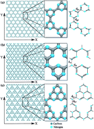
Understanding the mechanical properties of novel materials is essential in order to enable the design of applications. In this work we have investigated the mechanical and fracture patterns of three members of the two dimensional CN family (Fig. 1): graphene-based g-CN, triazine-based g-C3N4 and heptazine-based g-C3N4. We have carried out fully atomistic reactive molecular dynamics simulations considering different stretching directions (along their main axes) and at different temperatures. We expect this work to provide an extensive analysis on the behavior of graphitic CN structures under mechanical stress, as well as motivate further investigations.
 |
| Fig. 1 Structural schemes of the investigated sheets: (a) graphene-based g-CN; (b) triazine-based g-C3N4, and; (c) heptazine-based g-C3N4 membranes. The insets show their corresponding unit cell and highlights some important bond-lengths. | |
Methodology
All calculations were carried out with reactive classical molecular dynamics methods using the ReaxFF force field,35 as implemented in the LAMMPS package.36 ReaxFF was developed in order to simulate large systems while keeping an accurate description of bond formation and bond break processes. This method employs total energy description based on partial energy contributions, such as bond elongation, van der Waals forces and Coulomb interactions, among others. The ReaxFF ability to dynamically describe hybridization changes and charge redistribution (allowing the description of creating/breaking bonds), makes it suitable for the present study.
ReaxFF parameters are obtained from experiments and/or DFT calculations. The mean deviation between the heat of formation predicted by this method and experimental data is no larger than 2.9 kcal mol−1 for hydrocarbon systems.37 To further assess the suitability of the employed parameter set,35 we compared the predicted structures of β-C3N4 and graphitic-C3N4 with other values previously reported in the literature.4 The differences on bond-length and lattice parameter values were of 1% and 2% respectively, thus corroborating the adequacy of the used parameter set for this family of structures.
The investigated models consist of three different carbon nitride membranes called here: graphene-based g-CN, triazine-based g-C3N4 and heptazine-based g-C3N4, as shown in Fig. 1. The considered membranes in this work have dimensions around of 160 × 150 Å, where the g-CN structure has 6068 atoms, triazine-based g-C3N4 and heptazine-based g-C3N4 ones, 9240 and 8624 atoms, respectively. All structures were considered with periodic boundary conditions along the X and Y directions. To assure that each structure was at equilibrium before the start of the stretching process, we first thermalized them. In order to do this, we ran the molecular dynamics simulations under the NPT ensemble, i.e., with fixed number of atoms, pressure and temperature values. External pressure was set to zero, so we had no initial stress on any structure. The value of the chosen temperature was controlled during the stretching process through a Nosé–Hoover chain thermostat.38 Three different temperatures values were considered (10 K, 300 K and 600 K), in order to determine how dependent the mechanical properties are on thermal effects.
The stretching process was simulated through the gradual increase of the lattice parameter along the periodic directions. A timestep of 0.05 fs was used together with a constant strain rate of 10−6 per fs. The increased stretching is maintained until complete rupture of the membranes, which means typical simulation times of the order of 106 fs. The methodology used in this work has been successfully applied in the study of the mechanical properties of many other structures.39–44
From the simulated stretching processes we can obtain the stress–strain curves. In the linear region of the stress–strain curve we have calculated the Young's modulus, which can be defined as
|
 | (1) |
where
εi is the strain along direction
i and
σii is the in-plane virial stress tensor component along direction
i, defined as
|
 | (2) |
where
V is the volume of the membrane,
N is the number of atoms,
v the velocity,
r the position and
f the force per atom. As the membrane is only one atom-thick and atomic volumes are not very well-defined, we opt to calculate all Young's moduli as a function of this thickness
d, effectively writing the volume
V as
V =
Ad, where
A is the surface area of the membrane.
In order to have a better estimation of the spatial stress distribution during the stretching regime, we have also calculated the von Mises stress per atom i, defined as
|
 | (3) |
Results and discussions
The three distinct membranes, graphene-based g-CN, triazine-based g-C3N4 and heptazine-based g-C3N4 (Fig. 1) were stretched at a constant rate until complete rupture. Table 1 summarizes the critical strain values (i.e., strain values at the point where fracture starts) for the three structures at three different temperatures, and for two distinct strain directions. We can see a clear difference for these values for each structure. This can be explained by the considerable difference in the strain energies associated with each stretched membrane. In the case of graphene-based g-CN, there are C–C single bonds, which are naturally weaker than resonant C–N bonds. The presence of these bonds decreases the strain energy associated with the stretched membrane, therefore making it easier to fracture. In the cases of heptazine-based and triazine-based g-C3N4, both present the same types of bonds, i.e., single and double C–N bonds, but the pore density and, therefore, the number of these chemical bonds in their unit cells, is considerably different for each case. Heptazine-based structures show a lower density of chemical bonds than triazine-based structures, meaning a lower strain energy associated with the former. From these arguments, we can understand the variation on the critical strain values due to their different topologies. The decrease of the strain rate values with the increase of temperature is an expected effect, as higher thermal energy increases the fluctuations, thus making bond breaking easier.
Table 1 Critical strain values
Direction |
Temperature (K) |
g-CN |
Heptazine g-C3N4 |
Triazine g-C3N4 |
X |
10 |
0.132 |
0.149 |
0.183 |
Y |
10 |
0.172 |
0.178 |
0.204 |
X |
300 |
0.114 |
0.129 |
0.150 |
Y |
300 |
0.150 |
0.140 |
0.170 |
X |
600 |
0.100 |
0.104 |
0.130 |
Y |
600 |
0.120 |
0.120 |
0.137 |
The distinct morphologies of each membrane type lead to different fracture patterns. Also, these patterns depend on the direction of the applied strain. We applied strain along the two principal directions, X and Y, as defined in Fig. 2–4. Fig. 2 shows that, when stretching a graphene-based g-CN sheet along the X direction, stress accumulates on the C–C single bonds that are almost parallel to that direction. These are the first bonds to break. When the strain is applied along the Y direction, the C–C single bonds are parallel to the stretching direction, thus much less stress is built up before the fracturing process starts, ultimately breaking these single bonds.
 |
| Fig. 2 MD snapshots showing the stretch process considering (a and b) X and (c and d) Y directions for the g-CN membrane. The insets show the beginning of the fracture process. The von Mises stress values indicate the stress distribution during the process by color scale labeled in the figure. | |
 |
| Fig. 3 MD snapshots showing the stretch process considering (a and b) X and (c and d) Y directions for the triazine g-C3N4 membrane. The insets show the beginning of the fracture process. The von Mises stress values indicate the stress distribution during the process by color scale labeled in the figure. | |
 |
| Fig. 4 MD snapshots showing the stretch process considering (a and b) X and (c and d) Y directions for the heptazine g-C3N4 membrane. The insets show the beginning of the fracture process. The von Mises stress values indicate the stress distribution during the process by color scale labeled in the figure. | |
For triazine-based g-C3N4 membranes, as shown in Fig. 3, the stress also builts up mostly into the single bonds, in this case C–N. When stretching along the X direction, fracture yields rough edges, while stretching along the Y direction fracture yields very clean edges. This is due to the single C–N bonds which are aligned with that direction, breaking in succession.
A very similar behavior is observed in the case of heptazine-based g-C3N4 membranes, as shown in Fig. 4. This should be expected as, despite presenting larger macro-cycles, the heptazine-based membranes present a very similar structure to that of triazine-based membranes. The types of chemical bonds are almost the same, as well as, their alignment with the stretching directions, X and Y. Therefore, while the critical strain values vary significantly between each of these structures, due to the different density (number) of chemical bonds, the stress and fracture patterns are very similar.
Stress–strain curves for all the considered structures and temperatures are presented in Fig. 5. In general, the stress–strain curves start with a linear region where the Young's modulus can be calculated, going through a non-linear region and until the total rupture. Analyzing the stress–strain curves, we observe that for the graphene-based g-CN membranes a direct transition from linear regime to the fracture occurs. For the triazine g-C3N4 and heptazine g-C3N4, after the linear region, the stress is momentarily relieved and another linear region can be observed, leading to a complete fracture afterwards. This stress decrease can be attributed to an internal rearrangement of bond lengths and angles. Similar behavior in membranes formed by carbon, nitrogen and boron was observed using the Tersoff potential.45
 |
| Fig. 5 Stress–strain curves of the carbon nitride sheets (g-CN, heptazine g-C3N4 and triazine g-C3N4) for different directions and temperatures. | |
The Young's modulus for all considered structures, directions and temperatures were obtained by fitting the linear region of the stress–strain curves. For instance, considering the room temperature (300 K) and the X direction, the obtained value for g-CN was 1663 GPa Å, while for triazine g-C3N4 1668 GPa Å and heptazine g-C3N4 1247 GPa Å, as can be seen along with another results in Table 2. Our results are in good agreement with previously theoretical values obtained from a recent work with Tersoff potential for the case of triazine-based g-C3N4.45
Table 2 Young's modulus values
Structure |
Temperature (K) |
Young's modulus (GPa Å) |
Direction |
g-CN |
10 K |
1675 |
X |
Heptazine |
10 K |
1356 |
X |
Triazine |
10 K |
1890 |
X |
g-CN |
10 K |
1516 |
Y |
Heptazine |
10 K |
1397 |
Y |
Triazine |
10 K |
1920 |
Y |
g-CN |
300 K |
1663 |
X |
Heptazine |
300 K |
1247 |
X |
Triazine |
300 K |
1668 |
X |
g-CN |
300 K |
1349 |
Y |
Heptazine |
300 K |
1299 |
Y |
Triazine |
300 K |
1733 |
Y |
g-CN |
600 K |
1571 |
X |
Heptazine |
600 K |
1197 |
X |
Triazine |
600 K |
1578 |
X |
g-CN |
600 K |
1333 |
Y |
Heptazine |
600 K |
1229 |
Y |
Triazine |
600 K |
1575 |
Y |
Comparing the calculated value of the Young's modulus for graphene (3570 GPa Å (ref. 46)) with the values herein reported, CN membranes values are lower by 53% for g-CN and triazine g-C3N4 and 65% for heptazine g-C3N4. This decrease is due to differences in the chemical structure of the carbon nitride sheets when compared to graphene, namely the presence of pores, decreasing the density of chemical bonds, as well as the presence of single bonds.
Summary and conclusions
We have investigated the mechanical and fracture patterns of a series of two-dimensional CN structures: g-CN, triazine g-C3N4 and heptazine g-C3N4 (Fig. 1). The study was carried out through fully atomistic reactive molecular dynamics simulations using the ReaxFF force field. The Young's moduli for the carbon nitride membranes are smaller when compared with the Young's modulus for graphene. This can be understood by presence of pores and single C–N bonds in the carbon nitride membranes. More interestingly, graphene-based g-CN goes abruptly from elastic to brittle behavior, while triazine and heptazine g-C3N4 structures go through significant structural reconstructions with multiple elastic stages. This differentiated behavior can be explained by the differences in the density of chemical bonds and how the rings are oriented in relation to the stretching directions, resembling an arch-type effect recently reported to silicene membranes.42
Acknowledgements
This work was supported in part by the Brazilian Agencies CAPES, CNPq and FAPESP. The authors thank the Center for Computational Engineering and Sciences at Unicamp for financial support through the FAPESP/CEPID Grant # 2013/08293-7. J. M. S. acknowledges the support from CAPES through the Science Without Borders program (project number A085/2013).
References
- M. L. Cohen, Phys. Rev. B: Condens. Matter Mater. Phys., 1985, 32, 7988 CrossRef CAS.
- A. Y. Liu and M. L. Cohen, Science, 1989, 245, 841–842 CAS.
- A. Y. Liu and R. M. Wentzcovitch, Phys. Rev. B: Condens. Matter Mater. Phys., 1994, 50, 10362 CrossRef CAS.
- D. M. Teter and R. J. Hemley, Science, 1996, 271, 53–55 CAS.
- Y. Miyamoto, M. L. Cohen and S. G. Louie, Solid State Commun., 1997, 102, 605–608 CrossRef CAS.
- C. Niu, Y. Z. Lu and C. M. Lieber, Science, 1993, 261, 334–337 CAS.
- K. M. Yu, M. L. Cohen, E. Haller, W. Hansen, A. Y. Liu and I. Wu, Phys. Rev. B: Condens. Matter Mater. Phys., 1994, 49, 5034 CrossRef CAS.
- M. Terrones, P. Redlich, N. Grobert, S. Trasobares, W.-K. Hsu, H. Terrones, Y.-Q. Zhu, J. P. Hare, C. L. Reeves, A. K. Cheetham, H. W. K. Manfred Ruhle and D. R. M. Walton, Adv. Mater., 1999, 11, 655–658 CrossRef CAS.
- K. S. Novoselov, A. K. Geim, S. Morozov, D. Jiang, Y. Zhang, S. Dubonos, I. Grigorieva and A. Firsov, Science, 2004, 306, 666–669 CrossRef CAS PubMed.
- C. Berger, Z. Song, X. Li, X. Wu, N. Brown, C. Naud, D. Mayou, T. Li, J. Hass and A. N. Marchenkov, Science, 2006, 312, 1191–1196 CrossRef CAS PubMed.
- A. K. Geim, Science, 2009, 324, 1530–1534 CrossRef CAS PubMed.
- C. Lee, X. Wei, J. W. Kysar and J. Hone, Science, 2008, 321, 385–388 CrossRef CAS PubMed.
- R. Joshi, P. Carbone, F. Wang, V. Kravets, Y. Su, I. Grigorieva, H. Wu, A. Geim and R. Nair, Science, 2014, 343, 752–754 CrossRef CAS PubMed.
- X. Yang, C. Cheng, Y. Wang, L. Qiu and D. Li, Science, 2013, 341, 534–537 CrossRef CAS PubMed.
- L. Feng, L. Wu and X. Qu, Adv. Mater., 2013, 25, 168–186 CrossRef CAS PubMed.
- Y. Tao, Y. Lin, Z. Huang, J. Ren and X. Qu, Adv. Mater., 2013, 25, 2594–2599 CrossRef CAS PubMed.
- W. Auwärter, T. Kreutz, T. Greber and J. Osterwalder, Surf. Sci., 1999, 429, 229–236 CrossRef.
- P. Vogt, P. De Padova, C. Quaresima, J. Avila, E. Frantzeskakis, M. C. Asensio, A. Resta, B. Ealet and G. Le Lay, Phys. Rev. Lett., 2012, 108, 155501 CrossRef PubMed.
- Y. Zheng, J. Liu, J. Liang, M. Jaroniec and S. Z. Qiao, Energy Environ. Sci., 2012, 5, 6717–6731 CAS.
- X. Wang, X. Chen, A. Thomas, X. Fu and M. Antonietti, Adv. Mater., 2009, 21, 1609–1612 CrossRef CAS.
- Y. Wang, Y. Di, M. Antonietti, H. Li, X. Chen and X. Wang, Chem.
Mater., 2010, 22, 5119–5121 CrossRef CAS.
- E. Perim and D. S. Galvao, ChemPhysChem, 2014, 15, 2367–2371 CrossRef CAS PubMed.
- J. Li, C. Cao and H. Zhu, Nanotechnology, 2007, 18, 115605 CrossRef.
- Y. Zhao, D. Yu, H. Zhou, Y. Tian and O. Yanagisawa, J. Mater. Sci., 2005, 40, 2645–2647 CrossRef CAS.
- D. Wei, Y. Liu, Y. Wang, H. Zhang, L. Huang and G. Yu, Nano Lett., 2009, 9, 1752–1758 CrossRef CAS PubMed.
- M. Zelisko, Y. Hanlumyuang, S. Yang, Y. Liu, C. Lei, J. Li, P. M. Ajayan and P. Sharma, Nat. Commun., 2014, 5, 4284 CAS.
- G. Algara-Siller, N. Severin, S. Y. Chong, T. Björkman, R. G. Palgrave, A. Laybourn, M. Antonietti, Y. Z. Khimyak, A. V. Krasheninnikov and J. P. Rabe, Angew. Chem., 2014, 126, 7580–7585 CrossRef.
- A. Thomas, A. Fischer, F. Goettmann, M. Antonietti, J.-O. Müller, R. Schlögl and J. M. Carlsson, J. Mater. Chem., 2008, 18, 4893–4908 RSC.
- E. Kroke, M. Schwarz, E. Horath-Bordon, P. Kroll, B. Noll and A. D. Norman, New J. Chem., 2002, 26, 508–512 RSC.
- B. V. Lotsch, M. Döblinger, J. Sehnert, L. Seyfarth, J. Senker, O. Oeckler and W. Schnick, Chemistry, 2007, 13, 4969–4980 CrossRef CAS PubMed.
- M. Döblinger, B. V. Lotsch, J. Wack, J. Thun, J. Senker and W. Schnick, Chem. Commun., 2009, 1541–1543 RSC.
- F. Cui and D. Li, Surf. Coat. Technol., 2000, 131, 481–487 CrossRef CAS.
- M. Deifallah, P. F. McMillan and F. Corà, J. Phys. Chem. C, 2008, 112, 5447–5453 CAS.
- S.-D. Mo, L. Ouyang, W. Ching, I. Tanaka, Y. Koyama and R. Riedel, Phys. Rev. Lett., 1999, 83, 5046 CrossRef CAS.
- J. Budzien, A. P. Thompson and S. V. Zybin, J. Phys. Chem. B, 2009, 113, 13142–13151 CrossRef CAS PubMed.
- S. Plimpton, J. Comput. Phys., 1995, 117, 1–19 CrossRef CAS.
- A. C. van Duin, S. Dasgupta, F. Lorant and W. A. Goddard, J. Phys. Chem. A, 2001, 105, 9396–9409 CrossRef CAS.
- G. J. Martyna, M. E. Tuckerman, D. J. Tobias and M. L. Klein, Mol. Phys., 1996, 87, 1117–1157 CrossRef CAS.
- A. Nair, S. Cranford and M. Buehler, EPL, 2011, 95, 16002 CrossRef.
- S. W. Cranford and M. J. Buehler, Carbon, 2011, 49, 4111–4121 CrossRef CAS.
- A. P. Garcia and M. J. Buehler, Comput. Mater. Sci., 2010, 48, 303–309 CrossRef CAS.
- T. Botari, E. Perim, P. Autreto, A. C. van Duin, R. Paupitz and D. Galvao, Phys. Chem. Chem. Phys., 2014, 16, 19417–19423 RSC.
- B. D. Jensen, K. E. Wise and G. M. Odegard, J. Comput. Chem., 2015, 36, 1587–1596 CrossRef CAS PubMed.
- J. M. de Sousa, G. Brunetto, V. R. Coluci and D. S. Galvao, Carbon, 2016, 96, 14–19 CrossRef CAS.
- B. Mortazavi, G. Cuniberti and T. Rabczuk, Comput. Mater. Sci., 2015, 99, 285–289 CrossRef CAS.
- F. Liu, P. Ming and J. Li, Phys. Rev. B: Condens. Matter Mater. Phys., 2007, 76, 064120 CrossRef.
|
This journal is © The Royal Society of Chemistry 2016 |
Click here to see how this site uses Cookies. View our privacy policy here.